Báo cáo khoa học: A new highly toxic protein isolated from the death cap Amanita phalloides is an L-amino acid oxidase pdf
Bạn đang xem bản rút gọn của tài liệu. Xem và tải ngay bản đầy đủ của tài liệu tại đây (564.14 KB, 10 trang )
A new highly toxic protein isolated from the death cap
Amanita phalloides is an
L-amino acid oxidase
Taras Stasyk
1,2
, Maxim Lutsik-Kordovsky
1
, Christer Wernstedt
3
, Volodymyr Antonyuk
1
,
Olga Klyuchivska
1
, Serhiy Souchelnytskyi
4
, Ulf Hellman
3
and Rostyslav Stoika
1
1 Institute of Cell Biology, National Academy of Sciences of Ukraine, Lviv, Ukraine
2 Biocenter, Innsbruck Medical University, Austria
3 Ludwig Institute for Cancer Research Ltd, Uppsala University, Sweden
4 Karolinska Biomics Center, Karolinska Institutet, Stockholm, Sweden
Introduction
The death cap (Amanita phalloides) is known to be a
deadly poisonous mushroom as a result of the produc-
tion of several toxic substances. The first substance
was isolated in 1937 by Wieland [1] and was shown to
possess an oligopeptide structure. In further studies,
Wieland and Faulstich [2] revealed other toxic cyclo-
peptides, which were classified into two structural
groups (i.e. amanitine and phalloidine), with both
Keywords
Amanita phalloides; apoptosis; death cap;
L-amino acid oxidase; toxic protein
Correspondence
R. Stoika, Institute of Cell Biology, National
Academy of Sciences of Ukraine,
Drahomanov Street 14 ⁄ 16, 79005, Lviv,
Ukraine
Fax: +38 032 261 22 87
Tel: +38 032 261 22 87
E-mail:
Database
Nucleotide sequence data have been
submitted to the GenBank database under
the accession number GU220069
(Received 8 October 2009, revised 2
December 2009, accepted 21 December
2009)
doi:10.1111/j.1742-4658.2010.07557.x
A new highly cytotoxic protein, toxophallin, was recently isolated from the
fruit body of the death cap Amanita phalloides mushroom [Stasyk et al.
(2008) Studia Biologica 2, 21–32]. The physico-chemical, chemical and bio-
logical characteristics of toxophallin differ distinctly from those of another
death cap toxic protein, namely phallolysin. The interaction of toxophallin
with target cells is not mediated by a specific cell surface receptor. It
induces chromatin condensation, as well as DNA and nucleus fragmenta-
tion, which are typical for apoptosis. However, caspase III inhibitor [ben-
zyloxycarbonyl-Asp(OMe)-fluoromethylketone] did not stop toxophallin-
induced DNA fragmentation. Thus, toxophallin uses a caspase-independent
pathway of apoptosis induction. In the present study, we applied a comple-
mentary approach based on a combination of proteomics and molecular
biology tools for the protein identification of toxophallin. The primary
structure of toxophallin was partially studied via direct sequencing of its
tryptic peptides, followed by PCR-based cloning of the corresponding
cDNA. A subsequent bioinformatic search revealed a structural homology
of toxophallin with the
L-amino acid oxidase of the Laccaria bicolor mush-
room. This demonstrates the usefulness of our approach for the identifica-
tion of proteins in organisms with unknown genomes. We also found a
broad substrate specificity of toxophallin with respect to oxidizing selected
amino acids. Ascorbic acid inhibited the cytotoxic effect of toxophallin,
most likely as a result of scavenging hydrogen peroxide, which is the
product of oxidase catalysis. Thus, in addition to highly toxic cyclopeptides
and toxic lectin phallolysin, the death cap fruit body contains another
cytotoxic protein in the form of an enzyme, namely
L-amino acid oxidase.
Abbreviations
CM, carboxymethyl; MALDI-TOF, matrix-assisted laser desorption ionization time-of-flight.
1260 FEBS Journal 277 (2010) 1260–1269 ª 2010 The Authors Journal compilation ª 2010 FEBS
exhibiting different mechanisms of toxic action.
Although amanitine inhibits mRNA transcription,
phalloidine binds actin and suppresses the functions of
the cytoskeleton. These cyclopeptides are frequently
used as experimental tools in scientific studies because
their intracellular molecular targets and mechanisms of
action have been well characterized. In addition to
these toxic polypeptides, the death cap also contains
antitoxin antamanide, a cyclodecapeptide that blocks
the effects of phalloidine [1,2]. Another toxic polypep-
tide, phallolysin, possessing hemolytic activity, was
also detected in the fruit body of the death cap [3–7].
Its chemical properties and biological activity, as well
as its mechanisms of action, have been described previ-
ously [2,8].
Many mushroom species have been shown to con-
tain polypeptide substances that possess antitumor and
immunomodulating activity [9–11]. Lectin-like proteins
demonstrating antiproliferative activity towards
tumor cells were isolated from the mushrooms
Tricholoma mongolicum [12] and Agaricus bisporus [13].
Another protein with antineoplastic activity, volvarin,
which belongs to the family of ribosome inactivating
proteins type I, was isolated from the edible mush-
room Volvariella volvacea [14]. The poisonous mush-
room Boletus satanas Lenz contains a toxic lectin
bolesatine that inhibits protein synthesis both in vitro
and in vivo [15].
Recently, a new cytotoxic protein, toxophallin, was
isolated from the fruit body of the death cap
A. phalloides [16]. Its physico-chemical, chemical and
biological properties differ distinctly from those of the
other known toxic proteins of this mushroom. Toxo-
phallin was not bound by any target cell surface spe-
cific receptors. Furthermore, it induced apoptosis
(chromatin condensation, DNA and nucleus fragmen-
tation) but this was not blocked by caspase III inhibi-
tor [benzyloxycarbonyl-Asp(OMe)-fluoromethylketone]
[16]. In the present study, we carried out a more pre-
cise structural analysis of toxophallin by directly
sequencing its tryptic peptides, followed by PCR-based
cloning of cDNA. A bioinformatics approach allowed
us to demonstrate the sequence homology of toxophal-
lin with the recently identified l-amino acid oxidase of
Laccaria bicolor [17].
Results
Purification of toxophallin
The purification procedure consisted of four main
steps: (a) ammonium sulfate precipitation of total
protein from the juice of thawed and grinded mush-
rooms; (b) elimination of pigmented material from
the obtained protein bulk by ion-exchange chroma-
tography on a DEAE-cellulose column; (c) affinity
chromatography on the immobilized ovomucin to
remove cytolytic lectin, phallolysin; and (d) purifica-
tion of toxophallin by the repeated ion-exchange
chromatography on a carboxymethyl (CM)-cellulose
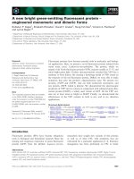
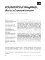
column. Native gel electrophoresis of water-soluble
proteins upon elimination of pigmented materials
revealed three main protein bands (Fig. 1). The prom-
inent band corresponds to phallolysin, the death cap
lectin with high cytolytic activity. Phallolysin was effi-
ciently removed from the protein extract using affinity
chromatography on the immobilized ovomucin. Pro-
tein exhibiting cytotoxic activity, and found in the
nonlectin fraction, was further purified by two-step
ion-exchange chromatography (see Materials and
methods). Purified toxophallin migrated as a
homogenous protein band by nondenaturing gel
electrophoresis (Fig. 1A). A single protein band of
55 kDa was also detected by SDS-PAGE (Fig. 1B).
The toxophallin purity was approximately 95%
according to native electrophoresis, and approxi-
mately 85% according to SDS-PAGE, presumably
because of partial protein degradation.
Protein characterization
Amino acid analysis revealed three cysteines, six
methionines and 36 proline residues in the toxophallin
molecule, which makes up approximately 7% of the
AB
Fig. 1. Electrophoretic study of extracted proteins of Amanita
phalloides. (A) b-alanin-acetate electrophoretic system, pH 4.5. (1)
Crude extract; (2) nonlectin proteins (not retained by the affinity
sorbents); and (3) cytotoxic protein purified by the ion-exchange
chromatography on CM-cellulose column. (B) SDS-PAGE in 14%
gel. (1) Molecular mass protein markers (Sigma) and (2) purified
protein (55 kDa) under study. Coomassi R-250 staining. Reproduced
with permission [16].
T. Stasyk et al. Cytotoxic
L-amino oxidase from A. phalloides
FEBS Journal 277 (2010) 1260–1269 ª 2010 The Authors Journal compilation ª 2010 FEBS 1261
amino acid residues present in this 55 kDa protein,
which consists of 503 amino acid residues (Table S1).
The relatively high content of proline residues suggests
a significant rigidity of the polypeptide chain of toxo-
phallin.
For MS analysis, toxophallin was in-gel digested
using trypsin, and the peptide mixture was analyzed by
matrix-assisted laser desorption ionization time-of-
flight (MALDI-TOF) MS. Sixty-nine tryptic peptides
were identified (Table S2) and used for the database
search. Because we could not find any homology with
other proteins in the databases by peptide mass finger-
printing, sequencing of the separated tryptic peptides
was carried out. Tryptic peptides of the toxophallin
were isolated by microbore reversed phase liquid chro-
matography (Fig. S1) and several isolated peptides
were subjected to Edman degradation. The amino acid
sequences of ten peptides that were sequenced, differ-
ing in length by five to 16 amino acid residues, are
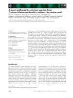
shown in Fig. 2A. According to the amino acid com-
position of toxophallin (Table S1), with 29 K and 14
R identified, 43 tryptic peptides could be expected.
This suggests that the other 26 peptides represent
modified peptides or miss cuts of the digestion.
Unexpectedly, all ten peptides sequenced by Edman
degradation were not modified because the masses cal-
culated from amino acid composition of the sequenced
peptides amounted to the values of the corresponding
peptides obtained by the MALDI-TOF MS. Direct
sequencing of the toxophallin N-terminus from sam-
ples after blotting onto poly(vinylidene difluoride)
membrane did not reveal any signal, thereby suggest-
ing that the N-terminus of the protein was blocked. In
total, sequenced peptides account for 20% of the
whole molecule, with 107 amino acids being identified
in the ten peptides analyzed.
To obtain an mRNA sequence of toxophallin, we
performed RT-PCR-based cloning. Ten oligonucleotide
primers were designed using the amino acid sequence
of the identified peptides. PCR reactions with different
combinations of primers were performed with cDNA
from mRNA isolated from the whole fruit body. The
primer combinations TTC CCA GAG ATC GAG
TCA ATG CGT (3¢-to5¢) and TCT GTC GTA CCA
ACC AGT TGA (5¢-to3¢), designed on the basis of
peptides 15 (FPEIESMR) and nine (STGWYDR),
respectively, resulted in a PCR product. This PCR
product was cloned and sequenced, as described in the
A
B
Fig. 2. Partial sequence of toxophallin. (A)
Amino acid sequence of ten identified
tryptic peptides of toxophallin (Edman
degradation analysis; Fig. 2). (B) Partial
nucleotide sequence and deduced amino
acid sequence of the toxophallin. The amino
acid translation is under the second
nucleotide of the corresponding codon. The
masses of underlined peptides correspond
to the masses of tryptic peptides (Table S2)
identified by MALDI-TOF MS.
Cytotoxic
L-amino oxidase from A. phalloides T. Stasyk et al.
1262 FEBS Journal 277 (2010) 1260–1269 ª 2010 The Authors Journal compilation ª 2010 FEBS
Materials and methods, and 429 nucleotides were iden-
tified (Fig. 2B). This sequence, in combination with
primers, corresponds to a polypeptide consisting of
158 amino acid residues, and comprises approximately
one-third of the molecule (approximately 503 amino
acid residues; Table S1). In the internal part of the
identified cDNA, sequences corresponding to two
other peptides sequenced by Edman degradation were
found: peptides 22 and 57 (Fig. 2). Moreover, the
obtained partial sequence of toxophallin was also con-
firmed by the MS data when comparing the in silico
tryptic digest of the translated amino acid sequence
with the list of masses of tryptic peptides obtained by
the MALDI-TOF analysis. In total, nine peptides from
the list of toxophallin tryptic peptides (numbers 9, 15,
19, 22, 27, 57, 58, 63, 65; Fig. 2 and Table S2), includ-
ing four peptides sequenced by Edman degradation,
matched the corresponding tryptic peptides of the
sequenced toxophallin mRNA fragment (see under-
lined peptides in Fig. 2), thereby unambiguously con-
firming our RT-PCR-based cloning strategy in
combination with MS and Edman sequencing.
A database search for similar protein sequences
was carried out using the blast algorithm. We found
sequence homology of toxophallin with the amine
oxidase of L. bicolor (Fig. S2). The partial amino acid
sequence deduced from the cloned mRNA fragment
was found to be related to two predicted proteins
from L. bicolor S238N-H82 according to the recently
published genome of this mushroom [17] (accession
numbers EDR00058.1 and EDR12198.1) with 49%
and 45% identities (i.e. the extent to which two
sequences are invariant) and 60% and 57% positives
(i.e. changes at a specific position of an amino acid
sequence that preserves the physico-chemical proper-
ties of the original residue), respectively. Moreover,
we could align the remaining six sequenced peptides
to the C-terminal part of the L. bicolor protein
EDR12198.1 (Fig. S2). A high degree of similarity
between the partial sequence of toxophallin and the
amine oxidases sequences available in the database
strongly suggests a putative amine oxidase activity of
toxophallin.
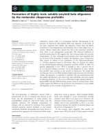
Biological activity of toxophallin
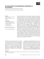
The cytotoxic activity of the purified toxophallin was
monitored by measuring its effect towards human leu-
kemia CEM-T4 and murine leukemia L1210 cells
(Fig. 3). Toxophallin possesses a distinct cytotoxic
effect (as detected by the trypan blue exclusion assay)
that was much stronger in the case of CEM-T4 cells
compared to L1210 cells. The IC
50
of purified
toxophallin was 0.5 lgÆmL
)1
. The IC
50
values with
respect to the action of toxophallin in the cell viability
test as estimated by the trypan blue exclusion assay
(0.5 lgÆmL
)1
), as well as by cell proliferation as deter-
mined by [
3
H]-thymidine incorporation (0.25–0.45
lgÆmL
)1
) [16], were of similar concentration depen-
dence, indicating that the activity of toxophallin is
cytotoxic, rather than antiproliferative. In a previous
study, we have shown that toxophallin promotes cell
death via apoptosis, which was demonstrated by a
DNA fragmentation assay performed in different
mammalian cell lines (murine leukemia L1210, mink
lung epithelial CCL-64, human lung carcinoma A549
and human breast carcinoma MCF-7 cells) [16]. The
proapoptotic action of toxophallin, as revealed in a
DNA-laddering bio-assay, was demonstrated by the
results of a cytomorphological study, using 4¢,6¢-
diamidino-2-phenylindole staining and terminal
0.0 0.5 1.0 1.5 2.0 2.5
0
20
40
60
80
100
A
B
L1210
CEMT4
% of trypan-positive (dead) cells
L-amino-acid oxidase (µg·mL
–1
)
0.0 0.5 1.0 1.5 2.0 2.5
0
20
40
60
80
100
L1210
CEMT4
Cell number (% of control)
L-amino-acid oxidase (µg·mL
–1
)
Fig. 3. Dose-dependent effect of toxophallin (L-amino acid oxidase)
towards target (human CEM-T4 and murine L1210) cells.
Approximately 300 000 cells of the L1210 line per well in 1 mL,
and 200 000 cells of the CEM-T4 line per well in 1 mL were
present at the beginning of the experiment. After 24 h, the tested
substances were added at different concentrations. The number of
viable cells was counted in the hemocytometric chamber. The ratio
of dead cells was defined subsequent to staining with trypan blue
(0.1%, w ⁄ v) and observation under a light microscope.
T. Stasyk et al. Cytotoxic
L-amino oxidase from A. phalloides
FEBS Journal 277 (2010) 1260–1269 ª 2010 The Authors Journal compilation ª 2010 FEBS 1263
deoxynucleotidyl-transferase-mediated dUDP nick-end
labeling (i.e. assay for apoptosis detection) [16]. The
time dependence of the effect of toxophallin as studied
using the trypan blue exclusion test and 4¢,6¢-
diamidino-2-phenylindole staining demonstrated that
toxophallin-induced cell death became noticeable after
5 h [16].
Taking into account that toxophallin of A. phalloides
displays structural homology with amine oxidase
isolated from L. bicolor, we have examined the amine
oxidase activity of toxophallin, as reported previously
[18]. The results obtained in that study are presented
in Table 1 (see also Table S3). Toxophallin did not use
benzylamine, ethanolamine, diethylamine, meta- and
para-phenylendiamine, ortho-, meta- and para-
aminophenols, or putrescin as a substrate for the
enzymatic reaction, which testifies to the absence of its
mono- and diamine oxidase activity. The highest oxi-
dase activity was observed towards dl-methionine and
l-methionine, l-phenylalanine, dl-norleucine, l-isoleu-
cine, l-arginine, l-tyrosine, and dl-leucine; oxidase
activity was relatively low towards dl-lysine and
l-lysine, dl-asparagine, dl-valine, l-histidine, dl -threo-
nine, dl-thryptophane, and l-glutamic acid; and there
was a lack of oxidase activity towards l-cysteine,
l-glycine, l-proline, l-oxyproline, dl-serine, and
dl-aspartic acid. These results indicate that the novel
toxic protein, purified from A. phalloides mushroom is
an l-amino acid oxidase.
Ascorbic acid (10 lgÆmL
)1
) inhibited the cytotoxic
effect (measured by the trypan blue exclusion test)
caused by toxophallin (l-amino acid oxidase) (Fig. 4).
The mechanisms of such inhibition could be based on
inactivating the H
2
O
2
that appears as a result of the
amine oxidase reaction and is toxic for cells. Both
ascorbic acid and reduced glutathione also inhibited
amine oxidase reaction in vitro (Fig. 5).
Discussion
When studying toxic proteins isolated from the fruit
bodies of the death cap A. phalloides, we detected a
novel cytotoxic protein that differed from all previ-
ously described toxic proteins from that mushroom
species. It differs distinctly from phallolysin, which
was isolated and characterized by Faulstich et al. [3,4]
and Seeger et al. [5–7]. Both proteins differ substan-
tially in their biological activity. Phallolysin is highly
toxic in animals, reaching a lethal dose at 40 lgÆkg
)1
in rabbits [3]. Its hemolytic activity towards rabbit
erythrocytes in vitro was 5 lgÆmL
)1
[16]. Toxophallin
was found to exhibit high toxicity towards various
mammalian cells; for example, in cells of A549 and
T47D lines, IC
50
= 0.25 lgÆmL
)1
and, in cells of
CCL-64 and MCF 7 lines, IC
50
= 0.45 lgÆmL
)1
[16].
Toxophallin preparations did not possess hemolytic
Table 1. Toxophallin is L-amino acid oxidase. Different amino acids
were studied as toxophallin substrates using an amine oxidase
enzymatic activity assay, as described in the Materials and
methods.
No. Substrate Relative activity
1
DL-tyrosine 1.0
2
L-tyrosine 1.9
3
DL-lysine 0.3
4
L-lysine 0.6
5
DL-asparagine 0.4
6
L-asparagine 0.8
7
DL-phenylalanine 1.3
8
L-phenylalanine 2.6
9
DL-methionine 2.7
10
L-methionine 3.6
012345
0
20
40
60
80
100
A
B
Cell number (% of control)
L-amino-acid oxidase (µg·mL
–1
)
Control
+ 10 µg·mL
–1
ascorbic acid
012345
0
10
20
30
40
50
Control
+ 10 µg·mL
–1
ascorbic acid
% of trypan-positive (dead) cells
L-amino-acid oxidase (µg·mL
–1
)
Fig. 4. Ascorbic acid inhibits cytotoxic effect of toxophallin (L-amino
acid oxidase) towards the murine L1210 cell line. The experiment
conditions are as in Fig. 3. Ascorbic acid (a concentration of
10 lgÆmL
)1
was selected as being the most effective with respect
to inhibiting the cytotoxicity of toxophallin) was added to cell
culture simultaneously with toxophallin used at different
concentrations. A statistical significant difference (P < 0.05) was
observed at a concentration of toxophallin of 5 lgÆmL
)1
.
Cytotoxic
L-amino oxidase from A. phalloides T. Stasyk et al.
1264 FEBS Journal 277 (2010) 1260–1269 ª 2010 The Authors Journal compilation ª 2010 FEBS
activity, whereas the hemolytic activity of phallolysin
reached 24 000 unitsÆmg
)1
[4].
In the present study, we used a complementary
approach (i.e. a combination of proteomic and molec-
ular biology tools) to identify biologically active pro-
teins in organisms with unknown genomes. The
sequence of toxophallin was partially studied by a
combination of MS and direct Edman sequencing of
tryptic peptides with a PCR-based cloning of the
cDNA. The high level of overlap between the
sequenced peptides and the cDNA indicated strong
support for the partial protein sequence obtained, and
allowed us to find a homology of toxophallin with the
l-amino acid oxidase of L. bicolor according to the
recently published genome of this mushroom [17].
The mRNA of toxophallin (2.1 kb) was detected
only in the stem and, to a lesser extent, in the cap of
A. phalloides fruit bodies by Northern blot analysis
using a RT-PCR fragment of the cloned toxophallin
cDNA as a probe for the hybridization reaction (data
not shown). Recently, the l-amino acid oxidase gene
of L. bicolor has been shown to be expressed at protein
level [19]. In this study, it was suggested that amine
oxidases are enzymes of cellular amino acid catabo-
lism, comprising potential candidates for a mechanism
that catalyses nitrogen mineralization from amino
acids at the ecosystem level. The distribution of
l-amino acid oxidase in the stem of the A. phalloides
mushroom fruit body fits very well with this hypothe-
sis. It should be noted that we also found a protein
possessing toxophallin-like activity in the Amanita
virosa fruit body (V. Antonyuk et al., in press).
A cross-linking receptor study did not reveal specific
receptor molecules for this protein on the surface of
target cells [16]. The cytotoxic effects were found to
develop relatively slowly because the first signs of cell
damage were observed only after 5 h of treatment.
Target cells underwent apoptosis subsequent to
toxophallin treatment and cell death did not depend
on the activation of the caspase cascade [16]. The most
pronounced destructive changes, namely condensation
of nuclear chromatin and DNA fragmentation, were
observed in the cell nucleus. Similar processes were
characteristic for cell damage caused by the ionizing
radiation, and these were mediated by generation of
reactive oxygen species [20]. Thus, it is suggested that
toxophallin induces cell damage indirectly via the gen-
eration of free radicals and oxidant agents that can
trigger cell impairment and apoptosis by a caspase-
independent pathway. l-amino acid oxidase enzymatic
activity of the toxophallin is well suited for such
action. Via the H
2
O
2
generated by the enzyme activity,
amine oxidases may act as a defense or attack mecha-
nism. l-amino acid oxidase has been described as one
of the most common components of snake venom
[21–23], as recently reviewed [24]. Although partially
purified toxophallin was accessible previously [25], its
specific enzymatic activity remained unknown at that
time.
Recently, a protective action of the radical scavenger
N-acetylcysteine upon treatment of A. phalloides poi-
soning was demonstrated [26]. It is possible that the
products of the l-amino acid oxidase (toxophallin)
enzymatic reaction could also be inactivated by this
agent.
Various biological systems, accompanied by an
increased production of reactive oxygen species, are
effective as potential anticancer remedies. Cytotoxicity
was observed as a result of the action of BSA oxidase
in the presence of spermine, and this was attributed to
H
2
O
2
and aldehyde production [27]. Increasing the
incubation temperature from 37 to 42 °C enhanced
cytotoxicity in tumor cells exposed to spermine metab-
olites. Moreover, it was found that multidrug-resistant
human melanoma cells were more sensitive than their
wild-type counterparts to H
2
O
2
and aldehydes [28].
The metabolites formed by BSA oxidase targeting
spermine were more toxic than exogenous H
2
O
2
and
acrolein, even though their concentration was lower
during the initial phase of incubation. The increase of
natural polyamines in malignant and actively prolifer-
0
0.1
0.2
0.3
0.4
0.5
13579
E525
Time (min)
Control
glutathione-SH
0.5 mg·mL
–1
Ascorbic acid
0.5 mg·mL
–1
Ascorbic acid,
glutathione-SH,
5 mg·mL
–1
Fig. 5. Ascorbic acid and reduced glutathione inhibit L-amine oxidase
activity of the toxophallin in vitro. The reaction mixture consisted of
0.15 mL of 0.1% aqueous solution of toxophallin and 2.5 mL of
0.3 m
M o-dianizidine solution to which 0.2 mL of 0.1% horseradish
peroxidase (RZ = 0.4–0.6) was added. The reaction was started by
adding 0.2 mL of 0.2% solution of
L-methionine, after which the
absorbance at 525 nm was measured in the spectrophotometer cuv-
ette at different time intervals. When ascorbic acid or glutathione-SH
were used, they were added at a final concentration of 0.5 and
5mgÆmL
)1
before adding L-methionine.
T. Stasyk et al. Cytotoxic
L-amino oxidase from A. phalloides
FEBS Journal 277 (2010) 1260–1269 ª 2010 The Authors Journal compilation ª 2010 FEBS 1265
ating cells has led the use of polyamine depletion as a
strategy for inhibiting cell growth [29]. Thus, in the
anticancer therapeutic strategy, there is increasing
interest in spermine oxidase, which specifically oxidases
spermine. Because putrescine was not active as a sub-
strate in the enzymatic reaction of toxophallin purified
from A. phalloides, this testifies to the absence of
mono- and diamine oxidase activity in that protein.
However, toxophallin demonstrated itself to be an
l-amino acid oxidase, which suggests that it should
not have a deficiency of substrates for its catalytic
activity. For definite conclusions on the anticancer
potential of toxophallin, additional investigations are
required.
It should be noted that deadly poisonous mush-
rooms, such as the death cap, contain various cyto-
toxic polypeptide compounds that possess different
mechanisms of toxic action. Thus, treatment for poi-
soning caused by these mushrooms should be complex
and include antidotes against all toxic compounds.
Because toxophallin is an l-amino acid oxidase, H
2
O
2
scavengers may be protective during its action in the
organism.
In conclusion, in the present study, a novel cytotoxic
protein was isolated from the death cap and character-
ized. The physico-chemical, chemical and biological
characteristics of this protein differ distinctly from
those of all previously described toxic substances
of A. phalloides, such as toxic cyclopeptides or
phallolysin. The isolated cytotoxic protein was shown
to be an enzyme, namely l-amino acid oxidase.
Materials and methods
Isolation and purification of cytotoxic proteins
from the death cap
Fruit bodies of A. phalloides mushrooms were collected in
the forests of Lviv region (Ukraine), and stored at
)20 °C until use (not longer than 1 month). The mush-
room fruit bodies were pressed, subjected to centrifugation
at 4000 g for 15 min, and the supernatant was collected.
Ammonium sulfate was added to 90% saturation of the
supernatant, and precipitated proteins were collected by
filtration. For elimination of dark colored pigment mate-
rial, the precipitate was dissolved in a small volume of
distilled water, dialyzed against buffer solution (50 mm
potassium phosphate buffer, pH 7.0 supplemented with
100 mm sodium chloride) and passed through a DEAE-
cellulose column (Serva, Heidelberg, Germany), equili-
brated with the same buffer. The fraction of unabsorbed
protein was collected and precipitated with ammonium
sulfate at 90% saturation.
For elimination of cytolytic lectin, phallolysin, crude pro-
tein fraction was passed through a column filled with affin-
ity sorbent, ovomucin immobilized on agarose [30],
equilibrated with NaCl ⁄ Pi. The unbound ‘nonlectin’ protein
fraction was collected, dialyzed against 30 mm sodium
acetate buffer (pH 5.3) and applied onto a CM-cellulose
column (CM-32; Whatman Biochem. Ltd., Maidstone,
UK), which was equilibrated with 30 mm sodium acetate
buffer (pH 5.3). The column was eluted stepwise with
100 mm sodium acetate buffer and, subsequently, with the
same buffer supplemented with 75 mm sodium chloride.
Protein possessing cytotoxic action was eluted with 75 mm
sodium chloride. This protein peak was collected, concen-
trated and subjected to re-chromatography on a CM-cellu-
lose column in 100 mm sodium acetate buffer (pH 5.3) and
75 mm sodium chloride. The main protein peak corre-
sponding to pure cytotoxic protein was collected, dialyzed
against distilled water and lyophilized.
Two electrophoretic systems were applied for the
evaluation of purity of isolated toxophallin: (a) disc-electro-
phoresis in 7.5% PAGE using the Reisfeld system in b-
alanine-acetate buffer (pH 4.5) and protein staining with
Amido Black 10B [31] and (b) SDS-PAGE in 14% slab gel
in a Laemmli buffer system [32] with protein visualization
by Coomassie Brilliant Blue R-250. Markers of protein
molecular mass (GE Healthcare, Uppsala, Sweden) were in
the range 14.4–94 kDa.
In-gel digestion, MS analysis and Edman
sequencing
Purified toxopallin sample was subjected to SDS-PAGE,
and protein bands were visualized by Coomassie Brilliant
Blue R-250 staining. The 55 kDa protein band was excised
from the gel and in-gel digested with modified trypsin of
sequence grade (Promega, Madison, WI, USA), as
described previously [33]. The peptide mixture was analyzed
by MALDI-TOF MS, using a Bruker Biflex III instrument
(Bruker Daltonics, Bremen, Germany) equipped with
delayed extraction and reflector. The sample was prepared
by the dried droplet technique, using alpha-cyano-4-
hydroxycinnamic acid as matrix. The instrument was
externally calibrated with angiotensin II (MH+1046.54)
and adrenocorticotropic hormone fragment 18–39
(MH+2465.20). The peptide mass fingerprint analysis was
performed using profound ( />and mascot ( For Edman
degradation, peptides were isolated by microbore reversed
phase liquid chromatography on a 1 · 150 mm Kromasil
C18 column using a SMART System (GE Healthcare).
Selected fractions were subjected to amino acid sequence
analysis using a Procise 494 instrument (PE-Biosystems,
Foster City, CA, USA), in accordance with the manufac-
turer’s instructions.
Cytotoxic L-amino oxidase from A. phalloides T. Stasyk et al.
1266 FEBS Journal 277 (2010) 1260–1269 ª 2010 The Authors Journal compilation ª 2010 FEBS
mRNA purification and cDNA synthesis
Total RNA was extracted using TRIzol Reagent (Life
Technologies, Grand Island, NY, USA), according to the
manufacturer’s instructions. cDNA from mRNA of the
fruit body was synthesized by a reverse transcriptase reac-
tion with Moloney murine leukemia virus reverse transcrip-
tase and random hexamer primers. Twenty microliters of
reaction mixture contained 1 lg of total RNA, 200 U of
Moloney murine leukemia virus reverse transcriptase (BRL,
Gaithersburg, MD, USA), 10 mm dithiotreitol (BRL),
10 mm of each dNTP (Pharmacia Biotechnology AB,
Stockholm, Sweden), 100 pM of random hexamers
(Boehringer Mannheim, Mannheim, Germany) and RNAse
inhibitor in the reverse transcriptase buffer (BRL). The
solutions were incubated at 37 °C for 90 min, and then
heated to 98 °C for 10 min, and cooled rapidly to 4 °C.
Cloning and sequencing of cDNA
The primers for PCR-based cloning were designed on the
basis of the peptide sequences identified by Edman sequenc-
ing. The selection of alternative codons was random. Five
pairs of complementary primers were used and 18 combina-
tions of 3¢-to5¢ and 5¢-to3¢ primers were employed. The
cycling program started with 0.5 min of denaturation at
95 °C, which was followed by 30 cycles consisting of
0.5 min of annealing at 38, 45 or 50 °C, 1.5 min of exten-
sion at 72 °C, and 0.5 min of denaturation at 95 °C. The
amplified DNA fragments were cloned in pCR-Script vector
(Stratagene, La Jolla, CA, USA), according to manufac-
turer’s instructions. Double-stranded DNA fragments were
sequenced in both directions with Big Dye Terminator
Cycle Sequencing Kit (Applied Biosystems, Foster City,
CA, USA) using an ABI Prism 310 Genetic Analyzer.
Sequences of PCR products translated into amino acid
sequences were also analyzed using gpmaw software (Light-
house Data, Odense, Denmark) after in silico tryptic diges-
tion of the corresponding peptides.
Cells: culturing and testing
Human lung carcinoma epithelial A549 cells, mink lung
epithelial CCL-64 cells, human breast adenocarcinoma
MCF-7 and T47D cells were obtained from the American
Type Culture Collection (Manassas, VA, USA). Murine
leukemia L1210 cells and human leukemia T-cells CEM-T4
line were obtained from the collection at R. E. Kavetsky
Institute of Experimental Pathology, Oncology and Radio-
biology (National Academy of Sciences of Ukraine, Kyiv).
Cells were cultured in DMEM (Sigma-Aldrich, St Louis,
MO, USA) supplemented with 10% fetal bovine serum and
100 unitsÆmL
)1
of gentamycin (Sigma-Aldrich). For mea-
surement of the cytotoxic effects of toxophallin, cells were
seeded in 24-well plastic dishes in DMEM in the presence
of 10% fetal bovine serum. After 24 h, tested substances
were added at different concentrations. The number of via-
ble cells was counted in the hemocytometric chamber at
various time intervals. The ratio of dead cells was defined
subsequent to staining with trypan blue (0.1%, w ⁄ v) and
observation under a light microscope.
Assay of enzymatic activity of amine oxidase
The enzymatic activity of toxophallin was measured as
described by Haywood and Large [18], with minor modifi-
cations. The mixture of 0.2 mL of 0.1% horseradish peroxi-
dase (RZ = 0.4–0.6) and 0.15 mL of 0.1% aqueous
solution of toxophallin was added to 2.5 mL of 0.3 mm
o-dianizidine solution, placed in the spectrophotometer
cuvette, and measured at a wavelength of 525 nm. The
reaction was started by adding various amine containing
compounds. The absorbance of the reaction mixture was
measured at different time intervals. The reaction was initi-
ated by adding 0.2 mL of 0.1–1% solution of various
amino compounds, and the absorbance was determined at
various time intervals. Enzymatic activity towards dl-tyro-
sine was considered as 1.0, and the corresponding activities
towards other amino compounds were calculated relative to
this value. When ascorbic acid or glutathione-SH was used
for inhibition of the enzymatic reaction, they were added at
a final concentration of 0.5 and 5 mgÆmL
)1
.
Statistical analysis
All experiments were repeated at least three times with
minimum three parallels. The standard deviation was
calculated, and the statistical significance of difference was
evaluated using Student’s t-test (P < 0.05).
Acknowledgements
The present study was supported by a grant awarded
by the Royal Swedish Academy of Sciences. T.S. was
also partially supported by a grant from the West-
Ukrainian BioMedical Research Center. The technical
assistance of Mrs Galina Shafranska during the cell
culturing is highly appreciated.
References
1 Wieland T (1983) The toxic peptides from Amanita
mushrooms. Int J Pept Protein Res 23, 257–276.
2 Wieland T & Faulstich H (1978) Amatoxins,
phallotoxins, phallolysin, and antamanide: the
biologically active components of poisonous Amanita
mushrooms. CRC Crit Rev Biochem 5, 185–260.
T. Stasyk et al. Cytotoxic L-amino oxidase from A. phalloides
FEBS Journal 277 (2010) 1260–1269 ª 2010 The Authors Journal compilation ª 2010 FEBS 1267
3 Faulstich H & Weckauf-Bloching M (1974) Isolation
and toxicity of two cytolytic glycoproteins from
Amanita phalloides mushrooms. Hoppe Seylers Z
Physiol Chem 355, 1489–1494.
4 Faulstich H, Buhring H-J & Seitz J (1983) Physical
properties and function of phallolysin. Biochemistry 22,
4574–4580.
5 Seeger R, Scharrer H & Haupt M (1973) Phallolysin,
ein hochmolekulares Toxin aus Amanita phalloides.
Experientia 29, 829–830.
6 Seeger R (1975) Demonstration and isolation of
phallolysin, a haemolytic toxin from Amanita phalloides.
Naunyn-Schmiedebergs Arch Pharmacol 287, 277–287.
7 Seeger R & Wachter B (1981) Rubescenslysin and
phallolysin release marker molecules from phospholipid
cholesterol liposomes. Biochim Biophys Acta 645, 59.
8 Herrmann M, Lorenz H-M, Voll R, Grunke M, Woith
W & Kalden JR (1994) A rapid and simple method for
the isolation of apoptotic DNA fragments. Nucleic Acid
Res 22, 5506–5507.
9 Borchers AT, Stern JS, Hackman RM, Keen CL &
Gershwin ME (1999) Mushrooms, tumors, and immu-
nity. Proc Soc Exp Biol Med 221, 281–293.
10 Hobbs Ch (2003) Medicinal Mushrooms: an Exploration
of Tradition, Healing, and Culture. Botanica Press,
Summertown, Tennessee. pp. 64–67.
11 Mizuno T, Saito H, Nishitoba T & Kawagishi H (1995)
Antitumor-active substances from mushrooms. Food
Rev Int 11, 23–61.
12 Wang HX, Ng TB, Liu WK, Ooi VE & Chang ST
(1995) Isolation and characterization of two distinct
lectins with antiproliferative activity from the cultured
mycelium of the edible mushroom Tricholoma
mongolicum. Int J Pept Protein Res 46, 508–513.
13 Parslew R, Jones K, Rhodes JM & Sharpe GR (1999)
The antiproliferative effect of lectin from edible mush-
room Agaricus bisporus on human keratinocytes: preli-
minary studies of its use in psoriasis. Brit J Dermatol
140, 56–60.
14 Yao QZ, Yu MM, Ooi LS, Ng TB, Chang ST, Sun SS
& Ooi VE (1998) Isolation and characterization of a
type 1 ribosome-inactivating protein from fruiting
bodies of the edible mushroom (Volvariella volvacea).
J Agr Food Chem 46, 788–792.
15 Kretz O, Creppy EE, Boulanger Y & Dirheimer G
(1989) Purification and some properties of bolesatine, a
protein inhibiting in vitro protein synthesis, from the
mushroom Boletus satanas Lenz (Boletaceae). Arch
Toxicol Suppl 13, 422–427.
16 Stasyk T, Lootsik M, Hellman U, Wernstedt C,
Souchelnytskyi S & Stoika R (2008) A new toxic pro-
tein from death cap Amanita phalloides: isolation and
study of cytotoxic activity. Studia Biologica 2, 21–32.
17 Martin F, Aerts A, Ahre
´
n D, Brun A, Danchin EG,
Coutinho PM, Henrissat B, Tuskan G & Grigoriev IV
(2008) The genome of Laccaria bicolor provides insights
into mycorrhizal symbiosis. Nature 452, 88–92.
18 Haywood G & Large P (1981) Microbial oxidation of
amines. Biochem J 199, 187–201.
19 Nuutinen JT & Timonen S (2008) Identification of
nitrogen mineralization enzymes, L-amino acid
oxidases, from the ectomycorrhizal fungi Hebeloma spp.
and Laccaria bicolor. Mycol Res 112, 1453–1464.
20 Mikkelsen RB & Wardman P (2003) Biological chemis-
try of reactive oxygen and nitrogen and radiation-
induced signal transduction mechanisms. Oncogene 22,
5734–5754.
21 Zhong SR, Jin Y, Wu JB, Jia YH, Xu GL, Wang GC,
Xiong YL & Lu QM (2009) Purification and character-
ization of a new L-amino acid oxidase from Daboia
russellii siamensis venom. Toxicon 54, 763–771.
22 Zhang L & Wu WT (2008) Isolation and characteriza-
tion of ACTX-6: a cytotoxic l-amino acid oxidase from
Agkistrodon acutus snake venom. Nat Prod Res 22,
554–563.
23 Jin Y, Lee WH, Zeng L & Zhang Y (2007) Molecular
characterization of l-amino acid oxidase from king
cobra venom. Toxicon 50, 479–489.
24 Doley R & Kini RM (2009) Protein complexes in snake
venom. Cell Mol Life Sci 66, 2851–2871.
25 Lutsik-Kordovsky MD, Stasyk TV & Stoika RS (2001)
Analysis of cytotoxicity of lectin and non-lectin proteins
from Amanita mushrooms. Exp Oncol 23, 43–45.
26 Ferna
´
ndez de Larrea C, Lozano M, Castro P & Nicola
´
s
JM (2008) Prothrombin index prolongation due to
N-acetylcysteine during the treatment of Amanita
phalloides poisoning. Med Clin (Barc) 131, 717–718.
27 Agostinelli E, Tempera G, Molinari A, Salvi M,
Battaglia V, Toninello A & Arancia G (2007) The
physiological role of biogenic amines redox reactions in
mitochondria. New perspectives in cancer therapy.
Amino Acids 33, 175–187.
28 Agostinelli E, Condello M, Molinari A, Tempera G &
Arancia G (2009) Cytotoxicity of spermine oxidation
products to multidrug resistant melanoma M14 ADR2
cells: sensitization by the MDL 72527 lysosomotropic
compound. Int J Oncol 35, 485–498.
29 Amendola R, Cervelli M, Fratini E, Polticelli F,
Sallustio DE & Mariottini P (2009) Spermine metabo-
lism and anticancer therapy. Curr Cancer Drug Targets
9, 118–130.
30 Antonyuk VA (1989) Method for obtaining affinity
sorbent for purification of lectins. Patent of USSR.
Number 1554961 from December 8, 1989.
31 Maurer HR (1971) Disc Electrophoresis and
Related Techniques of Polyacrylamide Gel Electrophore-
sis, 2nd edn. Walter de Gruyter Ltd, New York, NY.
32 Laemmli UK (1970) Cleavage of structural proteins
during the assembly of the head of bacteriophage T4.
Nature 277, 680–685.
Cytotoxic L-amino oxidase from A. phalloides T. Stasyk et al.
1268 FEBS Journal 277 (2010) 1260–1269 ª 2010 The Authors Journal compilation ª 2010 FEBS
33 Hellman U (2000) Sample preparation by SDS ⁄ PAGE
and in-gel digestion. EXS 88, 43–54.
Supporting information
The following supplementary material is available:
Fig. S1. Isolation of tryptic peptides of toxophallin by
microbore reversed phase liquid chromatography.
Fig. S2. Toxophallin is homological to amine oxidase
from Laccaria bicolor S238N-H82 (accession number
EDR12198, XP_001876462).
Table S1. Amino acid composition of toxophallin.
Table S2. Tryptic peptides of toxophallin.
Table S3. Substances tested as toxophallin substrates
in amine oxidase enzymatic activity assay, as described
in the Materials and methods.
This supplementary material can be found in the
online version of this article.
Please note: As a service to our authors and readers,
this journal provides supporting information supplied
by the authors. Such materials are peer-reviewed and
may be re-organized for online delivery, but are not
copy-edited or typeset. Technical support issues arising
from supporting information (other than missing files)
should be addressed to the authors.
T. Stasyk et al. Cytotoxic L-amino oxidase from A. phalloides
FEBS Journal 277 (2010) 1260–1269 ª 2010 The Authors Journal compilation ª 2010 FEBS 1269