Báo cáo khoa học: High-resolution crystal structures of the flavoprotein NrdI in oxidized and reduced states – an unusual flavodoxin pot
Bạn đang xem bản rút gọn của tài liệu. Xem và tải ngay bản đầy đủ của tài liệu tại đây (863.61 KB, 13 trang )
High-resolution crystal structures of the flavoprotein NrdI
in oxidized and reduced states – an unusual flavodoxin
Structural biology
Renzo Johansson
1
, Eduard Torrents
2
, Daniel Lundin
3
, Janina Sprenger
1
, Margareta Sahlin
3
,
Britt-Marie Sjo
¨
berg
3
and Derek T. Logan
1
1 Department of Biochemistry and Structural Biology, Lund University, Sweden
2 Cellular Biotechnology, Institute for Bioengineering of Catalonia, Barcelona, Spain
3 Department of Molecular Biology and Functional Genomics, Stockholm University, Sweden
Introduction
The ribonucleotide reductase (RNR) system is essential
for genome replication and repair in all free-living
organisms, comprising the enzymes that carry out the
first committed step of synthesis of the building blocks
of DNA, namely the conversion of ribonucleotides to
deoxyribonucleotides. RNRs have a highly diverse set
of radical generation, storage and transfer strategies,
and are divided into three classes on this basis [1–3].
Class I RNRs have a strict requirement for oxygen,
whereas the class II enzymes are indifferent to the
Keywords
crystal structure; flavin mononucleotide;
flavodoxin; NrdI; ribonucleotide reductase
Correspondence
D. T. Logan or B M. Sjo
¨
berg, Department
of Biochemistry and Structural Biology, Lund
University, Box 124, S-221 00 Lund;
Department of Biochemistry of Molecular
Biology and Functional Genomics,
Stockholm University, S-106 91 Stockholm,
Sweden
Fax: +46 46 222 4692; +46 8 16 64 88
Tel: +46 46 222 1443; +46 8 16 41 50
E-mail: ; britt-marie.
Website: ;
.
Database
Structural data for oxidized and reduced
NrdI are available in the Protein Data Bank
under the accession numbers 2XOD and 2XOE
(Received 12 July 2010, revised 10 August
2010, accepted 18 August 2010)
doi:10.1111/j.1742-4658.2010.07815.x
The small flavoprotein NrdI is an essential component of the class Ib ribo-
nucleotide reductase system in many bacteria. NrdI interacts with the class
Ib radical generating protein NrdF. It is suggested to be involved in the
rescue of inactivated diferric centres or generation of active dimanganese
centres in NrdF. Although NrdI bears a superficial resemblance to flavo-
doxin, its redox properties have been demonstrated to be strikingly differ-
ent. In particular, NrdI is capable of two-electron reduction, whereas
flavodoxins are exclusively one-electron reductants. This has been suggested
to depend on a lesser destabilization of the negatively-charged hydroqui-
none state than in flavodoxins. We have determined the crystal structures
of NrdI from Bacillus anthracis, the causative agent of anthrax, in the
oxidized and semiquinone forms, at resolutions of 0.96 and 1.4 A
˚
, respec-
tively. These structures, coupled with analysis of all curated NrdI
sequences, suggest that NrdI defines a new structural family within the
flavodoxin superfamily. The conformational behaviour of NrdI in response
to FMN reduction is very similar to that of flavodoxins, involving a pep-
tide flip in a loop near the N5 atom of the flavin ring. However, NrdI is
much less negatively charged than flavodoxins, which is expected to affect
its redox properties significantly. Indeed, sequence analysis shows a
remarkable spread in the predicted isoelectric points of NrdIs, from
approximately pH 4–10. The implications of these observations for class Ib
ribonucleotide reductase function are discussed.
Abbreviations
MR, molecular replacement; PDB, Protein Data Bank; RNR, ribonucleotide reductase; RNRdb, Ribonucleotide Reductase Database.
FEBS Journal 277 (2010) 4265–4277 ª 2010 The Authors Journal compilation ª 2010 FEBS 4265
degree of aerobicity and the class III RNRs are strictly
anaerobic. Class I RNRs are further divided into class
Ia and Ib based on differences in operon structure,
allosteric activity regulation and domain structure
[4–6], and recent phylogenetic studies demonstrate that
class Ib is restricted to the bacterial kingdom [7]. The
class Ia RNRs use a di-iron-oxo metal centre to gener-
ate a stable tyrosyl radical in protein NrdB (R2),
which is then reversibly tranported through a con-
served radical transfer pathway to the active site on
protein NrdA (R1) when required for catalysis [1]. The
class Ib homologue of NrdA is NrdE (or R1E) and
the class Ib protein NrdF (or R2F) is equivalent to
NrdB in class Ia. Class Ia RNRs are allosterically reg-
ulated with regard to both overall activity and sub-
strate specificity [2,3]. Class Ib RNRs are not regulated
for overall activity [5], and there is some ambiguity
concerning the nature of their metal centres. The first,
manganese-containing, RNR from Corynebacte-
rium ammoniagenes [8] was later shown to be a class Ib
RNR that was also functional in an Fe-containing
form [9,10]. Recently, the class Ib RNR from Escheri-
chia coli was also shown to be enzymatically active,
both as a Mn- and as a Fe-containing enzyme [11,12].
NrdI is a small flavodoxin-like protein whose gene is
found in all organisms where class Ib ribonucleotide
reductases (RNR) are present. It was first identified
in the mid-1990s as part of the nrdEF gene cluster in
Escherichia coli and Salmonella typhimurium [13]. In
these enterobacteria, it was found to code for a small
protein of 136 amino acids with a molecular mass of
15.3 kDa, normally forming an nrdHIEF operon struc-
ture. Subsequently, NrdI was shown to be involved in
the activity of class Ib RNR [14], having a stimulatory
effect on NrdEF activity. A decade later, NrdI was
demonstrated to be essential for class Ib RNR activity
in Streptococcus pyogenes [15], which contains two
redundant and simultaneously expressed class Ib gene
clusters: NrdHEF and NrdF*I*E*. The latter system
was not active in enzymatic assays in vitro but, in com-
plementation experiments, NrdF*I*E* was able to
restore lost class Ib RNR activity in a temperature-
sensitive E. coli strain. This led to the first proposal
that NrdI could be essential for maintenance of class
Ib RNR activity [15].
Recently, a thorough investigation of the potential
roles of NrdI in function of class Ib RNR in E. coli
has been carried out. Two non-mutually exclusive
hypotheses have been proposed [11,12]. In the first sce-
nario, NrdI is suggested to be involved in rescue of
active NrdF proteins whose Fe
III
-Fe
III
-Tyr° centres
have been reduced by one electron to produce the
inactive Fe
III
-Fe
III
-Tyr (met) form. This rescue would
be effected by the injection of two electrons in rapid
succession into the Fe
III
-Fe
III
centre to produce a
reduced Fe
II
-Fe
II
centre, which would then react with
molecular oxygen according to the well-characterized
assembly pathway [16] to regenerate active NrdF.
Importantly, NrdI was shown to differ significantly in
its redox properties from previously characterized Flds,
which typically alter the redox potentials for the ox ⁄ sq
and sq ⁄ hq couples of their FMN cofactors in such a
way that the flavin group becomes a one-electron
reductant. Flds normally stabilize near stoichiometric
amounts of the neutral sq form of FMN by shifting
the redox couple E
sq ⁄ hq
from )172 mV for free FMN
[17] to between )370 and )450 mV for the bound
form [18] and E
ox ⁄ sq
from )238 mV for the free form
to between )50 and )220 mV for the bound form [18].
By contrast, the protein environment of E. coli NrdI
maintains the redox potentials of the two couples at
very similar values, namely E
ox ⁄ sq
= )264 mV and
E
sq ⁄ hq
= )255 mV, respectively [11]. In this way,
FMN bound to E. coli NrdI may be made capable of
injecting two electrons in rapid succession into NrdF.
The interaction with NrdI was also shown to be spe-
cific for NrdF because no effect was seen on the class
Ia NrdB protein.
Given the ambiguity as to the nature of the redox-
active metal species in class Ib RNRs, an alternative
scenario has been investigated in which NrdI is
involved in the assembly of an active Mn
III
-Mn
III
-Tyr°
cofactor in E. coli NrdF [12]. The two proteins were
found to form a tight complex during nickel–nitrilotri-
acetic acid affinity chromatography. Aerobic incuba-
tion of fully-reduced NrdI with Mn
II
-reconstituted
NrdF led to the formation of active NrdF with 0.25
tyrosyl radicals per dimer. This was suggested to occur
through the reaction of the Mn
II
centre with two
equivalents of HO
2
)
produced by two successive one-
electron reductions of O
2
by NrdI
hq
bound to NrdF.
By contrast, aerobic incubation of NrdF reconstituted
with Fe
II
in the presence of NrdI led to a species with
only 13% of the specific activity, although it had 0.2
tyrosyl radicals per dimer. It was thus proposed that
NrdI is involved in the assembly of a Mn
III
-Mn
III
-Tyr°
cofactor in E. coli and that this is the true cofactor
in vivo. However, this hypothesis does not exclude the
possibility that the cofactor is Fe
III
–Fe
III
-Tyr° under
some growth conditions, and that NrdI could be
involved in maintenance of the cofactor under these
circumstances.
E. coli expresses a class Ia RNR during aerobiosis
that cannot be substituted for by its chromosomally
encoded class Ib RNR. This is in contrast to many
bacterial species that are dependent upon their class Ib
NrdI – an unusual flavodoxin R. Johansson et al.
4266 FEBS Journal 277 (2010) 4265–4277 ª 2010 The Authors Journal compilation ª 2010 FEBS
RNR for aerobic growth. It is therefore of interest to
study the structural and functional properties of class
Ib RNR from organisms such as the Bacillus cereus
group and, in the present study, we present the struc-
ture of NrdI from the human pathogen Bacillus an-
thracis, baNrdI [19]. Although the baNrdI protein is
highly similar to the B. cereus protein recently reported
in partially photoreduced forms [20], the previous
study concentrated on the structural effects on the
flavin of photoreduction during data collection. Given
the functional disparities between NrdI and normal
Flds, it is important to study the structural basis for
NrdI function. In the present study, we present the
crystal structures of baNrdI in the oxidized and chemi-
cally-reduced semiquinone forms. NrdI is shown to
have an unusually compact Fld fold, defining a new
structural class within the Fld family. The electrostatic
potential surface of baNrdI is shown to be strikingly
different to that of Flds. A bioinformatic analysis of a
large number of NrdI sequences shows that this effect
is general; indeed, on average, NrdI proteins are signif-
icantly basic and their electrostatic and redox proper-
ties can be expected to vary to a surprising degree.
Results
The crystal structure of baNrdI has been solved to
0.96 A
˚
resolution with the FMN cofactor in its oxi-
dized state and to 1.4 A
˚
with chemically-reduced
FMN. B. anthracis NrdI is unambiguously a member
of the Fld superfamily. The fold consists of a five-
stranded parallel b-sheet flanked by two a-helices on
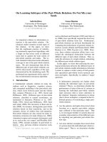
each side (Fig. 1). However, a search using the dali
server [21] indicates that NrdI is a structural outlier
within the Fld family. The closest structural neighbour
is Fld from Desulfovibrio desulfuricans [dsFld; Protein
Data Bank (PDB) code: 3F6R] [22], with a rmsd of
2.4 A
˚
for 113 alignable Ca atoms out of 117 in
baNrdI and 147 in dsFld. Very similar statistics are
obtained for a wide variety of Flds from diverse
organisms, whether short-chain or long-chain. How-
ever, B. anthracis NrdI is 30 residues shorter than a
typical short chain Fld and displays a more compact
fold. The truncations occur principally on the side of
baNrdI furthest from the flavin binding site. Helix a1
is shorter by five residues (seven versus 13), strand b2
by four residues (three versus seven) and strand b3by
four residues (five versus nine) (Fig. 1 and Table S1).
In addition, the loops between a1 and b2; a3 and b4;
and a4 and b5 are shortened compared to dsFld.
Analysis of 199 NrdI sequences extracted from the
Ribonucleotide Reductase Database (RNRdb) [7]
(Fig. S1) shows that NrdIs are fairly homogeneous in
length, with a median value of 141 and a SD of 13.
There is no division into short- and long-chain vari-
ants as there is for Fld. The minimum structural core,
with shortest loops, is apparently represented by the
Corynebacterium striatum sequence at 109 amino acids
(Fig. S1). Variations in length are essentially limited to
the termini and the loops between a1 and b2, between
b2 and b3, and the loop (residues 42–49) that interacts
with the flavin moiety of FMN. The first two variable
loops are distant from FMN. The variable length of
this ‘‘40s loop’’ is discussed below. By contrast,
beyond the FMN-binding loop, the NrdI structure is
extremely well conserved (Fig. S1), with no significant
insertions or deletions.
The electron density for the FMN cofactor is excel-
lent in both structures, and the high resolution of the
oxidized form allows confirmation of the protonation
40s loop
70s loop
β1
α1
α2
AB
α4
50s loop
90s loop
β1
α1
α2
α4
β2
β3
β4
β5
β2
β3
β4
β5
Fig. 1. (A) Overall structure of NrdI. The car-
toon is coloured as a rainbow from blue at
the N-terminus to red at the C-terminus to
emphasize the topology. For clarity, helix 1
is semi-transparent. The FMN cofactor is
shown in a stick representation. Lengths of
the secondary structure elements are given
in Table S1. (B) Structure of the most struc-
turally homologous standard flavodoxin, the
short chain protein from D. desulfuricans
(PDB ID 3F6R), for comparison. The repre-
sentation is as shown in (A). Prepared using
PYMOL.
R. Johansson et al. NrdI – an unusual flavodoxin
FEBS Journal 277 (2010) 4265–4277 ª 2010 The Authors Journal compilation ª 2010 FEBS 4267
state. In electron density maps calculated without
inclusion of explicit hydrogen atoms on the cofactor,
the difference electron density can clearly be seen at
2.5 r for many of the aliphatic hydrogen atoms of the
cofactor (Fig. 2A). By contrast, no hydrogen atoms
can be seen on N5, confirming the oxidized state of
FMN. The phosphate group of FMN is bound by the
N-terminus of a1 and the preceding P-loop. The flavin
moiety of FMN binds in a pocket formed by loops at
the C-terminal ends of b-strands 3 and 4 (loops 42–49
and 71–79, respectively), known in Fld as the W and
Y loops, or the 50s and 90s loops [23]. We refer to
these as the 40s and 70s loops, respectively, in baNrdI.
The flavin moiety is sandwiched between Trp74 on one
face and the side chain of Thr42 and the main chain
atoms of residues 42–44 on the other (Fig. 2A). The
isoalloxazine ring is completely buried and anchored
through seven hydrogen bonds between its carbonyl
(O2, O4) and amide (N3) groups and main-chain car-
bonyl and amide groups in the 40s and 70s loops. By
contrast, the dimethylbenzene ring is solvent-exposed.
The flavin binding pocket is capped by Phe45, whose
side chain lies perpendicular to the flavin moiety and
also makes an edge-on interaction to the stacking
Trp74.
The flavin environment in NrdI is considerably less
negatively charged than in Fld. For example, Clostrid-
ium beijerinckii Fld (cbFld) has a net charge of )14,
whereas in baNrdI it is only )4 (in the present study
the net protein charges always refer to the protein
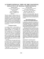
component only). Figure 2B shows the preponderance
of acidic side chains in the vicinity of the flavin in
cbFld, including two in the 50s loop itself. There are
three acidic residues within 6 A
˚
, and a further four
within 10 A
˚
. Overall, 26 negative charges are compen-
sated by only 12 positive charges. By contrast, in baNrdI,
18 negative charges are compensated for by 14
positive ones. The closest of these to the flavin, Asp76
in the 70s loop and Asp83 in helix a4, are 9 A
˚
distant
from the flavin (Fig. 2A). This has a remarkable effect
on the electrostatic energy landscape of NrdI com-
pared to Fld (Fig. 3). To test the generality of this
observation, we carried out an analysis of the length,
amino acid composition and calculated isoelectric
point of a representative set of 199 NrdI sequences
extracted from the RNRdb. A parallel analysis of 38
manually reviewed flavodoxin sequences from the
UniRef100 database () was
performed for comparison, confirming that NrdI
differs strongly from flavodoxins with respect to pI. The
median pI for NrdI sequences is 9.0 with a SD of 1.7
(Fig. 4A). However, the spread in values is wide,
ranging from 4.2 for Eubacterium biforme to 10.4 for
NrdI1 of S. pyogenes M1. Net charges vary remarkably,
from )15 to +15. The pI distribution is approximately
bimodal, with a major peak at pH 9.0–9.5 and a broader
peak at pH 5.0–5.5. By contrast, the 38 representative
flavodoxin sequences that have been analyzed are much
more homogeneous in pI: the median value is 4.5 with a
SD of only 0.6 (Fig. 4B).
70s loop
90s loop
40s loop
A
B
50s loop
W74
2.9
3.4
3.1
3.2
2.8
D83
E98
D105
F45
T43
α1
D76
G44
W90
D92
D58
D59
D57
M56
D98
E101
E120
E62
E63
E65
Fig. 2. (A) Electron density for the FMN cofactor and the 40s loop.
The grey mesh shows a r
A
-weighted 2|F
o
| ) |F
c
| map to 0.96 A
˚
cal-
culated using
SHELXL and contoured at 1.2 r around the FMN cofac-
tor and the 40s loop. Residues within 4 A
˚
of FMN that make
interactions with it are shown as thin lines. Particularly relevant
side chains, including all acidic side chains in the view, are shown
as sticks. Hydrogen bonds are shown as dashed lines. The green
mesh shows a similarly calculated F
o
) F
c
map to 1.1 A
˚
resolution
contoured at 2.5 r. For calculation of this map, hydrogen atoms
were included for the protein but omitted from the cofactor. (B)
The flavodoxin from C. beijerinckii in its oxidized state (PDB code:
4NLL) in the same orientation as baNrdI shown in (A). The repre-
sentation is identical to that shown in (A). The overall details of
FMN binding are very similar to baNrdI; however, note the prepon-
derance of acidic residues in the vicinity of the FMN binding site.
Prepared using
PYMOL.
NrdI – an unusual flavodoxin R. Johansson et al.
4268 FEBS Journal 277 (2010) 4265–4277 ª 2010 The Authors Journal compilation ª 2010 FEBS
Reduced baNrdI-FMN
The crystal structure of fully reduced baNrdI-FMN
was obtained by chemical reduction of crystals of oxi-
dized baNrdI using 500 mm sodium dithionite. The
largest conformational change in the protein is a pep-
tide flip between residues 44 and 45, resulting in the
orientation of the Gly44 carbonyl group towards N5,
which is protonated in the neutral sq radical form
(Fig. 5A). This peptide flip is accompanied by a slight
rearrangement of the whole loop from Thr42 to
Asn47. Interestingly, Thr42 undergoes a small shift,
and difference density appears at a level of approxi-
mately 4 r between its side chain and the flavin ring
(Fig. 5B). This density is also present at approximately
6 r in the maps of the chemically-reduced B. cereus
NrdI [20] as generated by the Electron Density Server
[24] ( although the authors
responsible for this entry did not interpret it. The
density is too close to Thr42 (1.7 A
˚
to Oc)tobea
water molecule, although it could be a loosely coordi-
nated metal ion. There is also a rotamer change in
Thr43 and a slight movement of Phe45 upon flavin
reduction. This confirms that the conformational
response of NrdI to reduction is very similar to that
observed in flavodoxins [18,25–27].
Flavin photoreduction as a result of X-ray
exposure
Røhr et al. [20] recently noted the need to take into
consideration the effects of radiation damage on the
geometry of flavin cofactors when analysing structures
where data were collected using synchrotron X-ray
sources. A significant distortion of the flavin was noted
in both NrdI
ox
and NrdI
sq
from B. cereus after esti-
mated radiation doses of 9 and 10 MGy, respectively.
Quantum mechanics simulations of the flavin geometry
in the protein context coupled to resonance Raman
experiments on the crystals suggested that both NrdI
ox
and NrdI
sq
had been reduced by one electron during
X-ray exposure, such that the flavins were now in the
FMN
°)
and FMNH
)
states respectively. With this in
mind, we investigated the effect of radiation damage in
the almost identical B. anthracis system. Using suitable
parameters for the I911-3 (NrdI
ox
) and I911-5 (NrdI
sq
)
beamlines at MAX-lab, Lund, Sweden), we arrived at a
dose estimate of approximately 2–4 MGy for baNrdI
ox
and 5–6 MGy for baNrdI
sq
, using the software rad-
dose [28]. In the last cycle of refinement, no restraints
were used in the refinement of the FMN geometry for
oxidized baNrdI. The ‘butterfly angle’ between the fla-
vin ring planes is 5.7°, which compares well with the
value of 4.8° reported for the oxidized B. cereus protein
after photoreduction, indicating that one-electron
reduction has also occurred in baNrdI. The resolution
of the sq form was not sufficiently high to allow unre-
strained refinement, and so a similar comparison is not
0
3.50–3.99
4.00–4.49
4.50–4.99
5.00–5.49
5.50–5.99
6.00–6.49
6.50–6.99
7.00–7.49
7.50–7.99
8.00–8.49
8.50–8.99
9.00–9.49
9.50–9.99
10.00–10.49
5
10
15
20
25
0
10
20
30
40
50
60
A
B
Fig. 4. Histograms of the distributions of predicted isoelectric point
for (A) NrdI and (B) Fld sequences. The sequences are colour-coded
according to the phylum of the source organisms. The pI groups to
which the NrdIs with determined structures belong are labelled:
Bant, B. anthracis; Bcer, B. cereus; Bsub, B. subtilis; Ecol, E. coli.
Produced using Google Docs ( />AB
Fig. 3. Electrostatic potentials for (A) baNrdI and (B) C. beijerinckii
flavodoxin. The potentials at the solvent accessible surface were
calculated using
APBS [53] and mapped onto the molecular surface
using
PYMOL. The colour scale in both panels runs from deep red at
)5 kTe
)1
to blue at +5 kTe
)1
. The FMN molecule is shown in a
space-filling representation. The molecular surface is semi-transpar-
ent and a grey cartoon of each molecule is shown for orientation.
The direction of view is into the side of the flavin plane. Prepared
using
PYMOL.
R. Johansson et al. NrdI – an unusual flavodoxin
FEBS Journal 277 (2010) 4265–4277 ª 2010 The Authors Journal compilation ª 2010 FEBS 4269
meaningful. However, distortion of the flavin geometry
as a result of accumulated photoreduction during data
collection can be seen in the anisotropic B-factors of
the flavin atoms in both oxidized and sq forms (Fig. 6).
With the isoallazine ring being fixed by its interactions
with the protein, the flavin distortion is concentrated
on the dimethylbenzene ring, which has greater free-
dom to distort from planar geometry.
Discussion
The crystal structures of NrdI from B. anthracis have
been solved in two functional states: in complexes with
oxidized and semiquinone FMN. The structures reveal
that NrdI is unambiguously a member of the Fld
superfamily, although it has the most compact fold of
a Fld seen to date, being shorter than the average
short-chain Fld. It can thus be considered to define a
new family within the Fld superfamily. The NrdI fam-
ily is not divided into short- and long-chain subfami-
lies: the region in and around the final strand b5,
where the insertion defining the long-chain Flds
occurs, is extremely highly conserved with regard to
secondary structure in NrdI sequences (Fig. S1).
Despite being an outlier in the Fld family, the FMN-
binding regions are more conserved than the rest of
the structure.
Correctly-folded baNrdI for functional and struc-
tural studies could only be obtained by including
FMN in the growth medium (in our case LB medium)
at a concentration of 60 lm, when overexpressed in
E. coli. In the absence of FMN NrdI was misfolded
and produced irreversibly in inclusion bodies. Without
FMN, NrdI from S. typhimurium, C. ammoniagenes,
B. anthracis and Deinococcus radiodurans also form
inclusion bodies during heterologous overexpression in
E. coli (E. Torrents, unpublished results). The observa-
tion is also in agreement with previously published
studies on E. coli NrdI, in which significant quantities
of functional protein could only be obtained by refold-
ing from inclusion bodies in the presence of FMN [11].
A general requirement for FMN in NrdI folding
would contrast with the behaviour of traditional Flds.
The dependence of Fld folding on FMN has been
studied, and the binding of FMN to native apo-Fld
was found to constitute the last step [29]. The autono-
mous formation of native apo-Fld is essential during
holo-Fld folding, and FMN does not act as a nucle-
ation site for folding. FMN can be removed from Fld
by acid treatment [30], despite affinity in the sub-
nanomolar range [29], also resulting in a stable apo-
protein. Conformational differences between apo- and
holo-Flds are small and confined to the 50s and 90s
loops [31,32]. Further experiments are required to
establish whether NrdI has a general requirement for
FMN for correct folding during overexpression.
NrdI is remarkably less negatively charged than
normal flavodoxins
The major function of the protein environment in
flavodoxins is modification of the redox potentials
W74
F45
G44
2.8
3.4
2.8
T43
T42
O2
FMN
AB
S69
3.2
2.1
2.4
Fig. 5. (A) Conformational change upon chemical reduction of baNrdI to the semiquinone state. The grey mesh shows a r
A
-weighted
2|F
o
| ) |F
c
| map to 1.4 A
˚
resolution calculated using REFMAC5 and contoured at 1.2 r around the FMN cofactor and the 40s loop. Reduced
baNrdI is shown in green and the 40s loop of the oxidized form is shown in blue for comparison. Hydrogen bonds between FMN and the
40s loop are shown as dashed lines. (B) The strong difference density that appears between the flavin and Thr42 in the crystal structure of
reduced baNrdI, which is also present in bcNrdI (Fig. S2). A 2|F
o
| ) F
c
| map contoured at 1.0 r and an |F
o
| ) |F
c
| map contoured at 3.0 r are
shown in grey and green, respectively. The height of the difference map peak is approximately 4 r. A marker atom has been placed in the
electron density to show the distances to potential coordinating atoms in the vicinity. Prepared using
PYMOL.
NrdI – an unusual flavodoxin R. Johansson et al.
4270 FEBS Journal 277 (2010) 4265–4277 ª 2010 The Authors Journal compilation ª 2010 FEBS
ox ⁄ sq and sq ⁄ hq couples from the rather similar values
of )172 mV and )238 mV, respectively, in the free fla-
vin in FMN [17] to the widely different values of )50
to 260 mV and )370 to )450 mV, respectively, for the
bound form [18]. Thereby, flavodoxins are made into
effective one-electron donors. The effects on redox
potentials occur primarily through (a) stabilization of
the sq form via a hydrogen bond between the N5 atom
and a carbonyl group in the 50s loop [18,25,26,33–36]
and (b) destabilization of the negatively-charged hq
form (FMNH
)
) through the lack of solvation in the
protein environment coupled to highly negative elec-
trostatic field, which reduces the protein’s association
rate with the hq form of FMN [18,36,37]. Replacement
of acidic residues by neutral or positively-charged ones
tends to increase the sq ⁄ hq potential [18,38,39],
although theoretical studies have shown that compen-
satory (de)protonation effects on other charged resi-
dues can make the effect difficult to predict [37]. By
contrast, modification of the conformational properties
of the 50s loop affects the ox ⁄ sq potential to a greater
degree [26,35,36].
The remarkably similar redox potentials of the
ox ⁄ sq and sq ⁄ hq couples in E. coli NrdI (ecNrdI) have
been attributed to a lesser destabilization of the sq
form, FMNH
)
, than what is normally the case in
flavodoxins [11]. The structure of baNrdI confirms this
hypothesis, and our bioinformatic analysis extends it,
with few exceptions, to the whole NrdI family. The
conformational changes observed between the oxidized
and reduced states are limited to a peptide flip between
residues 44 and 45 and a small rearrangement of the
40s loop. The high similarity of these changes to those
observed in Fld from several species strongly suggests
that the unusual redox potentials of NrdI are governed
more by protein electrostatics than by specific hydro-
gen bonds or other direct interactions with the flavin.
However, the wide spread in predicted pI values for
NrdI sequences is unexpected. The data point to the
interesting possibility of two distinct functional groups
with acidic and basic characters, respectively, which
obviously will have quite different effects on the poten-
tials of the FMN redox couples. Alternatively, the
charge variation in NrdI may be correlated with a sim-
ilar variation in the electrostatic properties of the
respective NrdF proteins, in particular at the interac-
tion area. The reason for the wide spread in predicted
pI is not obvious. Figure 4A shows the distribution
colour-coded by taxonomy. It can be seen that NrdIs
from the a- and c-proteobacteria belong almost exclu-
sively to the high-pI group, whereas the firmicutes and
actinobacteria occupy a wide range of pI values. Sev-
eral organisms in the latter two groups, including some
Bacillus species, encode more than one class Ib operon,
whereas the proteobacteria all encode only one. In
general, the NrdI pI values differ substantially within
organisms encoding two different nrdI genes. A phylo-
genetic tree of 91 representative NrdI sequences
(Fig. S3) shows that the NrdI phylogeny is not signifi-
cantly different from that based on NrdF sequences
[19] or on 16S rRNA, although actinobacteria and fir-
micutes, with two class Ib operons, generally have
NrdIs divided into two separate clusters. However, the
presence of a sequence in a genome provides no infor-
mation regarding if and when the protein is expressed,
and so further experiments are required to establish
the reason for the wide spread in electrostatic proper-
ties in an otherwise structurally conserved family.
E. coli NrdI, with pI = 9.4, belongs to the major
group of NrdI sequences with basic character, whereas
baNrdI, with pI = 5.4, belongs to the minor, acidic
group. These proteins also differ in their ability to sta-
bilize an FMN sq radical: a maximum of 28% can be
detected in ecNrdI, whereas, in baNrdI, the amount is
up to 60% (M. Sahlin & B M. Sjo
¨
berg, unpublished
results). Interestingly, this correlates with the predicted
pIs: that of baNrdI (net charge )4) is much closer to
A
B
Fig. 6. Depiction of the anisotropic movements of atoms in the
FMN molecule as represented by their anisotropic B-factors. (A)
NrdI
ox
, 0.96 A
˚
resolution, refined using SHELXL. (B) NrdI
sq
, 1.4 A
˚
,
refined using
REFMAC5. The anisotropic B-factors are represented by
thermal ellipsoids at 50% probability. The B-factors are coloured
from dark blue at 5.0 A
˚
2
to bright red at 12.8 A
˚
2
in (A) and from
7.7 A
˚
2
to 18.7 A
˚
2
using the same colour scheme in (B). Prepared
using
PYMOL.
R. Johansson et al. NrdI – an unusual flavodoxin
FEBS Journal 277 (2010) 4265–4277 ª 2010 The Authors Journal compilation ª 2010 FEBS 4271
that of a normal flavodoxin than that of ecNrdI (net
charge +4).
In E. coli NrdI, a neutral sq radical is produced when
NrdI is titrated anaerobically with dithionite [11]. In the
presence of NrdF, whether in the apo- or Mn-contain-
ing forms, an anionic sq radical is produced instead.
This behaviour was described as being potentially more
similar to that of flavoprotein oxidases than flavodoxins
[12]. However, the stabilization of an anionic sq form
would be favoured by the presence of a positively-
charged protein residue in the vicinity of the N1-C2-O2
atoms [40], as in glycolate oxidase [41]. No such residue
can be found in baNrdI and, indeed, the formation of
anionic sq should be disfavoured by the overall negative
charge of the protein. This points to influence of NrdF
on the flavin environment in the NrdF ⁄ I complex,
although the flavin N1-C2-O2 atoms would remain
inaccessible to residues from NrdF in the complex,
unless NrdF induces a conformational change in the 90s
loop to expose the flavin. We have identified the possi-
bility that a metal ion is trapped close to the N1-C2-O2
locus in chemically-reduced baNrdI and bcNrdI, in the
space made available by the conformational rearrange-
ment in the 40s loop induced by the peptide flip of
Gly44 (Fig. 5B). This may help to compensate the
negative charge in an anionic sq form.
The FMN-binding loop is a major site of
sequence variation in NrdI
NrdI sequences vary in length from 103 to 188 amino
acids. Intriguingly, one of the most variable loops in
NrdI is the one that interacts with the flavin ring of
FMN, namely the 40s loop. In the baNrdI structure, the
loop extends from Thr42 to Pro49 (i.e. eight residues).
At the other extreme, in mycobacteria, the loop is up to
15 residues longer, containing a high proportion of Pro
and Gly (Fig. S1). The role of this highly variable length
in a critical area of the structure is not clear. Short- and
long-chain flavodoxins differ in the presence or absence
of a 20 residues loop that splits the fifth b-strand [23],
although this is not the case in NrdI. The 50s loop is
slightly longer in long-chain Flds than in short-chain
ones, and it has been proposed that the inserted loop
stabilizes the longer 50s loop [42]. However, an extended
40s loop in NrdI does not appear to be correlated with
any other insertion. It might be hypothesized that the
extended loop contributes to increased affinity for
NrdI’s interaction partner NrdF because, although the
K
D
for the baNrdI ⁄ baNrdF complex is only 50 lm,
the affinity of the E. coli complex (with a loop longer
by four residues) is so high that interactions are
maintained during nickel–nitrilotriacetic acid affinity
chromatography [12]. The capping of the FMN bind-
ing site by Phe45 appears to be specific for Bacillus
and a few other species, and is correlated with the
exceptionally short loop found in baNrdI. This residue
is Gly in almost all other NrdI sequences. Thus, the
extended loop may close off the flavin binding site by
folding back over the core of the protein.
When the present paper was in revision, the crystal
structures of the complex between NrdI and NrdF
from E. coli in the oxidized and hq forms were pub-
lished [43]. This work confirmed our prediction that
the variable 40s loop will contribute to the different
affinities of NrdI for NrdF in various organisms. The
equivalent loop of NrdI in the E. coli complex forms
an important part of the molecular interface and
undergoes more significant conformational changes
upon reduction than in the Bacillus species, although
this may be influenced by its interactions with NrdF.
In summary, we have identified the NrdI protein as a
structural outlier within the flavodoxin family, having a
significantly more compact fold, although the structure
close to the flavin binding site is more conserved. The
40s loop is an important site of sequence variation in
the NrdI family. A very wide distribution in predicted
pI values has been identified, which may imply different
functional roles for NrdI in different organisms.
Materials and methods
Cloning of the nrdI gene
The B. anthracis nrdI was amplified by PCR from strain
Sterne 7700 pXO1
)
⁄ pXO2
)
genomic DNA as described
previously [19] using BanrdIup 5¢-A
CATATGTTAGTTG
CCTATGATTCTATG-3¢ and BanrdIlw 5¢-A
AAGCTTAT
TCAGTTCAATGTGTC-3¢, as a forward and reverse prim-
ers containing NdeI and HindIII restriction sites, respectively
(underlined). The PCR product was cloned in the pGEM-T
easy vector (Promega, Madison, WI, USA). After digestion
with NdeI and HindIII, the nrdI fragment (380 bp) was
ligated into pET22b generating plasmid pETS153.
Expression and purification
E. coli Rosetta(DE3) cells (Novagen, Madison, WI, USA)
containing pETS153 were grown in LB medium (Difco,
Franklin Lakes, NJ, USA) at 37 °C with 100 lgÆmL
)1
ampicillin, 17 lgÆmL
)1
chloramphenicol and 60 lm FMN
(Sigma, St Louis, MO, USA) until a A
550
of 0.5 was
reached, induced with 1 mm isopropyl thio-b-d-galactoside
for 4 h, collected by centrifugation, and disrupted in an
X-Press (BioX AB, Gothenburg, Sweden) in buffer 50 mm
Tris-HCl (pH 7.6), 30 mm KCl and protease inhibitors
NrdI – an unusual flavodoxin R. Johansson et al.
4272 FEBS Journal 277 (2010) 4265–4277 ª 2010 The Authors Journal compilation ª 2010 FEBS
(Roche Applied Science, Basel, Switzerland). All the pro-
tein purification steps were carried out at 4 °C. After high-
speed centrifugation, the protein concentration was
adjusted to 10 mgÆmL
)1
and the supernatant solution was
first precipitated with streptomycin sulfate (final concentra-
tion 1%) and, after a second centrifugation, with solid
ammonium sulfate to 45% saturation. After centrifugation,
the precipitate was dissolved in buffer A (50 mm Tris-HCl,
pH 7.6, and 30 mm KCl) and desalted by dialysis against
2 L of buffer A for 16 h. The dialyzed solution was diluted
with buffer A to 6 mgÆmL
)1
protein concentration and
loaded on HiLoad 16 ⁄ 10 Q-Sepharose High Performance
column (GE Healthcare, Milwaukee, WI, USA) on a Bio-
Logic DuoFlow System fast protein liquid chromatography
instrument (Bio-Rad, Hercules, CA, USA) previously equil-
ibrated with ten volumes of buffer A. NrdI protein was
eluted with a linear gradient of KCl (30–400 mm,
3mLÆmin
)1
) in buffer A. Fractions containing the NrdI
protein were pooled, concentrated using Centricon-10 (Mil-
lipore, Billerica, MA, USA) and loaded at 0.5 mLÆmin
)1
on a 24-ml Superdex-75 column equilibrated and eluted
with buffer 50 mm Tris-HCl (pH 7.6) and 200 mm KCl.
Each fraction was analyzed by PhastGel electrophoresis
(GE Healthcare), and fractions with the highest purity
(strong yellow colour) were pooled, concentrated using
Centricon-10 (Millipore), and finally freed from KCl by
washing with buffer A.
Crystallization and data collection
The protein solution used for crystallization was at
8mgÆmL
)1
in 50 mm Tris-HCl (pH 7.6). Screening for ini-
tial crystallization conditions was performed at the crystalli-
zation facility at MAX-lab. The PACT Premier and
JCSG+ screens (Molecular Dimensions Ltd, Newmarket,
UK) were carried out at 20 °C in 100 + 100 nL drops in
Greiner low-profile 96-well plates (Greiner Bio-One GmbH,
Frickenhausen, Germany). A hit in condition 55 (E7) of the
JCSG+ screen was refined using manual setups. The crys-
tal used for data collection on the oxidized form was grown
from a drop consisting of 1 lL of protein solution and
1 lL of a reservoir solution consisting of 10% (v ⁄ v) 2-pro-
panol, 0.2 m Zn acetate, 0.1 m Na cacodylate buffer (pH
6.5). Crystals appeared after one day and reached full size
after approximately 1 week. The crystal used for data col-
lection was approximately 0.2 · 0.1 · 0.1 mm in size. Crys-
tals were taken directly from the drop and flash-cooled in
the gas stream from an Oxford Diffraction CryoJet (Oxford
Diffraction Ltd, Oxford, UK). Data on the oxidized form
were collected to 0.96 A
˚
resolution at station I911-3 of the
MAX-II synchrotron (MAX-lab), using a 225 mm marMo-
saic CCD detector (Rayonix LLC, Evanston, IL, USA).
The X-ray wavelength was 0.7300 A
˚
. Data were collected,
in two passes, to 1.9 and 0.96 A
˚
, respectively. The crystal
belonged to space group P2
1
2
1
2
1
with cell dimensions as
shown in Table 1. For determination of the structure of
reduced NrdI, a crystal was soaked in a solution consisting
of 500 mm sodium dithionite in crystallization mother
liquor for 10 min before flash cooling. During this time, the
crystal colour changed from yellow to dark blue. Data were
collected at station I911-5 of the MAX-II synchrotron, with
an X-ray wavelength of 0.9789 A
˚
, in two passes, to 1.8 and
1.4 A
˚
, respectively. Diffraction data were integrated using
xds and scaled using xscale [44]. Data were further pro-
cessed using software from the ccp4 suite [45]. The crystals
contain one NrdI molecule in the asymmetric unit, resulting
in a Matthews volume of 2.05 A
˚
3
ÆDa
)1
and a solvent con-
tent of 40.0%.
Structure solution and refinement
The structure of oxidized baNrdI was solved by molecular
replacement (MR) using the unpublished coordinates of
Table 1. Data and structure quality statistics. Figures in parenthe-
ses refer to the highest resolution bin.
Oxidized Reduced
Unit cell dimensions (A
˚
) a = 42.80,
b = 45.62,
c = 56.33
a = 42.83,
b = 45.26,
c = 55.66
Data collection
X-ray wavelength (A
˚
) 0.7300 0.9077
Resolution range (A
˚
) 23.9–0.96
(0.98–0.96)
23.7–1.4
(1.44–1.4)
Completeness (%) 97.3 (77.8) 99.3 (98.7)
R
merge
(%) 3.9 (60.1) 9.3 (74.4)
<I> ⁄ <r(I)> 17.1 (2.0) 9.1 (1.5)
Number of observations 283 726 102 202
Number of unique reflections 65 490 21 820
Wilson B-factor (A
˚
2
) 11.1 21.5
Refinement
Resolution range (A
˚
) 25–0.96
(1.0–0.96)
26.7–1.4
(1.46–1.40)
R
model
(%) 12.5 (25.9) 14.7 (23.8)
R
free
(%) 15.2 (–) 19.2 (32.5)
Test set size, % (n) 3.0 (1965) 5.0 (1106)
Number of protein residues 118 118
Number of water molecules 185 120
Other small molecules 1 · cacodylate,
1 · Zn
1 · cacodylate,
3 · Zn
Mean isotropic B-factor (A
˚
2
) 13.4 (protein) 14.3 (protein),
7.3 (FMN) 10.5 (FMN)
34.2 (water) 39.2 (water)
Rmsd from ideal geometry
Bond length (A
˚
) 0.016 0.024
Bond angles 0.035 A
˚
a
1.99°
Ramachandran plot quality
Most favoured (%) 99.2 99.2
Additional allowed (%) 0.8 0.8
a
From DANG restraints in SHELXL.
R. Johansson et al. NrdI – an unusual flavodoxin
FEBS Journal 277 (2010) 4265–4277 ª 2010 The Authors Journal compilation ª 2010 FEBS 4273
NrdI from Bacillus subtilis (PDB code: 1RLJ), which has
48% sequence identity to baNrdI. The automated MR pipe-
line mrbump [46] was used. The best search model was gen-
erated by truncation of nonconserved side chains using
molrep from the ccp4 suite and the solution was found
using molrep as the MR search engine. The MR solution
had an R-factor of 36.0% and a free R-factor of 37.3% (cal-
culated using 3% of the data). The model was rebuilt by
iterated rounds of model building in coot [47] and refine-
ment in refmac5 [48]. The high-resolution limit for refine-
ment was increased gradually from 2.0 A
˚
to 1.1 A
˚
.
Anisotropic B-factors were introduced at 1.5 A
˚
. After con-
vergence of refinement in refmac5, R
model
and R
free
were
15.6% and 18.1%, respectively. At this point, the refinement
software was switched to shelxl [49]. Restraints for FMN
were generated from coordinates in the HIC-UP database
[50] using the prodrg server [51] To ensure convergence,
the resolution was reduced to 1.8 A
˚
and gradually increased
to 0.96 A
˚
. Riding hydrogen atoms were used throughout.
Anisotropic B-factors were introduced at 1.6 A
˚
resolution.
After shelxl refinement, R
model
and R
free
were 13.5% and
16.3%, respectively. Water molecules were introduced in
peaks over 4.0 r in the difference map fulfilling reasonable
distance and hydrogen bonding criteria to protein residues or
other water molecules. Refined water molecules were removed
if they had excessively high B-factors or electron density in
2F
o
) F
c
maps under 1.0 r.
The structure of reduced baNrdI was solved by direct
refinement of the oxidized structure against the dataset
from a reduced crystal. After the first round of refinement,
strong difference electron density was observed, indicating
a flip in the peptide bond between residues 44 and 45. The
solvent structure was modelled according to the same crite-
ria as for the oxidized protein and the coordinates were
refined using refmac5 [48] and phenix.refine [52].
Electrostatic potential calculations
Electrostatic potentials were calculated using the apbs plu-
gin [53] to pymol () using default
parameters throughout.
Sequence analysis
A set of 199 unique NrdI sequences was extracted from the
RNRdb database: sequences annotated as containing only
an NrdI fragment were immediately discarded. Sequences
from different strains of the same organism were then
removed to decrease redundancy. In cases where different
strains contained either one or two NrdI sequences, the
strain containing two sequences was retained. The remaining
sequences were aligned using clc sequence viewer 6.3
(CLC Bio, A
˚
rhus, Denmark; ). Four
sequences that were not marked as fragments in the data-
base, but which were evidently too short because they
lacked the first a-helix and b-strand, were then removed.
Isoelectric points, sequence lengths and amino acid compo-
sitions were calculated and tabulated using clc sequence
viewer 6.3 and overall statistics calculated using Microsoft
Excel (Microsoft Corp., Redmond, CA, USA). For compar-
ison, an analysis of 38 flavodoxin sequences extracted from
the UniProt100 database () was also
carried out.
Phylogenetic reconstruction
From the 277 unique NrdI sequences in the RNRdb [7], 91
representative sequences were chosen and aligned using
probcons, version 1.10 [54]. The sequences were chosen to
represent the full diversity of NrdI sequences except for
highly divergent sequences. A maximum likelihood tree was
estimated from 100 well-aligned positions using phyml, ver-
sion 3.0, the LG substitution model and four gamma cate-
gories [55]. Branch confidence was calculated using the
SH-like algorithm [56].
Estimation of isoelectric points
pI values of protein sequences were estimated using the
pI ⁄ Mw tool at the expasy web server (asy.
ch/tools/pi_tool.html).
Acknowledgements
This work was supported by grants from the Swedish
Research Council to B.M.S. and D.L. E.T. was
supported by grants from the Spanish Ministerio de
Ciencia e Innovacio
´
n (PI081062), the CONSOLIDER
(CSD2008-00013) and ERANET Pathogenomics.We
wish to thank Maria Ha
˚
kansson for help at the MAX-
lab crystallization facility and the staff at beamline
I911 at MAX-lab for assistance with data collection.
We thank Ilya Borovok for stimulating discussions.
References
1 Eklund H, Uhlin U, Fa
¨
rnega
˚
rdh M, Logan DT &
Nordlund P (2001) Structure and function of the radical
enzyme ribonucleotide reductase. Prog Biophys Mol Biol
77, 177–268.
2 Nordlund P & Reichard P (2006) Ribonucleotide reduc-
tases. Annu Rev Biochem 75, 681–706.
3 Torrents E, Sahlin M & Sjo
¨
berg BM (2008) The ribonu-
ceotide reductase family: genetics and genomics. In
Ribonucleotide Reductase (Andersson KK ed), pp.
17–77. Nova Science Publishers, New York.
4 Jordan A, Pontis E, Aslund F, Hellman U, Gibert I &
Reichard P (1996) The ribonucleotide reductase system
of Lactococcus lactis. Characterization of an NrdEF
NrdI – an unusual flavodoxin R. Johansson et al.
4274 FEBS Journal 277 (2010) 4265–4277 ª 2010 The Authors Journal compilation ª 2010 FEBS
enzyme and a new electron transport protein. J Biol
Chem 271, 8779–8785.
5 Eliasson R, Pontis E, Jordan A & Reichard P (1996)
Allosteric regulation of the third ribonucleotide reduc-
tase (NrdEF enzyme) from enterobacteriaceae. J Biol
Chem 271, 26582–26587.
6 Eriksson M, Jordan A & Eklund H (1998) Structure of
Salmonella typhimurium nrdF ribonucleotide reductase
in its oxidized and reduced forms. Biochemistry 37,
13359–13369.
7 Lundin D, Torrents E, Poole AM & Sjoberg BM (2009)
RNRdb, a curated database of the universal enzyme
family ribonucleotide reductase, reveals a high level of
misannotation in sequences deposited to Genbank.
BMC Genomics 10 , 589.
8 Willing A, Follmann H & Auling G (1988) Ribonucleo-
tide reductase of Brevibacterium ammoniagenes is a
manganese enzyme. Eur J Biochem 170, 603–611.
9 Fieschi F, Torrents E, Toulokhonova L, Jordan A,
Hellman U, Barbe J, Gibert I, Karlsson M & Sjo
¨
berg
BM (1998) The manganese-containing ribonucleotide
reductase of Corynebacterium ammoniagenes is a class
Ib enzyme. J Biol Chem 273, 4329–4337.
10 Huque Y, Fieschi F, Torrents E, Gibert I, Eliasson R,
Reichard P, Sahlin M & Sjo
¨
berg BM (2000) The active
form of the R2F protein of class Ib ribonucleotide
reductase from Corynebacterium ammoniagenes is a dif-
erric protein. J Biol Chem 275, 25365–25371.
11 Cotruvo JA Jr & Stubbe J (2008) NrdI, a flavodoxin
involved in maintenance of the diferric-tyrosyl radical
cofactor in Escherichia coli class Ib ribonucleotide
reductase. Proc Natl Acad Sci USA 105, 14383–14388.
12 Cotruvo JA Jr & Stubbe J (2010) An active dimanga-
nese(III)-tyrosyl radical cofactor in Escherichia coli class
Ib ribonucleotide reductase. Biochemistry 49, 1297–
1309.
13 Jordan A, Aragall E, Gibert I & Barbe J (1996)
Promoter identification and expression analysis of
Salmonella typhimurium and Escherichia coli nrdEF
operons encoding one of two class I ribonucleotide
reductases present in both bacteria. Mol Microbiol 19,
777–790.
14 Jordan A, A
˚
slund F, Pontis E, Reichard P & Holmgren
A (1997) Characterization of Escherichia coli NrdH.
A glutaredoxin-like protein with a thioredoxin-like
activity profile. J Biol Chem 272, 18044–18050.
15 Roca I, Torrents E, Sahlin M, Gibert I & Sjoberg BM
(2008) NrdI essentiality for class Ib ribonucleotide
reduction in Streptococcus pyogenes. J Bacteriol 190,
4849–4858.
16 Stubbe J & van Der Donk WA (1998) Protein radicals
in enzyme catalysis.
Chem Rev 98, 705–762.
17 Draper RD & Ingraham LL (1968) A potentiometric
study of the flavin semiquinone equilibrium. Arch Bio-
chem Biophys 125, 802–808.
18 Hoover DM, Drennan CL, Metzger AL, Osborne C,
Weber CH, Pattridge KA & Ludwig ML (1999)
Comparisons of wild-type and mutant flavodoxins from
Anacystis nidulans. Structural determinants of the redox
potentials. J Mol Biol 294, 725–743.
19 Torrents E, Sahlin M, Biglino D, Gra
¨
slund A &
Sjo
¨
berg BM (2005) Efficient growth inhibition of
Bacillus anthracis by knocking out the ribonucleotide
reductase tyrosyl radical. Proc Natl Acad Sci USA 102,
17946–17951.
20 Røhr AK, Hersleth HP & Andersson KK (2010) Track-
ing flavin conformations in protein crystal structures
with Raman spectroscopy and QM ⁄ MM calculations.
Angew Chem Int Ed Engl 49, 2324–2327.
21 Holm L & Sander C (1995) Dali: a network tool for
protein structure comparison. Trends Biochem Sci 20,
478–480.
22 Guelker M, Stagg L, Wittung-Stafshede P & Shamoo Y
(2009) Pseudosymmetry, high copy number and twin-
ning complicate the structure determination of Desulf-
ovibrio desulfuricans (ATCC 29577) flavodoxin. Acta
Crystallogr D Biol Crystallogr 65, 523–534.
23 Sancho J (2006) Flavodoxins: sequence, folding, bind-
ing, function and beyond. Cell Mol Life Sci 63, 855–
864.
24 Kleywegt GJ, Harris MR, Zou JY, Taylor TC, Wahlby
A & Jones TA (2004) The Uppsala Electron-Density
Server. Acta Crystallogr D Biol Crystallogr 60, 2240–
2249.
25 Smith WW, Burnett RM, Darling GD & Ludwig ML
(1977) Structure of the semiquinone form of flavodoxin
from Clostridium MP. Extension of 1.8 A resolution
and some comparisons with the oxidized state. J Mol
Biol 117, 195–225.
26 O’Farrell PA, Walsh MA, McCarthy AA, Higgins TM,
Voordouw G & Mayhew SG (1998) Modulation of the
redox potentials of FMN in Desulfovibrio vulgaris flavo-
doxin: thermodynamic properties and crystal structures
of glycine-61 mutants. Biochemistry 37 , 8405–8416.
27 McCarthy AA, Walsh MA, Verma CS, O’Connell DP,
Reinhold M, Yalloway GN, D’Arcy D, Higgins TM,
Voordouw G & Mayhew SG (2002) Crystallographic
investigation of the role of aspartate 95 in the modula-
tion of the redox potentials of Desulfovibrio vulgaris
flavodoxin. Biochemistry 41 , 10950–10962.
28 Paithankar KS & Garman EF (2010) Know your dose:
RADDOSE. Acta Crystallogr D Biol Crystallogr 66,
381–388.
29 Bollen YJ, Nabuurs SM, van Berkel WJ & van Mierlo
CP (2005) Last in, first out: the role of cofactor binding
in flavodoxin folding. J Biol Chem 280, 7836–7844.
30 Wassink JH & Mayhew SG (1975) Fluorescence titra-
tion with apoflavodoxin: a sensitive assay for riboflavin
5¢-phosphate and flavin adenine dinucleotide in mix-
tures. Anal Biochem 68, 609–616.
R. Johansson et al. NrdI – an unusual flavodoxin
FEBS Journal 277 (2010) 4265–4277 ª 2010 The Authors Journal compilation ª 2010 FEBS 4275
31 Genzor CG, Perales-Alcon A, Sancho J & Romero A
(1996) Closure of a tyrosine ⁄ tryptophan aromatic gate
leads to a compact fold in apo flavodoxin. Nat Struct
Biol 3, 329–332.
32 Martinez-Julvez M, Cremades N, Bueno M, Perez-Dor-
ado I, Maya C, Cuesta-Lopez S, Prada D, Falo F,
Hermoso JA & Sancho J (2007) Common conforma-
tional changes in flavodoxins induced by FMN and
anion binding: the structure of Helicobacter pylori
apoflavodoxin. Proteins 69, 581–594.
33 Watt W, Tulinsky A, Swenson RP & Watenpaugh KD
(1991) Comparison of the crystal structures of a flavo-
doxin in its three oxidation states at cryogenic tempera-
tures. J Mol Biol 218, 195–208.
34 Romero A, Caldeira J, Legall J, Moura I, Moura JJ &
Romao MJ (1996) Crystal structure of flavodoxin from
Desulfovibrio desulfuricans ATCC 27774 in two oxida-
tion states. Eur J Biochem 239, 190–196.
35 Ludwig ML, Pattridge KA, Metzger AL, Dixon MM,
Eren M, Feng Y & Swenson RP (1997) Control of oxi-
dation-reduction potentials in flavodoxin from Clostrid-
ium beijerinckii: the role of conformation changes.
Biochemistry 36, 1259–1280.
36 Ishikita H (2007) Influence of the protein environment
on the redox potentials of flavodoxins from Clostridium
beijerinckii. J Biol Chem 282, 25240–25246.
37 Ishikita H (2008) Redox potential difference between
Desulfovibrio vulgaris and Clostridium beijerinckii flavo-
doxins. Biochemistry 47, 4394–4402.
38 Zhou Z & Swenson RP (1995) Electrostatic effects of
surface acidic amino acid residues on the oxidation-
reduction potentials of the flavodoxin from Desulfovib-
rio vulgaris (Hildenborough). Biochemistry 34, 3183–
3192.
39 Goni G, Herguedas B, Hervas M, Peregrina JR, De la
Rosa MA, Gomez-Moreno C, Navarro JA, Hermoso
JA, Martinez-Julvez M & Medina M (2009) Flavodox-
in: a compromise between efficiency and versatility in
the electron transfer from photosystem I to ferredoxin-
NADP(+) reductase. Biochim Biophys Acta 1787, 144–
154.
40 Massey V (1994) Activation of molecular oxygen by
flavins and flavoproteins. J Biol Chem 269, 22459–
22462.
41 Lindqvist Y & Bra
¨
nde
´
n CI (1989) The active site of
spinach glycolate oxidase. J Biol Chem 264, 3624–3628.
42 Lo
´
pez-Llano J, Maldonado S, Bueno M, Lostao A,
A
´
ngeles-Jime
´
nez M, Lillo MP & Sancho J (2004) The
long and short flavodoxins: I. The role of the differenti-
ating loop in apoflavodoxin structure and FMN bind-
ing. J Biol Chem 279, 47177–47183.
43 Boal AK, Cotruvo JA Jr, Stubbe J & Rosenzweig
AC(2010) Structural Basis for Activation of Class Ib
Ribonucleotide Reductase. Science doi:10.1126/
science.1190187.
44 Kabsch W (2010) XDS. Acta Crystallogr D Biol Crys-
tallogr 66, 125–132.
45 Collaborative Computational Project n (1994) The
CCP4 suite: programs for protein crystallography. Acta
Crystallogr D Biol Crystallogr 50, 760–763.
46 Keegan RM & Winn MD (2007) Automated search-
model discovery and preparation for structure solution
by molecular replacement. Acta Crystallogr D Biol
Crystallogr 63, 447–457.
47 Emsley P, Lohkamp B, Scott WG & Cowtan K (2010)
Features and development of Coot. Acta Crystallogr 66,
486–501.
48 Murshudov GN, Vagin AA & Dodson EE (1997)
Refinement of macromolecular structures by the maxi-
mum-likelihood method. Acta Crystallogr D Biol Crys-
tallogr 53, 240–255.
49 Sheldrick GM (2008) A short history of SHELX. Acta
Crystallogr A 64, 112–122.
50 Kleywegt GJ & Jones A (1998) Databases in protein
crystallography. Acta Crystallogr D Biol Crystallogr 54,
1119–1131.
51 van Aalten DM, Bywater R, Findlay JB, Hendlich M,
Hooft RW & Vriend G (1996) PRODRG, a program
for generating molecular topologies and unique molecu-
lar descriptors from coordinates of small molecules.
J Comput Aided Mol Des 10, 255–262.
52 Adams PD, Afonine PV, Bunkoczi G, Chen VB, Davis
IW, Echols N, Headd JJ, Hung LW, Kapral GJ,
Grosse-Kunstleve RW et al. (2010) PHENIX: a com-
prehensive Python-based system for macromolecular
structure solution. Acta Crystallogr D Biol Crystallogr
66, 213–221.
53 Baker NA, Sept D, Joseph S, Holst MJ & McCammon
JA (2001) Electrostatics of nanosystems: application to
microtubules and the ribosome. Proc Natl Acad Sci
USA 98, 10037–10041.
54 Do CB, Mahabhashyam MS, Brudno M & Batzoglou S
(2005) ProbCons: Probabilistic consistency-based multi-
ple sequence alignment. Genome Res 15, 330–340.
55 Guindon S & Gascuel O (2003) A simple, fast, and
accurate algorithm to estimate large phylogenies by
maximum likelihood. Syst Biol 52, 696–704.
56 Anisimova M & Gascuel O (2006) Approximate likeli-
hood-ratio test for branches: a fast, accurate, and pow-
erful alternative. Syst Biol 55, 539–552.
Supporting information
The following supplementary material is available:
Fig. S1. Multiple sequence alignment of 199 NrdI
sequences extracted from the RNRdb.
Fig. S2. Electron density revealing a possible metal ion
in the crystal structure of the reduced form of B. cer-
eus NrdI at 1.15 A
˚
resolution.
NrdI – an unusual flavodoxin R. Johansson et al.
4276 FEBS Journal 277 (2010) 4265–4277 ª 2010 The Authors Journal compilation ª 2010 FEBS
Fig. S3. Maximum likelihood phylogeny of a represen-
tative choice of 91 NrdI proteins.
Table S1. Lengths of secondary structure elements in
baNrdI compared to the closest structural neighbour,
the flavodoxin from Anacystis nidulans (PDB code:
3F6R).
This supplementary material can be found in the
online version of this article.
Please note: As a service to our authors and readers,
this journal provides supporting information supplied
by the authors. Such materials are peer-reviewed and
may be re-organized for online delivery, but are not
copy-edited or typeset. Technical support issues arising
from supporting information (other than missing files)
should be addressed to the authors.
R. Johansson et al. NrdI – an unusual flavodoxin
FEBS Journal 277 (2010) 4265–4277 ª 2010 The Authors Journal compilation ª 2010 FEBS 4277