Gastric Carcinoma- New Insights into Current Management pdf
Bạn đang xem bản rút gọn của tài liệu. Xem và tải ngay bản đầy đủ của tài liệu tại đây (30.6 MB, 304 trang )
GASTRIC CARCINOMA-
NEW INSIGHTS INTO
CURRENT MANAGEMENT
Edited by Daniela Lazăr
Gastric Carcinoma- New Insights into Current Management
/>Edited by Daniela Lazăr
Contributors
Ekambaram Ganapathy, Devaraja Rajasekaran, Asokan Devarajan, Muhammad Farooq Shukkur, Muhammed Farooq
Abdul Shukkur, Sakthisekaran Dhanapal, Murugan Sivalingam, Eriko Maeda, Kuo-Wang Tsai, Chung-Man Leung,
Hung-Wei Pan, Takehiro Okabayashi, Yasuo Shima, Okan Akturk, Jolanta Czyzewska, Daniela Lazar, Elvira Garza
Gonzalez, Guillermo Perez Perez, Shinya Shimada, Masafumi Kuramoto, Jae Y. Ro, Sun - Mi Lee, Kyoung-Mee Kim
Published by InTech
Janeza Trdine 9, 51000 Rijeka, Croatia
Copyright © 2013 InTech
All chapters are Open Access distributed under the Creative Commons Attribution 3.0 license, which allows users to
download, copy and build upon published articles even for commercial purposes, as long as the author and publisher
are properly credited, which ensures maximum dissemination and a wider impact of our publications. After this work
has been published by InTech, authors have the right to republish it, in whole or part, in any publication of which they
are the author, and to make other personal use of the work. Any republication, referencing or personal use of the
work must explicitly identify the original source.
Notice
Statements and opinions expressed in the chapters are these of the individual contributors and not necessarily those
of the editors or publisher. No responsibility is accepted for the accuracy of information contained in the published
chapters. The publisher assumes no responsibility for any damage or injury to persons or property arising out of the
use of any materials, instructions, methods or ideas contained in the book.
Publishing Process Manager Danijela Duric
Technical Editor InTech DTP team
Cover InTech Design team
First published February, 2013
Printed in Croatia
A free online edition of this book is available at www.intechopen.com
Additional hard copies can be obtained from
Gastric Carcinoma- New Insights into Current Management, Edited by Daniela Lazăr
p. cm.
ISBN 978-953-51-0914-3
free online editions of InTech
Books and Journals can be found at
www.intechopen.com
Contents
Preface VII
Section 1 Preneoplastic Lesions and Early Gastric Cancer 1
Chapter 1 The Role of Endoscopy and Biopsy in Evaluating Preneoplastic
and Particular Gastric Lesions 3
Daniela Lazăr, Sorina Tăban and Sorin Ursoniu
Chapter 2 Management of Early Gastric Cancer 41
Takehiro Okabayashi and Yasuo Shima
Section 2 Risk and Protective Factors 53
Chapter 3 Risk Factors in Gastric Cancer 55
Jolanta Czyzewska
Chapter 4 Relevance of Host Factors in Gastric Cancer Associated with
Helicobacter Pylori 75
Elvira Garza-González and Guillermo Ignacio Pérez-Pérez
Chapter 5 Fetal-type Glycogen Phosphorylase (FGP)Expression in
Intestinal Metaplasia as a High Risk Factor of the Development
of Gastric Carcinoma 93
Masafumi Kuramoto, Shinya Shimada, Satoshi Ikeshima, Kenichiro
Yamamoto, Toshiro Masuda, Tatsunori Miyata, Shinichi
Yoshimatsu, Masayuki Urata and Hideo Baba
Chapter 6 Naringenin Inhibits Oxidative Stress Induced Macromolecular
Damage in N-methyl N-nitro N-nitrosoguanidine Induced
Gastric Carcinogenesis in Wistar Rats 111
Ekambaram Ganapathy, Devaraja Rajasekaran, Murugan
Sivalingam, Muhammed Farooq Shukkur, Ebrahim Abdul Shukkur
and Sakthisekaran Dhanapal
Section 3 Morphological and Molecular Aspects 127
Chapter 7 Gastric Carcinoma: Morphologic Classifications and
Molecular Changes 129
Sun-Mi Lee, Kyoung-Mee Kim and Jae Y. Ro
Chapter 8 Variants of Gastric Carcinoma: Morphologic and Theranostic
Importance 177
Sun-Mi Lee, Kyoung-Mee Kim and Jae Y. Ro
Chapter 9 DNA Methylation in Aggressive Gastric Carcinoma 223
Chung-Man Leung, Kuo-Wang Tsai and Hung-Wei Pan
Section 4 Diagnostic Tools, Prognosis and Management 243
Chapter 10 Imaging Findings of Gastric Carcinoma 245
Eriko Maeda, Masaaki Akahane, Kuni Ohtomo, Keisuke Matsuzaka
and Masashi Fukayama
Chapter 11 Prognosis in the Cancer of the Stomach 259
Okan Akturk and Cemal Ulusoy
Chapter 12 Gastric Carcinoma: A Review on Epidemiology, Current Surgical
and Chemotherapeutic Options 271
Rokkappanavar K. Kumar, Sajjan S. Raj, Esaki M. Shankar, E.
Ganapathy, Abdul S. Ebrahim and Shukkur M. Farooq
ContentsVI
Preface
Although gastric cancer was in the past the second most common cancer in the world, its
incidence has dropped to fourth place, after cancers of the lung, breast, and colon and
rectum. In most developed countries, rates of stomach cancer have shown a dramatically
decline over the past half century. Nevertheless, gastric cancer is still the second most
common cause of cancer-related death in the world, causing about 800,000 deaths
worldwide per year.
Gastric neoplasm is often either asymptomatic or it may cause only nonspecific symptoms
in its early stages; by the time symptoms occur, the tumor has often reached a locally
advanced stage or may have also metastasized, which is one of the main reasons for the
delayed diagnosis and relatively poor prognosis of this cancer.
Gastric cancer may often be multifactorial, involving both genetic predisposition (e.g
hereditary non-polyposis colorectal cancer, familial adenomatous polyposis, hereditary
diffuse gastric cancer and Peutz–Jeghers syndrome) and environmental factors, such as
Helicobacter pylori infection.
Multidisciplinary treatment approach is compulsory for stomach cancer, including
surgeons, gastroenterologists, medical and radiation oncologists, radiologists and
pathologists. Surgical resection represents the only modality that is potentially curative. In
the last years, in the case of early gastric cancer, endoscopic resection may replace the
surgical procedure.
Literature data have shown that the 5-year survival rate for curative surgical resection
ranges from 30-50% for patients with stage II disease and from 10-25% for patients with
stage III disease. Because these patients have a high likelihood of local and systemic relapse,
the treatment is completed by adjuvant chemotherapy.
Many trials have demonstrated a survival benefit for adjuvant chemotherapy or
chemoradiotherapy in patients with stage II/III gastric cancer. Patients with inoperable,
locally advanced gastric cancer should be treated with palliative chemotherapy; afterwards,
they may be reassessed for surgery if a good response is achieved. Patients with metastatic
disease should be considered for palliative chemotherapy, which improves survival. Recent
data have shown the benefit of adding targeted therapy to the chemotherapy schemes on
the survival of selected gastric cancer patients (e.g the addition of trastuzumab to
chemotherapy in patients with HER2-positive gastric cancer).
This book contains a comprehensive overview of most recent data concerning a multitude of
facets of the gastric cancer. The book highlights various aspects of gastric neoplasm, from
the epidemiology, preneoplastic lesions, the complex process of carcinogenesis, the risk and
protective factors, morphological and molecular changes, up to the modern diagnostic tools
and current management of early and advanced gastric cancer, revealing the valuable
contribution of the multidisciplinary treatment approach.
This publication is appropriate for students, clinicians and researchers in the field of
gastroenterology, oncology, pathology, immunology, genetics, molecular biology,
radiology, and many other specialties. They will find interesting data and hot topics in this
book, from fundamental research knowledge to clinical issues that may be helpful in daily
practice.
This book, written in an easy-to-read style, makes an insight into the diagnosis and
assessment of premalignant gastric lesions and into the current management of gastric
cancer in its early stages. The authors focus on novel risk factors in gastric carcinogenesis,
such as fetal-type glycogen phosphorylase expression in intestinal metaplasia and also new
discovered protective factors such as naringenin that inhibits oxidative stress induced
macromolecular damage in a model of gastric carcinogenesis in rats. Also, one of the topics
refers to the importance of the host factors in gastric neoplasm associated with H. pylori
infection. Furthermore, this publication is presenting the role of endoscopic and histological
assessment in order to obtain a proper diagnostic of the premalignant gastric lesions.
“Gastric cancer-new insights into current management” provides a detailed description of
the morphologic classification and of the molecular changes encountered in the case of this
tumor. Moreover, it depicts some rare histological types of gastric cancers that may help
young scientists recognize them. An important topic refers to the epigenetic gene regulation
mechanisms and the biological behavior of the tumors, focusing on DNA methylation
aspects in aggressive gastric neoplasm. The identification of the molecular mechanisms of
gastric carcinogenesis and its progression using recent advances in genomic science allows
finding markers for early detection of stomach cancer, and can provide better information
on tumor aggressiveness, prognosis and prediction of response to cancer therapy.
The publication reveals some distinct and particular imaging findings that may accompany
different histological types of gastric tumors and also, the most important factors
contributing to the prognosis of gastric cancer.
There are no screening tests available for diagnosis of gastric cancer, therefore patients
usually presents in late stages. The preoperative evaluation stratifies patients in those with
loco-regional, potentially resectable disease, and those with systemic involvement. The book
describes the role of different diagnostic tools in the preoperative assessment of patients
with gastric carcinoma. Currently, early gastric cancer is treated with endoscopic resection,
gastrectomy, antibiotic therapy for H.pylori infection and adjuvant treatment. Surgical
resection remains the curative treatment for local and locoregional cancer. The authors
highlights the fact that adjuvant chemoradiotherapy is an essential part of the treatment
schedule as 80% of the cases develop local recurrence.
“Gastric cancer-new insights into current management” represents an important tool for
clinicians in the process of continuing medical education by updating with novel
information offered by a valuable team of well-known scientist who belong to different
specialties. Moreover, it may open new and interesting gates for further research concerning
carcinogenesis, genetic and epigenetic alterations, signaling pathways, H. pylori infection,
PrefaceVIII
the discovery of protective factors against gastric cancer and of revolutionary therapies of
this tumor.
I wish to express my gratitude to all the scientists-authors and co-authors- who have
contributed to the elaboration of this comprehensive book, and also to the publisher with his
entire team, especially to Ms. Danijela Duric, for the support.
Finally, I wish to dedicate this book to my beloved parents who are my models in life and
profession.
Daniela Lazar
Assistant Professor of Gastroenterology,
Gastroenterology and Hepatology Department
University of Medicine and Pharmacy “Victor Babes”
Timisoara, Romania
Preface IX
Section 1
Preneoplastic Lesions and Early Gastric Cancer
Chapter 1
The Role of Endoscopy and Biopsy in Evaluating
Preneoplastic and Particular Gastric Lesions
Daniela Lazăr, Sorina Tăban and Sorin Ursoniu
Additional information is available at the end of the chapter
/>1. Introduction
Although incidence has declined in recent years, gastric cancer still represents the second
most frequent cause of cancer-related mortality in the world [1]. The prognosis of stom‐
ach cancer is related to the stage of disease at the time of diagnosis, with a good progno‐
sis associated with early gastric cancer [2]. Therefore, it is essential an early diagnosis of
gastric carcinoma, at present only about 10-20% of cancers being diagnosed in an early
phase [3]. A great interest has arisen in recent years in the detection and management of
premalignant conditions and early gastric cancer because of the high cure rate achieved
treating these lesions, compared with advanced gastric cancer. The well known multistep
cascade of carcinogenesis developed by Correa [4] is represented by superficial gastritis
followed by atrophic gastritis, intestinal metaplasia and increasing grades of dysplasia,
leading to gastric adenocarcinoma. Surveillance of the premalignant lesions could deter‐
mine an early detection of patients with disease progression, with the possibility of early
therapeutic intervention and improved survival of these patients [5].
Diagnosis and localization of premalignant lesions and early gastric cancer is difficult be‐
cause of the possible lack of evident gross endoscopic signs, even with the performance
of multiple random biopsies [6]. Another problem with conventional white light endo‐
scopic diagnosis of these lesions consists in finding the exact location of previously sam‐
pled sites for endoscopic or surgical treatment [7]. Recently developed new endoscopic
techniques have surpassed some of these drawbacks and have an improved accuracy of
diagnosing early cancers and precancerous lesions.
© 2013 Lazăr et al.; licensee InTech. This is an open access article distributed under the terms of the Creative
Commons Attribution License ( which permits unrestricted use,
distribution, and reproduction in any medium, provided the original work is properly cited.
2. Material and methods
In order to evaluate the role of endoscopy and biopsy in assessing preneoplastic gastric le‐
sions, we prospectively included in our study 96 consecutive patients with dyspeptic symp‐
toms, admitted at the Department of Gastroenterology of the County Emergency Hospital
Timisoara, Romania, between April 2010 and March 2011. The patients with various condi‐
tions which prevented satisfactory endoscopic examination were excluded from the study.
Previously, the patients were informed and given their written consent regarding the proto‐
col and the maneuvers of intervention included in the study.
All the endoscopic investigations were performed by senior endoscopists, with a conven‐
tional endoscope of the type Olympus Exera 140 (Japan). According to the criteria of the
Sydney system of endoscopic evaluation of the gastritis [8,9,10], we designed a protocol
(completed for each case) including: location of lesions, endoscopic aspect at the antral and
body level (normal, focal or diffuse erythematous gastritis, erosive gastritis, erosive-hemor‐
rhagic gastritis, atrophic gastritis, petechial gastritis), maintaining of certain particular ele‐
ments (hypertrophy of the folds, nodularity, etc.), the severity of gastritis on endoscopy
(mild, moderate, and severe).
For each case 5 biopsies were taken and processed: two biopsies from the antral level (A1 =
the small curvature; A2 = the large curvature), two biopsies from the gastric body (C1 = the
small curvature; C2 = the large curvature) and a biopsy from the gastric angle (U). More‐
over, all macroscopically visible lesions have been biopsied with specification of their loca‐
tion and clinical diagnosis.
Tissue fragments were processed in the same manner, with fixation in 4% formaldehyde,
paraffin inclusion and stained with hematoxylin-eosin. For histological identification of H.
pylori we utilized the Giemsa modified stain. Histochemical reactions AA-PAS pH 2.5 and
HID-AA allowed to appreciate the profile of mucins on sections examined. Morphological
investigation was performed by a pathologist with experience in digestive pathology.
Statistical analysis of data was performed in a computerized manner, on the folder created,
with specialized programs: Epi Info 6.04, SPSS 10 and Open Epi. This analysis consisted of:
• calculating the arithmetic means and standard deviations, for the quantitative variables;
• calculating the frequencies and percentages for the qualitative variables;
• statistical comparison of percentages with the χ
2
(square Chi) test;
• statistical estimation of results was performed using the criteria of decision of statistical
tests:
• p<0.05- significant differences
• p<0.01- very significant differences
• p<0.001- extremely significant differences.
Gastric Carcinoma- New Insights into Current Management4
3. Results
A total of 96 patients (58 females and 38 males) aged between 24 and 86 years (mean age
60.1±15.1 years) were included in the study. Age groups and gender distribution are shown
in Table 1 and Graphic 1.
Age groups
Males
(no. of cases; %)
Females
(no. of cases; %)
21-30 years 0 (0%) 6 (10.35%)
31-40 years 4 (10.53%) 0 (0%)
41-50 years 6 (15.79%) 12 (20.69%)
51-60 years 6 (15.79%) 8 (13.79%)
61-70 years 8 (21.05%) 20 (34.48%)
≥ 71 years 14 (36.84%) 12 (20.69%)
Total patients 38 (100%) 58 (100%)
Table 1. Age groups and gender distribution of cases
statistical estimation of results was performed using the criteria of decision
of statistical tests:
o p<0.05- significant differences
o p<0.01- very significant differences
o p<0.001- extremely significant differences.
3. Results
A total of 96 patients (58 females and 38 males) aged between 24 and 86 years
(mean age 60.1±15.1 years) were included in the study. Age groups and gender distribution
are shown in Table 1 and Graphic 1.
Tab. 1 Age groups and gender distribution of cases
Age groups
Males
(no. of cases; %)
Females
(no. of cases; %)
21-30 years 0 (0%) 6 (10.35%)
31-40 years 4 (10.53%) 0 (0%)
41-50 years 6 (15.79%) 12 (20.69%)
51-60 years 6 (15.79%) 8 (13.79%)
61-70 years 8 (21.05%) 20 (34.48%)
≥ 71 years 14 (36.84%) 12 (20.69%)
Total patients 38 (100%) 58 (100%)
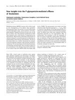
Graphic 1. Distribution of cases according to age groups and gender
0
6
4
0
6
12
6
8
8
20
14
12
0
2
4
6
8
10
12
14
16
18
20
21-30
years
31-40
years
41-50
years
51-60
years
61-70
years
≥ 71
years
Graphic Age groups and gender distribution of cases
Males
Females
Gastric biopsies (480 samples) were taken and processed from these patients (two
antral biopsies, two biopsies from the body, and one biopsy from the gastric angle for each
case).
Atrophic gastritis, defined endoscopically by the appearance of submucosal
vessels, giving rise to a mucosal vascular pattern similar to that found in the colon,
Graphic 1. Distribution of cases according to age groups and gender
Gastric biopsies (480 samples) were taken and processed from these patients (two antral bi‐
opsies, two biopsies from the body, and one biopsy from the gastric angle for each case).
Atrophic gastritis, defined endoscopically by the appearance of submucosal vessels, giving
rise to a mucosal vascular pattern similar to that found in the colon, sometimes associated
with other features, e.g, mucosal discoloration, smoothness, or flattened rugal folds, consti‐
tuted a rarely encountered entity in our study. In the cases studied we did not observe any
The Role of Endoscopy and Biopsy in Evaluating Preneoplastic and Particular Gastric Lesions
/>5
case of antral atrophic gastritis. In the gastric body, atrophic gastritis, was noted in 6 elderly
patients (Tab 2).
Endoscopic aspect
Antrum
No. of cases (%)
Body
No. of cases (%)
Focal erythematous gastritis 12 (12.5 %) 0 (0%)
Diffuse erythematous gastritis 54 (56.1 %) 22 (23%)
Erosive gastritis 4 (4.2 %) 0 (0%)
Erosive-hemorrhagic gastritis 2 (2.1 %) 0 (0%)
Petechial gastritis 22 (23 %) 10 (10.4%)
Atrophic gastritis 0 (0 %) 6 (6.3%)
Normal aspect 2 (2.1 %) 58 (60.3%)
Table 2. Frequency of gastritis diagnosed endoscopically
For this lesion we noted a poor correlation between the conventional endoscopic investiga‐
tion and histopathological examination (Tab. 3).
In accordance with the Sydney system, the morphological criteria of quantification applied
to cases with gastric atrophy are the following:
• 0 = absent;
• 1 = mild (disappearance of less than 25% of glands);
• 2 = moderate (disappearance of 25 - 50% of glands);
• 3 = severe (disappearance of over 50% of glands);
• 4 = biopsy inappropriate for histopathological interpretation.
Age groups
Antral atrophy score
(no. of cases; %)
Gastric body atrophy score
(no. of cases; %)
0 1 2 3 0 1 2 3
21-30 years 6 - - - 6 - - -
31-40 years 2 - 2 - 2 - 2 -
41-50 years 10 2 6 - 8 4 6 -
51-60 years 8 - 6 - 8 2 4 -
61-70 years 20 4 4 - 18 2 8 -
≥ 71 years 4 4 16 2 2 6 14 4
Total patients 50 (52.1%) 10 (10.4%)
34
(35.4%)
2 (2.1%) 44 (45.8%) 14 (14.6%) 34 (35.4%) 4 (4.2%)
Table 3. Distribution of the histological score of atrophy identified histopathologically
Gastric Carcinoma- New Insights into Current Management6
Atrophic chronic gastritis is characterized histopathologically by the numeric decrease in
glandular structures of the gastric mucosa and development of new metaplastic glands lined
by intestinal and/or pseudo-pyloric epithelium. We did not consider as real atrophy certain
modifications of the gastric mucosa that produce a false reduction in gastric glands, such as
the massive inflammatory infiltrate or the edema of the lamina propria.
From the total number of cases included in the study, we observed lesions of atrophic type
in 46 antral biopsies (48%) and 52 gastric body biopsies (54.2%).
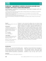
For antral location (Graphic 2) 10 cases with mild atrophy were noted (21.7%), 34 cases with
moderate atrophy (74%) and 2 cases with severe atrophy (4.3% - Fig. 2). Glandular atrophy
of gastric mucosa was observed much more frequently in patients with older ages (over 61
years). Biopsies noted with score 3 for atrophy pertain only to patients with ages ≥ 71 years.
Glandular atrophy was more frequently encountered in gastric body biopsies (but without
significant differences compared with the antrum, p=0.386), being predominant in patients
with average or old ages. Especially moderate and mild forms of atrophy were noted (14
cases with mild atrophy – 27%; 34 cases with moderate atrophy – 65.4% - Fig. 3; 4 cases with
severe atrophy – 7.6%). All patients with severe glandular atrophy pertain to the age group
≥ 71 years.
Figure 1. Congestive gastritis of gastric body, with mild atrophy of the mucosa
The Role of Endoscopy and Biopsy in Evaluating Preneoplastic and Particular Gastric Lesions
/>7
Figure 2. Antral chronic gastritis with severe atrophy and intestinal metaplasia. HE x 200.
Figure 3. Chronic gastritis of the gastric body with moderate atrophy. HE x 100.
Gastric Carcinoma- New Insights into Current Management8
Graphic 2 Histological evaluation of gastric atrophy
10
14
34 34
2
4
0
5
10
15
20
25
30
35
Mild atrophy Moderate atrophy Severe atrophy
Antrum Gastric body
Intestinal metaplasia (IM) represents the replacement of the surface and glandular
gastric epithelium by one composed of cells of the intestinal type (small or large intestine).
In conventional endoscopy, modifications such as nodules of yellow and white-
nacreous color, aspect like fish scales or diffuse granular are suggestive for intestinal
metaplasia of gastric mucosa. Such lesions were evident in 7 patients (4 males and 3
females) with ages between 54 and 76 years, with location in the gastric body.
In histopathological examination, preparations stained through usual morphological
methods do not allow for the certainty diagnosis, nor do they allow for classification of
intestinal metaplasia. For these reasons we used histochemical stain methods which give
exact information on the composition of the mucus synthesized by the modified glands,
respectively the neutral mucins, sialo- and sulfomucins.
Among histochemical methods recommended by the specialty literature, we used
staining methods PAS-AA at pH 2.5 and reaction with colloidal iron diamine-AA (HID-AA).
Type I intestinal metaplasia (complete) is characterized by relatively normal glandular
architecture, with straight crypts and glands lined by absorbing cells which do not secrete
mucus and goblet cells with flattened nuclei and with widened apical pole, these two cellular
types being encountered in approximately equal proportions. Occasionally, at the base of
the glands one can observe Paneth cells. We identified this form of intestinal metaplasia
with the PAS-AA stain, due to the presence of blue sialomucins in goblet cells (Fig. 4).
Reaction with paraphenyldiamine is negative.
Graphic 2. Histological evaluation of gastric atrophy
Intestinal metaplasia (IM) represents the replacement of the surface and glandular gastric
epithelium by one composed of cells of the intestinal type (small or large intestine).
In conventional endoscopy, modifications such as nodules of yellow and white-nacreous
color, aspect like fish scales or diffuse granular are suggestive for intestinal metaplasia of
gastric mucosa. Such lesions were evident in 7 patients (4 males and 3 females) with ages
between 54 and 76 years, with location in the gastric body.
In histopathological examination, preparations stained through usual morphological meth‐
ods do not allow for the certainty diagnosis, nor do they allow for classification of intestinal
metaplasia. For these reasons we used histochemical stain methods which give exact infor‐
mation on the composition of the mucus synthesized by the modified glands, respectively
the neutral mucins, sialo- and sulfomucins.
Among histochemical methods recommended by the specialty literature, we used staining
methods PAS-AA at pH 2.5 and reaction with colloidal iron diamine-AA (HID-AA).
Type I intestinal metaplasia (complete) is characterized by relatively normal glandular archi‐
tecture, with straight crypts and glands lined by absorbing cells which do not secrete mucus
and goblet cells with flattened nuclei and with widened apical pole, these two cellular types
being encountered in approximately equal proportions. Occasionally, at the base of the
glands one can observe Paneth cells. We identified this form of intestinal metaplasia with
the PAS-AA stain, due to the presence of blue sialomucins in goblet cells (Fig. 4). Reaction
with paraphenyldiamine is negative.
The Role of Endoscopy and Biopsy in Evaluating Preneoplastic and Particular Gastric Lesions
/>9
Figure 4. Type I intestinal metaplasia. AA-PAS x 200.
Type II intestinal metaplasia presents slight architectural modifications, with elongated and
tortuous crypts, with focal areas of foveolar hyperplasia and columnar cells in variable num‐
ber, which contain a mixture of neutral mucines and sialomucins, but not sulfomucins. The
proportion of the goblet cells is greater than in type I. PAS-AA positive reaction is translated
by mixed areas, PAS-positive and alcyanophil, representing neutral and acid mucines. The
positive material is located in the apical portion of epithelial cells, in the lumen of some
glands and in goblet cells (Fig. 5).
Type III of intestinal metaplasia is characterized morphologically by important glandular
distortions, with ramified glands, lined with columnar cells which secrete sulfomucins and
goblet cells secreting sialomucins. PAS reaction is negative, but the HID-AA reaction ap‐
pears intensely positive, through both dyeing solutions. The positive substrate appears in
goblet cells (blue), in the apical portion of columnar cells and in the lumen of metaplastic
glands (dark brown) (Fig. 6 and Fig. 7).
The prevalence of intestinal metaplasia identified histopathologically at the antral level was
of 20.8% (20 cases), and at the level of the gastric body of 25% (24 cases – Tab. 3) (p=0.492).
At the antral level we noted 18 cases with focal distribution (score given 1 and 2) and only
two cases with diffuse distribution (score 3) interesting almost entirely the gastric glandular
epithelium. Following the extension of intestinal metaplasia according to patients’ age, we
observed the great frequency of types II and III in patients over 61 years old. For gastric
body biopsies we did not encountered intestinal metaplasias with score 3, but 18 cases of
metaplasias with score 1 and 6 cases with score 2 were identified. These metaplastic trans‐
formations occur more frequently in older patients, but also in patients from the age groups
31-40 years and 41-50 years (Tab 4).
Gastric Carcinoma- New Insights into Current Management10
Figure 5. Type II intestinal metaplasia. AA-PAS x 200.
Figure 6. Type III intestinal metaplasia. HID-AA x 400.
The Role of Endoscopy and Biopsy in Evaluating Preneoplastic and Particular Gastric Lesions
/>11
Figure 7. Secretion of sulfomucins (brown) and sialomucins (blue) from intestinal metaplasia type III. HID-AA x 400.
In accordance with the Sydney system, the morphological criteria of quantification applied
to cases with intestinal metaplasia are the following:
• 0 = absent;
• 1 = mild (intestinal metaplasia in a focus of 1-4 glands);
• 2 =moderate (intestinal metaplasia in separate foci, but limited as extension);
• 3 = severe (intestinal metaplasia in over 50% from the gastric epithelium);
• 4 = biopsy inappropriate for histopathological interpretation.
Age groups
IM – antral biopsies
(no. cases; %)
IM – gastric body biopsies
(no. cases; %)
0 1 2 3 0 1 2 3
21-30 years 6 - - 6 - - -
31-40 years 4 - - - 2 - 2 -
41-50 years 18 - - - 16 2 - -
51-60 years 10 4 - - 12 2 - -
61-70 years 24 2 - 2 24 2 2 -
≥ 71 years 14 10 2 - 12 12 2 -
Total patients 76 (79.2%)
16
(16.6%)
2 (2.1%) 2 (2.1%) 72 (75%) 18 (18.7%) 6 (6.3%) 0 (0%)
IM –intestinal metaplasia
Table 4. Distribution of the histological score given to intestinal metaplasia
Gastric Carcinoma- New Insights into Current Management12
For both locations, type I intestinal metaplasia was the most frequent encountered type
(11.4% for antral biopsies and 15.6% for gastric body biopsies). The distribution of the three
types of intestinal metaplasia at the antrum and gastric body level, respectively, did not dif‐
fer significantly (p=0.560). Type II of intestinal metaplasia presented a relatively uniform
distribution in all age groups (Tab. 5).
Age groups
IM – antral biopsies IM – gastric body biopsies
I II III I II III
21-30 years - - - - - -
31-40 years - - - - 1 1
41-50 years - - 1 1 -
51-60 years 3 1 - 1 1 -
61-70 years - 3 1 3 - 1
≥ 71 years 8 2 2 10 1 3
Total patients 11 (11.4%) 6 (6.25%) 3 (3.1%) 15 (15.6%) 4 (4.2%) 5 (5.2%)
Table 5. Types of intestinal metaplasia
In our study we evaluated the incidence and types of epithelial dysplasia encountered in
patients with dyspeptic symptoms. In accordance with the Vienna classification, dysplastic
modifications were divided in low-grade dysplasia and high-grade dysplasia.
Histopathological examination of the 96 cases showed dysplastic lesions in 10 patients, prev‐
alence being of 10.4%.
Low-grade dysplasia, observed in 8 patients (Tab. 6, Graphic 3), is characterized by glandu‐
lar architecture mostly preserved, sometimes with the presence of pseudovilli, cystic dilated
glands or slightly irregular glands, with discrete intraluminal papillary projections or serrat‐
ed aspect. Glandular structures are lined with high, crowded cells, with or without mucous
vacuoles at the apical pole. The nuclei appear elongated and pseudostratified, discretely
pleomorphic, being situated in the lower half of the cytoplasm (Fig. 8 and Fig. 9). Mitotic
activity is discrete.
Age groups
Gastric epithelial dysplasia
Low-grade dysplasia High-grade dysplasia
21-30 years - -
31-40 years 1 -
41-50 years - -
51-60 years 1 -
61-70 years 2 1
≥ 71 years 4 1
Total patients 8 (8.33%) 2 (2.1%)
Table 6. Epithelial dysplasia in the cases studied
The Role of Endoscopy and Biopsy in Evaluating Preneoplastic and Particular Gastric Lesions
/>13
Graphic 3. The distribution of cases with epithelial dysplasia
00
1
0
00
1
0
2
1
4
1
0
1
2
3
4
21-30
years
31-40
years
41-50
years
51-60
years
61-70
years
≥ 71
years
Graphic The distribution of cases with epithelial dysplasia
Low-grade dysplasia
High-grade dysplasia
Fig. 8 Low-grade epithelial dysplasia. HE x 100.
Graphic 3. The distribution of cases with epithelial dysplasia
Figure 8. Low-grade epithelial dysplasia. HE x 100.
In all cases, dysplastic lesions were diagnosed histopathologically in the biopsies taken from the
antrum. From an endoscopic point of view, patients presented more frequently aspects of antral
diffuse erythematous gastritis (in 7 cases). In the case of a 66 year-old patient, the antral mucosa
did not show macroscopic modifications which were visible with conventional gastroscopy. Ep‐
ithelial dysplasia is observed mostly in patients in age groups 51-60 years, 61-70 years, and ≥ 71
years. In the cases studied we noted low-grade dysplastic lesions in a young patient, of age 36.
Gastric Carcinoma- New Insights into Current Management14
Only in 2 patients we noted high-grade dysplastic modifications. High-grade epithelial dys‐
plasia is characterized histopathologically by highly distorted glandular architecture, with
crowded, irregular and ramified glands, with frequent papillary intraluminal projections,
lined with stratified epithelium, with crowded, pleomorphic nuclei overlapping, with in‐
tense mitotic activity, losing of normal polarity, nuclei that touch the apical pole of the cell.
In the neoplastic epithelium, goblet cells and Paneth cells are absent (Fig. 10).
Figure 9. Low-grade epithelial dysplasia (detail). HE x 200.
Figure 10. High-grade epithelial dysplasia. HE x 200.
The Role of Endoscopy and Biopsy in Evaluating Preneoplastic and Particular Gastric Lesions
/>15