Báo cáo khoa học: Structural stability of the cofactor binding site in Escherichia coli serine hydroxymethyltransferase – the role of evolutionarily conserved hydrophobic contacts potx
Bạn đang xem bản rút gọn của tài liệu. Xem và tải ngay bản đầy đủ của tài liệu tại đây (418.02 KB, 10 trang )
Structural stability of the cofactor binding site in
Escherichia coli serine hydroxymethyltransferase – the role
of evolutionarily conserved hydrophobic contacts
Rita Florio
1
, Roberta Chiaraluce
1
, Valerio Consalvi
1
, Alessandro Paiardini
1
, Bruno Catacchio
1,2
,
Francesco Bossa
1,3
and Roberto Contestabile
1
1 Dipartimento di Scienze Biochimiche ‘A. Rossi Fanelli’, ‘Sapienza’ Universita
`
di Roma, Italy
2 CNR, Istituto di Biologia e Patologia Molecolari, ‘Sapienza’ Universita
`
di Roma, Italy
3 Centro di Eccellenza di Biologia e Medicina Molecolare (BEMM), ‘Sapienza’ Universita
`
di Roma, Italy
Introduction
Pyridoxal 5¢-phosphate (PLP)-dependent enzymes
comprise a vast and highly diversified group of cata-
lysts, whose action is required in a large number of
cellular processes. It is generally accepted that
PLP-dependent enzymes originated very early on in
evolution, before the three biological kingdoms
Keywords
cofactor binding site; conserved
hydrophobic contacts; pyridoxal phosphate;
serine hydroxymethyltransferase; urea-
induced denaturation
Correspondence
R. Contestabile, Dipartimento di Scienze
Biochimiche, ‘Sapienza’ Universita
`
di Roma,
Piazzale Aldo Moro 5, 00185, Roma, Italy
Fax: +39 0649917566
Tel: +39 0649917569
E-mail:
Website: />sito_biochimica/EN/index.html
(Received 3 September 2009, revised 12
October 2009, accepted 16 October 2009)
doi:10.1111/j.1742-4658.2009.07442.x
According to their fold, pyridoxal 5¢-phosphate-dependent enzymes are
grouped into five superfamilies. Fold Type I easily comprises the largest and
most investigated group. The enzymes of this group have very similar 3D
structures. Remarkably, the location of the cofactor in the active site,
between the two domains that form a single subunit, is almost identical in
all members of the group. Nonetheless, Fold Type I enzymes show very lit-
tle sequence identity, raising the question as to which structural features
determine the common fold. An important fold determinant appears to be
the presence of three evolutionarily conserved clusters of hydrophobic con-
tacts. A previous investigation, which used Escherichia coli serine hydrox-
ymethyltransferase, a well characterized Fold Type I member, demonstrated
the involvement of one of these clusters in the stability of the quaternary
structure. The present study focuses on the role of the same cluster in the
stability of the cofactor binding site. The investigation was carried out by
equilibrium denaturation experiments on serine hydroxymethyltransferase
forms in which the hydrophobic contact area of the cluster under study was
reduced by site-directed mutagenesis. The results obtained show that the
mutations clearly affected the process of pyridoxal 5¢-phosphate dissociation
induced by urea, reducing the stability of the cofactor binding site. We sug-
gest that the third cluster promotes the formation of a bridging structural
region that stabilizes the overall protein structure by connecting the two
domains, shaping the cofactor binding site and participating in the forma-
tion of the quaternary structure.
Structured digital abstract
l
MINT-7293394, MINT-7293405, MINT-7293418: eSHMT (uniprotkb:P0A825) and eSHMT
(uniprotkb:
P0A825) bind (MI:0407)bycosedimentation in solution (MI:0028)
Abbreviations
CHCs, conserved hydrophobic contacts; eSHMT, Escherichia coli serine hydroxymethyltransferase; PLP, pyridoxal 5¢-phosphate.
FEBS Journal 276 (2009) 7319–7328 ª 2009 The Authors Journal compilation ª 2009 FEBS 7319
diverged, from different protein ancestors that gener-
ated five independent families, corresponding to as
many different Fold Types [1,2]. The Fold Type I,
or aspartate aminotransferase family, is the largest,
functionally most diverse and best characterized. Its
members are made of dimers or multiple of dimers,
whose subunits are formed by a large domain and a
small domain. Despite the poor sequence similarity
among many Fold Type I enzymes, all members of
the family share the same basic protein architecture,
and are assembled with 17 structurally conserved
regions that form the heart of the domains [3]. The
presence of three clusters of evolutionarily conserved
hydrophobic contacts (CHCs; Fig. 1A) appears to be
one important structural feature determining the
native fold of Fold Type I enzymes [3]. Although
two of these clusters are located in the central cores
of the domains and presumably stabilize their scaf-
fold, the role of the third cluster is much less clear.
This cluster forms a hinge between two conserved
a-helices (which correspond to two structurally con-
served regions), located respectively at the beginning
and at the end of the large domain. Examination of
the contact network shows that, in this cluster, the
CHCs lie along one side of each helix, forming a
buried spine at positions i, i + 4 and i +7.
In a previous study investigating dimeric Escherichia
coli serine hydroxymethyltransferase (eSHMT; EC
2.1.2.1), a well characterized Fold Type I member, we
reported a site-directed mutagenesis study in which the
third cluster of CHCs was destabilized, reducing its
hydrophobic contact area [4]. The characterization of
the enzyme mutant forms (L85A, L276A and
L85A ⁄ L276A) under native conditions indicated that
the stability of the cluster is essential for the correct
quaternary assembly of the enzyme and is increased
by the binding of PLP and substrates. Indeed, the
two helices that form the cluster interact with the
N-terminal a-helix of the other subunit in the dimer
and are contiguous with two polypeptide loops, which,
in all Fold Type I enzymes, mediate the interactions
between the subunits and are involved in cofactor
binding, substrate binding and catalysis (Fig. 1). On
the other hand, the mutations did not affect either the
capability to bind the cofactor or the catalytic activity
of the enzyme. The monomeric form of the enzyme
(resulting from the double L85A⁄ L276A mutation)
binds PLP with comparable affinity with respect to the
dimeric wild-type form, suggesting that the subunit
structure of the monomer is more or less the same as
that in the dimer.
By contrast with the CHCs located in the core of
the large and small domains, the third hydrophobic
A
B
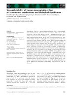
Fig. 1. (A) Cartoon representation of the crystal structure of eSHMT,
a Fold Type I enzyme, showing the residues involved in CHCs as
spheres. Subunits of eSHMT ternary complex with glycine and 5-for-
myl-tetrahydropteroylglutamate (Protein Data Bank code: 1dfo) are
represented in cyan and salmon. Residues forming CHCs are shown
only in one subunit. The residues of the clusters located in the large
and small domains are shown in magenta and orange, respectively.
The residues forming the third cluster are shown in red. The PLP–Gly
complex is represented by yellow sticks, with the phosphorus atom
depicted in orange, the oxygen atoms in red and the nitrogen atoms
in blue. The helices forming the third cluster interact with the N-ter-
minal helix of the other subunit in the dimer (shown in blue). Two
polypeptide loops (in green), which are contiguous with the helices
of the third cluster of CHCs, contribute residues that interact, at the
active site of the other subunit, with PLP and substrates. (B) Mono-
meric structure of eSHMT. Only the residues involved in the forma-
tion of the third CHCs are shown as spheres and the mutated
residues are indicated by arrows. The N-terminal tail (residues 1–61)
of the protein is coloured in orange, the large domain (residues 62–
211) in salmon, the interdomain segment (residues 212–279) in
green and the small domain in blue (for details, see Discussion).
Stability of cofactor binding site in SHMT R. Florio et al.
7320 FEBS Journal 276 (2009) 7319–7328 ª 2009 The Authors Journal compilation ª 2009 FEBS
cluster is not directly involved in the proper position-
ing or stabilization of the active site or PLP-binding
residues. Its location rather suggests a role in bridging
different structural regions of the protein in order to
stabilize its native overall fold. The present study
investigated the role of the third cluster of CHCs with
respect to the structural stability of the PLP-binding
site by means of urea-induced denaturation experi-
ments on eSHMT L85A, L276A and L85A ⁄ L276A
mutants. These mutations were carried out to reduce
the hydrophobic contact area in the cluster. Leu276 is
the most conserved residue in the cluster, with its posi-
tion being almost invariably occupied by a leucine resi-
due in Fold Type I enzymes [3]. Leu85 was chosen
because this residue shows the largest contact area
with Leu276.
Results
The structural stability of holo-eSHMT wild-type and
mutant forms was investigated by performing equilib-
rium unfolding experiments, using urea as denaturing
agent. The structural changes of the active site and
overall protein induced by urea were monitored by
measuring catalytic activity, the fraction of covalently
bound cofactor, intrinsic fluorescence emission, far-UV
CD and the sedimentation coefficient.
The reversibility of the unfolding process was ana-
lyzed by measuring the activity that denatured eSHMT
samples (23 lm in 8 m urea at 20 °C) were able to
recover after 4 h, subsequent to a ten-fold dilution
with buffer at 20 °C. In agreement with a previous
study [5], complete enzyme activity was recovered after
refolding. In this respect, comparable results were
obtained with all mutant forms.
Activity and internal aldimine measurements
Enzyme samples (2.3 lm) were incubated with increas-
ing urea concentrations in 50 mm NaHepes buffer (pH
7.2), containing 200 lm dithiothreitol and 100 lm
EDTA, at 20 °C for 15 h. The residual fractions of
catalytic activity and covalently bound cofactor (inter-
nal aldimine) were then measured and reported as a
function of denaturant concentration (Fig. 2). With all
enzyme forms, the loss of internal aldimine appears to
take place according to a sigmoid process. The
decrease in catalytic activity shows a more complex
behaviour. At a urea concentration in the range 0–1 m,
almost half of the activity is lost, apparently as a result
of an hyperbolic process. The complete loss of activity,
taking place at higher urea concentrations, appears to
follow a sigmoid course. In 1 m urea, almost all the
cofactor is bound to the enzyme as internal aldimine,
indicating that the loss of activity does not result from
the denaturation of the active site. These observations
suggest that urea might act as an enzyme inhibitor.
Indeed, experiments in which the kinetic parameters of
the catalyzed reaction were determined in the absence
or presence of 0.25, 0.50, 0.75 and 1 m urea clearly
demonstrated that urea is responsible for a mixed-type
Fig. 2. Dependence of catalytic activity and PLP covalent binding
to eSHMT on the urea concentration. The fractions (f) of retained
catalytic activity (open symbols) and internal aldimine (closed sym-
bols) were measured as a function of the urea concentration. The
reaction catalyzed by eSHMT was the aldol cleavage of
L-threo-3-
phenylserine into glycine and benzaldehyde. The profiles obtained
with the L85A (triangles), L276A (squares) and L85A ⁄ L276A (dia-
monds) mutants are compared in each panel with the profiles
obtained with the wild-type enzyme (circles). The lines through the
experimental points (dashed lines for wild-type and continuous lines
for mutant enzymes) are those obtained from the global nonlinear
least squares fitting of internal aldimine and activity data to
Eqns (1,2).
R. Florio et al. Stability of cofactor binding site in SHMT
FEBS Journal 276 (2009) 7319–7328 ª 2009 The Authors Journal compilation ª 2009 FEBS 7321
inhibition. The inhibitory effect of urea on catalytic
activity has been also reported for other enzymes and
is well documented in the literature [6,7].
Figure 2 shows that the denaturation profiles of the
L85A and L85A ⁄ L276A mutants are shifted toward
lower urea concentrations with respect to those of the
wild-type and L276A enzymes. To quantify this shift,
the internal aldimine and activity data of each enzyme
form were analyzed according to Eqn (1) (the equation
of a sigmoid curve) and Eqn (2) (which takes in
account both the inhibitory and denaturing effects of
urea), respectively, in a global least squares minimiza-
tion procedure in which the parameters of the sigmoid
denaturation processes contained in both equations
were shared (Fig. 2). The analysis gave a good global
fit of the data and showed that the midpoints of the
sigmoid transitions coincide and, in the case of the
L85A and L85A ⁄ L276A mutants, are approximately
0.3 m lower compared to the wild-type (Table 1).
Although the midpoint of the L276A mutant coincides
with that of the wild-type enzyme, it is clear from the
values of n given in Table 1 that the steepness of the
sigmoid transitions is lower. This indicates that the
mutation had the effect of lowering the cooperativity
of the denaturation process. The same consideration
can be made for the double L85A ⁄ L276A mutant.
Spectroscopic measurements
Intrinsic fluorescence emission and far-UV CD mea-
surements on wild-type and mutant eSHMTs were car-
ried out on protein samples (2.3 lm) incubated with
increasing denaturant concentrations (from 0 to 7.9 m)
in 50 mm NaHepes buffer (pH 7.2), containing 200 lm
dithiothreitol and 100 lm EDTA at 20 °C for 15 h
(Fig. 3). With the wild-type enzyme, a urea concentra-
tion rising up to 2.3 m determines the increase in rela-
tive fluorescence emission intensity (Fig. 3A). Part of
this increase clearly takes place with negligible dissoci-
ation of the cofactor (from 0 to 1 m urea) and appears
to coincide with the drop of catalytic activity attribut-
able to urea inhibition. The further increase of fluores-
cence matches the complete loss of catalytic activity
and the release of the cofactor, which relieves the
quenching of tryptophan fluorescence [5]. As previ-
ously shown, this represents the first of two steps in
the denaturation mechanism of eSHMT. At a urea
concentration of 2.3 m, the enzyme is known to exist
in the form of a denaturation intermediate [5,8,9],
which denatures completely and loses part of its fluo-
rescence emission as the urea concentration is
increased up to 7.9 m.
The fluorescence profiles obtained with the mutant
enzymes are similar in shape to that of wild-type
eSHMT. However, at a urea concentration in the
range 0–1 m, the relative fluorescence emission inten-
sity of the mutant forms is higher than that of the
wild-type enzyme. Moreover, with the L85A and
L85A ⁄ L276A mutants, the increase of fluorescence
emission that corresponds to the dissociation of the
cofactor is shifted towards lower urea concentrations
with respect to wild-type and L276A forms (Fig. 3).
This observation is reminiscent of that noted for the
internal aldimine and activity data, and indicates a
shift of the equilibrium between the native and the
intermediate forms in favour of the latter. At urea con-
centrations above 2.3 m, and as the protein unfolds
completely, the fluorescence emission profiles of all
enzyme forms coincide, suggesting that the equilibrium
between the denaturation intermediate and the
unfolded protein is not affected by the mutations.
The influence of the mutations on the far-UV CD
and average lambda (for a definition, see Experimental
procedures) profiles is not as clear as in the case of the
activity and internal aldimine data (Fig. 3E, F). The
CD signal and average lambda do not vary at a urea
concentration in the range 0–1 m. At a urea concentra-
tion of 2.3 m, approximately one-third of the far-UV
CD signal is lost, although very little change of the
average lambda is observed. This indicates that the
loss of cofactor that takes place in the first denatur-
ation step does not correspond to large structural
changes.
Sedimentation velocity measurements
In a previous study [4], we reported that the L85A,
L276A and L85A ⁄ L276A mutations affect the quater-
nary structure stability of
eSHMT. The wild-type
enzyme is a dimer with molecular mass of approxi-
mately 91 kDa and shows a single band in the
sedimentation coefficient distribution with a maximum
Table 1. Parameters obtained from the global best-fit of activity
and internal aldimine data. Parameters are expressed as the
mean ± SE determined by the global nonlinear least squares fitting
of data to Eqns (1,2), as detailed in the text. K
i
represents the cal-
culated inhibition constant, whereas cm
1
and n reflect, respectively,
the urea concentration midpoints and the steepness of the sigmoi-
dal curves.
K
i
(M) cm
1
(M) n
Wild-type 1.71 ± 0.05 1.88 ± 0.01 9.54 ± 0.47
L85A 1.30 ± 0.08 1.52 ± 0.01 11.19 ± 0.66
L276A 1.49 ± 0.05 1.88 ± 0.01 7.04 ± 0.34
L85A ⁄ L276A 1.38 ± 0.06 1.56 ± 0.01 6.84 ± 0.35
Stability of cofactor binding site in SHMT R. Florio et al.
7322 FEBS Journal 276 (2009) 7319–7328 ª 2009 The Authors Journal compilation ª 2009 FEBS
at 5.5 S ( s
20,w
), which is the value expected for a
hydrated dimer with an approximately spherical shape.
The hydrophobic cluster mutations shifted the equilib-
rium between dimeric and monomeric forms of the
enzyme in favour of the latter, which shows a sedimen-
tation coefficient of approximately 3 S. Interestingly,
the monomeric form of the enzyme was shown to bind
PLP. This observation is not unprecedented for Fold
Type I enzymes [10].
In the present study, ultracentrifugation experiments
were carried out aiming to analyse the effect of urea
on the sedimentation properties of wild-type and
mutant eSHMTs. Sedimentation velocity experiments
were performed in the presence of either 1 or 2.3 m
urea. These concentrations correspond to crucial
events in the denaturation mechanism: exposure of the
enzyme to 1 m urea results in the loss of half of the
catalytic activity, although all the cofactor is retained
at the active site as internal aldimine. In 2.3 m urea,
the cofactor is lost as the denaturation intermediate is
formed (Fig. 2). In Table 2, the results of the present
ultracentrifuge analysis are compared with those
obtained in the absence of urea [4]. In 1 m urea, the
wild-type enzyme is dimeric, but becomes mainly
monomeric when the urea concentration is increased
up to 2.3 m, as indicated by a predominant band with
a maximum at 3.1 S in the sedimentation coefficient
distribution (Fig. 4). The frictional ratio (f ⁄ f
0
) (i.e. the
ratio between the experimentally calculated friction
coefficient and the minimum friction coefficient of an
anhydrous sphere) of monomeric wild-type eSHMT is
Fig. 3. Spectroscopic analysis of urea-
induced unfolding of wild-type (d), L85A (D),
L276A (h) and L85A ⁄ L276A ()) enzyme
forms. All spectroscopic measurements
were carried out on enzyme samples at a
concentration of 2.3 l
M,in50mM NaHepes
(pH 7.2), containing 200 l
M dithiothreitol
and 100 l
M EDTA at 20 °C. Intrinsic fluores-
cence emission measurements at 336 nm
on wild-type and mutant forms were per-
formed with a 1 cm quartz cuvette upon
excitation at 295 nm. (A) Comparison
among relative fluorescence emission (F
r
)
and retained fractions (f ) of activity (s)
and internal aldimine (·) measured with
wild-type eSHMT as a function of the urea
concentration. (B–D) Relative fluorescence
denaturation profiles obtained with wild-type
and mutant enzymes. (E) Molar ellipticity at
222 nm ([h]
222
) calculated from far-UV CD
measurements carried out in a 0.2 cm
quartz cuvette. (F) Average lambda (k) pro-
files calculated from fluorescence emission
spectra acquired from 320–500 nm, with
excitation at 295 nm.
Table 2. Sedimentation coefficients calculated from ultracentrifuge
experiments on wild-type and mutant eSHMTs. The frictional ratio
(f ⁄ f
0
) is the ratio between the experimentally calculated friction
coefficient and the minimum friction coefficient of an anhydrous
sphere.
s
20, w
(S)
No urea 1
M urea 2.3 M urea
Wild-type 5.5 (f ⁄ f
0
= 1.2) 5.4 (f ⁄ f
0
= 1.2) 5.5 (17%)
3.1 (83%)
L85A 5.5 (f ⁄ f
0
= 1.2) 5.3 (88%)
2.9 (12%)
6.4 (10%)
2.8 (90%)
L276A 5.5 (f ⁄ f
0
= 1.2) 5.5 (77%)
3.1 (23%)
2.5 (f ⁄ f
0
= 1.5)
L85A ⁄ L276A 5.5 (66%)
3.3 (34%)
5.5 (35%)
3.3 (65%)
2.5 (f ⁄ f
0
= 1.5)
R. Florio et al. Stability of cofactor binding site in SHMT
FEBS Journal 276 (2009) 7319–7328 ª 2009 The Authors Journal compilation ª 2009 FEBS 7323
equal to that of its dimeric form (1.2) and close to that
of a spherical protein, suggesting that the subunit
dissociation took place without any large structural
denaturation.
With all mutant forms, 1 m urea determines the
partial dissociation of the dimer subunits (Fig. 4 and
Table 2). The extent of dissociation is greater for the
double mutant, which already exists as an equilibrium
mixture of monomers and dimers in the absence of
urea, and is smaller for the L85A mutant and interme-
diate for the L276A mutant. Nothing may be con-
cluded regarding the shape of the mutant monomeric
forms in 1 m urea because the frictional ratio cannot
be calculated when more than one sedimentation
species is present at equilibrium. However, a sedimen-
tation coefficient of approximately 3 S suggests that all
monomers have an approximately spherical shape in
1 m urea. It should be noted that, with all mutant
forms, the dissociation of subunits in 1 m urea takes
place without any loss of cofactor (Fig. 2), in agree-
ment with a substantial retention of the active site
native structure. By contrast, with the wild-type
enzyme, the dissociation of subunits is observed at a
much higher urea concentration range (1–2.3 m),
apparently in concomitance with the loss of cofactor.
In 2.3 m urea, the L276A and L85A ⁄ L276A mutant
forms exist as single sedimentation species with a coef-
ficient of 2.5 S. The frictional ratio calculated for this
species is 1.5, suggesting that the decrease in the sedi-
mentation coefficient is determined by the loss of
spherical shape as a result of a partial structural dena-
turation of the monomer. The L85A mutant in 2.3 m
urea is present as two sedimentation species with coef-
ficients of 2.8 S and 6.4 S. The 6.4 S species most
probably results from aggregation. The 2.8 S species,
which shows a broad coefficient distribution, may also
correspond to a partially denatured monomer.
PLP-binding properties of the monomeric
denaturation intermediate
The cofactor binding properties of monomeric wild-
type and mutant enzymes in 2.3 m urea (dissolved in
the same buffer used in the previous experiments) were
analysed in order to probe the structural features of
the active site. The visible absorption spectrum of
wild-type eSHMT shows a characteristic absorption
band with maximum at 420 nm, as a result of the pres-
ence of PLP bound at the active site as a protonated
internal aldimine [11]. The absorption spectra of the
enzymes (10 lm subunit concentration) in 2.3 m urea
were recorded and subtracted from the absorption
spectra acquired after 20 min subsequent to the addi-
tion of 100 lm PLP, allowing sufficient time for the
cofactor binding equilibrium to be reached (as indi-
cated by the fact that the absorption spectrum stopped
changing). The differential spectrum obtained with the
wild-type enzyme (Fig. 5) clearly results from the
development of a positive 420 nm absorbing band,
demonstratring that the addition of PLP determines
Fig. 4. Sedimentation velocity distributions
obtained with wild-type and mutant
eSHMTs. Sedimentation velocity measure-
ments were performed at 116 480 g on
2.5 l
M (subunit concentration) enzyme sam-
ples kept at 20 °Cin50m
M NaHepes buffer
(pH 7.2) in the absence (——) or presence
of 1
M (- - - -) and 2.3 M urea (ÆÆÆÆÆ).
Stability of cofactor binding site in SHMT R. Florio et al.
7324 FEBS Journal 276 (2009) 7319–7328 ª 2009 The Authors Journal compilation ª 2009 FEBS
the formation of a native-like internal aldimine. The
differential spectra recorded with the mutant enzymes
indicate the formation of a band with maximum
absorbance at lower wavelengths (at approximately
390–400 nm). Evidently, in 2.3 m urea, PLP binds to
the mutant enzymes, but without the formation of a
native-like internal aldimine, suggesting a structural
difference between the mutant active site and the
wild-type.
Discussion
Previous studies [5,8,9] show that the urea-induced
denaturation of wild-type eSHMT, from a native
dimer to a fully denatured monomer, takes place
according to a three-state mechanism (Scheme 1A), in
which an intermediate catalytically inactive apo-form
of the enzyme mostly accumulates at a urea concentra-
tion of approximately 2.3 m. The data obtained in the
present study show that, at this urea concentration,
the enzyme has lost most of the cofactor (Fig. 2) and
is in a monomeric form (Fig. 4 and Table 2). There-
fore, the urea-induced loss of cofactor and dissociation
of subunits appear to be simultaneous events of the
first denaturation step (N
2
¢
ÀPLP
þPLP
2I in Scheme 1A). The
dissociation of subunits apparently takes place without
any large denaturation of the monomer structure. This
is suggested by the fact that the enzyme monomers in
2.3 m urea retain a spherical shape, as indicated by the
frictional ratio (f ⁄ f
0
) and the sedimentation coefficient
calculated from the ultracentrifuge analysis (Table 2).
The minor change of the far-UV CD spectrum
observed when the urea concentration is increased up
to 2.3 m is therefore hardly attributable to a loss of
secondary structure, and may instead result from a
conformational change of the protein monomer. Upon
PLP binding, the native apo-eSHMT is known to
undergo a conformational change, which most proba-
bly results from a shift of the equilibrium between the
open and closed forms of the enzyme [12]. Urea might
act to shift this equilibrium in favour of the open
form, promoting PLP dissociation from the active site.
The wild-type enzyme in 2.3 m urea, which is mostly
monomeric and in the apo-form, maintains the capa-
bility to form a native-like internal aldimine if a large
excess of PLP is added (Fig. 5). Increasing the urea
concentration from 2.3 to 8 m determines the complete
unfolding of the enzyme (2I¢2U in Scheme 1A), with
a complete loss of secondary structure, as reported in
previous studies [5,8,9], and as can be deduced from
the spectroscopic data shown in Fig. 3. In this higher
urea concentration range, the denaturation profiles
obtained with the mutant enzymes do not differ from
those of wild-type eSHMT.
Mutations clearly influence the apparent denatur-
ation mechanism of the enzyme at a urea concentra-
tion in the range 0–2.3 m. Comparison of data
obtained from the ultracentrifuge analysis and from
the measurement of covalently bound cofactor shows
that, in contrast to that observed with the wild-type
enzyme, with the mutant forms, the urea-induced sub-
unit dissociation is detectable as a separate process
with respect to the dissociation of PLP. With all three
mutant forms, at a urea concentration of 1 m, subunit
dissociation is clearly visible (N
2
¢2N in Scheme 1B),
whereas the loss of cofactor is negligible and becomes
visible only at higher denaturant concentrations
(2N ¢
ÀPLP
þPLP
2I
0
; Scheme 1B). This observation agrees with
the previously published data, indicating that the
monomeric form of the enzyme is able to bind PLP
and that the third cluster of CHCs plays an important
role in the stabilization of the eSHMT quaternary
structure [4]. The extent of subunit dissociation in 1 m
Fig. 5. Analysis of PLP-binding properties of wild-type and mutant
enzymes in 2.3
M urea. Separate enzyme (20 lM) and PLP (200 lM)
samples were kept in 2.3
M urea at 20 °C for 15 h. Equal volumes
of enzyme and PLP samples were then mixed and absorption spec-
tra recorded after 20 min. Absorption spectra of enzyme samples
mixed with an equal volume of buffer containing 2.3
M urea were
subtracted from spectra acquired in the presence of PLP, generat-
ing the differential spectra shown: wild-type eSHMT (——), L85A
(- - - -), L276A (ÆÆÆÆÆ) and L85A ⁄ L276A (ÆÆ-ÆÆ) mutants.
A
B
Scheme 1. Presumed equilibrium denaturation mechanisms of
wild-type (A) and mutant (B) eSHMTs. N and U represent, respec-
tively, the native and the fully denatured forms of the enzyme
subunits. The denaturation intermediate formed by the wild-type
enzyme is indicated by I, whereas I¢ represents the partially dena-
tured intermediate formed by the mutant enzymes.
R. Florio et al. Stability of cofactor binding site in SHMT
FEBS Journal 276 (2009) 7319–7328 ª 2009 The Authors Journal compilation ª 2009 FEBS 7325
urea is higher for the double L85A ⁄ L276A form, lower
for the L85A form and intermediate for the L276A
mutant. This situation again reflects what is observed
for the same enzyme forms under native conditions [4].
A novel observation is that the mutations clearly
affected the process of PLP dissociation induced by
urea. The L85A and L85A ⁄ L276A mutations lowered
the midpoint of this transition and the L276A muta-
tion lowered its cooperativity (Table 1). Moreover,
upon PLP dissociation in 2.3 m urea, the monomeric
mutant enzymes appear to be partially denatured (I¢ in
Scheme 1B), as indicated by the frictional ratio and
the sedimentation coefficients calculated from the
ultracentrifuge analysis (Table 2). The extent of dena-
turation of this monomeric intermediate is not
expected to be very large because the average lambda
profiles of the wild-type and mutant enzymes are very
similar (Fig. 3). Nevertheless, the addition of excess
PLP to the partially denaturated monomer does not
result in the formation of a native internal aldimine.
Evidently, the concentration of the species at equilib-
rium cannot be altered by the addition of PLP, con-
firming that the mutant enzymes follow a different
denaturation mechanism with respect to wild-type
eSHMT. Taken together, these observations suggest
that, in the mutant enzymes, the urea-induced dissocia-
tion of PLP is favoured by the disruption of the third
cluster of CHCs. The mutations introduced with the
aim of reducing the hydrophobic surface contacts have
instead reduced the stability of the PLP binding site.
This conclusion points to a clear relationship between
the formation of the cluster and the structural integrity
of the active site.
By contrast to the CHCs located in the cores of the
large and small domains, the cluster under study is not
directly involved in the positioning or stability of any
active site or PLP-binding residue. Considering the
studies on eSHMT folding, we suggest that this cluster
may play a fundamental role in the folding mechanism
of the enzyme, which may be divided into two phases
[5,8,9,13]. In the first phase, the large and small
domains rapidly assume their native state, forming a
folding intermediate that has almost all of the native
secondary and tertiary structure, but is unable to bind
PLP. In this intermediate, the N-terminus and an
inter-domain segment remain exposed to solvent [8]
(Fig. 1B). In the second, slower phase, these structural
elements fold into the native structure, conferring the
enzyme with the capability of binding PLP. Because
the interdomain segment comprises the a-helix contrib-
uting L276 and the second a-helix (where L85 is
located) of the cluster is part of the large domain, it
follows that the last folding event may correspond to
the formation of the third cluster of CHCs. The for-
mation of the cluster presumably fastens together all
the structural components of the protein and confers
stability to the active site. The high degree of sequence
and structural conservation of the third cluster of
CHCs, as observed for the majority of fold-type I
enzymes, suggests that this function, which is hypothe-
sized for eSHMT, could be extended to the whole
superfamily.
Experimental procedures
Materials
Ingredients for bacterial growth were obtained from
Sigma-Aldrich (St Louis, MO, USA). Chemicals for the
purification of the enzymes were obtained from BDH
(Poole, UK); DEAE-sepharose and phenyl-sepharose were
obtained from GE Healthcare (Milwaukee, WI, USA). The
L85A, L276A and L85A ⁄ L276A mutant forms of the
E. coli glyA (SHMT encoding gene) were already available
from a previous study [4]. Wild-type and mutant forms of
eSHMT were purified as described previously [14]. PLP was
added to protein samples during the purification procedure,
but it was left out in the final dialysis step. All experiments
were performed at 20 °Cin50mm NaHepes buffer at pH
7.2, containing 200 lm dithiothreitol and 100 lm EDTA.
PLP was obtained from Sigma-Aldrich (98% pure). All
other reagents were obtained from Sigma-Aldrich.
Preparation of holoenzyme samples
We noted that different batches of purified e SHMT samples
contained variable holoenzyme ⁄ apoenzyme ratios. This
observation was made with either wild-type or mutant
forms of the enzyme. To carry out comparable experiments,
it was mandatory to prepare enzyme samples that con-
tained the same fraction of protein-bound PLP (possibly
close to saturation) and, at the same time, were devoid of
any excess of cofactor. Holoenzymes were then prepared
from apoenzyme samples, by the addition of PLP at the
concentration needed to obtain 98% saturation, as calcu-
lated on the basis of the related dissociation constant of
PLP binding equilibrium [4]. The subunit concentration of
the holoenzyme was calculated according to a molar
absorptivity value of e
280
= 44884 cm
)1
Æm
)1
[12]. Apoen-
zyme samples were prepared as described previously [5].
Unfolding and refolding experiments
Concentrated protein samples were diluted into urea solu-
tions (0–7.9 m in 50 mm NaHepes, pH 7.2, containing
200 lm dithiothreitol and 100 lm EDTA, at 20 °C) to
obtain a final protein concentration of 2.3 lm. Spectro-
Stability of cofactor binding site in SHMT R. Florio et al.
7326 FEBS Journal 276 (2009) 7319–7328 ª 2009 The Authors Journal compilation ª 2009 FEBS
scopic measurements, activity assays, measurements of
internal aldimine and ultracentrifuge analyses were carried
out after 15 h, which allows sufficient time to reach the
equilibrium [5].
The l-threo-3-phenylserine cleavage activity of urea-
incubated enzyme samples was measured after the direct
addition of 7.5 mm substrate, which corresponds to approx-
imately 16% saturation. The dilution of urea resulting from
substrate addition was taken into account.
The reversibility of the urea-induced unfolding was ana-
lyzed by means of refolding experiments in which denatured
enzyme samples (23 lm kept with 8 m urea in 50 mm
NaHepes, pH 7.2, containing 200 lm dithiothreitol and
100 lm EDTA for 15 h at 20 °C) were diluted ten-fold with
buffer at 20 °C. These conditions have been shown to give
the highest yield of refolded enzyme [5]. Four hours later,
after allowing sufficient time to complete the refolding pro-
cess [5], the enzyme activity of the refolded sample was
assayed upon a further 50-fold dilution of the sample into
the assay reaction mixture (the final enzyme concentration
in the activity assay was 0.05 lm). Ten-fold molar excess
PLP was added to refolded samples immediately before
dilution into the activity assay reaction mixture.
Activity assays
All assays were carried out at 20 °Cin50mm NaHepes
(pH 7.2), containing 200 lm dithiothreitol and 100 lm
EDTA. The rate of l-threo-3-phenylserine cleavage (3 lm
enzyme samples) was obtained from spectroscopic measure-
ment of benzaldehyde production at 279 nm, employing a
molar absorptivity value of e
279
= 1400 cm
)1
Æm
)1
[15,16].
Spectroscopic measurements
Fluorescence emission measurements were carried out with
a LS50B spectrofluorimeter (Perkin Elmer Life Sciences,
Waltham, MA, USA) using a 1 cm path length quartz
cuvette. Fluorescence emission spectra were recorded in the
range 320–500 nm (1 nm sampling interval; 5 nm emission
slit), with the excitation wavelength set at 295 nm (3 nm
excitation slit). Fluorescence measurements were expressed
as relative fluorescence intensity and average lambda (aver-
age lambda = R(I
i
· k
i
) ⁄ R(I
i
), where k
i
is the ith wave-
length and I
i
is the corresponding relative fluorescence
intensity).
Far-UV (190–250 nm) CD spectra were measured using a
0.2 cm path length quartz cuvette and expressed as the mean
residue ellipticity [Q], assuming a mean residue molecular
mass of 110 per amino acid residue. UV-visible spectra were
recorded with a double-beam Lambda 16 spectrophotometer
(Perkin Elmer Life Sciences). Kinetic measurements in the
activity assays were performed on a Hewlett-Packard 8453
diode-array spectrophotometer (Hewlett-Packard, Palo Alto,
CA, USA). All spectroscopic measurements were carried out
at 20 °Cin50mm NaHepes (pH 7.2), containing 200 lm
dithiothreitol and 100 lm EDTA.
Analytical ultracentrifugation analysis
Sedimentation velocity experiments were carried out at
20 °Cin50mm NaHepes buffer pH 7.2, containing 200 lm
dithiothreitol and 100 lm EDTA, on a Beckman XL-I ana-
lytical ultracentrifuge (Beckman Coulter, Fullerton, CA,
USA) equipped with absorbance optics and an An60-Ti
rotor. In the sedimentation velocity experiments, performed
at 116 480 g, the protein concentration was 2.5 lm . Data
were collected at 277 nm at a spacing of 0.003 cm with
three averages being obtained in a continuous scan mode.
Sedimentation coefficients and integration of data were
obtained using the software sedfit (provided by P. Schuck,
National Institutes of Health, Bethesda, MD, USA). The
values were reduced to water (S
20,w
) using standard proce-
dures. The buffer density and viscosity were calculated
using the software sednterp (lway.
com). The ratio f ⁄ f
0
was calculated from the diffusion coef-
ficient, which in turn is related to the spreading of the
boundary, using the software sedfit.
Internal aldimine measurements
Borohydride (BH
4
)
) is known to reduce imines and alde-
hydes rapidly and efficiently [17]. When the internal aldi-
mine (PLP-enzyme Schiff base) of PLP-dependent enzymes
is reduced with BH
4
)
, the cofactor is irreversibly attached
to the protein, giving an absorption spectrum with a band
at approximately 330 nm [18]. NaBH
4
was prepared as a
concentrated solution (5 m)in50mm NaOH and added to
proteins samples (2.3 lm in 8 mL of 50 mm NaHepes buf-
fer at pH 7.2, containing 200 lm dithiothreitol and 100 lm
EDTA) incubated with urea, so that its final concentration
was 83 mm. Thirty minutes after the addition of NaBH
4
,
protein samples were concentrated in Amicon Ultra centri-
fuge filters (30 kDa cut-off; Millipore, Billerica, MA, USA)
and diluted with 50 mm NaHepes buffer repeatedly in order
to eliminate low-molecular mass molecules, including the
reduced free cofactor. In the final dilution step, the buffer
was added to obtain a final volume of 2 mL and absorption
spectra were recorded. The A
335
⁄ A
280
ratio was calculated
to normalize the absorbance of the reduced internal aldi-
mine on the basis of protein concentration, and then
divided by the ratio found for a native, PLP-saturate
enzyme sample. The result obtained gave the fraction of
PLP molecules bound per monomer of enzyme.
Data analysis
The equilibrium unfolding profiles obtained from the mea-
surement of the internal aldimine and the catalytic activity
R. Florio et al. Stability of cofactor binding site in SHMT
FEBS Journal 276 (2009) 7319–7328 ª 2009 The Authors Journal compilation ª 2009 FEBS 7327
were expressed as fraction of the related measurements car-
ried out in the absence of urea and analyzed, respectively,
according to Eqns (1,2). Equation (1) describes a sigmoid
curve and conforms to a two-state denaturation process.
Equation (2) takes into account both the inhibitory and
denaturing effects of urea on the catalytic activity of the
enzyme. The enzyme in the presence of urea is assumed to
exist as a mixture of free, active enzyme and urea-bound
inactive enzyme at equilibrium. Both enzyme forms are also
assumed to denature according to an identical sigmoid pro-
cess (Eqn 1). In the equations, f represents the fraction of
either internal aldimine (Eqn 1) or activity (Eqn 2), n
reflects the steepness of the sigmoid transition, and c
m
is its
urea concentration midpoint. In Eqn (2), K
i
is the inhibi-
tion constant of the urea binding equilibrium.
f ¼ 1 À
urea½
n
c
n
m
þ urea½
n
ð1Þ
f ¼ 1 À
urea½
n
c
n
m
þ urea½
n
 1 urea½
urea½þK
i
ð2Þ
Internal aldimine and activity data were fitted in a global
least squares minimization procedure in which the parame-
ters of the sigmoid denaturation processes contained in
Eqns (1,2) were shared, using the software prism (Graph-
Pad Software Inc., La Jolla, CA, USA).
Acknowledgements
We thank Professor Verne Schirch for helpful discus-
sions. This work was supported by grants from the
Italian Ministero dell’Universita
`
e della Ricerca. Rita
Florio is the recipient of a fellowship from the Facolta
`
di Scienze Matematiche, Fisiche e Naturali of ‘Sapien-
za’ Universita
`
di Roma, Italy.
References
1 Mehta PK & Christen P (2000) The molecular evolution
of pyridoxal-5¢-phosphate-dependent enzymes. Adv
Enzymol Relat Areas Mol Biol 74, 129–184.
2 Grishin NV, Phillips MA & Goldsmith EJ (1995)
Modeling of the spatial structure of eukaryotic orni-
thine decarboxylases. Protein Sci 4, 1291–1304.
3 Paiardini A, Bossa F & Pascarella S (2004) Evolution-
arily conserved regions and hydrophobic contacts at the
superfamily level: the case of the fold-type I, pyridoxal-
5¢-phosphate-dependent enzymes. Protein Sci 13, 2992–
3005.
4 Florio R, Chiaraluce R, Consalvi V, Paiardini A,
Catacchio B, Bossa F & Contestabile R (2009) The role
of evolutionarily conserved hydrophobic contacts in the
quaternary structure stability of Escherichia coli serine
hydroxymethyltransferase. Febs J 276, 132–143.
5 Cai K, Schirch D & Schirch V (1995) The affinity of
pyridoxal 5¢-phosphate for folding intermediates
of Escherichia coli serine hydroxymethyltransferase.
J Biol Chem 270, 19294–19299.
6 Strambini GB & Gonnelli M (1986) Effects of urea and
guanidine hydrochloride on the activity and dynamical
structure of equine liver alcohol dehydrogenase.
Biochemistry 25, 2471–2476.
7 Rajagopalan KV, Fridovich I & Handler P (1961) Com-
petitive inhibition of enzyme activity by urea. J Biol
Chem 236, 1059–1065.
8 Cai K & Schirch V (1996) Structural studies on folding
intermediates of serine hydroxymethyltransferase using
single tryptophan mutants. J Biol Chem 271, 2987–2994.
9 Cai K & Schirch V (1996) Structural studies on folding
intermediates of serine hydroxymethyltransferase using
fluorescence resonance energy transfer. J Biol Chem
271, 27311–27320.
10 Montioli R, Cellini B, Bertoldi M, Paiardini A & Volt-
attorni CB (2009) An engineered folded PLP-bound
monomer of Treponema denticola cystalysin reveals the
effect of the dimeric structure on the catalytic properties
of the enzyme. Proteins 74, 304–317.
11 Schirch V, Hopkins S, Villar E & Angelaccio S (1985)
Serine hydroxymethyltransferase from Escherichia coli:
purification and properties. J Bacteriol 163, 1–7.
12 Malerba F, Bellelli A, Giorgi A, Bossa F & Contesta-
bile R (2007) The mechanism of addition of pyridoxal
5¢-phosphate to Escherichia coli apo-serine hydroxym-
ethyltransferase. Biochem J 404, 477–485.
13 Fu TF, Boja ES, Safo MK & Schirch V (2003) Role of
proline residues in the folding of serine hydroxymethyl-
transferase. J Biol Chem 278, 31088–31094.
14 Iurescia S, Condo I, Angelaccio S, Delle Fratte S &
Bossa F (1996) Site-directed mutagenesis techniques in
the study of Escherichia coli serine hydroxymethyltrans-
ferase. Protein Expr Purif 7, 323–328.
15 Ulevitch RJ & Kallen RG (1977) Studies of the reac-
tions of lamb liver serine hydroxymethylase with
L-phenylalanine: kinetic isotope effects upon quinonoid
intermediate formation. Biochemistry 16, 5350–5354.
16 Ulevitch RJ & Kallen RG (1977) Purification and char-
acterization of pyridoxal 5¢-phosphate dependent serine
hydroxymethylase from lamb liver and its action upon
beta-phenylserines. Biochemistry 16, 5342–5350.
17 Hajaˆ os A (1979) Complex Hydrides and Related Reduc-
ing Agents in Organic Synthesis. Elsevier Scientific Pub.
Co., New York: distributed in the USA by Else-
vier ⁄ North-Holland, Amsterdam; New York.
18 Hughes RC, Jenkins WT & Fischer EH (1962) The site
of binding of pyridoxal-5¢-phosphate to heart glutamic-
aspartic transaminase. Proc Natl Acad Sci USA 48,
1615–1618.
Stability of cofactor binding site in SHMT R. Florio et al.
7328 FEBS Journal 276 (2009) 7319–7328 ª 2009 The Authors Journal compilation ª 2009 FEBS