Báo cáo khoa học: Functional interaction of Escherichia coli heat-labile enterotoxin with blood group A-active glycoconjugates from differentiated HT29 cells docx
Bạn đang xem bản rút gọn của tài liệu. Xem và tải ngay bản đầy đủ của tài liệu tại đây (225.7 KB, 10 trang )
Functional interaction of Escherichia coli heat-labile
enterotoxin with blood group A-active glycoconjugates
from differentiated HT29 cells
Estela M. Galva
´
n, German A. Roth and Clara G. Monferran
Departamento de Quı
´
mica Biolo
´
gica – CIQUIBIC (CONICET), Facultad de Ciencias Quı
´
micas, Universidad Nacional de Co
´
rdoba, Argentina
The type I heat-labile toxin produced by enterotoxi-
genic Escherichia coli (LT-I), and cholera toxin (CT)
secreted by Vibrio cholerae, are responsible for the
diarrhea observed in traveller’s diarrhea and cholera,
respectively. These enterotoxins are the closest struc-
tural and functionally related members of the CT fam-
ily [1,2]. LT-I and CT are AB
5
toxins, in which the
pentameric B subunit [B subunit of E. coli heat labile
toxin (LT-B), B subunit of cholera toxin (CT-B)] medi-
ates toxin binding to membrane receptors on polarized
intestinal epithelial cells. Upon binding, the holotoxin
enters the cell and moves by retrograde transport to
the trans-Golgi and the endoplasmic reticulum [3,4].
The A subunit, responsible for the toxic activity,
undergoes controlled proteolytic cleavage and reduc-
tion in the endoplasmic reticulum, giving rise to the
fully active A
1
-peptide, which is translocated to the
cytoplasm [5]. ADP ribosylation of the a subunit of
the heterotrimeric GTP-binding protein by the A
1
-pep-
tide renders adenylylate cyclase irreversibly activated
and, consequently, increases cyclic AMP production,
leading to net fluid secretion [6,7].
Keywords
ABH glycoconjugates; differentiated HT29
cells; Escherichia coli heat-labile toxin;
glycosphingolipids; toxin receptors
Correspondence
C. G. Monferran, Departamento de Quı
´
mica
Biolo
´
gica, Facultad de Ciencias Quı
´
micas,
Universidad Nacional de Co
´
rdoba, Ciudad
Universitaria, Co
´
rdoba X5000HUA, Argentina
Fax: +54 351 4334074
Tel: +54 351 4334168 ⁄ 4334171
E-mail:
(Received 1 March 2006, revised 28 April
2006, accepted 22 May 2006)
doi:10.1111/j.1742-4658.2006.05368.x
Human colon adenocarcinoma cells (HT29-ATCC) and the clone HT29-
5F7 were cultured under conditions that differentiate cells to a polarized
intestinal phenotype. Differentiated cells showed the presence of junctional
complexes and intercellular lumina bordered by microvilli. Intestinal brush
border hydrolase activities (sucrase, aminopeptidase N, lactase and mal-
tase) were detected mainly in differentiated HT29-ATCC cells compared
with the differentiated clone, HT29-5F7. The presence of non-GM1 recep-
tors of Escherichia coli heat-labile enterotoxin (LT-I) on both types of dif-
ferentiated HT29 cells was indicated by the inability of cholera toxin B
subunit to block LT-I binding to the cells. Binding of LT-I to cells, when
GM1 was blocked by the cholera toxin B subunit, was characterized by
an increased number of LT-I receptors with respect to undifferentiated
control cells. Moreover, both types of differentiated cells accumulated
higher amounts of cyclic AMP in response to LT-I than undifferentiated
cells. Helix pomatia lectin inhibited the binding of LT-I to cells and the
subsequent production of cyclic AMP. LT-I recognized blood group
A-active glycosphingolipids as functional receptors in both HT29 cell lines
and the active pro-sucrase form of the glycoprotein carrying A-blood
group activity present in HT29-ATCC cells. These results strongly suggest
that LT-I can elicit an enhanced functional response using blood group
A-active glycoconjugates as additional receptors on polarized intestinal
epithelial cells.
Abbreviations
CT, cholera toxin; CT-B, B subunit of cholera toxin; LT-I, type I heat-labile toxin produced by enterotoxigenic Escherichia coli; LT-B, B subunit
of E. coli heat labile toxin; TEM, transmission electron microscopy.
3444 FEBS Journal 273 (2006) 3444–3453 ª 2006 The Authors Journal compilation ª 2006 FEBS
LT-I and CT bind with high affinity to the ganglio-
side GM1 in cell membranes and other systems [1].
Despite the high amino acid sequence and structural
homology, LT-B and CT-B are bacterial lectins that
also recognize non-GM1 carbohydrate structures with
different specificity. Numerous studies have shown that
LT-I binds glycosphingolipids and glycoproteins from
intestinal mucosal cells of several animal species [8–14],
although most of these interactions have no recognized
biological function. We have previously reported a dif-
ferential ability of glycosphingolipids and glycoproteins
obtained from pig and rabbit gastrointestinal tract tis-
sue to interact with LT-I, depending on the type of
ABH blood group determinant carried by these glyco-
conjugates. Conversely, CT showed almost no inter-
action with either blood group-active glycolipids or
glycoproteins [12–14]. Furthermore, LT-I recognized
ABH glycoconjugates among the abundant non-GM1
receptor population on rabbit intestinal brush border
membranes and was demonstrated to activate adenylate
cyclase, suggesting that ABH glycolipids and glycopro-
teins are LT-I functional receptors in rabbit intestine
[15]. Recently, we have demonstrated that LT-I binding
to blood group A-active glycosphingolipids from the
plasma membrane of human adenocarcinoma HT29
cells elicits a signal transduction pathway, resulting in
an increase of the cellular cyclic AMP levels [16].
HT29 cells and other few intestinal cell lines
undergo morphological and functional differentiation
in vitro. Under standard culture conditions, HT29 cells
are covered by irregular microvilli and devoid of tight
junctions. When HT29 cells are cultured under specific
conditions [17–21], they develop some features of
distinct pathways of enterocyte differentiation, charac-
terized basically by cell polarization. The plasma mem-
brane of enterocyte-like cells differentiated in vitro
exhibits two structural and functionally different
domains - apical and basolateral - separated by tight
junctions. The apical membrane is characterized by the
presence of microvilli containing peptidase and glyco-
hydrolase digestive enzymes, whereas the basolateral
membrane displays distinct surface protein markers
[22–25]. It is well known that enterocyte-like differenti-
ation overcomes the impaired glycosylation and
rapid degradation of the glycoprotein observed in the
undifferentiated stage, allowing the expression of
sucrase-isomaltase, which carries ABH blood group
determinants [26,27].
Because the natural target of LT-I is a polarized
intestinal cell, the purpose of this study was to investi-
gate the interaction of LT-I with non-GM1 receptors
of polarized HT29 cells. Toxic activity, triggered by
LT-I binding to additional receptors, was measured as
intracellular cyclic AMP accumulation. We also inves-
tigated the nature of alternate LT-I receptors in differ-
entiated cells.
Results
Characterization of differentiated HT29 cells
In order to analyze the interaction of LT-I with cells
that resemble the polarized enterocyte, HT29 cells
from American type culture collection (ATCC) (HT29-
ATCC), and the clone HT29-5F7, were grown under
conditions appropriate for stimulating intestinal differ-
entiation. Some structural and biochemical features of
the differentiated cells have been studied. Contrary to
that observed in undifferentiated HT29-ATCC cells,
cells at late confluence clearly showed, by phase-con-
trast microscopy, intercellular lumina that were visible
as vesicles or cysts between cells (Fig. 1A). By trans-
mission electron microscopy (TEM), it was clearly
evident that intercellular lumina were bordered by
abundant microvilli provided by surrounding cells
facing the medium, and junctional complexes between
cells were frequently observed (Fig. 1B–D). TEM sec-
tions also showed that confluent differentiated HT29-
ATCC cells were formed by three to four cell layers,
while undifferentiated cells at confluence had five to
Fig. 1. Morphological studies of differentiated HT29 cells. Phase
contrast micrograph (A) and thin sections (B–D) of postconfluent
cultures of HT29-ATCC cells (day 21) grown in RPMI-1640 contain-
ing 10% fetal bovine serum. Note the presence of intercellular
lumina (ICL) (A and B), apical brush border (arrows in C) and junc-
tional complexes between adjacent cells facing the lumen (arrows
in D). Magnification: A, ·40; B, ·4000; C, ·12 000; and D, ·20 000.
E. M. Galva
´
n et al. Interaction of LT-I with differentiated HT29 cells
FEBS Journal 273 (2006) 3444–3453 ª 2006 The Authors Journal compilation ª 2006 FEBS 3445
seven cell layers (data not shown). Similar structural
characteristics were also observed on HT29-5F7 cells
at late confluence (data not shown).
In order to characterize biochemically the differen-
tiated stage of HT29-ATCC and HT29-5F7 cells,
intestinal enzyme activities were measured in brush
border-rich membrane fractions. Table 1 shows that
maltase (EC.3.2.1.20), lactase (EC.3.2.1.23), sucrase
(EC.3.2.1.48) and aminopeptidase N (EC.3.4.11.2)
were active in differentiated HT29-ATCC cells, and
that lower amounts of maltase and aminopeptidase N
activity were present in polarized HT29-5F7. Together,
these results indicated that HT29 cells, when growing
under appropriate conditions, could acquire some mor-
phological and biochemical characteristics of entero-
cytes.
LT-I binding to differentiated HT29 cells and
cyclic AMP-induced production
Several concentrations of nontoxic LT-B and CT-B
were assayed for competitive inhibition on
125
I-labelled
LT-I binding to differentiated HT29 cells. Figure 2
shows that complete inhibition of LT-I binding to
HT29-ATCC cells was dependent on the LT-B concen-
tration, indicating the specificity of the
125
I-labelled
LT-I preparation. When CT-B was assayed at concen-
trations similar to those used for LT-B, toxin binding
was not blocked. From these results it is apparent that
most of the LT-I receptors are not shared with CT-B.
CT-B was able to block
125
I-labelled CT binding to
both differentiated HT29 cell types in a concentration-
dependent manner (results not shown).
In order to determine the number of LT-I receptors,
additional to GM1, on the cell membrane of polarized
and nonpolarized HT29-ATCC and HT29-5F7 cells,
we measured the binding of
125
I-labelled LT-I in the
absence and in the presence of CT-B. Saturation
curves performed at steady state showed that in polar-
ized and nonpolarized cells, there was little difference
in the binding of
125
I-labelled LT in the presence and
in the absence of CT-B (Fig. 3A,B). Moreover, Fig. 3
shows that the binding capacity for non-GM1 recep-
tors was approximately four times higher on differenti-
ated HT29-ATCC cells than on undifferentiated
control cells (Fig. 3A). Differentiated HT29-5F7 cells
also exhibited a significantly higher number of addi-
tional LT-I receptor sites with respect to the undiffer-
entiated stage (1600 versus 800 fmolÆ10
)6
cells)
(Fig. 3B). Helix pomatia lectin, which recognizes the
carbohydrate structure of blood group A, inhibited
125
I-labelled LT-I binding to differentiated HT29 and
HT29-5F7 cells in a dose-dependent manner (Fig. 4).
These results indicate that the differentiation process
increased the expression of non-GM1 receptors for
LT-I and that blood group A-active glycoconjugates
may be alternate LT-I receptors in both cell lines.
The functional response of differentiated cells to
LT-I was determined in terms of the cyclic AMP
Table 1. Activity of brush border membrane-associated enzymes
(mUÆmg
)1
protein). Sucrase (EC.3.2.1.48), maltase (EC.3.2.1.20),
lactase (EC.3.2.1.23) and aminopeptidase N (EC.3.4.11.2) activities
were measured in brush border membranes (P2 fractions) from un-
differentiated and differentiated HT29-ATCC and HT29-5F7 cells, as
described in the Experimental procedures. Values are the mean ±
SD of two experiments. ND, not detected.
Undifferentiated Differentiated
HT29-ATCC
Sucrase ND 6.75 ± 1.59
Maltase ND 29.2 ± 7.9
Lactase ND 4.41 ± 0.14
Aminopeptidase N ND 6.75 ± 2.09
HT29-5F7
Sucrase ND ND
Maltase ND 2.56 ± 0.52
Lactase ND ND
Aminopeptidase N ND 1.28 ± 0.72
Fig. 2. Effect of B subunits of cholera toxin (CT-B) and Escheri-
chia coli heat-labile toxin (LT-B) on the binding of
125
I-labelled
heat-labile enterotoxin (LT-I) to cells. HT29-ATCC cells grown at
confluence for 18 days were incubated with different concentra-
tions of CT-B or LT-B for 30 min at 4 °C and then further incubated
with
125
I-labeled LT-I (5.0 nM) for 60 min at 4 °C. Binding of
125
I-
labelled LT-I was determined as indicated in the Experimental pro-
cedures. Each point is the mean of triplicate determinations, with a
standard deviation 610%.
Interaction of LT-I with differentiated HT29 cells E. M. Galva
´
n et al.
3446 FEBS Journal 273 (2006) 3444–3453 ª 2006 The Authors Journal compilation ª 2006 FEBS
content, measured after incubation with increasing
concentrations of toxin. Figure 5 shows that differenti-
ated HT29-ATCC cells increased the cyclic AMP con-
tent compared with control cells, reaching a maximum
at a toxin concentration of 12 nm.
Table 2 shows that both differentiated HT29-ATCC
and HT29-5F7 cell lines accumulated twice the con-
tent of cyclic AMP, with respect to control cells, in
response to 10 nm LT-I acting on either the total or
the non-GM1 receptor population. When cells were
pre-incubated with H. pomatia lectin before the addi-
tion of LT-I, the cyclic AMP level was significantly
diminished. These results strongly suggest that LT-I
alternate receptors can elicit a functional response
in both polarized HT29-ATCC and HT29-5F7 cells
and, furthermore, that blood group A-active glyco-
conjugates could represent a major proportion of
the functional additional receptors to LT-I in both
cell lines.
Fig. 3. Binding of Escherichia coli heat-labile enterotoxin (LT-I) to
HT29 cells in culture. Differentiated and control HT29-ATCC (A)
and HT29-5F7 (B) cells were incubated with increasing concentra-
tions of
125
I-labelled LT-I for 60 min at 4 °C in the absence or in
the presence of 1.0 l
M unlabelled cholera toxin B subunit (CT-B).
The bound
125
I-labelled toxin was determined as described in the
Experimental procedures. Results have been corrected for the
nonspecific binding of
125
I-labelled LT-I. The levels of nonspecific
binding were not greater than 10% of total binding for each toxin
concentration. In all panels, each point represents the mean ± SD
of three experiments.
Fig. 4. Concentration-dependent effect of Helix pomatia lectin on
125
I-labelled LT-I binding to differentiated cells. Lectin was pre-incu-
bated with differentiated HT29-ATCC and HT29-5F7 cells for
30 min at 4 °C before the addition of
125
I-labelled LT-I (10 nM)and
then further incubated for 60 min at 4 °C. Bound toxin was meas-
ured as indicated in the Experimental procedures. Each point repre-
sents the mean of triplicate determinations ± SD.
Fig. 5. Intracellular cyclic AMP stimulated by Escherichia coli heat-
labile enterotoxin (LT-I) in HT29-ATCC cells. Undifferentiated and
differentiated HT29-ATCC cell monolayers were incubated with
increasing concentrations of LT-I in the presence of 1.0 l
M CTB for
90 min at 37 °C. Cyclic AMP was assayed as described in the
Experimental procedures. Each point is the mean of triplicate deter-
minations.
E. M. Galva
´
n et al. Interaction of LT-I with differentiated HT29 cells
FEBS Journal 273 (2006) 3444–3453 ª 2006 The Authors Journal compilation ª 2006 FEBS 3447
Presence of blood group A-active
glycoconjugates on differentiated HT29 cells
and interaction with LT-I
Because cell polarization involves marked changes in
the cell architecture as well as in the expression and
sorting of new membrane molecules, we investigated
the nature of ABH glycoconjugates able to bind LT-I
in the HT29-ATCC and HT29-5F7 differentiated cells.
Brush border membrane preparations (P2) from differ-
entiated cells were examined by western blotting for
the interaction with LT-I and for the presence of blood
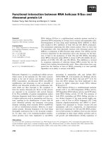
group A activity. Figure 6A shows that LT-I only
recognized one blood group A-active glycoprotein,
which was identified as pro-sucrase-isomaltase by reac-
tion with the corresponding antibody and the expected
relative migration after SDS ⁄ PAGE. In the P2 frac-
tions from differentiated HT29-5F7, no glycoprotein
with the ability to bind LT-I (data not shown), and no
glycoprotein carrying the blood group A determinant,
were detected.
Total lipid extracts from both differentiated HT29-
ATCC and HT29-5F7 cells were separated by HPTLC
and assayed for binding of the blood group A mAb
and LT-I by the TLC-overlay technique. Figure 6B
shows that LT-I recognized GM1 and several blood
group A-active glycosphingolipids, migrating more
slowly than GM1, from lipid extracts of both differen-
tiated cells. The ability of LT-I to interact more effi-
ciently with the complex glycosphingolipids carrying
the blood group A determinant from polarized cells is
similar to that previously observed with lipid extracts
from undifferentiated HT29 cells [16].
Discussion
LT-I is a major virulence factor of enterotoxigenic
E. coli, which colonizes human and animal intestines.
The toxic activity of LT-I on the target cell is mediated
by permanent activation of adenylate cyclase, which
increases the cyclic AMP level in intestinal mucosa
cells. Consequently, alteration in Na
+
and Cl
–
fluxes
in villus and crypt cells has been involved in the char-
acteristic symptoms of diarrhea.
The polarized HT29 cell model was used, in this
work, to study the interaction of LT-I with non-GM1
receptors. The undifferentiated HT29 parental cell line
contains a very small proportion of differentiated cell
types, which, under a pressure selection process,
emerge as one of mainly two differentiated polarized
enterocyte-like or mucus-secreting phenotypes [17].
The mechanisms by which biochemical conditions or
drug pressure induce survival of colon carcinoma cells
are currently under study [28–31]. In the present work,
enterocytic differentiation was induced in HT29-ATCC
parental cells and clone HT29-5F7, as detected by
ultrastructural and functional studies. At late conflu-
ence, cells were polarized, had well developed brush
border at the apical membrane and expressed several
intestinal enzymes from the mature enterocyte. The
Table 2. Cyclic AMP production elicited by Escherichia coli heat-
labile enterotoxin (LT-I) on HT29 cells. Effect of CT-B and Helix po-
matia lectin. Undifferentiated and differentiated HT29-ATCC and
HT29-5F7 cells were pre-incubated at 4 °C with 1.0 l
M CTB or
10 l
M H. pomatia lectin and then cells were further incubated with
10 n
M LT-I at 37 °C for 90 min. Intracellular cyclic AMP was meas-
ured as described in the Experimental procedures. Values are the
mean ± SD of two experiments.
Cells
Cyclic AMP (pmol ⁄ 10
6
cells)
LT-I LT-I + CT-B LT-I + HP
HT29-ATCC 670 ± 70 400 ± 50 48.0 ± 11
HT29 differentiated 1260 ± 130 915 ± 90 112.0 ± 28
HT29-5F7 1170 ± 120 830 ± 80 5.2 ± 0.4
HT29-5F7 differentiated 2270 ± 120 1600 ± 130 2.1 ± 0.3
AB
Fig. 6. Blood group A-active glycoconjugates from differentiated
HT29 cells and their ability to interact with Escherichia coli heat-
labile enterotoxin (LT-I). (A) Blood group antigenic activity and LT-
I-binding properties of brush border-enriched P2 fractions from dif-
ferentiated HT29-ATCC-P2 fractions were separated by SDS ⁄ PAGE
and electrotransferred to nitrocellulose. Nitrocellulose strips were
incubated with mouse monoclonal anti-(blood group A) or anti-(suc-
rase-isomaltase) (SI) Ig and then with horseradish peroxidase
(HRP)-conjugated monoclonal anti-mouse Ig. For LT-I binding, nitro-
cellulose strips were incubated with 5.0 n
M LT-I followed by incu-
bation with rabbit anti-LT-I Ig and HRP-conjugated Protein A. In all
cases, peroxidase was revealed by a chemiluminescent reaction.
(B) Blood group A activity and LT-I binding to glycosphingolipids
from HT29-ATCC and HT29-5F7 differentiated cells. HPTLC plates
were overlaid with anti-(blood group A) IgM and then with a secon-
dary HRP-conjugated antibody. Peroxidase was revealed with
0.05% 4-chloro-1-naphtol and 0.01% hydrogen peroxide as sub-
strate solution.
Interaction of LT-I with differentiated HT29 cells E. M. Galva
´
n et al.
3448 FEBS Journal 273 (2006) 3444–3453 ª 2006 The Authors Journal compilation ª 2006 FEBS
morphological features of differentiated HT29-ATCC
and HT29-5F7 cells observed in this work closely
resembled that previously reported [21,22,29,30]. Func-
tionally, differentiation was accompanied by the
expression of aminopeptidase N, lactase, maltase and
sucrase activities. Sucrase-isomaltase is localized at the
apical brush border membranes of HT29 cells differen-
tiated in RPMI [22] and by glucose deprivation [26,27].
LT-I binds to the high-affinity receptor, GM1, and
to alternate receptors (glycosphingolipid and gycopro-
teins) from several cell membranes [8–10,12–16].
We have previously described that ABH-active
glycoconjugates could act as alternate LT-I receptors
on intestinal brush border membranes from pig and
rabbits and in undifferentiated HT29-ATCC cells
[12–16]. In the present work, we found that specific
LT-I binding to differentiated intestinal cells is not sig-
nificantly diminished in the presence of a molar excess
of CT-B (Fig. 3), which may reflect a very low contri-
bution of GM1 to LT-I binding on cells. Saturation
curves performed on differentiated HT29-ATCC and
HT29-5F7 cells showed that
125
I-labelled CT maximally
bound 74 and 28 fmolÆ10
)6
cells, respectively (data not
shown), supporting the idea of the existence of an
unbalanced ratio between alternate ⁄ GM1 receptor sites
in HT29 cells. By using the polarized HT29-ATCC cell
line and the HT29-5F7 clone we demonstrated an
increased expression (two- to four-fold) of non-GM1
LT-I receptor sites with respect to the undifferentiated
control cells.
The dose-dependent inhibition of LT-I binding by
H. pomatia lectin clearly indicates that LT-I recognized
blood group A-active glycoconjugates on the cell sur-
face of undifferentiated [16] and differentiated HT29
cells (Fig. 4). Although no direct quantification of
blood group A-active glycoconjugates on the cell sur-
face was performed, we assumed that the higher num-
ber of LT-I receptor sites on differentiated cells should
result from a greater number of blood group A-active
glycoconjugates on the cell surface. Differentiation of
adenocarcinoma cell lines (e.g. HT29, Caco-2) to an
enterocyte like-status involves a change in morphologi-
cal features, such as the development of brush border
membranes. A great increase of the brush border
membrane surface in differentiated cells (Fig. 1) may
increase the number of receptor sites provided by
blood group A-glycosphingolipid and blood group
A-glycoprotein sucrase-isomaltase (the latter on the
HT29-ATCC plasma membrane).
Polarized cells were also capable of inducing an
increase in the intracellular cyclic AMP level in
response to LT-I concentrations higher than 6 nm.
This effect was observed, even at 10 nm toxin, when
the number of occupied binding sites of differentiated
and control cells were similar. We have no clear
explanation for this observation and further studies are
necessary to add new insight into the mechanism of
the toxin action on these cells. However, we speculate
that the enhanced cyclic AMP production can be rela-
ted to the polarized status of cells, which may allow a
more efficient coupling of the secondary signal path-
ways triggered by the toxin in respect to nonpolarized
HT29 cells. For CT, it has been shown that a small
percentage of the cell-bound toxin is converted to A1
peptide over a period of time during which the full
activation of adenylate cyclase is reached [6]. Because
CT binding to differentiated cells was completely
blocked by 100 nm CT-B in the present work (results
not shown), we attributed cyclic AMP accumulation in
polarized cells to the action of LT-I on low-affinity
non-GM1 LT-I receptors. Apparently, these alternate
receptors account for 70% of the total cyclic AMP
response to LT-I in both polarized cell lines (Table 2).
Using toxin overlay assays, we found that several
blood group A-glycosphingolipids from HT29-ATCC
and HT29-5F7 cell lines, migrating more slowly than
GM1, efficiently bound LT-I. These results, together
with the inhibitory effect of H. pomatia on toxin
action, indicated that glycoconjugates bearing the
blood group A determinant are additional receptors to
LT-I in HT29-ATCC and HT29-5F7 cells. We have
recently reported that blood group A-active glycosp-
hingolipids, migrating more slowly than GM1, are
additional LT-I receptors in parental HT29 cells and
that these non-GM1 receptors may account for 50%
of the cyclic AMP response elicited by the toxin in
these cells [16]. The results from this work indicate that
blood group A-active glycosphingolipids are major
functional LT-I alternate receptors in HT29-ATCC
and HT29-57 cells.
Even though glycosphingolipid distribution in
polarized cells was not investigated in this work, we
speculate that polarized HT29 cells have glycosphingo-
lipid-enriched brush border membranes resembling the
mature enterocyte [32]. Interestingly, a glycoprotein
band present in the brush border-enriched membrane
preparation from HT29-ATCC cells bound LT-I. This
glycoprotein was identified as the glycosylated blood
group A-active pro-sucrase-isomaltase by western blot
assays (Fig. 5). The glycosylated pro-form of sucrase-
isomaltase has been clearly detected in the enterocytic
differentiated HT29 cells carrying the A blood group of
the human donor [26,27]. Furthermore, the results of
the present work suggest that this glycoprotein may
function as an LT-I receptor on human intestinal brush
border membranes. Sucrase-isomaltase has already
E. M. Galva
´
n et al. Interaction of LT-I with differentiated HT29 cells
FEBS Journal 273 (2006) 3444–3453 ª 2006 The Authors Journal compilation ª 2006 FEBS 3449
been postulated as a glycoprotein receptor of LT-I on
intestinal brush border membranes from several animal
species, but we have detected this interaction between
LT-I and blood group-A active-sucrase-isomaltase in
porcine and rabbit intestines [13,14]. The present
results, together with our earlier findings, support the
idea that the blood group A determinant (mostly of the
type 2 oligosaccharide chain) from glycosphingolipids
and glycoproteins may actually be involved in the car-
bohydrate structure recognized by LT-I. Recently, the
fine structural basis of the interaction of a hybrid
between CT and LT-I and a type 2 blood group A pen-
tasaccharide, which involves a novel binding site at the
toxin molecule, was established [33].
Several epidemiological studies have demonstrated a
relationship between ABH blood group status and
high risk of developing cholera [34–37]. Recently, a
study was carried out to eilucidate the relationship of
the ABH blood group, immunity and susceptibility
to symptomatic and asymptomatic infections with
V. cholerae [38]. An association has also been observed
in the occurrence of diarrhea after ingestion of E. coli-
producing LT-I in volunteers [39]. LT-I and, to a
much lesser degree, CT, interacted with ABH glyco-
conjugates from human and animal intestinal mucosa
[12–14], and furthermore, some of these interactions
have proved to be functional [15,16]. These interac-
tions may have relevance in the clinical outcome of
diarrhea caused by LT-I and CT in relation to the
blood group of the patient.
Regarding differentiated HT29 cells as intestinal
model system, it is apparent that enterocyte-like differ-
entiated HT29 cells provide a useful in vitro model to
evaluate the functional role of interactions between
bacterial virulence factors and intestinal polarized cells.
Experimental procedures
Cell culture
The human colon adenocarcinoma HT29 parental cell line
(HT29-ATCC) was grown in Dulbecco’s modified Eagle’s
medium (D-MEM) containing 10% heat-inactivated fetal
bovine serum. Enterocytic differentiation was performed as
described by Hekmati et al. [21]. Briefly, cells were switched
to RPMI-1640 containing 10% inactivated fetal bovine
serum, replated four times in this medium and then exam-
ined at late confluence (18–21 days). After HT29-ATCC
cells reached confluence, RPMI-1640 was changed every
day. Clone HT29-5F7, which was selected by resistance to
5-fluoruracil (kindly donated by Dr T. Lesuffleur, INSERM
U560, Lille, France) was usually grown in D-MEM, con-
taining 10% inactivated fetal bovine serum, and examined
at early confluence (undifferentiated) or at late confluence
(12 days) when the cells exhibit a polarized phenotype
[23,30]. Antibiotics (100 UÆmL
)1
penicillin, 100 lgÆmL
)1
streptomycin) were added to both D-MEM and RPMI-
1640. Cell lines were maintained at 37 °C in a humidified
atmosphere containing 5% CO
2
. Cell number was deter-
mined by Trypan blue exclusion in a hemocytometer.
Toxin-binding assay
LT-I was iodinated by a stoichiometric method with chlor-
amine T [40], as described previously [11], and the specific
activity for the iodinated LT-I was 3.0 lCiÆlg
)1
. Biological
activity of the
125
I-labelled LT-I preparation was 90%,
measured as the percentage of
125
I-labelled LT-I total pro-
tein able to specifically bind to GM1-containing membranes
(rat red blood cells or NHI 3T3 fibroblasts).
Toxin binding to cells in culture was assayed as previ-
ously described [16]. Briefly, cells were incubated in serum-
free D-MEM buffered with 25 mm Hepes or RPMI-1640
containing 0.01% BSA without (total binding) or with
unlabeled LT-B (1.0 lm) before the addition of
125
I-labelled
toxin (3.0 lCiÆlg
)1
). After 60 min at 4 °C, cells were
washed, solubilized with NaOH and the radioactivity was
counted. Nonspecific binding was measured as the binding
of
125
I-labelled toxin in the presence of an excess of unlabe-
led LT-B.
To assay nonspecific binding of
125
I-labelled LT-I or
competitive inhibition by unlabelled LT-B or CT-B, the B
subunits of toxin were incubated with cells for 30 min at
4 °C and then further incubated with
125
I-labelled LT-I for
60 min at 4 °C. The blocking effect of
125
I-labelled LT
binding by H. pomatia lectin was also determined by pre-
incubation of cells with lectin, as indicated for B subunits
of toxins.
Toxin-stimulated accumulation of intracellular
cyclic AMP
The toxin-stimulated accumulation of intracellular cyclic
AMP was determined as described previously [16]. Briefly,
cells were pre-incubated without or with CT-B (1.0 lm), or
H. pomatia lectin (10 lm), at 4 °C. LT-I was then added
for 90 min at 37 °C. Finally, cells were treated with 0.1 m
HCl and the dried acid extracts were assayed for cyclic
AMP by RIA (Immunotech SA, Marseille, France), accord-
ing to instructions of the manufacturer.
Electron microscopy
TEM was performed as follows. Cell monolayers were fixed
in 2% glutaraldehyde and then postfixed in 1% OsO
4
.
After dehydration in graded ethanol solutions, the cells
were embedded in Epon. Ultrathin sections were contrasted
Interaction of LT-I with differentiated HT29 cells E. M. Galva
´
n et al.
3450 FEBS Journal 273 (2006) 3444–3453 ª 2006 The Authors Journal compilation ª 2006 FEBS
with uranyl acetate and lead citrate. Thin sections were
examined in a Jeol EX 1220 transmission electron micro-
scope (Jeol, Tokyo, Japan).
Hydrolase assays
Brush border-enriched membrane fractions (P2) were pre-
pared according to Trugnan et al. [27]. Briefly, cells were
scraped in Tris-mannitol buffer, pH 7.1, containing prote-
ase inhibitors (1.0 lgÆmL
)1
antipain, 17.5 lgÆmL
)1
benzami-
dine, 1.0 mm phenylmethylsulfonyl fluoride, 1.0 lgÆmL
)1
pepstatin, 10 lgÆmL
)1
aprotinin and 1.0 lgÆmL
)1
leupep-
tin). Cells were disrupted by sonication and then CaCl
2
was
added (to 18 mm). The homogenate was centrifuged (950 g,
10 min; Rotor Type 50, Beckman Instruments, Fullerton,
CA, USA) and the supernatant was centrifuged again
(33 500 g, 30 min) to yield the P2 fraction. Proteins were
measured by the method of Lowry et al. [41].
Glycohydrolases (sucrase, maltase and lactase) and ami-
nopeptidase N activities were determined in P2 fractions
according to Messer and Dalqvist [42] and Maroux et al.
[43], respectively. The enzyme activities are expressed as milli-
units (mU) per mg of protein. One unit is defined as the acti-
vity that hydrolyzes 1.0 l mol of substrate per min at 37 °C.
ABH phenotyping of cellular glycoconjugates and
toxin-binding assays
To detect blood group-active and toxin-binding glycopro-
teins, P2 fractions were separated by 7.5% SDS ⁄ PAGE,
electrotransferred to nitrocellulose sheets, and immuno-
stained as previously described [16]. The sucrase–isomaltase
complex was identified using a mouse anti-(human sucrase-
isomaltase) IgG (kindly donated by Dr A. Quaroni, Ithaca,
NY, USA) followed by a horseradish peroxidase-conju-
gated secondary antibody. Peroxidase was detected with
an enhanced chemiluminiscence immunodetection system
(Amersham Biosciences, Uppsala, Sweden).
Total lipids from cells were extracted and separated using
HPTLC. Glycolipids that bind either the toxins or the anti-
(blood group) IgM were immunodetected, essentially as
previously described [16].
Acknowledgements
We thank Dr W. S. Dallas (Glaxo Wellcome Research,
NC, USA) for providing the LT-I producing- bacterial
strains, Dr J. D. Clements (Tulane University, New
Orleans, LA, USA) for kindly donating LT-B, Dr
The
`
cla Lesuffleur (INSERM U560, France) for provi-
ding the HT29-5F7 clone and Dr Andrea Quaroni
(Cornell University, NY, USA) for providing mouse
monoclonal anti-human intestinal hydrolases. This
work was supported partly by grants from Consejo
Nacional de Investigaciones Cientı
´
ficas y Te
´
cnicas
(CONICET), Agencia Nacional de Promocio
´
n Cientı
´
fi-
ca y Tecnolo
´
gica (BID 1201 ⁄ OC-AR, PICT 05–10607)
and Secretarı
´
a de Ciencia y Te
´
cnica de la Universidad
Nacional de Co
´
rdoba (SeCyT-UNC), Argentina. EMG
was a fellow from CONICET and GAR and CGM are
senior career investigators from CONICET.
References
1 Spangler BD (1992) Structure and functions of cholera
toxin and the related Escherichia coli heat-labile entero-
toxin. Microbiol Rev 56, 622–647.
2 Finkelstein RA, Burks MF, Zupan A, Dallas WS, Jacob
CO & Ludwig DS (1987) Epitopes of the cholera family
of enterotoxins. Rev Infect Dis 9, 544–561.
3 Bastiaens PI, Majoul IV, Verveer PJ, Soling HD &
Jovin TM (1996) Imaging the intracellular trafficking
and state of AB5 quaternary structure of cholera toxin.
EMBO J 15, 4246–4253.
4 Torgersen ML, Skretting G, van Deurs B & Sandvig K
(2001) Internalization of cholera toxin by different
endocytic mechanisms. J Cell Sci 114, 3737–3747.
5 Tsai B, Ye Y & Rapoport T (2002) Retro-translocation
of proteins from the endoplasmic reticulum into the
cytosol. Nat Rev Mol Cell Biol 3, 246–255.
6 Fishman PH (1990) Mechanism of action of cholera
toxin. In ADP-Ribosylating Toxins and G Proteins:
Insights Into Signal Transduction. (Moss J & Vaughan
M, eds), pp. 127–140. American Society for Micro-
biology, Washington DC.
7 Moss J & Vaughan M (1981) Mechanism of action of
choleragen and E. coli heat-labile enterotoxin: Activa-
tion of adenylate cyclase by ADP-ribosylation. Mol Cell
Biochem 37, 75–90.
8 Teneberg S, Hirst TR, Angstro
¨
m J & Karlsson K-A
(1994) Comparison of the glycolipid-binding specificities
of cholera toxin and porcine Escherichia coli heat-labile
enterotoxin: identification of a receptor-active non-gang-
lioside glycolipid for the heat-labile toxin in infant rab-
bit small intestine. Glycoconjug J 11, 533–540.
9 Griffiths SL & Crithley DR (1991) Characterization of
the binding sites for Escherichia coli heat-labile toxin
type I in intestinal brush borders. Biochim Biophys Acta
1075, 154–161.
10 Zemelman BV, Chu SHW & Walker WA (1989) Host
response to Escherichia coli heat-labile enterotoxin via
two microvillus membrane receptors in the rat intestine.
Infect Immun 57, 2947–2952.
11 Monferran CG, Roth GA & Cumar FA (1990) Inhibi-
tion of cholera toxin binding to membrane receptors by
pig gastric mucin-derived glycopeptides: Differential
effect depending on the ABO blood group antigenic
determinants. Infect Immun 58, 3966–3972.
E. M. Galva
´
n et al. Interaction of LT-I with differentiated HT29 cells
FEBS Journal 273 (2006) 3444–3453 ª 2006 The Authors Journal compilation ª 2006 FEBS 3451
12 Barra JL, Monferran CG, Balanzino LE & Cumar FA
(1992) Escherichia coli heat-labile enterotoxin preferen-
tially interacts with blood group A-active glycolipids
from pig intestinal mucosa and A- and B-active glyco-
lipids from human red cells compared to H-active
glycolipids. Mol Cell Biochem 115, 63–70.
13 Balanzino LE, Barra JL, Monferran CG & Cumar FA
(1994) Differential interaction of Escherichia coli heat-
labile toxin and cholera toxin with pig intestinal brush
border glycoproteins depending on their ABH and
related blood group antigenic determinants. Infect
Immun 62, 1460–1464.
14 Balanzino LE, Barra JL, Galva
´
n EM, Roth GA &
Monferran CG (1999) Interaction of cholera toxin and
Escherichia coli heat-labile enterotoxin with glycoconju-
gates from rabbit intestinal brush border membranes:
Relationship with ABH blood groups determinants.
Mol Cell Biochem 194, 53–62.
15 Galva
´
n EM, Roth GA & Monferran CG (1999) Partici-
pation of ABH glycoconjugates in the secretory
response to Escherichia coli heat- labile toxin in rabbit
intestine. J Infect Dis 180, 419–425.
16 Galva
´
n EM, Diema CD, Roth GA & Monferran CG
(2004) Ability of Blood Group A-Active Glycosphingo-
lipids to Act as Escherichia coli Heat-Labile Enterotoxin
Receptors in HT-29 cells. J Infect Dis 189, 1556–1564.
17 Zweibaum A, Pinto M, Chevallier G, Dussaulx E, Tria-
dou N, Lacroix B, Haffen K, Brun JL & Rousset M
(1985) Enterocytic differentiation of a subpopulation of
the human colon- tumor cell line HT-29 selected for
growth in sugar-free medium and its inhibition by glu-
cose. J Cell Physiol 122, 21–29.
18 Huet C, Sahuquillo-Merino C, Coudrier E & Louvard
D (1987) Absorptive and mucus.secreting subclones
isolated from a multipotent intestinal cell line (HT-29)
provide new models for cell polarity and terminal differ-
entiation. J Cell Biol 105, 345–357.
19 Augeron C & Laboisse CL (1984) Emergence of Perma-
nently Differentiated Cell Clones in a Human Colonic
Cancer Cell Line in Culture after Treatment with
Sodium Butyrate. Cancer Res 44, 3961–3969.
20 Lesuffleur T, Kornowski A, Luccioni C, Muleris M,
Barbat A, Beaumatin J, Dussaulx E, Dutrillaux B &
Zweibaum A (1991) Adaptation to 5-fluorouracil of the
heterogeneous human colon tumor cell line HT-29
results in the selection of cells committed to differentia-
tion. Int J Cancer 49, 721–730.
21 Hekmati M, Polak-Charcon S & Ben-Shaul Y (1990) A
morphological study of a human adenocarcinoma cell
line (HT29) differentiating in culture. Similarities to
intestinal embryonic development. Cell Differ Dev 31,
207–218.
22 Polak-Charcon S, Hekmati M & Ben Shaul Y (1989)
The effect of modifying the culture medium on cell
polarity in a human colon carcinoma cell line. Cell
Differ Dev 26, 119–129.
23 Lesuffleur T, Violette S, Vasile-Pandrea I, Dussaulx E,
Barbat A, Muleris M & Zweibaum A (1998) Resistance
to high concentrations of methotrexate and 5-fluoroura-
cil of differentiated HT-29 colon-cancer cells is restricted
to cells of enterocyte phenotype. Int J Cancer 76, 383–
392.
24 Pinto M, Robine-Leon S, Appay MD, Kedinger M,
Triadou N, Dessaulx E, Lacroix B, Simon-Assmann P,
Haffen K, Fogh J et al. (1983) Enterocyte-like differen-
tiation and polarization of the of the human colon car-
cinoma cell line Caco2 in culture. Biol Cell 47, 323–330.
25 Hauri H-P, Sterchi EE, Bienz D, Fransen JAM &
Marxer A (1985) Expression and Intracellular Transport
of Microvillus Membrane Hydrolases in Human intest-
inal epithelial cells. J Cell Biol 101, 838–851.
26 Chantret I, Chevallier G, Dussaulx E & Zweibaum A
(1987) A and H Blood Group Antigens as Markers of
Sucrase-Isomaltase from the Enterocyte-like Differen-
tiated Human Colon Carcinoma Cell Lines HT-29 and
Caco-2. Cancer Res 47, 1426–1433.
27 Trugnan G, Rousset M, Chantret I, Barbat A & Zwei-
baum A (1987) The Posttranslational Processing of
Sucrase-Isomaltase in HT-29 Cells is a function of Their
State of Enterocytic Differentiation. J Cell Biol 104,
1199–1205.
28 Violette S, Poulain L, Dussaulx E, Pepin D, Faussat
AM, Chambaz J, Lacorte JM, Staedel C & Lesuffleur T
(2002) Resistance of colon cancer cells to long-term
5-fluorouracil exposure is correlated to the relative level
of Bcl-2 and Bcl-X(L) in addition to Bax and p53 sta-
tus. Int J Cancer 98, 498–504.
29 Violette S, Festor E, Pandrea-Vasile I, Mitchell V,
Adida C, Dussaulx E, Lacorte JM, Chambaz J,
Lacasa M & Lesuffleur T (2003) Reg IV, a new member
of the regenerating gene family, is overexpressed in
colorectal carcinomas. Int J Cancer 103, 185–193.
30 Leteurtre E, Gouyer V, Rousseau K, Moreau O, Barbat
A, Swallow D, Huet G & Lesuffleur T (2004) Differen-
tial mucin expression in colon carcinoma HT-29 clones
with variable resistance to 5-fluorouracil and methotrex-
ate. Biol Cell 92, 145–151.
31 Gout S, Laine MC, Tavernier G, Block MR & Jacqu-
ier-Starlin M (2004) Early enterocytic differentiation of
HT-29 cells: biochemical changes and strength increases
of adherens junctions. Exp Cell Res 299, 498–510.
32 Simons K & van Meer G (1988) Lipid sorting in epithe-
lial cells. Biochemistry 27, 6197–6202.
33 Holmner A, Lebens M, Teneberg S, Angstrom J, O
¨
kvist
M & Krengel U (2004) Novel binding site identified in a
hybrid between cholera toxin and heat-labile entero-
toxin: 1.9 A
˚
crystal structure reveals the details. Struc-
ture 12, 1655–1667.
Interaction of LT-I with differentiated HT29 cells E. M. Galva
´
n et al.
3452 FEBS Journal 273 (2006) 3444–3453 ª 2006 The Authors Journal compilation ª 2006 FEBS
34 Barua D & Paguio AS (1977) ABO blood groups and
cholera. Ann Hum Biol 4, 489–492.
35 Chaudhuri AS & De S (1977) Cholera and blood-
groups. Lancet 2, 404.
36 Glass RI, Holmgren J, Haley CE, Khan MR, Svenner-
holm A-M, Stoll BJ, Belayet Hossain KM, Black RE,
Yunus M & Barua D (1985) Predisposition for cholera
of individuals with O blood group. Am J Epidemiol 121,
791–796.
37 Levine MM, Nalin DR, Rennels MB, Hornick RB,
Sotman S, van Blerk G, Hughes TP & O’Donnell S
(1979) Genetic susceptibility to cholera. Ann Hum Biol
6, 369–374.
38 Harris JB, Khan AI, LaRocque RC, Dorer DJ, Chow-
dhury F, Faruque ASG, Sack DA, Ryan ET, Quadri F
& Calderwood SB (2005) Blood group, immunity, and
risk of infection with Vibrio cholerae in an area of ende-
micity. Infect Immun 73, 7422–7427.
39 Black RE, Levine MM, Clements ML, Hughes T &
O’Donnell S (1981) Association between O blood group
and occurrence and severity of diarrhoea due to Escheri-
chia coli. Trans R Soc Trop Med Hyg 81, 120–123.
40 Roth J (1975) Methods for assessing immunologic and
biologic properties of iodinated peptide hormones.
Methods Enzymol 37B, 223–233.
41 Lowry OH, Rosebrough NJ, Farr AL & Randall RJ
(1951) Protein measurement with the Folin phenol
reagent. J Biol Chem 193, 265–275.
42 Messer M & Dahlqvist A (1966) A one-step ultramicro-
method for the assay of intestinal dissacharidases. Anal
Biochem 14, 376–392.
43 Maroux S, Louvard D & Baratti J (1973) The amino-
peptidase from hog intestinal brush border. Biochim
Biophys Acta 321, 282–295.
E. M. Galva
´
n et al. Interaction of LT-I with differentiated HT29 cells
FEBS Journal 273 (2006) 3444–3453 ª 2006 The Authors Journal compilation ª 2006 FEBS 3453