Báo cáo khoa học: Oxidative stress and apoptotic events during thermal stress in the symbiotic sea anemone, Anemonia viridis potx
Bạn đang xem bản rút gọn của tài liệu. Xem và tải ngay bản đầy đủ của tài liệu tại đây (1.69 MB, 13 trang )
Oxidative stress and apoptotic events during thermal
stress in the symbiotic sea anemone, Anemonia viridis
Sophie Richier
1
,Ce
´
cile Sabourault
1
, Juliette Courtiade
1
, Nathalie Zucchini
3
, Denis Allemand
1,2
and Paola Furla
1
1 UMR 1112 UNSA-INRA ROSE, Nice-Sophia Antipolis University, Nice, France
2 Centre Scientifique de Monaco, Monaco
3 UMR 1112 UNSA-INRA ROSE, Sophia-Antipolis, France
Over the past several decades, symbiotic invertebrates
such as cnidarians, sponges and mollusks have been
regularly affected by a phenomenon known as ‘bleach-
ing’. This event has been observed all around the world
and involves principally the mass expulsion of unicel-
lular photosynthetic symbionts from animal tissue.
Bleaching phenomenon has been widely reported in the
ecologically and economically important tropical corals
reef, but several other invertebrates such as giant clams,
gorgonians and sea anemones have also been affected.
Previous studies have established a causal link
between environmental stresses, such as elevated tem-
perature, ultraviolet light, pathogen infection or pol-
lution, and symbiosis disruption (reviewed in [1]).
Mass symbiont expulsion has, however, been most fre-
quently associated with elevated seawater temperature,
generally considered to be the primary stress causing
worldwide bleaching [2]. General mechanisms have
been proposed to explain the thermal sensitivity of
symbiotic cnidarians including symbiont photoinhibi-
tion [3–6], cell degradation [7] and cell death [8,9].
However, despite the importance of the phenomenon,
the underlying molecular mechanisms associated with
symbiosis breakdown remained undetermined.
Oxidative stress is one molecular pathway that has
been suggested to cause bleaching. A pro-oxidant per-
iod is experienced daily by invertebrates harboring pho-
tosynthetic symbionts due to the high concentration of
oxygen produced throughout photosynthesis [10–13].
The light-dependent hyperoxic state induces high fluxes
of reactive oxygen species (ROS) such as O
À
2
and OH
•
[14] produced largely from mitochondria and chloro-
plasts. This increase in ROS is counterbalanced by an
efficient antioxidant capacity in the host and symbiont
cells [12,13,15,16]. The first hypothesis of oxidative
stress involvement in the bleaching event was proposed,
Keywords
apoptosis; bleaching; caspase; cnidarian;
oxidative stress
Correspondence
P. Furla, UMR 1112 ROSE, Nice-Sophia
Antipolis University, Parc Valrose, BP 71,
F-06108 Nice Cedex 2, France
Fax: +33 4 92 07 65 63
Tel: +33 4 92 07 68 30
E-mail:
(Received 6 April 2006, revised 30 June
2006, accepted 11 July 2006)
doi:10.1111/j.1742-4658.2006.05414.x
Symbiosis between cnidarian and photosynthetic protists is widely distri-
buted over temperate and tropical seas. These symbioses can periodically
breakdown, a phenomenon known as cnidarian bleaching. This event can be
irreversible for some associations subjected to acute and ⁄ or prolonged envi-
ronmental disturbances, and leads to the death of the animal host. During
bleaching, oxidative stress has been described previously as acting at mole-
cular level and apoptosis is suggested to be one of the mechanisms involved.
We focused our study on the role of apoptosis in bleaching via oxidative
stress in the association between the sea anemone Anemonia viridis and
the dinoflagellates Symbiodinium species. Characterization of caspase-like
enzymes were conducted at the biochemical and molecular level to confirm
the presence of a caspase-dependent apoptotic phenomenon in the cnidarian
host. We provide evidence of oxidative stress followed by induction of
caspase-like activity in animal host cells after an elevated temperature stress,
suggesting the concomitant action of these components in bleaching.
Abbreviations
AFC, 7-amino-4-trifluromethylcoumarin; CARD, caspase recruitment domain; CHO, adelhyde; DEVD, Asp-Glu-Val-Asp; IETD, Ile-Glu-Thr-Asp;
ROS, reactive oxygen species; TUNEL, dUTP nick end labeling.
4186 FEBS Journal 273 (2006) 4186–4198 ª 2006 The Authors Journal compilation ª 2006 FEBS
and later supported, by Lesser and coworkers [17–20].
These studies demonstrated the role of ROS production
in the temperature-induced bleaching. During thermal
stress, although the enzymatic antioxidant defenses are
induced [18,21–23], the additional amount of ROS pro-
duction causes a large increase in cellular damage such
as protein carbonylation [21,22,24], lipid peroxidation
[24] and DNA degradation [23].
ROS could be involved in cell death by two path-
ways: (a) they could cause oxidative stress that leads
to massive cellular damage [25] and they could be
involved in necrosis or in so-called postmitochondrial
phase of apoptosis [26]; or (b) they could be involved
in the initiation phase of apoptosis contributing to cell
death signaling [27].
Programmed cell death is known to model tissue dur-
ing embryogenesis, to remove damaged cells, protect
against pathogen infection, and regulate cell numbers
and tissue homeostasis. Program cell death is character-
istic of all multicellular animals and can be extended
now to the most basal metazoan phyla as porifera and
cnidaria with occurrence of apoptosis and homologues
of caspases and Bcl2 proteins [28]. Moreover, apoptosis
has been remarkably well conserved throughout meta-
zoan phyla both in terms of morphological features
and of the genes controlling the process. Recently, mor-
phological indicators of programmed cell death or
apoptosis have been observed in a symbiotic sea anem-
one, Aiptasia pallida, subjected to thermal stress [8,9],
leading to the proposal of a new molecular pathway for
bleaching induction. Furthermore, DNA cleavage
analysis [8,23] and increased expression of p53, a pro-
apoptotic protein expression [23], confirmed tempera-
ture-induced DNA damage in symbiotic cnidarians,
which in turn could activate the apoptotic cascade.
Upstream to specific morphological modifications,
apoptosis is also characterized by activation of highly
selective cysteine aspartate-specific proteases, known as
‘caspases’, which are constitutively expressed as pro-
enzymes with little catalytic activity and are activated
following apoptotic stimulation. Evidence of caspase 3-
like expression in cnidarians was first obtained in
Hydra vulgaris by Cikala et al. [29] with caspase activ-
ity measurements and gene characterization. Recently,
evidence of caspase-like involvement in Hydractinia
echinata metamorphosis [30] and a caspase gene in the
sea anemone Aiptasia pallida [31] has been shown. To
date however, no relation between heat stress and ca-
spase activity has ever been established.
In this study, we examined the biological effects of
heat stress on the sea anemone Anemonia viridis, living
in symbiosis with the unicellular dinoflagellate, Symb-
iodinium sp. commonly known as zooxanthellae. The
first aim of this study was to characterize caspase-like
activity and clone a putative caspase cDNA in sea
anemone tissues. In the second part of the work, we
tested the effect of hyperthermal stress on antioxidant
induction and on apoptotic markers (caspase-like acti-
vation and ⁄ or DNA degradation) in order to demon-
strate the concomitant involvement of oxidative stress
and apoptosis in a thermally induced bleaching event.
Results
Detection of caspase-like activity in tissue
extracts of A. viridis
In order to test for the presence of caspase-like activity
in the symbiotic sea anemone A. viridis, Asp-Glu-Val-
Asp (DEVD)-dependent (Fig. 1A) and Ile-Glu-Thr-Asp
(IETD)-dependent (Fig. 1B) protease activities were
tested in animal host cells (ectoderm and gastroderm)
and in freshly isolated zooxanthellae. For both sub-
strates, high protease activities were measured in the
animal host while only low activities were measured in
the freshly isolated zooxanthellae extracts. Moreover,
in the host extracts, IETD substrate presented a two-
fold higher rate of 7-amino-4-trifluoromethylcoumarin
(AFC) cleavage than DEVD-AFC substrate.
Addition of the inhibitors DEVD-adelhyde (CHO)
and IETD-CHO specific for the caspases 3 and 8,
respectively, completely abolished both protease activit-
ies in the ectodermal and gastrodermal tissue extracts
(Fig. 2). Table 1 summarizes the IC
50
for both inhibi-
tors, obtained by incubating extracts in each specific
substrate. In both tissue extracts, DEVD- and IETD-
dependent protease activities showed the same
sensitivity for the DEVD-CHO competitive substrate.
Surprisingly, the DEVD-CHO inhibitor had a higher
effect on IETD-dependent protease activity (3 nm) than
on the DEVD-dependent protease activity (15–20 nm).
For both tissue extracts and protease substrates, the
IETD-CHO inhibitor showed higher IC
50
values with a
predictably higher sensitivity of IETD-dependent activity
to this inhibitor. Although inhibition of IETD-depend-
ent protease activity by IETD-CHO was similar in
ectodermal and gastrodermal cells, inhibition of DEVD-
dependent protease activity by the same inhibitor was
lower in ectodermal cells than in gastrodermal cells.
Identification of a caspase-like cDNA from
A. viridis
To confirm the presence of caspases in A. viridis
tissues, cDNA encoding a caspase 3-like protein was
isolated from ectodermal cells. Using a PCR
S. Richier et al. Apoptosis and oxidative stress in bleaching
FEBS Journal 273 (2006) 4186–4198 ª 2006 The Authors Journal compilation ª 2006 FEBS 4187
approach with degenerate primers based on two
highly conserved caspase 3 domains, we obtained a
1627 bp sequence named AvCasp3 (accession number
DQ097195) containing an open reading frame of
1239 bp (Fig. 3). The predicted amino acid sequence
of 413 amino acids (Fig. 3) is highly conserved with
vertebrate caspase 3 sequences (Fig. 4) and was there-
fore named caspase 3-like. By homology with known
vertebrate caspases, we determined that the long form
of this sequence contains a prodomain, a large (p20)
and a small (p10) subunit. We identified two potential
cleavage sites at aspartate residues 164 and 172 for
cleaving the prodomain, and a potential cleavage site
at Asp306 for the cleavage between the large and
small subunits. The prodomain presents a caspase
recruitment domain (CARD) consisting of six alpha
helices (Fig. 3) [32]. The large subunit contains highly
conserved LS ⁄ THG and QACXG sequences sur-
rounding histine (His255) and cysteine (Cys294) resi-
dues of putative active site. The substrate binding site
is highly conserved and composed of Arg337, Ser343,
Gln296 and Arg198 (Fig. 3).
Large and small subunit sequences from various
vertebrate and invertebrate caspase 3 or caspase 7
sequences were aligned with the A. viridis caspase 3-
like sequence. The phylogenetic comparison (Fig. 4)
shows that AvCasp3 and other cnidarian sequences
delineate a specific branch more closely related to exe-
cutioner vertebrate caspases 3 or 7 than to caspases
from other invertebrates models (Drosophila melano-
gaster and Caenorhabditis elegans).
Effect of heat stress on apoptosis-like induction
in animal tissue of A. viridis
In order to study the effect of a heat stress (+8 °C
above ambient) on caspase-like activities, DEVD- and
IETD-dependent protease activities were measured in
the animal extracts of A. viridis throughout the stress
(7 days at 25 °C). In the ectodermal tissue, the DEVD-
dependent protease activity decreased while IETD-
dependent activity did not vary significantly (Fig. 5A).
In the gastrodermal tissue (Fig. 5B), both activities
increased. The DEVD-dependent activity was twofold
higher than controls (17 °C) after 48 h of stress, while
the IETD-dependent activity was 1.5-fold higher. After
7 days at 25 °C, activities in both tissues were restored
to control levels.
To confirm the induction of a specific caspase-like
activity and not a generic protease activity, all meas-
urements were also performed in the presence of
1 lm DEVD-CHO and 10 lm IETD-CHO (data not
shown). In these conditions, caspase-like activity was
totally abolished. The induction of an apoptosis-like
phenomenon in A. viridis subjected to a heat stress was
confirmed by the analysis of DNA fragmentation in
anemone tissues, using a dUTP nick end labeling
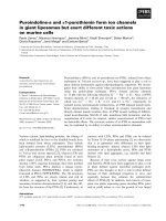
(TUNEL) assay. Figure 6 shows the increase of end-
labeling DNA after 48 h of thermal stress (25 °C).
DNA fragmentation occurred largely in the gastroder-
mal tissue harboring the zooxanthellae (Fig. 6B).
Effect of heat stress on antioxidant defenses and
bleaching in animal tissue of A. viridis
The occurrence of oxidative stress in the animal tissue
(ectoderm and gastroderm) and freshly isolated zoox-
anthellae of A. viridis was monitored during heat
stress (+8 °C above the control temperature) by the
A
0
2
4
6
8
10
12
Ectoderm Gastroderm Zooxanthellae
DEVD-AFC cleavage (pmol
.
min
-1
)
B
0
2
4
6
8
10
12
Ectoderm Gastroderm Zooxanthellae
IETD-AFC cleavage (pmol
.
min
-1
)
Fig. 1. Caspase-like activities in host epithelial tissues (ectoderm
and gastroderm) and zooxanthella extracts of A. viridis maintained
in control condition (+17 °C). Caspase 3-like (A) and caspase 8-like
(B) activities were assayed by fluorometric method using, respect-
ively, Ac-DEVD-AFC and Ac-IETD-AFC as substrates. One hundred
and twenty-five micrograms of protein have been tested for ecto-
derm, gastroderm and zooxanthella extracts. Results are expressed
as means ± SE of at least six independent tissue extractions from
distinct sea anemones.
Apoptosis and oxidative stress in bleaching S. Richier et al.
4188 FEBS Journal 273 (2006) 4186–4198 ª 2006 The Authors Journal compilation ª 2006 FEBS
measurement of oxygen radical-scavenging capacities
(Fig. 7). The ectodermal antioxidant capacity did not
change significantly over the stress period while the
gastrodermal antioxidant capacity increased starting at
6 h, peaking at 24 h at 2.5-fold higher than control
values and decreasing after 48 h. In the zooxanthellae,
the oxygen radical-scavenging capacity decreased signi-
ficantly after 24 h.
Because the stressed organisms present an evident
loss of pigmentation during the stress, concomitant
analyses have been conducted on whole tentacles of
A. viridis to highlight the bleaching event. Figure 8
shows a rapid decrease of chlorophyll (a+c
2
) content
in the first days of the stress period that became
significant with a two times decrease at the end of the
kinetic.
Discussion
In this study, we have investigated a pathway for sym-
biosis breakdown (bleaching) in the symbiotic associ-
ation A. viridis during an elevated temperature stress.
We have also demonstrated connections between oxi-
dative stress and host programmed cell death during
the bleaching event.
Characterization of caspase-like activities
in A. viridis
In order to test for the presence of programmed cell
death or apoptosis in A. viridis subjected to heat stress,
we measured protease activities using mammalian
caspase substrates. In control conditions, we measured
high specific protease activities in the animal compo-
nent (ectoderm and gastroderm) of A. viridis while, in
freshly isolated zooxanthellae an activity was almost
undetectable. The presence of high caspase-like activ-
ities, in animal tissues of control animals, could be
0
20
40
60
80
100
120
0.1 1 1 0 100 1000 10000 100000
Inhibitor concentration (n
M
)
Ac-IETD-AFC
cleavage activity (%)
B
0
20
40
60
80
100
120
140
160
0.1 1 1 0 100 1000 10000 100000
Inhibitor concentration (n
M
)
Ac-DEVD-AFC
cleavage activity (%)
A
Ectodermal extracts
0
20
40
60
80
100
120
0.1 1 10 100 1000 10000 100000
Inhibitor concentration (n
M
)
Ac-IETD-AFC
cleavage activity (%)
D
Ac-DEVD-AFC
cleavage activity (%)
0
20
40
60
80
100
120
140
160
0.1 1 1 0 100 1000 10000 100000
Inhibitor concentration (n
M
)
C
Gastrodermal extracts
Fig. 2. Inhibition of DEVD- and IETD-dependent protease activities in animal tissue of A. viridis by commercial synthetic peptide inhibitors.
One hundred and twenty-five micrograms of ectodermal (A,B) and gastrodermal (C,D) extracts were incubated with the fluorochromic
caspase substrate Ac-DEVD-AFC (A,C) or Ac-IETD-AFC (B,D) and with the competitive inhibitors Ac-DEVD-CHO (d) or Ac-IETD-CHO (s).
Results are expressed as means ± SE of at least six independent tissue extractions from distinct sea anemones.
Table 1. IC
50
of the competitive substrate inhibitors Ac-DEVD-CHO
and Ac-IETD-CHO on the DEVD-dependent and IETD-dependent
protease activities in the two animal tissue extracts of A. viridis.
Tissue extracts
Protease
substrates
Ac-DEVD-CHO
IC
50
(nM)
Ac-IETD-CHO
IC
50
(nM)
Ectodermal Ac-DEVD-AFC 20 45190
Ac-IETD-AFC 3 172
Gastrodermal Ac-DEVD-AFC 15 495
Ac-IETD-AFC 3 105
S. Richier et al. Apoptosis and oxidative stress in bleaching
FEBS Journal 273 (2006) 4186–4198 ª 2006 The Authors Journal compilation ª 2006 FEBS 4189
related to the high regeneration ability of cnidarians.
In fact, the role of apoptosis in development and
regeneration has been determined not only in verte-
brates (i.e., bone regeneration; reviewed in [33]) but
also in invertebrates such as cnidarians and flatworms
[34–36]. Apoptosis is considered a necessary character-
istic of all self-renewing tissues and its presence has
been detected not only in stressed organisms but also
in healthy ones. Mire and Venable [34] reported that
up to 10% of cells from the sea anemone Haliplanella
lineata contained TUNEL-labeled nuclei even under
control conditions.
Moreover, protease activities related to animal
extracts display several properties characteristic of
caspases, the critical central molecules of apoptotic
pathways. First, they were activated by two polypep-
tides, DEVD and IETD, which are used to distinguish
some of the caspase classes in mammalian cells.
DEVD-AFC is generally cleaved by caspase 3, which
belongs to executioner caspases [37] while, IETD-AFC
Fig. 3. Nucleotide and deduced amino acid sequence of caspase 3-like cDNA of A. viridis (AvCasp3). Putative prodomain sequence appears
in italic characters, the small subunit in regular type. The large (p20) subunit in bold and the small subunit fit between the two domains.
Residues boxed are the component of substrate binding site. Asterisks indicate the His and Cys residues of the putative active site located
in the large subunit. The six a -helix components of the prodomain are underlined.
Apoptosis and oxidative stress in bleaching S. Richier et al.
4190 FEBS Journal 273 (2006) 4186–4198 ª 2006 The Authors Journal compilation ª 2006 FEBS
is cleaved by caspase 8, an initiator caspase [38]. In
control specimens of A. viridis, IETD-dependent prote-
ase activity was two times higher than the DEVD-
dependent one, suggesting a predominantly caspase
8-like activity in the animal tissue. Secondly, the spe-
cificity of detected caspases has been tested using their
respective competitive substrates: DEVD-CHO and
IETD-CHO [39]. The inhibition of the two protease
activities by competitive substrates strengthens the
involvement of a caspase-like activity, in animal tissue
of A. viridis, avoiding interference by generic proteas-
es. In the two animal cell layers, the effect of the two
inhibitors was similar. Although we measured a higher
efficiency of DEVD-CHO for IETD-dependent activity
in both tissues of A. viridis (Table 1), in the literature
this inhibitor was found to be highly effective on both
caspase 3 and caspase 8 activities but still more speci-
fic to caspase 3 [39]. DEVD peptide, which was devel-
oped as a caspase 3 inhibitor, is also a fairly potent
inhibitor of caspases 1, 4 and 7, and is not conse-
quently selective for a particular caspase [40]. This
seems to indicate that tetrapeptide-based inhibitors are
unlikely to achieve the specificity required to allow
selective inhibition of caspases. However, compared to
results related to inhibitor specificity performed on
mammalian cells, we can conclude that there are at
least two original caspase-like activities in the animal
tissue of A. viridis.
Previous work has already highlighted the presence
of a caspase-like activity in the hydrozoan Hydra vul-
garis using DEVD substrate and DEVD-CHO inhib-
itor. The presence of such an enzyme in cnidarians was
confirmed by gene sequencing first, in H. vulgaris with
3A and 3B Casp, sharing a high degree of identity
with, respectively, C. elegans CED3 and human
Casp 3, respectively [29]. More recently a caspase 3-
like cDNA has been sequenced in both the anemone
A. pallida sharing high identity with 3B casp of H. vul-
garis [31] and in Hydractinia vulgaris [30]. We also
confirmed the presence of a caspase 3-like cDNA in
A. viridis host tissues. We obtained a full-length cDNA
sequence from ectodermal tissue with a deduced amino
acid sequence that is closely related to vertebrate exe-
cutioner caspases 3 ⁄ 7. All of the conserved residues
involved in the catalytic mechanism of caspases are
present in AvCasp3, as well as cleavage sites identified
by homology with vertebrate caspases. However, this
sequence also possess a long prodomain homologous
to the CARD domain [41], mostly similar to the initi-
ator caspases 2, 8, 9 and 10 [42]. The characteristics
have been described in Acasp from the sea anemone
A. pallida [31]. Acasp (large and small subunits) shares
an 81% identity and 91% similarity with AvCasp3
but only a 40% identity and 57% similarity with the
caspase 3B from H. vulgaris. As cnidarian caspase
3-like sequences shared both characteristics of execu-
Rattus norvegicus Casp3
Mus musculus Casp3
Homo sapiens Casp3
Gallus gallus Casp3
Xenopus laevis Casp3
Danio rerio Casp3
Salmo salar Casp3B
Xenopus laevis Casp7
Rattus norvegicus Casp7
Homo sapiens Casp7
Hydra vulgaris Casp3B
Aiptasia pallida Casp3-like
Anemonia viridis Casp3-like
D. melanogaster Casp3
Homo sapiens Casp8
Mus musculus Casp8
C. elegans Casp3
100
100
95
44
97
81
91
99
85
74
29
33
45
35
0. 5
Fig. 4. Phylogenetic comparison of A. viridis
caspase with caspase sequences from ver-
tebrates, invertebrates caspase 3 and from
vertebrates caspase 7. Vertebrate caspase 8
sequences have been used as an outgroup.
The tree was derived from alignments of
p10 and p20 domains excluding the pro-
domain.
S. Richier et al. Apoptosis and oxidative stress in bleaching
FEBS Journal 273 (2006) 4186–4198 ª 2006 The Authors Journal compilation ª 2006 FEBS 4191
tioner (caspases 3 and 7) and initiator caspases
(CARD domain, caspases 2, 8, 9, 10), this suggests
that cnidarian caspase 3-related enzymes may be
considered as potential ancestors of other metazoan
and vertebrate executioner caspases [31]. This has also
been suggested for H. vulgaris caspase 3-like [29] and
by Wiens et al. [43] for sponge caspase 3-like enzymes.
Furthermore, the caspase 3-like gene we described in
A. viridis appears more related to vertebrates than to
other invertebrate biological models such as nematodes
and flies [44]. This could be explained by the basal
position of cnidarians on the metazoan tree and by the
extensive gene loss in protostomes.
Caspase-like activity and thermal stress
After 2 days of heat stress, the increase in at least two
caspase-like activities detected in the animal tissue and
the DNA fragmentation induction in the gastrodermal
cells suggest the involvement of apoptotic events dur-
ing the first hours of high temperature treatment. It
also confirms previous work, which has already dem-
onstrated the induction of apoptosis in cnidarians sub-
jected to heat stress [8,9]. Because the present data
constitute the first evidence of caspase activation under
heat stress in cnidarian, further experiments are
required to exclude the hypothesis of involvement of
this latter enzyme in mechanisms other than the apop-
totic cascade. Nonapoptotic functions of caspase 3
have been described recently in human nervous tissue
[45,46]. Dunn et al. [8,9] have recently reported an
increase in morphological apoptotic indicators in the
sea anemone Aiptasia sp. incubated at high tempera-
ture. These authors reported a high frequency of cells
with apoptosis-like morphology predominantly in the
gastrodermal host cells and from the first hour of
exposure. A similar time-dependent pattern has been
Ectoderm
Gastroderm
control condition (17°C)
stress condition (25°C)
20 µm
Mesoglea
A B
Fig. 6. Temperature-induced DNA fragmen-
tation in tentacle tissue of A. viridis. DNA
fragmentation in specimen maintained at
17 °C (A, control condition) or incubated at
25 °C for 48 h (B, stress condition) was
revealed by TUNEL staining with DAB ⁄ H
2
O
2
substrate. Arrows indicate the different cell
layers.
0
50
100
150
200
250
300
0 6 24 48 168
Time (hours)
Gastrodermal rate
of AFC cleavage (% of control)
B
**
0
50
100
150
200
250
300
0 6 24 48 168
Time (hours)
A
***
DEVD-dependent protease activity
IETD-dependent protease activity
Ectodermal rate
of AFC cleavage (% of control)
Fig. 5. Temperature-induced protease activity in host epithelial tis-
sue of A. viridis. One hundred and twenty-five micrograms of ecto-
dermal (A) and gastrodermal (B) tissue extracts were incubated
with the Ac-DEVD-AFC (Caspase 3; black bars) and Ac-IETD-AFC
(Caspase 8, white bars) substrates. The assays were performed
during the increase temperature treatment (25 °C) and are
expressed as percentage of control (temperature incubation
17 °C) ± SE of at least five independent tissue extractions from dis-
tinct sea anemones. Asterisks indicate significant differences
between control and stress conditions (P<0.05;
ANOVA).
Apoptosis and oxidative stress in bleaching S. Richier et al.
4192 FEBS Journal 273 (2006) 4186–4198 ª 2006 The Authors Journal compilation ª 2006 FEBS
observed in our study, however, the apoptotic events
appear later in the heat stress (with a high activity
reported at 48 h). The difference between the two stud-
ies could be related to a species-specific sensitivity or
to the temperature range. Moreover, the later induc-
tion of necrotic events observed by Dunn et al. [9]
could be correlated with the caspase-like activity
decrease measured in A. viridis gastrodermal tissue
after 7 days of treatment. The decrease in ectodermal
caspase-like activity observed after 6 h was, however,
not related to necrotic cell death because it concerned
only DEVD-dependent protease activity. It could be
the result of caspase inhibition by a still undetermined
mechanism. Implication of different molecules is
suggested, such as inhibitor of apoptosis protein and
heat shock proteins [47,48].
Caspase-like activity and oxidative stress
After the finding of caspase-like activities and a
response by these enzymes to heat stress, parallel ana-
lyses were conducted to follow the occurrence of oxi-
dative stress in the stressed organisms. Variations of
antioxidant defenses and caspase activities were then
compared.
An increase in caspase 8-like activity appears after
24 h of gastrodermal antioxidant defense induction.
High protein damage has been shown in previous stud-
ies and supports the occurrence of an oxidative stress
period in heat stressed A. viridis [24]. We suggest that
apoptosis induction could be the consequence of the
previous oxidative stress event, a phenomenon that is
well established in vertebrates [27]. Increases in ROS
are the consequence of electron transport chain impair-
ments, principally in mitochondria and chloroplasts
and can directly and ⁄ or indirectly cause caspase activa-
tion (reviewed in [25,27,49]). The delay observed in
our study between the antioxidant induction and the
caspase-like activation largely supports this hypothesis.
Bleaching event and oxidative stress
Several studies have underlined the effect of thermal
stress on symbiotic cnidarians [1,2] and several hypo-
theses have been suggested to explain the mechanisms
of symbiosis breakdown, focused on the respective
implication of both partners in the phenomenon. In
the zooxanthella, heat stress has been demonstrated to
reduce the photosynthetic rate by decreasing the effi-
ciency of the photosystem II [4–6] and ⁄ or by causing
damage to the Calvin cycle [50,51]. Moreover, in situ
degeneration of zooxanthellae has been reported in
corals [52–54] and sea anemones [8,9,55]. Heat stress
was also documented to induce host cell degeneration
[8,9,53,56] and ⁄ or gastrodermal detachment [7,52,57].
Nevertheless, independently of the resulting effect of
the thermal stress, all mechanisms point to the involve-
ment of oxidative stress in the early stages of symbiosis
breakdown. In fact, several authors have suggested the
involvement of ROS production in the zooxanthella
photoinhibition (reviewed in [1]), gastrodermal cell
detachment [7], and host and symbiont degeneration
[9,23]. However, molecular mechanisms linked to ther-
mal stress induction, production of ROS and its phy-
siological consequences (e.g., photoinhibition and cell
degeneration) are still unclear. In previous work,
Richier et al. [24] showed the occurrence of oxidative
0.0
0.5
1.0
1.5
2.0
2.5
3.0
0 6 24 48 168
Time (hours)
Relative antioxidant activity
*
*
*
*
Ectodermal extracts
Zooxanthellae extracts
Gastrodermal extracts
Fig. 7. Temperature-induced antioxidant activity in tentacle tissue
of A. viridis. Relative antioxidant activities were measured in 1 lg
of ectodermal (black bars), gastrodermal (grey bars) and zooxanthel-
la (white bars) extracts by fluorometric assay during the increase
temperature treatment (25 °C) and are expressed relative to control
condition (temperature incubation 17 °C) ± SE of at least five inde-
pendent tissue extractions from distinct sea anemones. Asterisks
indicate significant differences between control and stress condi-
tions (P<0.05;
ANOVA).
0.0
0.5
1.0
1.5
2.0
2.5
3.0
3.5
0 50 100 150 200
Time (hours)
Chlorophyll (a+c
2
)
content (µg
·
mg
-1
)
*
Fig. 8. Chlorophyll a and c
2
content in A. viridis total tentacle extract
during thermal stress (25 °C). Results are expressed as means ± SE
of at least three independent chlorophyll extractions from distinct
sea anemone tentacles. Asterisks indicate significant differences
between control and stress conditions (P<0.05;
ANOVA).
S. Richier et al. Apoptosis and oxidative stress in bleaching
FEBS Journal 273 (2006) 4186–4198 ª 2006 The Authors Journal compilation ª 2006 FEBS 4193
stress in the animal host and its time course of appear-
ance. While symbiotic sea anemones seem to be more
resistant to thermal stress than nonsymbiotic species,
there was nonetheless an increase in oxidative attack
on proteins, as evidenced by the carbonylation of pro-
tein in A. viridis after a thermal increase of 8 °Cin
gastrodermal cells where zooxanthellae are housed
[24]. The present study supports these previous results
and shows the induction of antioxidant defenses exclu-
sively localized within the animal compartment and
more precisely in gastrodermal cells. These results sug-
gest the induction of antioxidant defenses in the gastro-
dermal compartment in order to counteract the
increase in cellular damage. By contrast, a decrease in
global antioxidant defenses in the zooxanthellae was
observed during the experiment. Previous results
obtained by Lesser and coworkers [18,19] and Richier
et al. [24] showed a slight increase in the activity of the
antioxidant enzymes superoxide dismutase and ascor-
bate peroxidase in zooxanthellae. Nevertheless, a glo-
bal decrease in zooxanthellae antioxidant defenses
during thermal stress would suggest two hypotheses:
(a) a dysfunction of zooxanthella metabolism induced
by necrosis or programmed cell death as suggested by
Dunn et al. [8,9]; or (b) a decrease in antioxidant def-
enses following a chlorophyll decrease as demonstrated
by Shick et al. [58]. In this thermal stress experiment
(+8 °C), we have observed a significant decrease of
chlorophyll content after 7 days incubation. Moreover,
visible bleaching of the experimental animals occurred
as the heat stress incubation progressed. This result
supports the induction of a bleaching event as a conse-
quence of an oxidative stress period.
In conclusion, our results contribute to the under-
standing of the mechanisms involved in coral bleaching
events in host cells of symbiotic cnidarians. Gastroder-
mal cells appear to be the predominant location of
thermal stress impact. In the first hour of stress, the
gastrodermal cells undergo oxidative stress, which is
rapidly followed by apoptotic events and completed by
occurrence of bleaching. The gastrodermal cell death is
then hypothesized to be responsible of zooxanthella
expulsion and ⁄ or gastrodermal cell detachment. Fur-
ther investigation will, however, be necessary to link
gastrodermal cell death with zooxanthella photoinhibi-
tion and expulsion. Finally, this work contributes to
the investigation of the evolutionary conservation and
the role of apoptosis in basal metazoans.
Experimental procedures
Unless otherwise specified, all chemicals were obtained
from Sigma-Aldrich (St Louis, MO).
Biological materials
Specimens of the Mediterranean sea anemone, Anemonia
viridis (Forska
˚
l), were collected in Villefranche-sur-mer
(France) and maintained in a closed-circuit seawater aquar-
ium at 17 ± 1 °C. Half of the aquarium seawater was
changed every week. A metal halide lamp (HQI-TS, 400 W;
Philips, Eindhoven, the Netherlands) provided light, at a
constant saturating irradiance of 250 lmolÆquanta m
)2
Æs
)1
on a 12 h light ⁄ 12 h dark regime.
Experimental designs
Three tanks, each containing one anemone, were heated
from 17 ± 1 °C (control condition) to 25 ± 1 °C (stress
condition) over 2 h and maintained at 25 °C for 7 days.
During the course of the experiment, all tanks were main-
tained under identical illumination conditions (250 lmolÆ
quanta m
)2
Æs
)1
,12h⁄ 12 h L ⁄ D) and the sea anemones were
not fed. Five to ten tentacles from each specimen were sam-
pled from each aquarium after 6, 24, 48 and 168 h after the
initial temperature increase. The experiment was repeated
twice with distinct sea anenome specimens.
Tissue extractions
The three cellular compartments of the symbiotic associ-
ation (host ectoderm, host gastroderm and zooxanthellae)
were extracted from tentacles according to Richier et al.
[13], avoiding any contamination between zooxanthellae
protein and the host gastrodermal cell. Each extract was
prepared at 4 °C in the appropriate medium for the subse-
quent analyses.
Oxygen radical-scavenging assay
The oxygen radical-scavenging activities of different tissue
extracts were determined using a fluorometric assay accord-
ing to Naguib [59]. Tissue extractions were performed in a
medium containing 50 mm phosphate buffer (pH 7.0),
2mm phenylmethylsulfonyl fluoride and 10 lgÆml
)1
prote-
ase inhibitor cocktail (P8340, Sigma). The volume corres-
ponding to 1 lg of protein extract was then incubated with
75 mm phosphate buffer (pH 7.0), 15 nm fluorescein as
fluorescent probe, 3 mm 2,2¢-azobis (2-amidino-propane)
dihydrochloride as the peroxyl radical generator, and 1 lm
Trolox as antioxidant standard. The decay of fluorescence
signal was recorded by a spectrofluorometer (Safas,
Monaco) at an excitation ⁄ emission wavelength of
520 ⁄ 495 nm every minute for a total duration of 45 min.
Caspase assay
Caspase 3-like and caspase 8-like activities were assayed
fluorometrically using the specific substrates Ac-DEVD-
Apoptosis and oxidative stress in bleaching S. Richier et al.
4194 FEBS Journal 273 (2006) 4186–4198 ª 2006 The Authors Journal compilation ª 2006 FEBS
AFC (N-acetyl-Ile-Glu-Val-Asp-7-amino-4-trifluoromethyl-
coumarin) and Ac-IETD-AFC (N-acetyl-Ile-Glu-Thr-Asp-
7-amino-4-trifluoromethylcoumarin), respectively (Biosource
International, Cliniscience, Montrouge, France). Tissue
extractions were performed in a medium containing 25 m m
Hepes (pH 7.5), 5 mm MgCl
2
,5mm 1,4-dithiothreitol,
2mm phenylmethylsulfonyl fluoride and 10 lgÆml
)1
prote-
ase inhibitor cocktail (P8340, Sigma). A quantity of 125 lg
of ectodermal, gastrodermal and zooxanthella extracts was
incubated in a reaction buffer containing 50 lm specific
probe, 100 mm Hepes (pH 7.5), 10% (v ⁄ v) sucrose, 0.1%
(v ⁄ v) CHAPS, 10 mm dithiothreitol and dimethylsulfoxide.
The AFC fluorescence was measured in a spectrofluorome-
ter (Safas, Monaco) at an excitation ⁄ emission wavelength
of 400 ⁄ 505 nm every 3 min for 21 min for animal compart-
ments and extended to 60 min for zooxanthellae extracts.
Caspase-like activities were expressed in picomol of AFC
cleavage per minute, according to a standard curve
obtained from AFC (A8401, Sigma). For inhibition experi-
ments, the competitive peptide (inhibitor) Ac-DEVD-CHO
or Ac-IETD-CHO were added to the reaction buffer prior
the addition of the fluorometric substrate.
RNA extraction
RNA from the ectodermal compartment was extracted
from six tentacles of A. viridis according to a modified pro-
tocol of Trizol extraction (Invitrogen, Carlsbad, CA). The
tentacles were isolated and immediately dried with blotting
paper. The gastrodermal cells were scraped at 4 °C with
forceps in order to effectively separate the ectodermal cell
layer, which was dissolved in 2 mL Trizol using a glass ho-
mogenizer. Homogenate was then processed in accordance
with Invitrogen recommendations, followed by final addi-
tional treatment using chloroform. After the precipitation
step using 70% ethanol, the RNA pellet was finally air
dried and dissolved in RNAse-free water. The RNA was
quantified by measuring the absorbance at 260 nm (Safas
UVmc2 spectrophotometer).
RT-PCR
Total RNA from the ectodermal fraction of A. viridis was
reverse transcribed using oligodT primer and Superscript II
reverse transcriptase (Invitrogen). Degenerate primers were
designed from two highly conserved regions of caspase 3
amino acid sequences from phylogenetically different
organisms (Hydra vulgaris AAF98012, Homo sapiens
AAH15799, Aiptasia pallida DQ218058): CniCaspF,
5¢-CAYGGNGARGARGGRAT-3¢ and CniCaspR 5¢-AT
NGANGGDATYTGYTTYTT-3¢). A volume of 0.5 lLof
ectodermal cDNA was PCR amplified using CniCaspF
(300 nm), CniCaspR (300 nm) and Platinum TAQ poly-
merase (Invitrogen). PCR products were analyzed by 2%
(w ⁄ v) agarose gel electrophoresis, then subcloned into
pGEM-T Easy vector (Promega, Madison, WI) and se-
quenced (Macrogen Inc, Seoul, South Korea).
Rapid amplification of cDNA ends
To further obtain the full-length cDNA sequence of A. vir-
idis caspase, 5¢⁄3¢ RACE-PCR kit and Expand Long
Template DNA Polymerase (both from Roche, Mannheim,
Germany) were used. For 3¢-RACE, the gene specific prim-
ers were: AvCasp1F (5¢-CTTGGCGAAACTCAGTCAAT
GG-3¢) and AvCasp2F (5¢-CTGCTGACAATGATGACG
AGAG-3¢). For 5¢-RACE, the gene specific primers
were: AvCasp1R (5¢-GTCAGCAGATCTGTGGTTTTG-3¢),
AvCasp2R (5¢-CCATTG ACTGA GTTTCG CCAA G-3¢). PCR
products were cloned into pGEM-T Easy vector and
sequenced.
Sequence analysis
The blast sequence analysis program [60] was used for ini-
tial comparisons. Multiple alignment of large (p20) and
small (p10) caspase 3 domains from vertebrates (Homo
sapiens P42574, Mus musculus P70677, Rattus norvegicus
NP037054, Gallus gallus AF083029, Danio rerio
AAH78310, Salmo salar AAY28972, Xenopus laevis
P55866) and invertebrates (Hydra vulgaris AAF98012,
Aiptasia pallida DQ218058, Drosophila melanogaster
AAD54071, Caenorhabditis elegans P42573), of vertebrate
caspase 7 sequences (Homo sapiens AAH15799, Rattus
norvegicus NP071596, Xenopus laevis AAH78049) and of
vertebrate caspase 8 sequences as an outgroup (Homo sap-
iens Q14790, Mus musculus AF067834) was performed with
clustalx program [61]. A phylogenetic tree was derived
from alignments using the Neighbour Joining method and
the mega3 software [62]. Secondary structure (alpha heli-
ces) of the corresponding peptide was predicted using the
PSIPRED server [63].
TUNEL assay
DNA fragmentation was identified in situ by terminal de-
oxynucleotidyl transferase mediated dUTP nick end labe-
ling (TUNEL) labeling [64]. Tentacle bags of A. viridis [65]
were fixed with 4% (w ⁄ v) paraformaldehyde in a fixation
buffer (450 mm NaCl, 10 mm KCl, 58 mm MgCl2, 100 mm
Hepes pH 7.8) overnight at 4 °C. Tissues were then dehy-
drated in ethanol series, cleared with toluene and embedded
in paraplast. Sections of 8 lm-thick were attached to
Silane-Prep slides, deparaffinized with xylene, and rehydrat-
ed in ethanol series. Sections were successively incubated in
proteinase K (1 ngÆml
)1
in TE buffer) for 15 min at room
temperature and in TdT buffer (140 mm cacodylate, 1 mm
cobalt chloride, 30 mm Tris pH 7.4) for 5 min. End-labeling
was carried out in 45 lL TUNEL-labeled dUTP (Roche
S. Richier et al. Apoptosis and oxidative stress in bleaching
FEBS Journal 273 (2006) 4186–4198 ª 2006 The Authors Journal compilation ª 2006 FEBS 4195
Diagnostic) and 5 lL terminal deoxynucleotidyl transferase
(TdT, Roche Diagnostic) solution for 30 min at 37 °C. Sec-
tions were then washed in the stop solution (300 mm NaCl,
30 mm sodium citrate) and incubated with biotin horserad-
ish peroxidase complex (TUNEL POD, Roche Diagnostic)
for 15 min. After two 15 min washes in NaCl ⁄ P
i
, the detec-
tion of DNA fragmentation was followed under the micro-
scope by addition of diamino-3,3¢ benzidine ⁄ H
2
O
2
substrate
for 5 min. The color reaction was stopped by rinsing in dis-
tilled water. This experiment was repeated three times.
Chlorophyll measurement
Chlorophyll content (a + c
2
) was measured in at least three
tentacles of each specimen of A. viridis according to Jeffrey
and Humphrey [66].
Presentation of results
Results are presented as means ± standard errors and nor-
malized by the protein content of each fraction. Protein
was determined by the Biorad (Hercules, CA) protein assay
using BSA as standard protein. The results were validated
by one-way anova with Fisher post hoc test and were con-
sidered statistically significant when P < 0.05.
Acknowledgements
We are grateful to the Daniel Jouvance Company and
the Provence Alpes Cotes d’Azur region for their finan-
cial support to S. R. We would like to thank V. Weis
for her scientific advice, her help with caspase cloning
and improvement of the English in the manuscript.
References
1 Hoegh-Guldberg O (1999) Climate change coral bleach-
ing and the future of the world’s coral reefs. Mar
Freshw Res 50, 839–866.
2 Coles SL & Brown BE (2003) Coral Bleaching – Capa-
city for Acclimatization and Adaptation. Adv Mar Biol
46, 183–223.
3 Hoegh-Guldberg O & Smith GJ (1989) The effect of
sudden changes in temperature, light and salinity on the
population density and export of zooxanthellae from
the reef corals Stylophora pistillata Esper and Seriato-
pora hystrix Dana. J Exp Mar Biol Ecol 129, 279–303.
4 Iglesias-Prieto R, Matta JL, Robins WA & Trench R
(1992) Photosynthetic response to elevated temperature
in the symbiotic dinoflagellate Symbiodinium microadria-
ticum in culture. Proc Natl Acad Sci USA 89, 10302–
10305.
5 Warner ME, Fitt WK & Schmidt GW (1996) The
effects of elevated temperature on the photosynthetic
efficiency of zooxanthellae in hospite from four different
species of reef coral: a novel approach. Plant Cell
Environ 19, 291–299.
6 Warner ME, Fitt WK & Schmidt GW (1999) Damage
to photosystem II in symbiotic dinoflagellates: a deter-
minant of coral bleaching. Proc Nat Acad Sci USA 96,
8007–8012.
7 Sawyer SJ & Muscatine L (2001) Cellular mechanisms
underlying temperature-induced bleaching in the tropi-
cal sea anemone Aiptasia pulchella. J Exp Biol 204,
3443–3456.
8 Dunn SR, Bythell JC, Le Tissier MDA, Burnett WJ &
Thomason JC (2002) Programmed cell death and cell
necrosis activity during hyperthermic stress-induced
bleaching of the symbiotic sea anemone Aiptasia sp.
J Exp Mar Biol Ecol 272, 29–53.
9 Dunn SR, Thomason JC, Le Tissier MDA & Bythell JC
(2004) Heat stress induces different forms of cell death
in sea anemones and their endosymbiotic algae depend-
ing on temperature and duration. Cell Death Differ 11,
1213–1222.
10 D’Aoust BG, White R, Wells JM & Olsen DA (1976)
Coral–algal associations: capacity for producing and
sustaining elevated oxygen tensions in situ. Undersea
Biomed Res 3, 35–40.
11 Crossland CJ & Barnes DJ (1977) Gas-exchange studies
with the staghorn coral Acropora acuminata and its
zooxanthellae. Mar Biol 40, 185–194.
12 Dykens JA & Shick JM (1982) Oxygen production by
endosymbiotic algae controls superoxyde dismutase
activity in their animal host. Nature 297 , 579–580.
13 Richier S, Merle PL, Furla P, Pigozzi D, Sola F &
Allemand D (2003) Characterization of superoxide
dismutases in anoxia- and hyperoxia-tolerant
symbiotic cnidarians. Biochim Biophys Acta 1621,
84–91.
14 Dykens JA, Shick JM, Benoit C, Buettner GR &
Winston GW (1992) Oxygen radical production in the
sea anemone Anthopleura elegantissima and its endosym-
biotic algae. J Exp Biol 168, 219–241.
15 Dykens JA (1984) Enzymic defenses against oxygen tox-
icity in marine cnidarians containing endosymbiotic
algae. Mar Biol Lett 5, 291–301.
16 Shick JM & Dykens JA (1985) Oxygen detoxification in
algal-invertebrate symbioses from Great Barrier reef.
Oecologia 66, 33–41.
17 Lesser MP & Shick JM (1989) Effects of irradiance and
ultraviolet radiation on photoadaptation in the zoox-
anthellae of Aiptasia pallida: primary production, photo-
inhibition, and enzymic defenses against oxygen
toxicity. Mar Biol 102, 243–255.
18 Lesser MP, Stochaj WR, Tapley DW & Shick JM
(1990) Bleaching in coral reef anthozoans: effects of
irradiance, ultraviolet radiation, and temperature on the
Apoptosis and oxidative stress in bleaching S. Richier et al.
4196 FEBS Journal 273 (2006) 4186–4198 ª 2006 The Authors Journal compilation ª 2006 FEBS
activities of protective enzymes against active oxygen.
Coral Reefs 8, 225–232.
19 Lesser MP (1996) Elevated temperatures and ultraviolet
radiation cause oxidative stress and inhibit photosyn-
thesis in symbiotic dinoflagellates. Limnol Oceanogr 41,
271–283.
20 Lesser MP (1997) Oxidative stress causes coral bleach-
ing during exposure to elevated temperature. Coral
Reefs 16, 187–192.
21 Downs CA, Fauth JE, Halas JC, Dustan P, Bemiss J &
Woodley CM (2002) Oxidative stress and seasonal coral
bleaching. Free Rad Biol Med 33, 533–543.
22 Brown BE, Downs CA, Dunne RP & Gibb SW (2002)
Exploring the basis of thermotolerance in the reef coral
Goniastrea aspera. Mar Ecol Prog Ser 242, 119–129.
23 Lesser MP & Farrell JH (2004) Exposure to solar radia-
tion increases damage to both host tissues and algae
symbionts of corals during thermal stress. Coral Reefs
23, 367–377.
24 Richier S, Furla P, Plantivaux A, Merle PL & Allemand
D (2005) Symbiosis-induced adaptation to oxidative
stress. J Exp Biol 208, 277–285.
25 Halliwell B & Gutteridge JMC (1999) Free Radicals in
Biology and Medicine Oxford Science Publications,
Oxford.
26 Armstrong JS (2006) Mitochondrial membrane permea-
bilization: the sine qua non for cell death. Bioessays 28,
253–260.
27 Fleury C, Mignotte B & Vayssiere JL (2002) Mitochon-
drial reactive oxygen species in cell death signaling.
Biochimie 84, 131–141.
28 David CN, Schmidt N, Schade M, Pauly B, Alexandr-
ova O & Bottger A (2005) Hydra and the Evolution of
Apoptosis. Integr Comp Biol 45, 631–638.
29 Cikala M, Wilm B, Hobmayer E, Bo
¨
ttger A & David
CN (1999) Identification of caspases and apoptosis in
the simple metazoan Hydra: Supplementary material.
Curr Biol 9, 959–962.
30 Seipp S, Wittig K, Stiening B, Bottger A & Leitz T
(2006) Metamorphosis of Hydractinia echinata
(Cnidaria) is caspase-dependent. Int J Dev Biol 50,
63–70.
31 Dunn SR, Phillips WS, Spatafora JW, Green DR &
Weis VM (2006) Highly conserved caspase and Bcl-2
homologues from the sea anemone Aiptasia pallida:
Lower metazoans as models for the study of apoptosis
evolution. J Mol Evol 63, 95–107.
32 Nicholson DW & Thornberry NA (1997) Caspases:
killer proteases. Trends Biochem Sci 8, 299–306.
33 Hock JM, Krishnan V, Oniya JE, Bidwell JP, Milas J &
Stanislaus D (2001) Osteoblast apoptosis and bone turn-
over. J Bone Miner Res 16, 75–84.
34 Mire P & Venable S (1999) Programmed cell death dur-
ing longitudinal fission in a sea anemone. Invertebr Biol
118, 319–331.
35 Miller DJ & Ball EE (2000) The coral Acropora
: what it
can contribute to our knowledge of metazoan evolution
and the evolution of developmental processes. Bioessays
22, 291–296.
36 Hwang JS, Kobayashi C, Agata K, Ikeo K & Gojobori
T (2004) Detection of apoptosis during planarian
regeneration by the expression of apoptosis-related
genes and TUNEL assay. Gene 333, 15–25.
37 Talanian RV, Quinlan C, Trautz S, Hackett MC,
Mankovich JA, Banach D, Ghayur T, Brady KD &
Wong WW (1997) Substrate specificities of caspase
family proteases. J Biol Chem 272, 9677–9682.
38 Grutter M (2000) Caspases: Key players in programmed
cell death. Curr Opin Struct Biol 10, 649–655.
39 Garcia-Calvo M, Peterson EP, Leiting B, Ruel R,
Nicholson DW & Thornberry NA (1998) Inhibition of
human caspases by peptide-based and macromolecular
inhibitors. J Biol Chem 273, 32608–32613.
40 Stennicke HR & Salvesen GS (1998) Properties of the
caspases. Biochem Biophys Acta 1387, 17–31.
41 Hofmann K, Bucher P & Tschopp J (1997) The CARD
domain: a new apoptotic signaling motif. Trends Bio-
chem Sci 22, 155–156.
42 Srinivasula SM, Saleh A, Ahmad M, Fernandes-Al-
nemri T & Alnemri ES (2001) Isolation and assay of
caspases. Methods Cell Biol 66, 1–27.
43 Wiens M, Krasko A, Perovic S & Muller WE (2003)
Caspase-mediated apoptosis in sponges: cloning and
function of the phylogenetic oldest apoptotic proteases
from Metazoa. Biochim Biophys Acta 1593, 179–189.
44 Kortschak RD, Samuel G, Saint R & Miller DJ (2003)
EST Analysis of the Cnidarian Acropora millepora
reveals extensive gene loss and rapid sequence diver-
gence in the model invertebrates. Curr Biol 13, 2190–
2195.
45 Gulyaeva NV (2003) Non-apoptotic functions of
caspase-3 in nervous tissue. Biochemistry (Mosc) 68,
1171–1180.
46 Oomman S, Strahlendorf H, Dertien J & Strahlendorf J
(2006) Bergmann glia utilize active caspase-3 for differ-
entiation. Brain Res 1078, 19–34.
47 Nashmias B, Yaqoub A & Ben-Yehuda D (2004) The
inhibitor of apoptosis protein family (IAPs): an emer-
ging therapeutic target in cancer. Semin Cancer Biol 14,
231–243.
48 Beere HM (2004) ‘The stress of dying’: the role of heat
shock proteins in the regulation of apoptosis. J Cell Sci
117, 2641–2651.
49 Skulachev VP (1998) Cytochrome c in the apoptotic
and antioxidant cascades. FEBS Lett 423, 275–280.
50 Jones RJ, Hoegh-Guldberg O, Larkum AWD &
Schreiber U (1998) Temperature-induced bleaching of
corals begins with impairment of the CO
2
fixation
mechanism in zooxanthellae. Plant Cell Environ 21,
1219–1230.
S. Richier et al. Apoptosis and oxidative stress in bleaching
FEBS Journal 273 (2006) 4186–4198 ª 2006 The Authors Journal compilation ª 2006 FEBS 4197
51 Leggat W, Whitney S & Yellowlees D (2004) Is coral
bleaching due to the instability of the zooxanthellae
dark reactions? Symbiosis 37, 137–155.
52 Brown BE, Le Tissier MDA & Bythell JC (1995)
Mechanisms of bleaching deduced from histological stu-
dies of reef corals sampled during a natural bleaching
event. Mar Biol 122, 655–663.
53 Le Tissier MDA & Brown EB (1996) Dynamics of solar
bleaching in the intertidal reef coral Goniastrea aspera
at Ko Phuket, Thailand. Mar Ecol Prog Ser 136, 235–
244.
54 Strychar KB, Coates M, Sammarco PW & Piva TJ
(2004) Bleaching as a pathogenic response in scleracti-
nian corals, evidenced by high concentrations of apop-
totic and necrotic zooxanthella. J Exp Mar Biol Ecol
304, 99–121.
55 Suharsono RK & Brown BE (1993) Cellular and ultra-
structural changes in the gastroderm of the temperate
sea anemone Anemonia viridis as a result of increased
temperature. Mar Biol 116, 311–318.
56 Glynn PW & D’Croz L (1990) Experimental evi-
dence for high temperature stress as the cause of
El Nin
˜
o-coincident coral mortality. Coral Reefs 8,
181–191.
57 Gates RD, Baghdasarian G & Muscatine L (1992) Tem-
perature stress causes host cell detachment in symbiotic
Cnidarians: implications for coral bleaching. Biol Bull
182, 324–332.
58 Shick JM, Lesser MP, Dunlap WC, Stochaj WR, Chal-
ker BE & Wu Won J (1995) Depth-dependent responses
to solar ultraviolet radiation and oxidative stress in the
zooxanthellate coral Acropora microphthalma. Mar Biol
122, 41–51.
59 Naguib YMA (2000) A fluorimetric method for meas-
urement of oxygen radical-scavenging activity of water-
soluble antioxydants. Anal Biochem 284, 93–98.
60 Altschul SF, Gish W, Miller W, Myers EW & Lipman
DJ (1990) Basic local alignment search tool. J Mol Biol
215, 403–410.
61 Thompson JD, Gibson TJ, Plewniak F, Jeanmougin F
& Higgins DG (1997) The CLUSTALX windows inter-
face: flexible strategies for multiple sequence alignment
aided by quality analysis tools. Nucleic Acids Res 25,
4876–4882.
62 Kumar S, Tamura K & Nei M (2004) MEGA3: inte-
grated software for molecular evolutionary genetics
analysis and sequence alignment. Brief Bioinform 5,
150–163.
63 McGuffin LJ, Bryson K & Jones DT (2000) The
PSIPRED protein structure prediction server. Bioinfor-
matics 16, 404–405.
64 Gavrieli Y, Sherman Y & Ben-Sasson S (1992) Identifi-
cation of programmed cell death in situ via specific
labeling of nuclear DNA fragmentation. J Cell Biol 119,
493–501.
65 Be
´
nazet-Tambutte
´
S, Allemand D & Jaubert J (1996)
Permeability of the oral epithelial layers in cnidarians.
Mar Biol 126, 43–53.
66 Jeffrey SW & Humphrey GF (1975) New spectrophoto-
metric equations for determining chlorophylls a, b, c1
and c2 in higher plants, algae and natural phytoplank-
ton. Biochem Physiol Pflanzen 167, 191–194.
Apoptosis and oxidative stress in bleaching S. Richier et al.
4198 FEBS Journal 273 (2006) 4186–4198 ª 2006 The Authors Journal compilation ª 2006 FEBS