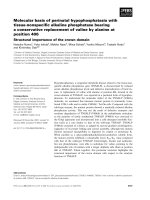
Báo cáo khoa học: Structural basis of charge transfer complex formation by riboflavin bound to 6,7-dimethyl-8-ribityllumazine synthase docx
Bạn đang xem bản rút gọn của tài liệu. Xem và tải ngay bản đầy đủ của tài liệu tại đây (559.32 KB, 7 trang )
Structural basis of charge transfer complex formation by riboflavin
bound to 6,7-dimethyl-8-ribityllumazine synthase
Michael Koch
1
, Constanze Breithaupt
1
, Stefan Gerhardt
1,
*, Ilka Haase
2
, Stefan Weber
3
, Mark Cushman
4
,
Robert Huber
1
, Adelbert Bacher
2
and Markus Fischer
2
1
Abteilung Strukturforschung, Max-Planck-Institut fu
¨
r Biochemie, Martinsried, Germany;
2
Lehrstuhl fu
¨
r Organische Chemie und
Biochemie, Technische Universita
¨
tMu
¨
nchen, Garching, Germany;
3
Institut fu
¨
r Experimentalphysik, Freie Universita
¨
t Berlin,
Germany;
4
Department of Medicinal Chemistry and Molecular Pharmacology, Purdue University, West Lafayette, IN, USA
The a mino acid residue tryptophan 27 of 6,7-dimethyl-
8-ribityllumazine synthase of the yeast Schizosaccharomyces
pombe was replaced by tyrosine. The structures of the W27Y
mutant protein in complex with riboflavin, the substrate
analogue 5-nitroso-6-ribitylamino-2,4(1H,3H)-pyrimidin-
edione, and the product analogue 6-carboxyethyl-7-oxo-
8-ribityllumazine, were determined by X-ray crystallography
at resolutions o f 2.7–2.8 A
˚
. Whereas the indole system of
W27 forms a coplanar p-complex with riboflavin, the cor-
responding phenyl ring in the W27Y mutant establishes only
peripheral contact with the heterocyclic ring system of the
bound riboflavin. These findings provide an explanation for
the absence of the long wavelength shift in optical absorption
spectra of riboflavin bound to the mutant enzyme. The
structures of the mutants are i mportant tools for the inter-
pretation of the unusual physical properties of r iboflavin in
complex w ith l umazine s ynthase.
Keywords: biosynthesis of riboflavin; crystallization;
6,7-dimethyl-8-ribityllumazine synthase; mutagenesis; ribo-
flavin b inding.
The biosynthesis of vitamin B
2
(riboflavin) in eubacteria and
fungi has been studie d in considerable detail [1,2]. In brief,
GTP cyclohydrolase II affords 2,5-diamino-6-ribosylamino-
4(3H)-pyrimidinone. R eduction of the ribose side c hain,
deamination and dephosphorylat ion a fford 5 -amino-6-ribi-
tylamino-2,4(1H,3H)-pyrimidinedione (1), which is conver-
ted into 6,7-dimethyl-8-ribityllumazine ( 3) by condensation
with 3,4-dihydroxy-2-butanone 4-phosphate (2) obtained
from ribulose 5-phosphate by a sigmatropic migration of
the terminal phosphoryl carbinol group and elimination of
formate (Fig. 1).
6,7-Dimethyl-8-ribityllumazine synthase (lumazine syn-
thase) catalyses the formation o f the direct precursor of
vitamin B
2
[3]. The lumazine synthases from yeasts and fungi
are C
5
-symmetric homopentamers [4–7], whereas plants and
many bacteria form lumazine synthases of 6 0 identical
subunits with icosahedral 532 symmetry [7–11]. The three-
dimensional structures of these hollow, icosahedral particles
are best described as dodecamers of pentamers. The subunit
fold of all lumazine synthases that have been reported is very
similar. A central four-stranded b-sheet is flanked on both
sides by two a-helices. The active sites of lumazine synthases
are invariably located at each respective interface between
adjacent subunits in the pentamer modules.
The binding of substrate and product analogues has been
studied with the lumazine synthases of Aquifex aeolicus,
Magnaporte grisea, Saccharomyces cerevisiae, Schizosaccha-
romyces pombe and Sp inacia o leracea [5–7,9]. Analogues of
1 and 3 are invariably bound via their ribityl side chain in an
extended conformation.
Surprisingly, the pure enzyme of S. pombe shows an
intense yellow colour after purification with a r atio of 6 : 1
of riboflavin/6,7-dimethyl-8-ribityllumazine bound in the
active site, due to the relatively high affinity of the enzyme
for the final product of the biosynthetic pathway.
Inthewild-typeenzymeofS. pombe, the heterocyclic
moieties of various ligands, including riboflavin, have been
shown to form coplanar p-complexes with the indole ring o f
tryptophan 27 [5]. In general, such p-stacking interactions
are known to play an important role in the m odulation of
cofactor reactivities [12–16]. An example is found in
flavodoxins, which utilize a flavin mononucleotide m olecule
as a cofactor in a highly conserved binding site containing
tryptophan and tyrosine residues [17,18]. Coordination of
flavin mononucleo tide i n a p-stacked configuration with
these aromatic amino acid side chains stabilizes the oxidized
redox state o f the flavin c ofactor and appears to disfavour
the formation of the electron rich hydrochinone form.
Furthermore, p-stacking interactions play a role in protein
binding of flavins. For example, in the recently discovered
flavoprotein dodecin, a pair of tryptophans facilitates the
formation of a unique tetrade comprising of a pair of
riboflavins with an antiparallel staggering of their isoallox-
azine moieties, sandwiched by the indole groups of the
symmetry-related tryptophans [19].
Correspondence to M. Fischer, Lehrstuhl fu
¨
r Organische Chemie und
Biochemie, Technische Universita
¨
tMu
¨
nchen, Lichtenbergstr. 4,
D-85747 Garching, Germany. Fax: +49 89 28913363,
Tel.: +49 89 28913336, E-mail: markus.fi
Abbreviations: CEOL, 6-carboxyethyl-7-oxo-8-ribityllumazine;
NORAP, 5-nitroso-6-(
D
-ribitylamino)-2,4(1H,3H)-pyrimidinedione;
NRAP, 5-nitro-6-(
D
-ribitylamino)-2,4(1H,3H)-pyrimidinedione.
*Present address: AstraZeneca, A lderleyPark,Macclesfield,
SK10 4TG , UK.
(Received 2 6 March 2004, revised 7 June 20 04,
accepted 11 June 2004)
Eur. J. Biochem. 271, 3208–3214 (2004) Ó FEBS 2004 doi:10.1111/j.1432-1033.2004.04253.x
Inthelumazinesynthase,suchap-stacked topology
correlates with a substantially modified optical absorption
spectrum of bound riboflavin. Specifically, the absorbance
of the protein-bound vitamin e xtends to wavelengths above
500 n m, and the relative intensity of t he optical transitions
at 445 and 370 nm is inverted compared to free riboflavin in
aqueous solution. These features a re less pronounced in a
W27Y mutant and virtually absent in a W27G mutant of
the protein [20]. Evi dence for p-stacking interactions of W27
or other a romatic a mino acid residues such a s t yrosine a nd
phenylalanine at the respective position with riboflavin is
also provided by time-resolved EPR experiments from
which t he triplet parameters o f riboflavin are obtained ([21]
andS.Weber,C.W.M.Kay,E.Schleicher,I.Hasse,
M. Koch, R. Huber, A. Bacher & M . F ischer, unpublished
results). The extent of p-orbital overlap i nfluences the flavin
triplet delocalization, which is reflected in the triplet zero-
field splitting parameters. Riboflavin bound to wild-type
and m utant lumazine synthases thus represents an ideal
system to specifically study such p-stacking interactions of
flavins in a protein environment.
In order to p rovide the structural basis for further studies
of the physical properties of riboflavin in complex with
lumazine synthase, we have determined the three-dimen-
sional structures of t he W27Y mutant protein complexed
with riboflavin, 6-carboxyethyl-7-oxo-8-ribityllumazine
(CEOL, 5; Fig. 2) and 5-nitroso-6-(
D
-ribitylamino)-
2,4(1H,3H)-pyrimidinedione (NORAP, 6;Fig.2)atresolu-
tions of 2.80, 2.75 and 2.70 A
˚
, respectively.
Experimental procedures
Materials
CEOL and NORAP were synthesized using published
procedures [22,23]. Riboflavin was obtained from Sigma.
Protein purification and crystallization
The W27Y mutant of S. pombe lumazine synthase was
cloned, expressed and purified as described previously [20].
After purification in the absence of riboflavin, less than 20%
of the purified mutant protein c ontained bound riboflavin
[5]. In order to obtain saturation with riboflavin, the protein
was cocrystallised with riboflavin, and the crystals were
subsequently soaked with riboflavin. Cocrystallization
experiments with the substrate analogue NORAP a nd the
product a nalogue CEOL were carried out by mixing
purified mutant enzyme (11 mgÆmL
)1
)in20m
M
potassium
phosphate (pH 7.0) and 50 m
M
potassium chloride with
stock solutions of the inhibitors to a final 10-fold molar
excess of the correspond ing inhibitor. Crystals were grown
at 18 °C b y the sitting drop vapor diffusion method by
mixing 2 lL of the protein-inhibitor s olution with 2 lLof
reservoir solution ( 0.1
M
sodium citrate, pH 5.0, contain ing
0.7
M
ammonium dihydrogen phosphate) and equilibrating
against reservoir solution.
Data collection
X-ray data of the riboflavin-b ound mutant enzyme W27Y
as well as of the two inhibitor complex structures were
collected on a MARResearch ( Norderstedt, Germany) 345
imaging plate detector system mounted on a R igaku RU-
200 rotating a node (Brandt Instruments, Prairieville, LA,
USA) operated at 50 mA and 100 kV with k ¼ CuK
a
¼
1.542 A
˚
The d ata s ets o f the crystals that diffracted up to a
resolution of 2.7 A
˚
were integrated, scaled, and merged
using the
DENZO
and
SCALEPACK
program packages [24].
Data collection statistics are shown in Table 1.
Structure solution and refinement
Initial phases of the riboflavin c omplex and the two
inhibitor complexes were determined by difference Fourier
Fig. 2. Inhibitors of 6,7-dimethyl-8-ribityllumazine synthase. 5,
6-Carboxyethyl-7-oxo-8-ribityllumazine (CEOL), 6, 5-nitroso-6-(
D
-
ribitylamino)-2,4(1H,3H)-pyrimidinedione (N ORAP), an d 7,5-nitro-
6-(
D
-ribitylamino)-2,4(1H,3H)-pyrimidinedione (NRAP).
Fig. 1. Terminal reactions in the pathway o f riboflavin biosynthesis.
(A) 3,4-dihydroxy-2-butanone 4-phosphate synthase; (B) 6,7-dimethyl-
8-ribityllumazine synthase; (C) riboflavin synthase; 1, 5-amino-6-ribi-
tylamino-2,4(1H,3H)-pyrimidinedione; 2, 3,4-dihydroxy-2-butanone
4-phosphate; 3, 6,7-dimethyl-8-ribityllumazine and 4, riboflavin.
Ó FEBS 2004 Structural basis of charge transfer formation (Eur. J. Biochem. 271) 3209
synthesis using the lumazine s ynthase wild-type structure [5]
as template. After initial rigid body minimization, refine-
ment was performed by alternating model building carried
out with the program
O
[25] and crystallographic refinement
using
CNS
[26]. The refinement procedure included posi-
tional r efinement and restrained temperature factor refine-
ment. Finally, water molecules were inserted a utomatically
and c hecked manually by inspection of the F
o
-F
c
map. For
all three models, noncrystallographic symmetry restraints
were applied. The ligands were not included in the model
during the first cycles of refinement; thereafter CEOL and
NORAP could be easily built into the clearly defined
electron density in contrast to riboflavin, which, due to its
low occupancy, exhibited only w eak e lectron density. Due
to disorder, residues 159 and the N-terminal residues 1–12
remained undetermined in the electron density map. Ster-
eochemical parameters of the structures were calculated
with
PROCHECK
[27]. F igures were designed with
MOLSCRIPT
[28],
BOBSCRIPT
[29] and
RASTER
3
D
[30].
Results and discussion
In contrast to lumazine synthases from other organisms
studied [6–8,11], the enzyme from S. pombe binds riboflavin
with relatively high affinity. This is believed to be due to a
p-complex formation between the bound ligand and the
adjacent tryptophan residue 27 [20]. In mutant proteins,
namely W27Y and W27F, riboflavin is l ess tightly bound as
compared to the wild-type protein. The three W27Y mutant
lumazine synthase structures in complex with riboflavin,
CEOL and NORAP were solved by difference Fourier
synthesis using the coordinates of the riboflavin-bound
wild-type structure from S. pombe. After refinement, more
than 90% of the r esidues lie in the most favoured region of
the Ramachandran plot in all three structures.
Crystals containing riboflavin belong to the space group
C222
1
with unit cell constants a ¼ 111.6 A
˚
,b¼ 145.1 A
˚
,
c ¼ 129.2 A
˚
. T he asymmetric unit contained one pentamer
(Fig. 3 ). Crystals of the inhibitor complexes belong to the
same space group with cell dimensions of a ¼ 111.1 A
˚
,
b ¼ 144.9 A
˚
,c¼ 128.3 A
˚
(CEOL) and a ¼ 111.2 A
˚
,b¼
144.8 A
˚
,c¼ 127.8 A
˚
(NORAP), respectively. The mono-
mers of S. pombe lumazine synthase consist o f 1 59 residues
that were well defined i n all structures with the exception of
residue 159 a nd the N-terminal residues 1–12 that r emained
undetermined in the electron density map (Fig. 4).
The five ac tive sites o f lumazine synthase are located at
the interfaces between each adjacent pair of monomers
(Fig. 3 ). Thus, residues of two adjacent monomers con-
tact the ligands that bind into the substrate binding
pocket. Y27, H94 and W63 of one monomer form most
of the substrate binding site, and L119 and H142 of the
second monomer close the pocket f rom the opposite side
(Figs 5 a nd 6).
Table 1. X-ray data-processing and r efinement statistics. RMSD, root m ean square deviations of temperat ure factors of bonded a tom s.
Data set W27Y-riboflavin W27Y-CEOL W27Y-NORAP
Number of unique reflections 25 184 26 937 28 707
Multiplicity
a
2.7 (2.1) 3.9 (3.8) 3.9 (3.8)
Limiting resolution (A
˚
) 2.80 2.75 2.70
Completeness of data (%) 96.7 (91.6) 99.4 (99.5) 99.9 (100.0)
R
merge
(%)
b
8.5 (37.3) 8.0 (51.7) 10.9 (48.2)
I/r 7.0 (2.0) 15.5 (2.6) 11.6 (2.6)
R
cryst
/R
free
(%)
c
20.4/22.2 20.6/23.0 19.1/21.2
Non hydrogen protein atoms 5550 5550 5550
Number of water molecules 28 – 58
Non hydrogen ligand atoms 135 115 100
Non hydrogen ion atoms 25 25 25
RMSD [bonds (A
˚
)/angles (°)/
bonded Bs (A
˚
2
)]
0.008/1.34/2.33 0.009/1.40/2.07 0.009/1.42/2.21
Mean temperature factors
(protein/ligand/ion/solvent)
49.3/75.4/51.0/47.5 60.6/52.9/68.9/– 43.7/38.1/43.0/40.2
a
Values in parentheses correspond to the highest resolution shell between 2.95 and 2.80 A
˚
(W27Y-riboflavin), 2.83–2.75 A
˚
(W27Y-CEOL)
and 2.78–2.70 A
˚
(W27Y-NORAP).
b
R
merge
¼ S
h
S
I
|I
i
(h) ) <I(h)>|/S
h
S
i
I
i
(h).
c
R
cryst
¼ S
h
||F
o
(h)| ) |F
c
(h)||/S
h
|F
o
(h)|.
Fig. 3. Overall X-ray structure of W27Y mutant 6,7-dimethyl-8-ribi-
tyllumazine synthase – the pentameric assembly. Pentameric assembly
of the 6 ,7- dim eth yl- 8-r ibit yl lumazine synthase m utant W27Y from
S. pombe. T he inhibitor CEOL is shown as a ball-and-stick model.
Subunits: A, r ed; B, light gre en; C, green; D, blu e; E, violet.
3210 M. Koch et al.(Eur. J. Biochem. 271) Ó FEBS 2004
Comparing the different wild-type and W27Y structures
in complex with the different ligands, significant changes of
the side chain conformation are observed for residue H94
(Fig. 7 ). H94 is highly conserved i n all known lumazine
synthase sequences and is assumed to be involved in the
initial proton transfer steps of catalysis [9]. The orientation
of H94 var ies according to the bound ligand but is nearly
independent of the nature of residue 27. In the case of the
two substrate analogue complexes of the w ild-type and the
W27Y mutant proteins, H94 is moved closer to the plane of
the ligand, NORAP (6; Fig. 2) in the W27Y-mutant protein
and 5-nitro-6-(
D
-ribitylamino)-2,4(1H,3H)-pyrimidinedione
(NRAP) (7; Fig. 2 ), in the wild-type enzyme (distance
between the C
c
-atom of H94 and the N5-atom o f
N(O)RAP: wild-type enzyme: 5.5 A
˚
; W27Y mutant:
5.5 A
˚
) than in the two corresponding complexes with the
larger product analogue C EOL (5; Fig. 2) c omprising two
annealed 6-membered rings (distance between the C
c
-atom
of H94 and the N 7-atom of CEOL: wild-type enzyme:
6.5 A
˚
;W27Ymutant:6.1A
˚
).
The smallest distance between t he C
c
-atom of H94 and
the inhibitor plane (N5-atom of riboflavin) is found in the
wild-type structure with bound riboflavin with a value of
4.9 A
˚
. Moreover, the imidazole ring of H94 is packed nearly
parallel against the riboflavin, contributing t o the observed
stacking interactions between the sandwiched riboflavin and
H94 and residue 27 (distances b etween the planes of about
4A
˚
)[5].
In the CEOL and NORAP structures of the W27Y
mutant and in all three ligand-bou nd structures of the wild-
type enzyme [5], the positions of the C
a
-atoms and the
aromatic planes of residues Y27 and W27, respect ively, are
almost identical (distance betwee n the ring system of the
ligand and the aromatic planes o f a mino acid 27: 3.5 A
˚
[5],
Fig. 7). In the W27Y mutant structu re with bound
riboflavin, however, the C
a
-trace dev iates from the other
structures by 0.6 A
˚
, and the aromatic r ing is very fl exible. In
the substrate a nd product a nalogue complexes of the
mutant and the wild-type protein, s tacking interactions ta ke
place between the ligand and Y27 or W27, respectively.
This leads to a fixed orientation of the Y27 side chain,
parallel to t he ligand r ing s ystem w ith we ll defined e lectron
densities for these two ligands (Figs 5 and 6). The r ibityl
side chain is bound in the same manner a s already described
for the S. pombe wild-type structure [5]. The mutant protein
binds riboflavin less tightly ( K
d
:12.0l
M
[20]); as com pared
to the wild-type protein (K
d
:1.2l
M
[20]); (for optical
properties see [20]). The bound riboflavin i n complex with
the mutant protein is less well defined than the two o ther
ligands and t hus, its position c annot be determined reliably.
This prevents aromatic stacking and thus the fixation of
Y27.
For the S. pombe wild-type e nzyme, a significant long-
wavelength optical absorbance extending well beyond
Fig. 4. Secondary structure arrangement of one lumazine synthase
monomer and one neighbouring monomer (shown i n lighter col ours). At
the subunit interface, in the active site, the inhibitor CEOL (green) and
the mutated W27 residue (y ellow) are shown.
Fig. 5. Stereo view of t he active site of W27Y 6,7-dimethyl-8-ribityllumazine synthase from S. pombe with bound substrate analogue NORAP (green)
intheactivesite.The final (2F
o
-F
c
)-OMIT map of the inhibitor was calculated at 2.7 A
˚
resolution.
Ó FEBS 2004 Structural basis of charge transfer formation (Eur. J. Biochem. 271) 3211
500 n m has been observed [20]. This feature is much less
pronounced in the W 27Y mutant. One possible reason for
this finding is that the phenyl r ing of Y 27 in the mutant is
rotated such t hat the coplanarity of its p-system and that of
the r iboflavin’s isoalloxazine r ing is reduced, w hereas in the
wild-type enzyme the aromatic rings W27 and riboflavin are
almost perfectly coplanar. Nearly perfect p-stacking inter-
actions between a tyrosine residue and a flavin have been
observed, for example, in flavodoxin from Desulfovibrio
vulgaris [31]. However no extended long-wavelength optical
absorption has been found in th at system [32]. Taking
together these observations with our results, we conclude
that the absence of long-wavelength absorption in the
W27Y m utant of S. pombe lumazine synthase is not due to
the d ifferent orientation o f the Y27’s phenyl ring but rather
due to the reduced p-orbital o verlap as a consequence of the
smaller size of the phenyl ring of Y 27 as compared to the
indole r ing of W27. Clearly further biophysical studies are
required to substantiate these notions.
The crystal structure of the lumazine synthase from
A. aeolicus was the first structure witho ut any ligand i n t he
active site [8]. The superpositions of the a mino acid residues
in the a ctive s ite of t he A. aeolicus enzymewith the ones of the
wild-type and th e mutant e nzyme of S. pombe in Fig. 8
shows t hat t he phenyl ring of residue F 22 in the A. aeolicus
enzyme is rotated by 30° as compared to the o rientation of
the aromatic residue in W27 in the S. pombe wild-type
enzyme, f or which coplanarity between the aromatic p lanes
Fig. 6. Stereo view of the active site of W27Y 6,7-dimethyl-8-ribityllumazine synthase from S. pombe with bound product analogue CEOL (green) i n
theactivesite.The fi nal (2F
o
-F
c
)-OMIT map of the inhibitor was c alcu lated at 2 .8 A
˚
resolution.
Fig. 7. Stereo drawing of the active sites of the wild-type and W27Y mutant 6,7-dimethyl-8-ribityllumazine-synthase–ligand complexes from S. pombe.
CEOL complexes are shown in green for the W27Y mutant and in cyan for th e wild-typ e enzym e, substrate analogue complexes in orange for the
W27Y mutant with bou nd NORAP and i n yellow for the N RAP-bound wild-type enzyme, the riboflavin-bound enzymes are shown in blue
(mutant) and violet (wild-type), resp ect ively. The p osition of residue H94 changes according t o the bound ligand, independently of the nature of
residue 27. The positions of the aromatic planes of the residues Y27 and W27 are almost identical for five of the six structures. The C
a
-position of
Y27 i n the ri boflavin-bound mutant enzyme differs from the position of residue 2 7 in t he other proteins.
3212 M. Koch et al.(Eur. J. Biochem. 271) Ó FEBS 2004
of the aromatic ligand is observed [9]. The orientation of
residue Y27 in the S. pombe W27Y mutant is not coplanar to
the aromatic plane (see above). Furthermore, the position of
the indole ring o f W 27 in th e wild-type e nzyme is not fixed
after elimination of riboflavin. Hence, it can b e c oncluded
that the orientation of Y27 in the mutant resembles the
situation in t he protein w ithout ligand. This is the reason for
the lower content of riboflavin in the riboflavin–mutant
complex compared t o the riboflavin–wild-typ e complex.
The interaction of the N -terminal residue P8 with W27 in
the wild-type complex is missing in the mutant complex
where the N-terminal region is not defined in the electron
density. The p–p-stacking i nteraction between the aromatic
residue 27 and the pyrimidine system presumably contri-
butes substantially to the substrate-binding energy [9]. Here,
this binding energy is expected to be lowered, leading to a
reduced affinity for the ligand.
The residue H142 shows a smaller, but still recognizable
deviation in the two structures with bound riboflavin as
compared to the structures w ith other bound inhibitors.
H142 is assumed t o form a salt bridge to the phosphate ion
of the second substrate, 3,4-dihydroxy-2-butanone-4-phos-
phate, during catalysis and is itself stabilized in its position
by D145 [9]. In the s ubstrate a nd pr oduct a nalogue
complexes, a phosphate ion is bound to the phosphate
binding site of the second substrate [5] that exhibits no direct
contact with the substrate and product analogues. The
much larger riboflavin, which is intuitively supposed to be
unable t o bind into the pocket, moves the position of H142
with respect to the other bound ligands.
Our findings clearly demonstrate that W27 in the wild-type
enzyme plays an essential role in substrate fixation by
p-orbital overlap of the indole ring of W27 with the aromatic
ring(s) in the substrate. The different extent of p–pinteraction
mediated by residue 27 in the wild-type and in various
mutants (W27Y, W27F, W27H) correlates favorably with
the different amounts of riboflavin s pecifically bound to the
protein [20]. Furthermore, the parallel alignment of the
isoalloxazine ring of ribofl avin and the aromatic side chain of
residue 27 in lumazine synthase manifests i tself in t he unusual
spectral properties of the wild-type and mutant complexes
indicating that partial p–p charge transfer between the r ings
has t aken place even in the ground state. Stacking inter-
actions are a well known structu re motif in flavoproteins but
also, for example, in riboflavin analogues in the solid state
[33,34] where the intimate overlap of the isoalloxazine core
provides an energetically favored packing mode. That this
ring stacking can be manipulated by specific site-selective
mutagenesis makes the lumazine synthase an ideal model
system for studying flavin-binding to proteins at a molecular
level and thus may contribute to an understanding of the
fundamentally different reaction mechanisms catalysed by
flavoproteins.
Acknowledgements
We thank Richard Feicht, Sebastian Schwamb and Thomas
Wojtulewicz for skillful help in protein preparation. This work was
supported by the Deutsche Forschungsgemeinschaft, the Fonds der
Chemischen Industrie, th e Hans–Fischer– Gesellschaft e.V ., and by
NIH g rant GM51469.
References
1. Ba cher, A ., Eberhardt, S., E isen reich, W., Fischer, M., Herz , S.,
Illarionov, B., Kis, K . & Richter, G. (2001) Biosynthesis of ribo-
flavin. Vi tamins Hormones 61 , 1–49.
2. Ba cher, A., Eberhardt, S., Fischer, M., Kis, K. & Richter, G.
(2000) Biosynthesis of vi tamin B
2
(Riboflavin). Annu . R ev. Nutr.
20, 1 53–167.
3. Neuberger, G. & Bacher, A. (1986) Biosynthesis of riboflavin.
Enzymatic formatio n of 6, 7-dimethyl-8-ribityllumazine by heavy
Fig. 8. Stereo view of the active sites of the S. pombe 6,7-dimethyl-8-ribityllumazine synthase W27Y mutant complexed with riboflavin (blue), the
S. pombe 6,7-dimethyl-8-ribityllumazine synthase wild-type e nzyme complexed with riboflavin (violet) and the A. aeolicus lu mazine syn thase with no
bound ligand (orange). The residue F22 from A. aeolicus has an orientation 30° bent to the orientation of W27 in the S. pombe wild-type enzyme,
which is coplanar to t he aromatic plane of bound aromatic ligands [9]. The orientation of Y27 i n the S. pombe W27Y mutant resembles the
A. aeolicus apoprotein more closely than the S. pombe wild-type enzyme complex. This relates to the high er dissociation constant in the riboflavin–
mutant complex compared t o the riboflavin–wild-type complex.
Ó FEBS 2004 Structural basis of charge transfer formation (Eur. J. Biochem. 271) 3213
riboflavin synthase from Bacillus subtilis. Biochem. Biophys. Res.
Commun. 139 , 1111–1116.
4. Braden, B.C., Vekilovsky, C.A., Cauerhff, A.A. , Polikarpov, I. &
Goldbaum, F.A. ( 2000) Divergence in ma cromolecular assembly:
X-ray crystallographic structure analysis of lumazine synthase
from Brucella abor tus. J. Mol. Biol. 297, 1031–1036.
5. Gerhardt, S., Haa se, I., S teinbacher, S., Kaiser, J.T., Cushman,
M., Bacher, A., Huber, R. & Fischer, M. (2002) The structural
basis of riboflavin binding to Schizosaccharomyces pombe 6,7-di-
methyl-8-ribityllumazine synthase. J. Mol. Biol. 318, 1317–1329.
6. Meining, W., Mo
¨
rtl, S., Fischer, M., Cushman, M., B acher, A. &
Ladenstein, R . ( 2000) T he atom ic st ru cture o f p entameric l u ma-
zine synthase from Sacc haromyces cerevisiae at 1.85 A
˚
resolution
reveals the binding mode of a phosphonate intermediate analogu e.
J. Mol. Biol. 299, 1 81–197.
7. Persson, K., Schneider, G ., Doug las, B.J., Viitanen, P.V. & San-
dalova, T. (1999) Crystal structure analysis of a pentameric fungal
and icosahedral plant lumazine synthase reveals the structural
basis of diferences in assembly. Protein S ci. 8, 2355–2365.
8. Zhang, X., Meining, W., Fischer, M., Bacher, A. & Ladenstein, R.
(2001) X-ray structure analysis and crystallographic refinements of
lumazine synthase from the hyperthermophile Aquifex aeolicus at
1.6 A
˚
resolution: determinants of thermostability revealed from
structural compar isons. J. Mo l. Biol. 306 , 1099–1114.
9. Zhang, X., Meining, W., Cushman, M., Haase, I., Fischer, M.,
Bacher, A. & Ladenstein, R. (2003) The catalytic mechanism of
lumazine sy nthase. A structur e based model o f the reaction cata-
lyzed by l umazine synthase from Aquifex aeolicus. J. Mol. Biol.
328, 1 67–182.
10. Ritsert,K.,Huber,R.,Turk,D., Ladenstein, R., Schmidt-Ba
¨
se,
K. & Bacher, A. (1995) Studies on the lumazine synthase/ribo-
flavin s ynthase c omplex of Bacillus s ubtilis: c rystal structure ana-
lysis of r econstituted, icosahedral beta-subunit capsids with bound
substrate analogue inhibitor at 2.4 A
˚
resolution. J. M ol. Biol. 253,
151–167.
11. Lade nstein, R., Me yer, B., Hu ber, R., Labisch inski, H., Barte ls,
K., Bartunik, H .D., Bachman n, L., L udwig, H.C. & Bach er, A .
(1986) Heavy riboflavin synthase from Bacillus subtilis.Particle
dimensions, crystal packing a nd molecu lar symme try. J. Mol. Biol.
187, 8 7–100.
12. Breinlinger, E. C., K eenan, C.J. & Rotello, V.M. (1998) M odula-
tion of flavin recognition and redox properties through donor
atom p-interactions. J. Am. Chem. Soc. 120, 8606–8609.
13. Breinlinger, E.C. & R otello, V.M. ( 1997) Model systems for fl a-
voenzyme activity. Modulation of flavin redox potentials through
p-stacking interactions. J. Am . Chem. Soc. 119 , 1165–1166.
14. Hu nter, C.A. & Sanders, J.K.M. (1990) The n ature of p–p inter-
actions. J. Am. Chem. Soc. 112, 5525–5534.
15. Lostao, A., Gomez-Moreno, C., Mayhew, S.G. & Sancho, J.
(1997) Differential stabilization of the three FMN redox forms by
tyrosine 94 and tryptophan 57 in flavodoxin from Anabaena and its
influence o n the re dox potentials. Biochemistry 36, 14334–14344.
16. Zhou, Z. & Swenson, R.P. (1996) The cumulative electrostatic
effectofaromaticstackinginteractions and the n egative electro-
static environ ment of the flavin mononucleotide b in ding site is a
major determinant of the reduction potential for the flavodoxin
from Desulfovibrio vulgaris [Hildenborough]. Biochem ist ry 35,
15980–15988.
17. Vervoort, J ., M ull er, F. , L eG all, J., B acher, A. & S edlmaier, H.
(1985) Carbo n-13 and n itrogen- 15 nuclear-magnetic-resonance
investigation on Desulfovibrio vulgaris flavodo xin. Eur. J.
Biochem. 15 1, 49–57.
18. Stockman, B.J., Richardson, T.E. & Swenson, R.P. (1994) Struc-
tural changes c aused by site-directed mutagenesis of tyrosine- 98
in Desulfovibrio vulgaris flavodoxin delineated by 1H and 15N
NMR spectroscop y: implications for redox potential modulation.
Biochemistry, 33, 15298–15308.
19. Bieger, B., Essen, L.O. & Oesterhelt, D. (2003) Crystal structure of
halophilic dodecin: a n ovel, dodecameric flavin binding protein
from Halobacterium s alinarum. Structure 11 , 375–385.
20. Fischer, M., Haase, I., Feicht, R., Richter, G., G erhardt, S.,
Changeux, J.P., Huber, R. & B acher, A. (2002) Biosynthesis of
riboflavin. 6,7-Dimethyl-8-ribityllumazine synthase of Schizo-
saccharomyces pombe. Eur. J. Biochem. 269 , 519–526.
21. Schleicher, E., Kowalczyk, R.M., Kay, C.W.M., Hegemann, P.,
Bacher, A., Fischer, M., Bittl, R., Richter, G. & Weber, S. (2004)
OnthereactionmechanismofadductformationinLOVdomains
of the p lant blue light receptor photo tropin. J. Am. Chem. Soc.,
in press.
22. Cushman,M.,Yang,D.,Gerhardt,S.,Huber,R.,Fischer,M.,
Kis, K . & B acher, A. (2 002) Design, synthesis, and e valuation of
6-carboxyalkyl and 6-phosphonoxyalkyl derivatives of 7-oxo-
8-ribitylaminolumazines as inhibitors of riboflavin synthase and
lumazine synthase. J. Org. Che m. 67, 5 807–5816.
23. Plaut, G.W.E. & Harvey, R.A. (1971) The enzymatic synthesis of
riboflavin. Me th. Enzymol . 18, 5 15–538.
24. Otwinowsky, Z.M.W. (1993) DENZO: A film-processing p rog ram
for macromolecular crystallography. Yale University Press, New
Haven, CT.
25. Jones, T.A., Zou, J.Y., Cowan, S.W. & Kjeldgaard, M. (1991)
Improved methods for building protein models in electron density
maps and the location of erro rs in these models. Acta Crystallogr.,
Sect. A : Found. Crysta llogr. 47, 110–119.
26. Brunger, A.T., A dams, P.D., Clore, G.M., D eLano, W.L., Gros,
P., Grosse-Kunstleve, R.W., Jiang, J.S., Kuszewski, J., Nilges, M.,
Pannu, N.S., Read, R.J., Rice, L.M., S imonson, T. & Warren,
G.L. (1998) Crystallography & NMR system: a new software suite
for macromolecular structure determination. Acta Crystallogr.,
Sect. D: B iol. Crystallogr. 54, 905–921.
27.Laskowski,R.A.,Macarthur,M.W.,Moss,D.S.&Thornton,
J.M. (1993 ) PROCHECK – a program to check t he stereochem-
ical quality o f protein structures. J. Appl. Cryst. 26, 283–291.
28. Kraulis, P.J. (1991) Molscript – a program to produce both
detailed and schematic plots of protein structures. J. Appl. C ryst.
24, 9 46–950.
29. Esnouf, R.M. (1997) An extensively modified version of Molscript
that includes gr eatly enha nced coloring capabilities. J. Mol. Graph.
Model. 15 , 112–113.
30. Merritt, E.A. & Murphy, M.E.P. (1994) Raster3d, Version 2.0 – a
program for photorealistic molecular graphics. Ac ta Crystallogr.,
Sect. D: B iol. Crystallogr. 50, 869–873.
31. Walsh, M.A., McCarthy, A., O’Farrell, P.A., McArdle, P.,
Cunningham, P.D., Mayhew, S.G. & Higgins, T.M. (1998)
X-ray crystal structure of the Desulfovibrio vulgaris (Hilden-
borough) apoflavodoxin-riboflavin complex. Eur. J. Biochem. 258,
362–371.
32. Dubourdieu, M., Le Gall, J. & Favaudon, V. (1975) Physio-
chemical properties of fl avodoxin from Desulfovibrio v ulgaris.
Biochim. Bi ophys. Acta 376, 519–532.
33. Ebitani, M., In, Y., Ish ida, T., Sakaguchi, K ., Flippen-Anderson,
J.L. & Karle I.L. (1993) Structures of ribofl avin tetraacetate and
tetrabutyrate – molecular packing mode of riboflavin tetrac arb-
oxylate and its extensive stacking and hydrogen-bonding
characteristics. Ac ta Crystallogr., Se ct. A: F ound. Crystallogr. 49,
136–144.
34. Wang,M.&Fritchie,C.J.J.(1973)Geometryoftheunperturbed
flavin nucleus. The crystal structure of 10-methylisoalloxazine.
Acta Crystallogr., Sect. B 29, 2040–2045.
3214 M. Koch et al.(Eur. J. Biochem. 271) Ó FEBS 2004