Báo cáo khoa học: Differential regulation of fatty acid amide hydrolase promoter in human immune cells and neuronal cells by leptin and progesterone pdf
Bạn đang xem bản rút gọn của tài liệu. Xem và tải ngay bản đầy đủ của tài liệu tại đây (361.45 KB, 11 trang )
Differential regulation of fatty acid amide hydrolase promoter in
human immune cells and neuronal cells by leptin and progesterone
Mauro Maccarrone
1,2
, Valeria Gasperi
3
, Filomena Fezza
1
, Alessandro Finazzi-Agro
`
3
and Antonello Rossi
3
1
Department of Biomedical Sciences, University of Teramo, Italy;
2
IRCCS C. Mondino, Mondino-Tor Vergata-Santa Lucia
Center for Experimental Neurobiology, Rome, Italy;
3
Department of Experimental Medicine and Biochemical Sciences,
University of Rome Tor Vergata, Rome, Italy
We have shown recently that in human T lymphocytes,
leptin stimulates activity and e xpression o f the e ndocan-
nabinoid-degrading enzyme fatty acid amide hydrolase
(FAAH), through S TAT3 (signal transducer and activator
of transcription 3) and its CRE (cAMP response element)-
like t ranscriptional t arget i n t he FAAH p romoter [Maccar-
rone, M., Di Rienzo, M., Finazzi-Agro
`
,A.,&Rossi,A.
(2003) J. Biol. C hem. 278, 13318–13324]. We have also
shown that progesterone, alone or additively with leptin,
up-regulates the FAAH gene in human T-cells, through the
Ikaros transcription factor [Maccarrone, M., Bari, M., Di
Rienzo, M., Finazzi-Agro
`
, A., & Rossi, A. (2003) J. Biol.
Chem. 278, 32726–32732]. Here, we extend these observa-
tions to immortalized human lymphoma U937 cells, where
stimulation o f FAAH by leptin (up to 300% of the
controls) involves binding to a leptin receptor (K
d
¼
2.0 ± 0.1 n
M
, B
max
¼ 382 ± 5 fmolÆmg protein
)1
, appar-
ent molecular mass of 110 kDa), and stimulation by
progesterone involves an intracellular receptor of
120 kDa. Unlike FAAH, the other proteins of the endo-
cannabinoid system are not modulated by the two hor-
mones. Interestingly, human neuroblastoma CHP100 cells
also have a leptin receptor ( 110 kDa, K
d
¼ 2.2 ± 0.2 n
M
,
B
max
¼ 339 ± 8 fmolÆmg protein
)1
), a progesterone rec-
eptor ( 120 kDa), STAT3 and Ikaros, yet their FAAH is
not activated by leptin or progesterone. These data, corro-
borated by transient expression and electrophoretic mobil-
ity-shift assays, demonstrate an unprecedented cell-specific
regulation of the FAAH gene, which has important impli-
cations for t he control o f tone a nd activity of AEA along the
neuroimmune axis.
Keywords: endocan nabinoids; i mmune system; l eptin; neu-
rons; progesterone.
Leptin is the 16-kDa nonglyco sylated product of the obese
gene, which is secreted by adipose cells, is released into
the circulation, and transported across the blood–brain-
barrier into t he central nervous system, where it regulates
energy homeostasis [1]. Leptin also serves systemic
functions, a part from those related to food intake and
energy expenditure in mammals, including regulation of
fertility [ 2] and modulation of immune response [ 3]. These
two actions might be interconnected in humans because
leptin alters the production from T lymphocytes of T
helper 1 (Th1) and Th2 cytokines [4], which are critical
in regulating embryo implantation and materno-fetal
exchanges [5,6]. Leptin signaling is mediated by the long
isoform of the leptin receptor (LR) via different pathways,
among which those mediated by the signal transducer and
activator of transcription (STAT) m embers have a
prominent role [7,8]. Recently, we have shown that leptin
reduces the levels of the endocannabinoid anandamide
(N-arachidonoylethanolamine, AEA) in human p eripheral
T lymphocytes by up-regulating the activity and expres-
sion of the AEA hydrolase (fatty acid amide hydrolase,
FAAH) in these cells [9]. The e ffect of leptin occurred
through a STAT3-dependent activation of a CRE (cAMP
response element)-like site in the promoter region of
FAAH gene [9]. Remarkably, leptin has been shown to
reduce the levels of AEA also in t he hypothalamus of
ob/ob mice [10], suggesting that this compound partakes
in the neural circuitry regulated by leptin [11,12]. We have
also shown that the activity and expression of FAAH in
human T cells is up-regulated b y progesterone, which
increases t he nucle ar levels of the Ikaros t ranscription
factor [13,14], and hence its binding to a specific site in
the promoter region of the FA AH gene [15]. Progesterone
is a c ritical hormone both f or fertility and immune
response [5,6]. In addition, progesterone is known to
regulate a number of activities in the central nervous
system [16,17].
Correspondence to M. Maccarrone, Department of Biomedical
Sciences, University of Teramo, Piazza A. M oro 45, 64100 Teramo,
Italy. Fax: +39 0861 412583; Tel.: +39 0861 266875;
E-mail:
Abbreviations:AEA,anandamide(N-arachidonoylethanolamine);
AMT, AEA membrane transporter; CAT, chloramphenicol acetyl-
transferase; CRE, cAMP-response element; FAAH, fatty acid amide
hydrolase; GAR/M-AP, goat anti-rabbit/mouse Igs conjugated with
alkaline phosphatase; IGF-IR, insulin-like g rowth factor I receptor;
Ik, Ikaros; (s)LR, (soluble) leptin receptor; NAPE, N-acylphosphati-
dylethanolamine; NAT, N-acyltransferase; PL D, ph ospholipase D;
PR, progesterone receptor; STAT, signal transducer and activator of
transcription.
Enzymes: Fatty acid amide hydrolase (E C 3.5.1.4); phosphatidylcho-
line phosphatidohydrolase (EC 3.1.4.4).
(Received 4 June 2004, revised 31 August 2004,
accepted 8 October 2004 )
Eur. J. Biochem. 271, 4666–4676 (2004) Ó FEBS 2004 doi:10.1111/j.1432-1033.2004.04427.x
Based o n this background, we sought to extend our
previous observations on the effect of leptin and
progesterone on FAAH activity and expression in
primary human T lymphocytes to immortalized human
lymphoma U937 cells. These are a model of peripheral
immune cells shown to dispose AEA through an active
FAAH and a selective AEA membrane transporter
(AMT) [18,19]. In addition, we inves tigated whether
leptin and progesterone can modulate the Ôon demandÕ
synthesis of AEA through N-acyltransferase [20] and
N-acylp hosphatidylethanolamine (NAPE)-specific phos-
pholipase D (PLD) [21]. In the light of the manifold
actions of AEA in the neuroimmune axis [ 22,23], we
sought to investigate the effect of leptin and progesterone
also on FAAH, A MT, NAT and P LD of human
neuroblastoma CHP100 cells, a model of neuronal cells
shown to metabolize AEA [18,19]. We report unprece-
dented evidence that leptin and progesterone up-regulate
FAAH, but not AMT, NAT or PLD, in human immune
cells, w hile they do not in human neuronal cells.
Therefore, the central actions of these two hormones do
not seem to involve the endocannabinoid system but
instead may play a role in the effects of leptin and
progesterone in the i mmune system. This d ifferential
regulation of FAAH suggests that the same factors can
lead to a c ell-specific control o f the tone [24] and the
activity of AEA a long the n euroimmune axis, with
important pathophysiological implications. In addition,
bearing in mind that F AAH has amidase or esterase
activity on many bioactive substrates [22,24], it can b e
anticipated that t he reported findings might have rele-
vance beyond AEA and related endocannabinoids.
Materials and methods
Materials
Chemicals were of the purest a nalytical grade. Leptin
(human recombinant), anandamide (N-arachidonoyletha-
nolamine, AEA), progesterone and m ifepristone (RU486)
were purchased from Sigma Chemical C o. (St Louis, MO,
USA). [
3
H]AEA (223 C iÆmmol
)1
),
125
I-labeled leptin
(2200 CiÆmmol
)1
)and[
3
H]CP55.940 {5-(1,1¢-dimethyh ep-
tyl)-2-[1R,5R-hydroxy-2R-(3-hydroxypropyl) cyclohexyl]-
phenol; 126 Ci mmol
)1
} were from NEN Life Science
Products, Inc. (Boston, MA, USA). [
3
H]N-arachidonoyl-
phosphatidylethanolamine (200 CiÆmmol
)1
)wasfromARC
(St Louis, MO, USA), and 1 ,2-di[1-
14
C]palmitoyl-phos-
phatidylcholine (111 mCiÆmmol
)1
) was from Amersham
Pharmacia Biotech (Uppsala, Sweden). Anti-FAAH poly-
clonal Igs were raised in rabbits against the conserved
FAAH sequence VGYYETDNYTMPSPAMR [25] conju-
gated to ovalbumin, a nd were prepared by Primm S.r.l.
(Milan, Italy). M ouse monoclonal antibodies against actin,
STAT3, phospho-STAT3, leptin receptor (LR) and pro-
gesterone re ceptor (PR), LR and PR blocking peptides, and
rabbit anti-(Ikaros serum) were from Santa Cruz Biotech-
nology ( Santa Cruz, CA, USA). Anti-human insulin-like
growth factor I receptor (anti-IGF-IR) and human leptin
receptor/Fc chimeras (soluble LR, sLR) were purchased
from R & D Systems (Minneapolis, MN, USA). According
to the m anufacturer’s instructions, 1.5 lgÆmL
)1
of
sLR are enough to n eutralize the effects o f 10 n
M
( 150 ngÆmL
)1
) leptin, and in this study we used a
two-fold excess of each neutralizing agent [9]. Goat anti-
rabbit and goat anti-mouse Igs conjugated to alkaline
phosphatase (GAR-AP an d GAM-AP) w ere from Bio-Rad
(Hercules, CA, USA).
Cell culture and treatment
Human lymphoma U937 cells (ATCC, Mannassas, VA,
USA) were cultured in RPMI 1640 medium (Gibco, P aisley,
UK), supplemented with 25 m
M
Hepes, 2.5 m
M
sodium
pyruvate, 100 UÆmL
)1
penicillin, 100 lgÆmL
)1
streptomycin
and 10% heat-inactivated fetal bovine serum [18]. Human
neuroblastoma CHP100 cells (ATCC, Mannassas, VA,
USA) were cultured in a 1 : 1 mixture of MEM (Eagle’s
minimal essential medium plus Earle’s salts) and Ham’s
F-12 media (Flow Laboratories Ltd, Irvine, UK), supple-
mented with 15% heat-inactivated fetal bovine serum,
sodium bicarbonate (1.2 gÆL
)1
), 15 m
M
Hepes buffer, 2 m
M
L
-glutamine and 1% nonessential amino acids [18]. Both
CHP100 and U937 cells were maintained at 37 °Cin
humidified 5% CO
2
atmosphere. Incubation of U937 or
CHP100 cells with leptin, alone or in the presence of
different compounds, or with progesterone was performed
at 37 °C in humidified 5% CO
2
atmosphere, at the indicated
concentrations for 24 h. In the case of progesterone, cells
were treated for 1 h in serum-free medium, then heat-
inactivated fetal bovine serum was added at a final
concentration of 10%, as reported [15]. Controls were
incubated w ith v ehicles alone. Cell viability after each
treatment was tested by Trypan blue dye exclusion, and was
found to be higher than 90% in all cases.
Anandamide hydrolase activity and expression
Fatty acid amide h ydrolase (EC 3.5.1.4; FAAH) activity
was a ssayed a t pH 9.0 by the reversed-phase HPLC method
described previously [18] using 1 0 l
M
[
3
H]AEA as substrate.
Cell homogenates (20 lg per lane) were prepared as
described previously [18] and were subjected to SDS/PAGE
(12%), under reducing conditions. Rainbow molecular
mass markers (Amersham Pharmacia Biotech, Bucking-
hamshire, UK) were phosphorylase b (97.4 kDa) and
bovine serum albumin (66.0 kDa). For immunochemical
analysis, gels were electroblotted onto 0.45 lm n itrocellu-
lose filters (Bio-Rad), and FAAH was visualized with anti-
FAAH polyclonal Ig (1 : 200), using GAR-AP diluted
1 : 2000 as second antibody [9]. Actin was immunodetected
with monoclonal anti-actin Ig (1 : 500), u sing GAM-AP
diluted 1 : 2000 as second antibody [9]. The same anti-
FAAH Igs were used to further quantify FAAH protein by
enzyme-linked i mmunosorbent assay (ELISA). Wells were
coated with cell homogenates (20 lg per well), which were
then reacted with polyclonal anti-FAAH Igs ( diluted
1 : 300), as first antibody, and with GAR-AP, diluted
1 : 2000, as second antibody [9]. Color development of the
alkaline phosphatase reaction was measured at 405 nm,
using p-nitrophenyl phosphate as substrate. The A
405
values
could not be converted into FAAH concentrations because
the purified enzyme was not available to m ake c alibration
curves. However, the ELISA test was linear in the range
Ó FEBS 2004 Differential regulation of the human FAAH promoter (Eur. J. Biochem. 271) 4667
0–50 lg per well of cell homogenate and its specificity for
FAAH was validated by antigen competition experiments
[9]. RT-PCR was performed using total RNA isolated from
U937 or CHP100 cells (10 · 10
6
cells) by means of the
SNAprogesterone
TM
Total RNA Isolation Kit (Invitrogen,
Carlsbad, CA, USA), as described [9]. RT-PCR reactions
were performed using 100 ng of total RNA, for the
amplification of FAAH, or 0.4 ng, for 18S rRNA, and
the EZ rTth RNA PCR k it (PerkinElmer, Norwalk, CO,
USA). The amplification parameters were as follows: 2 min
at 95 °C, 45 s at 95 °C, 30 s at 55 °C, and 30 s at 60 °C.
Linear amplification was observed after 20 cycles. The
primers were the following: (+) 5¢-TGGAAGTCCTCCA
AAAGCCCAG (–) 5¢-TGTCCATAGACACAGCCCTT
CAG, for FAAH; (+) 5¢-AGTTGCTGCAGTTAAAA
AGC (–) 5¢-CCTCAGTTCCGAAAA CCAAC, for 18S
rRNA.
Five microliters o f the reaction mixture were electro-
phoresed on a 6% polyacrylamide gel, w hich was then dried
and subjected to autoradiograp hy [9]. In some experiments,
the RT-PCR products were excised from the gel and counted
in a LKB1214 Rackbeta scintillation counter (Amersham
Pharmacia Biotech). Products were validated by size deter-
mination and sequencing, as described previously [18].
Analysis of
N
-acyltransferase, phospholipase D,
anandamide transporter and leptin receptor binding
An N-acyltransferase (NAT) assay was performed as
described [20], u sing 1,2-di[1–
14
C]palmitoyl-phosphatidyl-
choline (1 · 10
6
d.p.m. per test) as sub strate and measuring
the formation of N-[
14
C]palmitoyl-phosphatidylethanol-
amines by high performance TLC on silica gel plates
(Sigma Chemical Co.). 1,2-Dipalmitoyl,N-palmitoyl-phos-
phatidylethanolamine was used as a standard, and NAT
activity was expressed as pmol N-palmitoyl-phosphatidyl-
ethanolamine formed per min per mg protein [20]. The
activity of phospholipase D (phosphatidylcholine phos-
phatidohydrolase, EC 3.1.4.4; PLD) was a ssayed in cell
homogenates as described [21], by measuring the release of
[
3
H]AEA from [
3
H]N-arachidonoylphosphatidylethanol-
amine (100 l
M
) as s ubstrate [ 26]. PLD activity was
expressed as pmol AEA released per min per mg protein.
The uptake of 500 n
M
[
3
H]AEA by intact cells (2 · 10
6
per
test) through AMT was studied as described [18], and was
expressed as p mol AEA taken up per min p er mg protein.
The binding of
125
I-labeled leptin to U937 or CHP100 cells
was analyzed by rap id filtration assays [27]. In this case,
apparent dissociation constant (K
d
) and maximum binding
(B
max
) values were calculated f rom saturation curves in the
range 0–12 n
M
, elaborating the binding data through
nonlinear regression analysis with the
PRISM
3program
(GraphPAD Sofware for Science, San Diego, CA, USA) [9].
Unspecific binding was determined in the presence of
100 n
M
Ôcold Õ leptin [27]. The expression of leptin receptor
(LR) and of p rogesterone receptor (PR) in human cells was
assessed by Western blot analysis, performed as detailed
above for FAAH, with monoclonal anti-LR or anti-PR Igs
(diluted 1 : 5 00) as first antibody, a nd GAM-AP diluted
1 : 2000 as second antibody [9]. The specificity of anti-LR
and anti-PR Igs was ascertained by using t he corresponding
blocking peptides in competition assays [18].
Western blot analysis of protein phosphorylation and
nuclear levels of Ikaros
For the analysis of total STAT3 and of the corresponding
phosphorylated (activated) form, whole cell extracts were
prepared as reported p reviously [8]. For the determination
of Ikaros isoforms, nuclear extracts were prepared from
U937 or CHP100 cell suspensions as reported [13]. In all
cases, whole cell lysates and nucle ar extracts (50 lgprotein)
were loaded onto 10% SDS/polyacrylamide gels, and were
then electroblotted onto 0.45-lm nitrocellulose filters (Bio-
Rad), as d escribed above for FAAH. For immun odetection,
the specific first antibody was d iluted 1 : 1000, and the
appropriate second antibody (GAM-AP or GAR-AP) was
diluted 1 : 2000 [9]. Protein content was normalized before
loading onto the gel [28], and equal loading of extracts was
verified by Ponceau staining [ 13]. Cytosolic levels of
phospho-STAT3 and nuclear levels of total Ikaros isoforms
were quantified by ELISA, performed by coating each well
with 25 lg protein/sample, then reacted with monoclonal
anti-(phospho-STAT3 Ig) or polyclonal anti-Ikaros Igs
(1 : 1000) and GAM-AP or GAR-AP (1 : 2000), respect-
ively, as reported [15 ].
Construction of chloramphenicol acetyltransferase
expression vectors and transient transfection
Sequence information for the upstream regulatory region of
the FAAH gene was downloaded from GenBan k (region:
gi|11423254 : 644582–754250, International Human Gen-
ome P roject) and the proximal p romoter region of basepairs
from +1 to )107 (+1 being the first nucleotide of the
FAAH mRNA) was assembled using synthetic oligonucleo-
tides (Amersham Pharmacia Biotech). The DNA was
gel-purified and subcloned into the PstI/XbaIsitesof
pCAT3-Basic vector ( Promega Corporation, Madis on,
WI, USA). The same strategy was used to introduce
mutations in the recombinant plasmids bearing the promo-
ter region. The nucleotide sequences of all constructs were
verified by dideoxynucleotide chain termination sequencing
with a S equenase kit 2.0 (USB, Cleveland, OH, USA).
Human U937 or C HP100 cells (1 · 10
6
per test) were
transfected in triplicate using TransFast
TM
Transfection
Reagent (Promega Corporation), according to the manu-
facturer’s instructions. Typically, cells were washed in
phosphate-buffered saline and resuspended in 0.5 mL of
serum-free medium, then they were mixed with 0.5 mL of
serum-free medium containing 2 lg of total DNA and the
TransFast
TM
Transfection Reagent, at a charge ratio of 1 : 1
with respect to DNA. Transfection efficiency was monitored
by use o f 0.5 lg thymidine kinase b-galactosidase construct
(Clontech, Palo Alto, CA, USA). After transfection, the
medium was replaced with complete growth medium, and
cells were harvested 48 h later. For chloramphenicol acetyl-
transferase (CAT) activity assays, cellular extracts were
prepared as described above for FAAH, and different
aliquots w ere used for CAT assays, for b-galactosidase
(b-gal) activity d etermination, a marker of transfection
efficiency, and for protein quantitation. CAT activity was
determined using the Quan-T-CAT assay system
(Amersham Life Sciences), whereas the activity of b-gal
was assayed using the b-Galactosidase Enzyme System
4668 M. Maccarrone et al. (Eur. J. Biochem. 271) Ó FEBS 2004
(Promega Corporation). The values of CAT activity were
normalized to b-gal activity and to the protein content and
the relative CAT values were the average of at least three
independent experiments, each performed i n duplicate.
Nuclear extracts and electrophoretic mobility-shift
assays
Nuclear extracts were prepared according to S chreiber et al.
[29] with the modifications reported by Lee et al.[30].
Electrophoretic mobility-shift assay (EMSA) experiments
were performed as described [30], d eriving the sequence for
the wild-type CRE-like s ite bandshift from the FAAH
promoter region: )61 5 ¢-CCC GGC TGATCCAGTCCG-3¢
)44 (site in bold typeface). The sequence for the mutated site
was the same used for the transfection experiments, i.e.
)61 5¢-CCCGGCAAATCAAAGTCCG-3¢ )44 (mutated
nucleotides are in italic typeface). Th e Ikaros oligonucleo-
tide was )76 5¢-AGGCGG GCGTGGGATCCCGGCTG-
3¢ )54 (site in bold), w hereas the oligonucleotides used for
the cold competitions were 5¢-CTCGCAGCCTGGGAA
GATAAGTGG-3¢ (Ikaros site derived from vasoactive
intestinal peptide receptor-1 promoter), and )76 5¢-AGG
CGG GC GTTTTTTCCCGGCTG-3¢ )54, which is t he
mutatedsiteusedforthetransfectionexperiments(the
mutated nucleotides are in italic tyepface) [31]. In all
oligonucleotides, the numbers refer to positions in the
FAAH promoter. The complexes were resolved o n non-
denaturing 6% polyacrylamide gels in 0.5· TBE buffer
(0.45
M
Tris/borate, 10 m
M
EDTA, pH 8.0) for 1 h at
14 VÆcm
)1
, and were autoradiographed overnight.
Statistical analyses
Data reported in t his paper are the mean (± SD) of at least
three independent determinations, each in duplicate. Statis-
tical analysis was performed using the nonparametric
Mann–Whitney U-test, elaborating experimental data b y
means of the
INSTAT
3 program (GraphPAD Software for
Science).
Results
Leptin and progesterone stimulate FAAH activity and
expression in human U937 cells but not in CHP100 cells
In previous studies, w e have shown that leptin and
progesterone up-regulate FAAH activity and expression in
human peripheral lymphocytes [9,15]. Here, we extend these
observations to human U937 cells, where treatment with
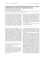
leptin for 24 h enhanced FAAH activity in a dose-depend-
ent manner (Fig. 1A). FAAH activation reached statistical
significance (P<0.05) at 2.5 n
M
leptin and a 300%
maximum at 10 n
M
. Therefore, the last concentration was
chosen to further investigate the effect of leptin on FAAH.
FAAH activation by 10 n
M
leptin (corresponding to
150 ngÆmL
)1
) was fully prevented by soluble leptin
receptor (sLR), used at neutralizing concentration of
3 lgÆmL
)1
.Instead,ÔmockÕ antibodies against IGF-IR were
ineffective at the same concentration (not shown). Also,
progesterone increased the activity of FAAH, in a dose-
dependent manner (Fig. 1B), at the same concentrations
used in the previous investigation [15]. At a dose of 1 l
M
,
progesterone enhanced FAAH activity and expression to
240% of the untreated control after 24 h of treatment
(Fig. 1B), and this effect was fully reversed by the synthetic
antiprogestinic compound RU486 (Fig. 1B), used at the
blocking concentration of 10 l
M
[15]. Western blot analysis
of U937 cell extracts s howed that specific anti-FAAH Igs
recognized a single immunoreactive band of the molecular
size expected for FAAH, the intensity of which was higher
in U937 cells treated w ith 10 n
M
leptin or 1 l
M
progesterone
than in controls (Fig. 1C). On the other hand, U937 cells
treatedwith10n
M
leptin or 1 l
M
progesterone expressed
the same levels of actin as the controls (Fig. 1C), ruling out
that the d ifferent levels of FAAH in these cells might be due
to unequal loading of proteins. The same anti-FAAH Igs
were used to further quantify FAAH content by ELISA,
which showed that leptin (Fig. 1A) and progesterone
(Fig. 1B) increased FAAH protein in human U937 cells
in parallel to the increase of enzymic activity. RT-PCR
amplification of cDNA of U937 cells showed a single band
of the expected molecular size f or FAAH gene, which
increased in cells treated with 10 n
M
leptin or 1 l
M
progesterone (Fig. 1C). Under the same experimental
conditions, the expression of the 18S rRNA gene was
unaffected (Fig. 1C). Liquid scintillation counting of
RT-PCR products showed that leptin (Fig. 1 A) and prog-
esterone (Fig. 1B) increased FAAH mRNA in a dose-
dependent manner in human U937 cells, in a way parallel to
that of enzymic activity a nd protein content. The effect of
leptin and progesterone on FAAH activity and expression
was ad ditive, in fact treatment of U937 cells for 24 h with
10 n
M
leptin and 1 l
M
progesterone enhanced FAAH
activity, protein level and mRNA content up to 500%,
450% and 490% of the controls, respectively (Table 1). On
the other hand, the same concentrations of leptin and
progesterone which enhanced FAAH activity and e xpres-
sion were ineffective on the activity of AMT, NAT and
PLD in U937 cells, when used either alone or in combina-
tion (Table 2). It should be recalled that U937 cells do not
express functional cannabinoid receptors [18].
Unlike U 937 cells, FAAH activity and expression in
human neuroblastoma CHP100 cells was not affected by
leptin and p rogesterone, used either alone or in combina-
tion, neithe r was the activity of AMT, NAT and PLD in the
same cells (Figs 1A,B,D and data not shown). It should be
recalled that also CHP100 cells are d evoid o f c annabinoid
receptors [18].
Both U937 and CHP100 cells have leptin receptor
and progesterone receptor
Human U937 and CHP100 cells were able to bind
125
I-labeled leptin according to a saturable process (Fig. 2A)
that yielded apparent dissociation constants (K
d
)of
2.0 ± 0.1 and 2.2 ± 0.2 n
M
and maximum binding (B
max
)
of 382 ± 5 and 339 ± 8 fmolÆmg protein
)1
, f or U937 cells
and CHP100 cells, respectively. These values are in agree-
ment with previous reports on LR of human hepatic cells
[32], and on LR stably transfected into different cell types
[27,33]. ÔColdÕ leptin fully displaced
125
I-labeled leptin from
its binding site, when used at 100 n
M
(Fig. 2 A). Moreover,
Western blot analysis showed that specific anti-LR Igs
Ó FEBS 2004 Differential regulation of the human FAAH promoter (Eur. J. Biochem. 271) 4669
recognized a s ingle immunoreactive b and in U937 and
CHP100 cell extracts, with an apparent molecular mass of
110 kDa (Fig. 2B). This value is consistent with the
expected molecular mass of the long form of LR [1,33].
Western blot a nalysis also showed that specific anti-PR Igs
recognized a single immunoreactive band with the same
intensity i n U937 and CHP100 cells, with an apparent
molecularmassof 120 kDa (Fig. 2C). This value is
consistent with the expected molecular mass of t he full
length form of PR [17].
Activation of downstream signals by binding to LR or PR
In previou s studies, we have shown that up-regulation of
FAAH activity and expression in human T lymphocytes by
Fig. 1. Effect of leptin a nd progesterone on
FAAH activity and expression. (A ) Effect of
leptin on the activity of FAAH in human U937
andCHP100cellsandontheproteincontent
and the mRNA of FAAH in U937 cells. These
cells were incubated for 24 h with leptin alone,
or wit h 10 n
M
( 150 ngÆmL
)1
)leptininthe
presence of 3 lgÆmL
)1
soluble leptin r ec eptor
(sLR) (100% activity in U 937 cells is
350 ± 30 pmolÆmin
)1
Æmg protein
)1
;protein
content, 0.280 ± 0.040 A
405
and mRNA level,
11 000 ± 1300 c.p.m; 100% activity in
CHP100 cells, 25 ± 3 pmolÆmin
)1
Æmg pro-
tein
)1
). (B) Effect of progesterone o n the s ame
samples a s in (A). Cells w ere incubated with
progesterone alone, or with 1 l
M
progesterone
inthepresenceof10l
M
RU486. In both
panels: * P < 0.05 vs. control, **P <0.01vs.
control and #P <0.01vs.10n
M
leptin or vs.
1 l
M
progesterone, r espectively. In both pan-
els, vertical bars represent SD values. (C) Up-
per p anel, Western blot analysis of U937 cells,
treated with 10 n
M
leptin or 1 l
M
progesterone
and reacted with specific anti-FAAH (top) or
anti-actin (bottom) Igs. The positions of
FAAH and actin are indicated to the right.
Lower panel, RT-PCR analysis of cDNA of
the s ame samples as in (C, upper panel). T he
expected sizes of the amplicons (199 base pairs
for F AAH and 258 b ase pairs for 18S rRNA)
are indicated to the right. (D) Same as in (C) on
CHP100 cell s. Data shown in (C,D) are rep -
resentative of three independent experiments.
Table 1. Modulation of FAAH expression in human U937 cells by leptin
and progesterone. Activity, 100 % ¼ 350 ± 30 pmolÆmin
)1
Æmg pro-
tein
)1
; Protein, 0.280 ± 0.040 A
405
;mRNAlevel,11000±1300
c.p.m.
Treatment of
U937 cells
FAAH Expression (%)
Activity Protein mRNA
Control 100 100 100
Leptin (10 n
M
)
for 24 h
300 ± 35* 270 ± 30* 310 ± 35*
Progesterone (1 l
M
)
for 24 h
240 ± 26* 220 ± 25* 240 ± 25*
Leptin (10 n
M
)+
progesterone (1 l
M
)
for 24 h
500 ± 45*# 450 ± 50*# 490 ± 45*#
*P < 0.01 vs. control; #P < 0.01 vs. 10 n
M
leptin or vs. 1 l
M
progesterone.
Table 2. Effect of leptin and progesterone on the activity of AEA
membrane transporter (AMT), N-acyltransferase (NAT) and phos-
pholipase D (PLD) in human U937 cells.
Treatment of
U937 cells AMT NAT PLD
Control 100
a
100
b
100
c
Leptin (10 n
M
)
for 24 h
100 ± 10 90 ± 10 100 ± 10
Progesterone (1 l
M
)
for 24 h
90 ± 10 90 ± 10 110 ± 10
Leptin (10 n
M
)+
progesterone (1 l
M
)
for 24 h
100 ± 10 100 ± 10 100 ± 10
a
100%, 150 ± 20 pmolÆmin
)1
Æmg protein
)1
;
b
100%, 14 ± 3 pmolÆ
min
)1
Æmg protein
)1
;
c
100%, 90 ± 10 pmolÆmin
)1
Æmg protein
)1
.
4670 M. Maccarrone et al. (Eur. J. Biochem. 271) Ó FEBS 2004
leptin [9] or p rogesterone [15] occurs through phosphory-
lation of STAT3, or increase of the nuclear content of the
transcription factor Ikaros, respectively. Therefore, the
levels of phospho-STAT3 and Ikaros were determined in
leptin-treated and progesterone-treated U937 and CHP100
cells. The nonphosphorylated, inactive form of STAT3 was
present in both cell-types, yet the active phospho-STAT3
increased only in U937 c ells treated with 1 0 n
M
lepti n
(Fig. 3A). In addition, nu clear l evels o f I karos isoforms Ik1,
Ik2 and Ik3 were increased by treatment of U937 cells, but
not of CHP100 cells, for 24 h with 1 l
M
progesterone
(Fig. 3B). Quantitative a nalysis of phospho-STAT3 and of
total Ikaros isoforms by ELISA corroborated the Western
blot data, showing that phospho-STAT3 and Ikaros
increased up to 300 and 250% of the untreated
controls in leptin-treated or progesterone-treated U937
cells, respectively (Fig. 3C). Neither phospho-STAT3 nor
Ikaros increased in CHP100 cells under the same experi-
mental conditions (Fig. 3 D).
Analysis of the
FAAH
promoter
The human F AAH gene has been located on chromosome 1
[34] and the latter has been completely sequenced. There-
fore, we have inspected this chromosome to gain insight on
the FAAH promoter features. Human FAAH promoter was
found to lack TATA boxes and, like many genes bearing
this feature, it had a proximally positioned SP1 site
(Fig. 4A). Moreover, there was another S P1 site in the
reverse orientation 100 nucleotides upstream (Fig. 4A), a
feature w hich resembles t he structure of the mouse proximal
promoter [35]. Inspection of the promoter sequence did not
show any obvious binding site for S TAT3, however, it had a
CRE-like site ( Fig. 4A), which is a target for transcriptional
regulation based on STAT3 tethering [ 36]. In addition, the
FAAH promoter had an Ikaros consensus site located at
position )66, upstream the CRE-like element (Fig. 4A).
Transient transfections using constructs containing both the
SP1 and the CRE-like ()107 to +1) sites driving the CAT
reporter gene in U937, but not in CHP100, cells showed that
the )107 construct w as up-regulated by leptin (Fig. 4 B, wt
and wt + L). Disruption o f this site by mutation a bolished
the leptin up-regulation (Fig. 4B, mutL and mutL + L).
Also progesterone up-regulated the reporter CAT gene in
U937, but not in CHP100, cells if under control of the
region between t he two SP1 sites of the FAAH upstream
region (Fig. 4B, wt and wt + P). Moreover, mutation of
the I karos c onsensus s ite abolished FAAH up-regulation by
progesterone (Fig. 4B, mutIk and mutIk + P). T he effect of
leptin and progesterone on FAAH promoter was a dditive
(Fig.4B,wt+L+P).
To confirm that leptin and progesterone act through a
CRE-like element and an Ikaros consensus site, respectively,
EMSA experiments were performed using nuclear extracts
prepared from U937 or CHP100 cells, untreated or treated
for 24 h w ith 10 n
M
leptin or 1 l
M
progesterone. As shown
in Fig. 5A, complex formation was only found with
oligonucleotides containing the CRE-like site of the FAAH
promoter. C omplex formation was not seen when the
mutant oligonucleotide (bearing the same mutation used
for the transient transfection experiment) was used as a
32
P-labeled probe, neither wa sitobservedinCHP100cells
under the same experimental conditions (Fig. 5A). On the
other hand, specificity o f the binding to the Ikaros s ite was
confirmed b y u sing a cold c ompetitor, which corresponds to
an established functional Ik site derived from the vasoactive
intestinal peptide receptor-1 promoter [31], and the mutated
site used for transient transfection experiments. Cold
competitor and m utated site, r espectively, abolished or
had no effect on the retarded complex in U937 cells
(Fig. 5B). Conversely, no retarded complex was seen in
CHP100 cells under the same experimental conditions
(Fig. 5 C).
Discussion
In this study we show that leptin and progesterone
stimulate, independently (Fig. 1A,D) or additively
Fig. 2. Characterization of leptin receptor (LR) and prog esterone
receptor (PR). (A) Saturation curves of the binding of
125
I-labeled
leptin to human U937 or CHP100 cells, alone or in the presence of
100 n
M
ÔcoldÕ leptin (data of the ÔcoldÕ leptin experiment with CHP100
cells were su perimposable to those o f U937 c ells, and we re omitted for
the sake of clarity). Western blot analysis of cell extracts (20 lgper
lane), reacted with anti-LR (B) or anti-PR (C) Igs. M olecular mass
markers and the position of LR and PR are indicated to the right. Data
shown in (B,C) are representative of three independent experiments. In
(A), vertical bars represent SD v alues.
Ó FEBS 2004 Differential regulation of the human FAAH promoter (Eur. J. Biochem. 271) 4671
(Table 1), FAAH activity and expression in human lym-
phoma U937 cells, but not in human neuroblastoma
CHP100 cells. FAAH activation occurs via a leptin
receptor-mediated activation of STAT3 signaling and/or
via a progesterone receptor-mediated increase in nuclear
levels of Ikaros, which in turn lead s to up-regulation of a
CRE-like site or of an Ikaros binding site, respectively, in
the FAAH promoter.
Leptin modulates FAAH activity and expression in U937
cells at the s ame circulating levels shown to m odulate
immune cell response [4]. These concentrations of leptin
were found to saturate the binding sites in U937 cells
(Fig. 2A), with calculated b inding constants (K
d
and B
max
)
similar to those of the leptin receptor [32,33]. This obser-
vation, together with the ability of ÔcoldÕ leptin to fully
displace
125
I-labeled leptin, strongly suggests that U937 cells
have an authentic LR. Western blot analysis further
corroborates this hypothesis, showing that specific anti-
LR Igs recognized a single band (Fig. 2B) of the expected
molecular mass of the long isoform of L R [ 1,27]. These data
extend our recent observations on primary human T-cells [9]
and suggest that regulation by leptin, being conserved in
immortalized cells, has a critical role for the immune
function. Also the effects of progesterone, used a t circula-
ting levels of 1 l
M
( 0.3 lgÆmL
)1
), on FAAH activity in
U937 cells extend our previous observations on T l ympho-
cytes [15], and here we show for the first time that these cells
have the full length form of PR (Fig. 2C). As this form is
known to function as a transcriptional activator of pro-
gesterone-responsive genes [17,37,38], this finding gives
ground to the hypothesis that the effects of progesterone on
FAAH gene expression were mediated through its bin ding
to PR [15]. On the other hand, leptin and progesterone were
ineffective on AEA transport through AMT, and on AEA
synthesis through NAT and PLD in U937 cells (Table 2),
suggesting that FAAH was the only Ôcheck pointÕ for the
effect of both leptin and progesterone. These observations
are in k eeping with the hypothesis that FAAH is the key-
regulator of AEA levels in vivo, indeed FAAH knockout
mice show 15-fold higher levels of AEA than wild-type
littermates [39], and AEA levels in human blood inversely
correlate with FAAH activity in peripheral lymphocytes
[40].
The mechanism of F AAH activation b y leptin and
progesterone was further investigated. Binding of leptin to
the l ong isoform of LR is known to trigger different
signaling pathways, among which STAT-dependent signal
transduction is prominent [7,8]. In human T lymphocytes,
we have shown recently that leptin activated (i.e. phos-
phorylated) only STAT3 [9], which is preferentially activa-
tedalsoinothercelltypes[33,36]andtissues([1]and
references therein). Phospho-STAT3 was up-regulated by
leptin also in U 937 cells (Fig. 3A,C), where the FAAH
promoter did not contain STAT3 DNA binding motifs, yet
it did contain a CRE-like element between the two SP1 sites
(Fig. 4A). Such CRE-like elements have been shown
recently to be tethered by STAT3, thus, leading to a novel
type of up-regu lation of gene transcription [36]. Transfec-
tion experiments using FAAH promoter constructs with
mutated CRE-like elements (mutL) revealed that indeed
these sites confer STAT3 responsiveness ( Fig. 4B). EMSA
analysis further corroborated this c onclusion (Fig. 5A).
FAAH promoter also revealed a binding site for Ikaros, a
member of the K ruppel family o f Ôzinc fingerÕ DNA-
binding proteins that acts as critical transcriptional regu-
lator of lymphocyte ontogeny and differentiation [14].
Ikaros comprises eight different isoforms, Ik1 through Ik3
localizing to the nucleus and Ik4 through Ik8 localizing to
the cytosol [14]. Recently Ik1, 2 and 3, the only isoforms
which exhibit high DNA binding affinity [14], have been
Fig. 3. Activation of downstream signals by
leptin (L) or progesterone (P). Human U937 or
CHP100 cells were treated for 24 h with 10 n
M
leptin (A) or 1 l
M
progesterone (B), then
lysates (50 lg o f protein) were immunoblotted
with the specific antibody against the inactive
(total) or active (phosphorylated, phospho)
form of STAT3 (A), or against Ikaros iso-
forms Ik1, Ik2 and Ik3 (B). The positions of
(phospho) proteins are indicated to the right.
These data are representative of three inde-
pendent experiments. Quantitative analysis by
ELISA of the same samples (25 lgofprotein)
shown in (A,B) is reported in (C,D), where
*P < 0.01 vs. control (CTR), and vertical
bars represent SD values.
4672 M. Maccarrone et al. (Eur. J. Biochem. 271) Ó FEBS 2004
detected in the nucleus of human peripheral blood mono-
nuclear cells [13,14], where they are essential in contro lling
the activation of granzyme B promoter by the glucocorti-
coid, dexamethasone [13]. Here, we demonstrated that
circulating levels of progesterone (1 l
M
, i.e. 0.3 lgÆmL
)1
)
increased the nuclear levels of these Ikaros i soforms in
U937 cells 2.5-fold over the controls (Fig. 3B,C). This is
noteworthy, because homo- and h eterodimer formation
between Ik1, 2 and 3 is k nown t o greatly increase their
affinity for DNA and their con sequent ability to activate
transcription [13,14]. Transfection experiments using
FAAH promoter constructs with mutated Ikaros (mutIk)
revealed that this site indeed confers progesterone respon-
siveness (Fig. 4B), and EMS A analysis corroborated this
conclusion (Fig. 5B). Taken together, it c an be concluded
that in human U937 cells leptin by binding to LR activates
STAT3, which in turn up-regulates FAAH gene transcrip-
tion by tethering to a CRE-like site in the FAAH promoter.
In addition, progesterone activates the FAAH gene by
stimulating the binding of Ikaros to DNA, thus, enhancing
its transcriptional activity on the FAAH promoter. Activa-
tion of the FAAH promoter by leptin may be additive to
that of progesterone at the two different sites (Table 1 and
Fig. 4).
Fig. 4. Analysis of FAAH promoter by transient expression. (A) Proximal upstream r egion of human FAAH gene. Left and right handed arrows
denote SP1 sites in the – and + strands, respectively. Pentagonal box: Ikaros (Ik) site; oval box: CRE-like site. (B) 5¢ Flanking regions of the human
FAAH gen e we re cloned in the PstI/XbaI sites of pCAT3-basic vector, wt: )107 to +1 upstream region containing the two SP1 sites fl anking t he
Ikaros site and the CRE-like site; mutL: same as wt but with the mutated CRE-like sequence; mutIk: same as wt but with mutated Ikaros site;
mutated sites are in w hite. Human U 937 cells and CHP100 cells w ere transfected with the aforementioned constru cts and left untreated or treated
with leptin or progesterone. Transfection efficiency was monitored by the use of thymidine kinase b-galactosidase construct. The values of CAT
activity were normaliz ed to b-gal activity and to the protein content and are expressed as percentage with r espect to the activity of the empty vector,
pCAT3 basic, which was set to 100%. *P < 0.01 vs. control, # P < 0.01 vs. leptin or progesterone alone, and h orizontal bars r epresent SD values.
Ó FEBS 2004 Differential regulation of the human FAAH promoter (Eur. J. Biochem. 271) 4673
A major finding of this investigation is that FAAH
activity and expression in human neuroblastoma CHP100
cells was not up-regulated by leptin and/or progesterone,
although these cells use LR, PR, S TAT3 and Ikaros. Th is
unprecedented observation gives support to previous
reports showing that in mouse hypothalamus FAAH
activity is not regulated by leptin [10], whereas it is in
mouse peripheral lymphocytes [9]. However, the molecular
reasons for the l ack of responsiveness of neuronal c ells
remain unclear and different hypotheses can be put forward.
On one hand, it can be suggested that coactivators triggered
by leptin or progesterone in immune cells are missing in
neuronal cells. On the other hand, it is also possible that
silencers of FAAH gene expression are present in neuronal
cells but not in immune cells. While both hypotheses are in
keeping with the tissue-specificity of FAAH promoter
regulation observed recently in vitro in mouse cells [41],
the observation that background FAAH activity ( 25 vs.
350 pmolÆmin
)1
Æmg protein
)1
with 10 l
M
[
3
H]AEA as
substrate; Fig. 1) and expression (Fig. 1C,D) in CHP100
cells is significantly lower than that in U937 cells seems to
speak in favour of a constitutive repression of FAAH gene
in the former cells. To the best of our knowledge, this is the
first demonstration that FAAH is differentially regulated in
neuronal and immune cells by the same factors; a finding
which may have important implications for the regulation
of the tone and activity of AEA in the neuroimmune axis.
In particular, it seems noteworthy that leptin [42,43] and
progesterone [44,45] play roles in survival, death and
proliferation of immune and neuronal cells; processes in
which AEA and the endocannabinoid system are also
involved [19]. In particular, le ptin and progesterone, by
stimulating AEA degradation through FAAH, might
prevent apoptosis induced by AEA in i mmune cells [19],
whereas they should be unable to protect by the same
mechanism neuronal cells [19]. Supporting this is the
observation that progesterone enhances AEA hydrolase in
immune cells by increasing nuclear levels of Ikaros – this
factor is crucial for the in vivo enhancer function of TrkA,
the nerve growth factor receptor [46]. Trk receptors are
suppressed by AEA in breast and prostate cancer cells [47],
suggesting a possible functional link between Ikaros, AEA
tone and Trk receptors in peripheral cells. Finally, it should
be pointed out that genes in ad dition to FAAH may be
differentially regulated by l eptin and/or progesterone in
U937 cells and CHP100 cells, potentially extending the
implications of the reported findings beyond the endocann-
abinoid system.
In conclusion, the results reported here represent the first
evidence of a differential regulation of FAAH gene expres-
sion in immune and neuronal cells, suggesting cell-specific
links between networks controlled by leptin o r progesterone
and the peripheral endocannabinoid system. Conversely,
the central actions of the two hormones do not seem to
involve modulation of endocannabinoid metabolism.
Acknowledgements
We wish to thank Drs Monica Bari and Natalia Battista for their expert
assistance in cell isolation and culture. T his study was partly s upported
by Ministero dell’Istruzione, dell’Universita
`
edellaRicerca(Cofin
2003) and by Istituto Superiore di Sanita
`
(III AIDS project), Rome.
References
1. Ahima, R.S. & Flier, J.S. (2000) Leptin. Annu. Rev. Physiol. 62,
413–437.
2. Cunningham, M.J., Cl ifton, D. K. & Steiner, R.A. (1999) Leptin’s
actions on the reproductive axis: perspectives and mechanisms.
Biol. Reprod. 60, 216–222.
3. Matarese, G., La Cava, A., Sanna, V., Lord, G.M., Lechler, R.I.,
Fontana, S., & Zappac osta, S. (2002) Balan cing susceptibility
infection and autoimmunity: a role for leptin? Trends Immunol. 23,
182–187.
4. Lord, G .M., Matarese, G. , Ho wa rd, J.K., Baker, R.J., Bloom,
S.R. & Lechler, R.I. (1998) Leptin modulates the T-cell immune
response and reverses starvation-induced immunosuppression.
Nature 394, 8 97–901.
5. Piccinni, M.P., Giudizi, M.G., Biagiotti, R., Beloni, L., Gianna-
rini, L., Sampognaro, S., Parronchi, P., Manetti, R., Annunziato,
F.,Livi,C.,Romagnani,S.&Maggi,E.(1995)Progesterone
favors the development of human T helper cells producing Th2-
type cytokines and promotes both IL-4 production and membrane
Fig. 5. Analysis of FAAH promoter by electrophoretic mobility-shift
assay (EMSA). EMSA experiments were performed with 3 lgofU937
or CHP100 nuclear extracts, prepared before (–) and after (+ ) sti-
mulation with 10 n
M
leptin (A) or 1 l
M
progesterone (B,C). (A)
Complex formation (indicated by an arrow) was performed with
oligonucleotides as
32
P-labeled probes for mutated (mutL) or wild-type
(wt) CRE-like site. (B) Complex formation (in dicated by an arrow) in
U937 cells, performed with
32
P-labeled oligo nucleotide c orresp onding
to the Ikaros site. L ane O represents
32
P-labeled oligonucleotide a lone,
which contained the Ikaros site; competitor wild type (wt) was a cold
oligonucleotide containing the Ikaros site derived from vasoactive
intestinal peptide receptor-1 promoter; competitor mutant (mut) was a
cold oligonucleotide containing the mutated Ikaros site used in
transfection experiments. (C) Same as in (B) with CHP100 cells.
4674 M. Maccarrone et al. (Eur. J. Biochem. 271) Ó FEBS 2004
CD30 expression in established Th1 cell clones. J. Immunol. 155,
128–133.
6. Piccinni, M.P., Beloni, L., Livi, C., Maggi, E., Scarselli, G. &
Romagnani, S. (1998) Defective production of both leukemia
inhibitory factor and type 2 T-helper cytokines by decidual T cells
in unexplained recurrent abortions. Nature Med. 4, 1020–1024.
7. Bjorbaek, C., Buchholz, R.M., Davis, S.M., Bates, S.H., Pierroz,
D.D., G u, H., Neel, B.G., Myers, M.G. Jr & Flier, J. S. (2001)
Divergent roles of SHP-2 in ERK activation by leptin receptors.
J. Biol. Chem. 276, 4747–4755.
8. Schrager, J.A., Der Minassian, V. & Marsh, J .W. (2002) HIV Nef
increases T cell E RK MAP kinase activity. J. Biol. Chem. 277,
6137–6142.
9. Maccarrone, M., Di Rienzo, M., Fina zzi-Agro
`
,A.&Rossi,A.
(2003) Leptin activates the Anandamide Hydrolase p romoter in
human T lymphocytes through STAT3. J. Biol. Chem. 278,
13318–13324.
10. Di Marzo, V., Goparaju, S.K., W ang, L., Liu, J., Batkai, S., Jarai,
Z.,Fezza,F.,Miura,G.I.,Palmiter,R.D.,Sugiura,T.&Kunos,
G. (2001) Leptin-regulated endocannabinoids are involved in
maintaining food intake. Nature 410, 822–825.
11. Harvey, J . & Ashford, M.L. (2003) Leptin in the CNS: much more
than a satiety signal. Neuropharmacology 44, 845–854.
12. Veniant, M.M. & LeBel, C.P. (2003) Leptin: from animals to
humans. Curr. Pharm. Des. 9, 811–818.
13. Wargnier, A., Lafaurie, C., Legros-Maida, S., Bourge, J F.,
Sigaux, F., Sasportes, M. & Pauli, P. (1998) Down-regulation of
human granzyme B expression by glucocorticoids. Dex-
amethasone inhibits binding to the Ikaros and AP-1 regulatory
elements o f the granzyme B promoter. J. Biol. Chem. 273, 35326–
35331.
14. Sun, L., Heerema, N ., Crotty, L., Wu, X., Navara, C., Vassilev,
A., Sensel, M., Reaman, G.H. & Uckun, F.M. (1999) Expression
of dominant-negative and mutant isoforms of the antileukemic
transcription factor Ikaros in infant acute lymphoblastic leukemia.
Proc.NatlAcad.Sci.USA96, 680–685.
15. Maccarrone,M.,Bari,M.,Di Rienzo, M ., Finazzi-Agro
`
,A.&
Rossi, A. (2003) Progesterone activates fatty Acid Amide
Hydrolase (FAAH) promoter in human T lymphocytes
through the transcription factor Ikaros. J. Biol. Chem. 278, 32726–
32732.
16. Belelli, D. & Herd, M.B. (2003) T he contraceptive agent Provera
enhances GABA (A) recept or-mediated in hibitory neu ro-
transmission in th e rat hippocampus: e vidence for endogenous
neurosteroids? J. Neurosci. 23, 10013–10020.
17. Sakamoto, H ., Shikimi, H ., Ukena, K . & Tsutsui, K. (2003)
Neonatal expression of progesterone recepto r isoforms in the
cerebellar Purkinje cell in rats. Neurosci. Lett. 343, 163–166.
18. Maccarrone, M., van der Stelt, M., Rossi, A ., Veldink, G.A.,
Vliegenthart, J.F.G. & Finazzi-Agro
`
, A. (1998) Anandam ide
hydrolysis by h uman cells in culture and brain. J. Biol. Chem. 273,
32332–32339.
19. Maccarrone, M. & Finazzi Agro
`
, A. (2003) The endocannabinoid
system, anandamide and the regulation of mam malian cell
apoptosis. Cell Death D iffer. 10, 946–955.
20. Cadas, H., di Tomaso, E. & Piomelli, D. (1997) Occurrence and
biosynthesis of end ogenous cannabinoid precursor, N -arachido-
noyl phosphatid ylethanolamine, in rat brain. J. Neurosci. 17,
1226–1242.
21. Moesgaard, B., Petersen, G., Jaroszewski, J.W. & Hansen, H.S.
(2000) Age dependent accu mula tion of N-acyl-ethanolamine
phospholipids in ischem ic rat brain . A ( 31) P NM R and en zyme
activity study. J. Lipid Res. 41, 985–990.
22. Bisogno, T., De Petrocellis, L. & Di Marzo, V. (2002) Fatty acid
amide hydrolase, an enzyme with many bioactive substrates.
Possible therapeutic implications. Curr. Pharm. Des. 8, 533–547.
23. Parolaro, D., Massi, P., Rubino, T. & Monti, E. (2002)
Endocannabinoids in the immune system and cancer. Pros-
taglandins Leukot. Essent. Fatty Acids 66, 319–332.
24. Cravatt, B.F. & Lichtman, A.H. (2003) Fatty acid amide hydro-
lase: an emerging therapeutic target in the endocannabinoid sys-
tem. Curr. Opin. Chem. Biol. 7, 469–475.
25. Giang, D.K. & Cravatt, B.F. (1997) Molecular characterization of
human and mouse fatty acid amide hydrolases. Proc. Natl Ac ad.
Sci. USA 94, 2238–2242.
26. Okamoto, Y., Morishita, J., Tsuboi, K., Tonai, T. & Ueda, N.
(2004) Molecular characterization of a phosph olipase D
generating anandamide and its congeners. J. Biol. Chem. 279,
5298–5305.
27. Tartaglia,L.A.,Dembski,M.,Weng,X.,Deng,N.,Culpepper,J.,
Devos, R., Richards, G.J., Campfield,L.A.,Clark,F.T.,Deeds,J.,
Muir,C.,Sanker,S.,Moriarty,A.,Moore,K.J.,Smutko,J.S.,
Mays, G.G., Wool, E.A., Monroe,C.A.&Topper,R.I.(1995)
Identification and expression cloning of a leptin receptor, OB-R.
Cell 83, 1263–1271.
28. Bradford, M.M. (1976) A rapid and sensitive method for the
quantitation of microgram quantities of protein utilizing the
principle of protein-dye binding. Anal. Biochem. 72, 248–254.
29. Schreiber, E., Muller, M.M. & Schaffner, W. (1989) Rapid
detection of octamer binding proteins with Ômini-extractsÕ,
prepared from a small number of cells. Nucleic Acids Res. 17,
6419.
30. Lee, J H., Jang, S I., Markova, N.G. & Steinert, P.M. (1996) The
proximal promoter of the h uman transglutaminase 3 gene.
Stratified squamo us e pithelial-specific expression in cultured
cells is mediated by binding of Sp1 and ets transcription factors
to a proximal promoter element. J. Biol. Chem. 271, 4561–
4568.
31. Dorsam, G. & Goetzl, E.J. (2002) Vasoactive intestinal peptide
receptor-1 (VPAC -1) is a novel gene target of the hemolympho-
poietic t ranscr iption factor Ikaros. J. Biol. Chem. 277, 13488–
13493.
32. Cohen, B., Novick, D. & Rubinstein, M. (1996) Modulation of
insulin activities by leptin. Science 274, 1185–1188.
33. Wang, Y., Kuropatwinski, K.K., White, D.W., Hawley, T.S.,
Hawley, R.G., Tartaglia, L.A. & Baumann, H. (1997) Leptin
receptor action in hepatic cells. J. Biol. Chem. 272, 16216–16223.
34. Wan,M.,Cravatt,B.F.,Ring,H.Z.,Zhang,X.&Francke,U.
(1998) Conserved chromosomal location and genomic structure of
human and mouse fatty-acid amide hydrolase genes and evalua-
tion of clasper as a ca ndidate neurological mutation. Genomics 54,
408–414.
35. Waleh, N.S., Cravatt, B.F., Apte-Deshpande, A., Terao, A. &
Kilduff, T.S. (2002) Transcriptional regulation of the mous e fatty
acid amide hydrolase gene. Gene 291, 203–210.
36. Niehof, M., Stree tz, K., Rakemann, T., Bischoff, S.C., Manns,
M.P., Horn, F. & Trautwein, C. (2001) In terleukin-6-in duced
tethering of STAT3 to the LAP/C/EBPbeta promoter suggests a
new m echanism of transcription al regulation by STAT3. J. Biol.
Chem. 276, 9016–9027.
37. Tora, L., Gronemeyer, H., Turcotte, B., Gaub, M.P. & Chambon,
P. (1998) The N-terminal region of the chicken progesterone
receptor specifies target gene ac tivation. Na ture 333, 185–188.
38. Wen,D.X.,Xu,Y.F.,Mais,D.E.,Goldman,M.E.&McDonnell,
D.P. (1994) The A and B isoforms o f th e h uman progesterone
receptor operate through d istinct signaling pathways within targe t
cells. Mol. Cell Biol. 14, 8356–8364.
39. Cravatt, B.F., Demarest, K., Patricelli, M.P., Bracey, M.H.,
Giang, D.K., Martin, B.R. & Lichtman, A.H. (2001) Super-
sensitivity t o anandamide a nd enhanced end ogenous cannabinoid
signaling in mice lacking fatty acid amide hydrolase. Proc. Natl
Acad. Sci. USA 98, 9371–9376.
Ó FEBS 2004 Differential regulation of the human FAAH promoter (Eur. J. Biochem. 271) 4675
40. Maccarrone, M., Bisogno, T., Valensise, H., Lazzarin, N., Fezza,
F., Manna, C., Di Marzo, V. & Finazzi-Agro
`
, A. (2002) Low fatty
acid amide hydrolase and high anandamide levels are associated
with failure to achieve an ongoing pregnancy after IVF and
embryo transfer. Mol. Hum. Reprod. 8, 188–195.
41. Puffenbarger, R.A., K apulina, O., Howell, J.M. & Deutsch, D .G.
(2001) Characterizatio n of the 5¢-sequence of the mouse fatty a cid
amide hydrolase. Neurosci. Lett. 314, 21–24.
42. De Falco, J., Tomishima, M., Liu, H., Zhao, C., Cai, X., Marth,
J.D., Enquist, L. & Friedman, J.M . (2001) Virus-assisted m apping
of neural inputs to a feeding center in the h ypothalam us. Science
291, 2608–2613.
43. Sanna,V.,DiGiacomo,A.,LaCava,A.,Lechler,R.I.,Fontana,
S., Zappacosta, S., & M atarese, G. (2003) Leptin surge precedes
onset of autoimmune encephalomyelitis and correlates with
development of pathogenic T cell responses. J. Clin. Invest. 111,
241–250.
44. Sakamoto, H., Ukena, K. & Tsutsui, K. (2001) Effects of pro-
gesterone synthesized de novo in the developing Purkinje cell on its
dendritic growth and synaptogenesis. J. Neurosci. 21 , 6221–6232.
45. Ghoumari, A.M., Dusart, I., El-Etr, M., Tronche, F., Sotelo, C.,
Schumacher, M. & Baulie u, E.E. (2003) M ifepristone (RU4 86)
protects Purkinje cells from cell death in organotypic slice cultures
of postnatal rat and mouse cerebellum. Proc. Natl A cad. Sci. USA
100, 7953–7958.
46. Lei, L., Ma, L., Nef, S., Thai, T. & Parada, L.F. (2001) mKlf7, a
potential transcriptional regulator of TrkA nerve growth fac tor
receptor e xpression in sensory and sympathetic neurons. Devel-
opment 128, 1147–1158.
47. Melck, D., De Petrocellis, L., Orlando, P., Bisogno, T., Laezza, C.,
Bifulco, M. & Di Marzo, V. (2000) Suppression of nerve growth
factor Trk r eceptors and prola ctin receptors by e ndocan nabinoids
leads to inhibition of human breast and prostate cancer cell pro-
liferation. Endocrinology 141, 118–126.
4676 M. Maccarrone et al. (Eur. J. Biochem. 271) Ó FEBS 2004