Báo cáo khoa học: Transphosphatidylation activity of Streptomyces chromofuscus phospholipase D in biomimetic membranes docx
Bạn đang xem bản rút gọn của tài liệu. Xem và tải ngay bản đầy đủ của tài liệu tại đây (288.57 KB, 8 trang )
Transphosphatidylation activity of
Streptomyces chromofuscus
phospholipase D in biomimetic membranes
Karim El Kirat
1
, Annie-France Prigent
2
, Jean-Paul Chauvet
3
, Bernard Roux
1
and Franc¸oise Besson
1
1
Laboratoire de Physico-Chimie Biologique, UMR CNRS 5013, Villeurbanne, Lyon, France;
2
Laboratoire de Biochimie et
Pharmacologie, UMR INSERM 585, Villeurbanne, Lyon, France;
3
Laboratoire d’Inge
´
nierie et de Fonctionnalization des Surfaces,
UMR CNRS 5621, Ecully, Lyon, France
The phospholipase D (PLD) from Streptomyces chromo-
fuscus belongs to the superfamily of PLDs. All the enzymes
included in this superfamily are able to catalyze both
hydrolysis and transphosphatidylation activities. However,
S. chromofuscus PLD is calcium dependent and is often
described as an enzyme with weak transphosphatidylation
activity. S. chromofuscus PLD-catalyzed hydrolysis of
phospholipids in aqueous medium leads to the formation of
phosphatidic acid. Previous studies have shown that phos-
phatidic acid–calcium complexes are activators for the
hydrolysis activity of this bacterial PLD. In this work, we
investigated the influence of diacylglycerols (naturally
occurring alcohols) as candidates for the transphosphati-
dylation reaction. Our results indicate that the transphos-
phatidylation reaction may occur using diacylglycerols as a
substrate and that the phosphatidylalcohol produced can be
directly hydrolyzed by PLD. We also focused on the surface
pressure dependency of PLD-catalyzed hydrolysis of
phospholipids. These experiments provided new informa-
tion about PLD activity at a water–lipid interface. Our
findings showed that classical phospholipid hydrolysis is
influenced by surface pressure. In contrast, phosphatidyl-
alcohol hydrolysis was found to be independent of surface
pressure. This latter result was thought to be related to
headgroup hydrophobicity. This work also highlights the
physiological significance of phosphatidylalcohol produc-
tion for bacterial infection of eukaryotic cells.
Keywords: phospholipase D; Langmuir films; transphos-
phatidylation; Streptomyces chromofuscus; diacylglycerol.
Phospholipase D (PLD) catalyzes the hydrolysis of the
phosphoester bond between the phosphatidyl moiety and
the choline headgroup of phosphatidylcholine (PtdCho),
liberating choline and phosphatidic acid (PA). More
precisely, PLD catalyzes the cleavage of the P-O bond of
PtdCho, as demonstrated previously [1]. The mechanism of
this reaction involves a molecule of water for the nucleophile
substitution on the phosphatidyl–enzyme intermediate.
When the nucleophile is an alcohol, a phosphatidylalcohol
is produced. This latter activity is called transphosphatidy-
lation and is specific for the PLD [2].
The PLD from Streptomyces chromofuscus belongs to the
PLD superfamily, together with some endonucleases, some
helicases, and some lipid synthases. Most of these enzymes
are capable of catalyzing the hydrolysis and/or formation of
phosphodiester bonds [3]. However, S. chromofuscus PLD
also presents interesting characteristics that allow it to be
distinguished from other enzymes of the PLD superfamily.
Yang & Roberts [4] have recently shown that S. chromo-
fuscus PLD does not bear the classical HKD motif and this
may explain its strong dependency on calcium. Phosphatase
activity has also been attributed to S. chromofuscus PLD,
which was able to catalyze para-nitrophenyl-phosphate
hydrolysis but was inactive on lipidic phosphomonoesters,
such as PA [5].
Most work concerning S. chromofuscus PLD has focused
on its hydrolytic activity and especially on its activation by
anionic lipids [6]. An involvement of this bacterial PLD on
the aggregation, leakiness and fusion of vesicles has also
been demonstrated [7]. PLD-catalyzed hydrolysis has been
measured using several techniques and with biomimetic
substrates [8]. In the case of phospholipid hydrolysis, the
production of PA has been determined by radioactive assay
using radiolabelled PtdCho, by pH-stat and by [
1
H] NMR
[9], by a choline oxidase electrode [10], by the Langmuir
film technique on phospholipidic monolayers [11], and
by polarization modulation infrared reflection-absorption
Correspondence to F. Besson, Laboratoire de Physico-Chimie Bio-
logique, UMR CNRS 5013, Bat. Chevreul, 43 Bd du 11/11/1918,
F-69622 Villeurbanne, UCB-Lyon 1, France.
Fax: + 33 4 72431543, Tel.: + 33 4 72431542,
E-mail: f-besson@univ-lyon1fr
Abbreviations: MLV, multilamellar vesicles; PA, phosphatidic acid;
PLD, phospholipase D; PtdCho, phosphatidylcholine;
Pam
2
(Pam[
3
H]N)GroChoP,
L
-a-dipalmitoyl-[2-palmitoyl-
9,10-
3
H(N)]-sn-glycero-3-phosphocholine; PamOleGroEtP,
1-O-palmitoyl-2-O-oleoyl-sn-glycero-3-phosphoethanol; PamOle-
GroBuP,1-O-palmitoyl-2-O-oleoyl-sn-glycero-3-phosphobutanol;
Myr
2
GroChoP,
L
-a-dimyristoyl-sn-glycero-3-phosphocholine;
Myr
2
Gro, 1,2-dimyristoyl-rac-glycerol; Myr
2
GroPA,
L
-a-dimyristoyl-
sn-glycero-3-phosphatidic acid; PamLinGroEtnP-HNE, 1-O-palmi-
toyl-2-O-linoleoyl-sn-glycero-3-phosphoethanolamine-4-hydroxy-
nonenal; SM, sphingomyelin from bovine brain.
(Received 11 July 2003, revised 17 September 2003,
accepted 22 September 2003)
Eur. J. Biochem. 270, 4523–4530 (2003) Ó FEBS 2003 doi:10.1046/j.1432-1033.2003.03841.x
spectroscopy at the air–water interface [12]. All of these
studies concluded that PLD is activated by PA and calcium.
However, no factor has as yet been identified which
enhances the transphosphatidylation reaction. PLD from
S. chromofuscus is often described as an enzyme that has
only weak transphosphatidylation activity. However, Fried-
man et al. [13] have shown that this PLD is able to catalyze
an intramolecular transphosphatidylation reaction within
lyso-PtdCho. This activity is responsible for the formation
of cyclic lyso-PA, which can be further hydrolyzed by PLD
to produce lyso-PA. In other words, PLD from S. chromo-
fuscus is able to form transphosphatidylation products and
can also catalyze their hydrolysis.
In the present work, the S. chromofuscus PLD trans-
phosphatidylation activity was investigated in model mem-
branes (i.e. vesicles and monolayers). Our results highlight
the involvement of diacylglycerols in a naturally occuring
transphosphatidylation reaction catalyzed by PLD. More-
over, monolayer experiments allowed us to estimate phos-
phatidylalcohol hydrolysis under PLD action at the air–
water interface. Comparison with the results obtained on
lipidic vesicles led to the elaboration of a model mechanism
of transphosphatidylation reaction catalyzed by S. chromo-
fuscus PLD.
Materials and methods
Materials
L
-a-Dipalmitoyl-[2-palmitoyl-9,10-
3
H(N)]-sn-glycero-3-pho
sphocholine [Pam
2
(Pam[
3
H]N)GroChoP] was purchased
from NEN (Life Science Products, Inc., Boston, MA,
USA). Coomassie Brilliant Blue R and TLC aluminium
sheets (Silica gel 60F
254
) were from Merck (Darmstadt,
Germany). 1-O-palmitoyl-2-O-oleoyl-sn-glycero-3-phos-
phoethanol (PamOleGroEtP)and1-O-palmitoyl-2-O-
oleoyl-sn-glycero-3-phosphobutanol (PamOleGroBuP)
were purchased from Biomol Research Laboratories Inc.
(Plymouth Meeting, PA, USA).
L
-a-Dimyristoyl-sn-glyc-
ero-3-phosphocholine (Myr
2
GroChoP), 1,2-dimyristoyl-
rac-glycerol (Myr
2
Gro),
L
-a-dimyristoyl-sn-glycero-3-phos-
phatidic acid (Myr
2
GroPA), sphingomyelin from bovine
brain (SM) and PLD from S. chromofuscus were purchased
from Sigma Chemical Co. and used without further
purification. SDS/PAGE analysis of PLD gave the same
three bands as those obtained by Geng et al.[14].Triswas
purchased from Roche Diagnostics. 1-O-palmitoyl-2-O-
linoleoyl-sn-glycero-3-phosphoethanolamine-4-hydroxynonenal
(PamLinGroEtnP-HNE) was a gift from M. Guichardant
(Laboratoire de Biochimie et Pharmacologie, UMR
INSERM 585, INSA de Lyon, France).
Monolayer technique
All experiments were performed at a constant temperature
of 21 ± 0.1 °C. The film balance was built by R&K
(Wiesbaden, Germany) and equipped with a Wilhemy-type
surface-pressure measuring system. The subphase was
aqueous buffer containing 120 l
M
CaCl
2
,150m
M
NaCl,
and 10 m
M
Tris/HCl, pH 8.0. The calcium concentration
was sufficient to allow maximum PLD activity [11].
Phospholipids were spread at the air–water interface in
hexane/ethanol (9 : 1, v/v), at a concentration of 0.175 m
M
,
to reach a final quantity of 8.75 nmol of lipids. After 15 min
of solvent evaporation, the monolayer was compressed to a
lateral pressure of 35 mNÆm
)1
to obtain a control p-A
isotherm. Then, the pressure was fixed at 30 mNÆm
)1
and
theenzyme(15 lg of protein) was injected into the subphase
after monolayer stabilization. The subphase was stirred
using a magnetic stirrer spinning at 100 r.p.m. Surface
compressional moduli were calculated from the pressure-
area data obtained from the monolayer compressions, using
the following equation [15]:
Ks ¼ÀAxdp=dA
where A is the molecular area at the indicated surface
pressure p. High Ks values correspond to low interfacial
elasticity among packed lipids forming a monolayer [16].
This suggests that the higher the Ks value of a
monolayer, the greater the monolayer rigidity.
Vesicle preparation
Lipids (i.e. Myr
2
GroChoP and Myr
2
Gro) were dissolved in
chloroform at 5 m
M
. The lipid mixture, containing varying
molar ratios of Myr
2
Gro, were dried under nitrogen for 2 h.
Then, the lipidic film was resuspended by vigorous agitation
for 1 min in 120 l
M
CaCl
2
, 150 m
M
NaCl and 100 m
M
Tris/HCl, pH 8.0, to achieve a 5 m
M
final concentration of
total lipids. The lipid suspension was frozen in liquid
nitrogen for 5 min and then heated for 10 min at 40 °Cina
thermostated bath. This vortex freeze-warming was repea-
ted three times to obtain the multilamellar vesicles (MLV).
Radioactive assay
PLD activity on lipidic bilayers was determined using
mixed radiolabelled vesicles of Myr
2
GroChoP/Myr
2
Gro,
at various ratios, with 20 lCi Pam
2
(Pam[
3
H]N)GroChoP,
in a final volume of 200 lL. The MLV were prepared in
exactly the same way as described above (Vesicle prepar-
ation) to achieve the same final concentration (5 m
M
). To
measure PLD activity, vesicles (corresponding to 2 lCi)
were incubated with 1 lg of PLD at 37 °C in a final
volume of 200 lL. The reaction was stopped with 2 mL
of chloroform/methanol/0.1
M
HCl (1 : 1 : 0.002, v/v/v)
and 1 mL of 1
M
HCl containing 5 m
M
EDTA. The tubes
were agitated vigorously and then centrifuged at 400 g for
5min at 4°C. The organic phases containing the lipids
were then transferred to a TLC plate (Silica gel 60F
254
)
with Myr
2
GroChoP,Myr
2
GroPA, PamOleGroEtP,
PamOleGroBuP and Myr
2
Gro standards. After develop-
ment in ethyl acetate/isooctane/acetic acid (90 : 50 : 20,
v/v/v), the plate was developed using Coomassie Brilliant
Blue R250 [17]. The spots were then scraped off and
counted for radioactivity determination (Wallac Winspec-
tral
TM
1414 Liquid Scintillation Counter; Wallac, Turku,
Finland).
Calcium content determination
Plasma emission spectroscopy (Service Central d’Analyse,
CNRS, Vernaison, France) was used to determine the
calcium content of buffers.
4524 K. El Kirat et al. (Eur. J. Biochem. 270) Ó FEBS 2003
Results
Influence of surface pressure on PLD-catalyzed
hydrolysis of phospholipids
In these experiments, PLD activity was recorded as
described previously [11]. First, the compression isotherm
of the lipid was measured and then the monolayer was
stabilized at a constant surface pressure. Under these
conditions, and after PLD injection into the subphase, the
apparent molecular area of the monolayer decreases with
time, indicating the formation of a lipidic product with
a smaller polar head and thus presenting a lower molecular
area than the phospholipidic substrate. For example,
if Myr
2
GroChoP (60 A
˚
2
per molecule at 30 mNÆm
)1
surface pressure) is the substrate, then it will be converted
into Myr
2
GroPA (42 A
˚
2
per molecule at 30 mNÆm
)1
)atthe
air–water interface under PLD-catalyzed hydrolysis [11].
Therefore, this reaction, at a constant surface pressure, will
lead to a decrease of molecular area with time.
Three lipids (PtdCho, SM and PamOleGroBuP)were
tested for PLD interfacial activity (Fig. 1). All of these lipids
are substrates of S. chromofuscus PLD. For the two
naturally occuring lipids – PtdCho and SM (Fig. 1A,B) –
PLD activity is strongly dependent on surface pressure. The
representation of PLD activity as a function of surface
pressure (Fig. 2) shows sigmo curves for these two lipids,
with a cut-off at % 25 mNÆm
)1
for PtdCho and SM.
As shown in Fig. 1C, PLD-catalyzed hydrolysis of
PamOleGroBuP is not dependent on surface pressure, as
the slope of apparent molecular area decrease was similar at
different pressures. According to Fig. 2, PLD-catalyzed
hydrolysis of PamOleGroBuP always occurs with approxi-
mately the same slope, even at high surface pressures. Lee
et al. [18] have previously described the headgroup insertion
of phosphatidylalcohols within the hydrophobic core of
membrane bilayers. This phenomenon is obviously a result
of the unusual hydrophobicity of the phosphatidylalcohols’
headgroup. Such hydrophobic interactions, whilst driving
the alkyl headgroup of PamOleGroBuP into the membrane,
lead to an unusual exposure of the phosphate group at the
membrane–water interface. Hence, it may be postulated
that, regardless of the surface pressure, the phosphate group
of PamOleGroBuP would always be exposed to the water
and that this would enhance PLD activity.
Fig. 1. Influence of surface pressure on phospholipase D (PLD)-cata-
lyzed hydrolysis of phospholipids. Three lipids were tested as substrates
for PLD activity at the air–water interface: phosphatidylcholine (A),
sphingomyelin from bovine brain (B) and 1-O-palmitoyl-2-O-oleoyl-
sn-glycero-3-phosphobutanol (C). After isotherm measurement, the
monolayer was stabilized at constant surface pressure: 10 (d), 15 (n),
20 (m), 25 (j)and30(h)mNÆm
)1
. Then, the enzyme was injected into
the subphase and PLD activity was recorded continuously as the
apparent molecular area decreases with time. The apparent molecular
area was normalized to compare the different reactions. The subphase
was 120 l
M
CaCl
2
,150m
M
NaCl and 10 m
M
,TrispH8.0,andthe
temperature was fixed at 21 °C.
Fig. 2. Influence of surface pressure on phospholipase D (PLD) activity
at the air–water interface. PLD activity was expressed in min
)1
as the
velocity of normalized apparent molecular area decrease. These values
were calculated from the curves given in Fig. 1 for phosphatidylcholine
(j), sphingomyelin from bovine brain (h)and1-O-palmitoyl-2-O-
oleoyl-sn-glycero-3-phosphobutanol (m). Subphase and temperature
were as described in the legend to Fig. 1.
Ó FEBS 2003 S. chromofuscus PLD activation by diacylglycerol (Eur. J. Biochem. 270) 4525
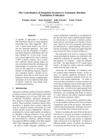
Influence of phospholipid headgroup hydrophobicity
on PLD activity
In these experiments, we measured PLD activity at the air–
water interface using substrates with different degrees of
headgroup hydrophobicity (Fig. 3). These results allow
comparison to be made between a naturally occuring
phospholipid, such as PtdCho, and phospholipids present-
ing an aliphatic chain as a headgroup, such as PamOle-
GroEtP,PamOleGroBuP and PamLinGroEtnP-HNE.
The isotherms of these three atypical phospholipids were
measured. PamOleGroEtP and PamOleGroBuP present
approximately the same molecular area (60 and 70 A
˚
2
per
molecule, respectively) for a surface pressure of 30 mNÆm
)1
.
The molecular area of PamLinGroEtnP-HNE was % 90 A
˚
2
per molecule (data not shown). Their Ks values were also
calculated from the p-A isotherms for a surface pressure of
30 mNÆm
)1
. According to previously published results
[19,20], a surface pressure of 30 mNÆm
)1
is thought to
approximate to the internal pressure of biological mem-
branes. Ks values were found within surface pressures of
70–80 mNÆm
)1
, which reveal a compressibility similar to
that of Myr
2
GroChoP. As shown in Fig. 3, PtdCho is
slowly hydrolyzed at a surface pressure of 30 mNÆm
)1
, while
the nonclassical phospholipids are rapidly converted into
PA. Morever, EtP, which presents only a two-carbon chain
headgroup, seems to be a better substrate of PLD than
PtdCho. When the hydrophobic headgroup is composed of
at least four carbons, which is the case for PamOleGroBuP
and PamLinGroEtnP-HNE, the rate of their PLD-cata-
lyzed hydrolysis is maximal (60 times higher than that of
PtdCho). One may postulate that the acyl-chain headgroup
has a tendency to penetrate into the hydrophobic core of the
membrane and that this could expose the phosphate group
to PLD activity. Under these conditions, phosphatidyl-
alcohols would always be hydrolyzed by PLD activity as
soon as they were synthesized within membranes.
Properties of mixed Myr
2
GroCho
P
/Myr
2
Gro monolayers
Diacylglycerols are naturally occuring alcohols that are
transiently produced by phospholipase C in biological
membranes [21]. Therefore, we can assume that these
neutral lipids could serve as natural nucleophiles within
membranes in a PLD-catalyzed transphosphatidylation
reaction. In order to test this hypothesis, mixed monolayers
of Myr
2
GroChoP/Myr
2
Gro were prepared and compressed
at the air–water interface (Fig. 4A).
These results indicate that Myr
2
Gro presents a molecular
area of % 40 A
˚
2
per molecule. This value is consistent with
the small polar head of Myr
2
Gro. The isotherms show that
increasing the amount of Myr
2
Gro in Myr
2
GroChoP
monolayers leads to a decrease of the mixed film molecular
area. The isotherms present a modification of slope during
compression (Fig. 4A). This could be the result of an
Fig. 3. Influence of the headgroup hydrophobicity on phospholipase D
(PLD) activity. Phosphatidylcholine (a), 1-O-palmitoyl-2-O-oleoyl-
sn-glycero-3-phosphoethanol (b), 1-O-palmitoyl-2-O-linoleoyl-sn-
glycero-3-phosphoethanolamine-4-hydroxynonenal (c) and 1-O-pal-
mitoyl-2-O-oleoyl-sn-glycero-3-phosphobutanol (d) were tested as
substrates for PLD activity at a constant surface pressure of
30 mNÆm
)1
. The apparent molecular area was normalized to compare
the different reactions. The chemical structure of PamLinGroEtnP-
HNE is given within the figure. Subphase and temperature were as
described in the legend to Fig. 1.
Fig. 4. Pressure-area isotherms (A) and surface compressional modulus
(B) of
L
-a-dimyristoyl-sn-glycero-3-phosphocholine (Myr
2
GroChoP)
monolayers containing an increasing molar percentage of 1,2-dimyris-
toyl-rac-glycerol (Myr
2
Gro). Myr
2
Gro molar ratios in Myr
2
GroChoP
monolayer were: a (0%); b (5%); c (10%); d (20%); e (40%); f (50%);
and g (100%). The surface compressional modulus (Ks) value was
calculated from pressure-area isotherms and was plotted as a function
of surface pressure. Subphase and temperature were as described in the
legend to Fig. 1.
4526 K. El Kirat et al. (Eur. J. Biochem. 270) Ó FEBS 2003
expanded-liquid to condensed-liquid phase transition that
seems to occur for low pressure values at high Myr
2
Gro
percentages. This transition could be explained by the highly
ordered chain organization induced by Myr
2
Gro at the air–
water interface, which would lead to a global rigidization
of the monolayer. It should also be borne in mind that
diacylglycerol induces lateral segregation of lipids, which
leads to domains enriched in diacylglycerol with a low
headgroup steric encumbrance [22].
Surface compressional moduli were calculated from the
p-A isotherms for each monolayer in order to estimate
membrane rigidity caused by the presence of Myr
2
Gro in
the mixtures (Fig. 4B). These results confirm the phase
transition caused by Myr
2
Gro mixed with Myr
2
GroChoP
in monolayers. This transition corresponds to a sudden
change in the slope of the curve; for example, % 17 mNÆm
)1
in surface pressure for the Myr
2
GroChoP/Myr
2
Gro 90 : 10
(mol/mol) mixture. The surface pressure value of this
transition decreases with increasing amounts of Myr
2
Gro in
the monolayer. Morever, the curves show that the presence
of Myr
2
Gro does not change the physical properties of the
monolayer until a molar percentage of 5–10 is reached.
Above this limit, Myr
2
Gro induces an increase in Ks values,
indicating an increased rigidity of the monomolecular film.
Influence of Myr
2
Gro on PLD activity at the air–water
interface
Mixed Myr
2
GroChoP/Myr
2
Gro monolayers were com-
pressed and stabilized at a surface pressure of 30 mNÆm
)1
.
PLD was injected into the subphase and its activity against
the lipidic monolayers was measured by monitoring the
decrease in apparent molecular area, along with time, at
30 mNÆm
)1
(Fig. 5A).
These results indicate an increase of PLD-catalyzed
Myr
2
GroChoP hydrolysis occuring in the presence of
Myr
2
Gro. By increasing the initial Myr
2
Gro content in
the monolayer, the apparent molecular area decrease seems
to occur more rapidly and with a greater slope (Fig. 5). PLD
activation starts at low percentages (1.25 molar percentage)
of Myr
2
Gro and seems to be maximal between 3 and 5
molar percentage. In previous work [11], we reported that
PLD activity is dependent on membrane compressibility, so
we represented the slope of the reaction and the variation of
the monolayer compressibility (Ks) as a function of
Myr
2
Gro molar percentage (Fig. 5B). This result showed
that 3 molar percentage is almost sufficient for the maximal
activation of PLD, while the Ks value is only slightly
modified. As shown previously, only low values of Ks
(ranging from 30 to 60 mNÆm
)1
) are able to induce maximal
PLD activity [11]. Therefore, it seems that the Myr
2
Gro-
induced activation of PLD is not dependent on membrane
compressibility, but may occur through a transphosphati-
dylation activity. The effect of another alcohol on PLD
activity at the air–water interface was also tested to
determine a transphosphatidylation mechanism. Monolay-
ers of octanol (OctOH) mixed with Myr
2
GroChoP were
prepared and compressed to 30 mNÆm
)1
. After pressure
stabilization, PLD was injected into the subphase. In this
case, we obtained PLD activities of % 0.006 min
)1
and
0.0128 min
)1
for 4 and 10 molar percentage OctOH within
the monolayer, respectively. These values are in the range of
those obtained for Myr
2
Gro (Fig. 5B) and no change of the
Myr
2
GroChoP/OctOH isotherms could be observed as
compared to Myr
2
GroChoP alone.
Influence of Myr
2
Gro on PLD activity in vesicles
van Blitterswijk & Hilkmann [21] have proved the existence
of a transient lipid, called bis(PA), in mammalian cells,
which is produced via the PLD-catalyzed transphosphati-
dylation reaction in the presence of diacylglycerol. In order
to detect bis(PA) production in biomimetic membranes
incubated with S. chromofuscus PLD, we prepared vesi-
cles, containing different molar percentages of Myr
2
Gro
mixedwithMyr
2
GroChoP, in the presence of
Pam
2
(Pam[
3
H]N)GroChoP radiolabelled on its sn-2 fatty
acid. The reaction was stopped at different time-points using
a concentrated EDTA solution, lipids were separated by
TLC, and the levels of diacylglycerol, bis(PA) and PA were
measured by radioactivity. Low initial percentages of
Myr
2
Gro mixed in Myr
2
GroChoP vesicles were tested,
Fig. 5. Influence of 1,2-dimyristoyl-rac-glycerol (Myr
2
Gro) molar per-
centage in the monomolecular film of
L
-a-dimyristoyl-sn-glycero-3-
phosphocholine (Myr
2
GroChoP) on phospholipase D (PLD) activity.
The apparent molecular area was normalized to compare the different
reactions at a constant surface pressure of 30 mNÆm
)1
.TheMyr
2
Gro
molar ratio in the Myr
2
GroChoP monolayer was: a (0%); b (1.25%);
c (2.5%); d (3%); e (5%); f (10%). Figure 5B shows PLD activity (d)
and Ks (h) dependency on the Myr
2
Gro molar ratio. Ks values were
reported from Fig. 4 and PLD activity was expressed as the decrease in
slope of normalized molecular area observed in Fig. 5A. Subphase and
temperature were as described in the legend to Fig. 1.
Ó FEBS 2003 S. chromofuscus PLD activation by diacylglycerol (Eur. J. Biochem. 270) 4527
but no significant production of radiolabelled lipids was
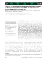
observed (data not shown). However, at 10–30% Myr
2
Gro
initially present in Myr
2
GroChoP vesicles, we observed the
simultaneous production of PA, diacylglycerol and bis(PA)
(Fig. 6). Increasing the initial content of Myr
2
Gro led to
higher amounts of each of these three lipids. Therefore,
diacylglycerol was considered as an activator for PLD
activity. Furthermore, it was impossible to produce PA in
amounts greater than 30% (Fig. 6A). This is because of the
shape of this lipid, which is not compatible with the
existence of a bilayer in the presence of calcium. Under these
conditions, lipidic structures enriched in PA would be
stabilized in inverted micelles that have a tendency to form
aggregates. In such aggregates, PLD would not be able to
reachthepolarheadofPtdChotocatalyzeitshydrolysis.
Diacylglycerol is the second major product of this
reaction (Fig. 6B). However, only small amounts of this
neutral lipid were detected after the reaction. Under optimal
conditions, i.e. 30% of Myr
2
Gro initially present in the
vesicles, 5% radiolabelled diacylglycerol is detected after
10 min vs. 30% radiolabelled PA. Radioactive diacylglyc-
erol can be produced in two different ways: first, through the
PLD-catalyzed hydrolysis of bis(PA), which can lead, in
theory, to equal production of radiolabelled PA or diacyl-
glycerol, as bis(PA) is symmetric; or, second, via the
phosphatase activity of S. chromofuscus PLD that would
have converted PA into diacylglycerol. However, as PA is
not a substrate for S. chromofuscus PLD phosphatase
activity, according to the observations of Zambonelli &
Roberts [5], this latter explanation is not feasible. Concern-
ing bis(PA) (Fig. 6C), it seems that it is only produced in
small amounts, from 0.6 to 0.7% of the radioactivity
recovered, indicating that it is a reaction intermediate, which
cannot accumulate in membranes.
Discussion
The monolayer technique is a powerful method for assaying
the PLD-catalyzed hydrolysis of various lipids. This method
requires only small amounts of lipids and provides infor-
mation on the compressibility of lipidic membranes.
Another important advantage of this technique is the
possibility of forming monolayers with lipids that cannot
form vesicles. This is because the monolayer interface is
planar, unlike the liposome interface which is curved. Under
these conditions, PLD activity can lead to total hydrolysis of
phospholipids, which cannot be obtained when vesicles are
used. Furthermore, all the lipids spread at the air–water
interface are in contact with the subphase, whereas in
vesicles, lipids can divide between the two leaflets of the
bilayer. In the latter case, only a fraction of the lipids are
accessible to proteins.
Phosphatidylalcohols are substrates of
S. chromofuscus
PLD
Surface pressure dependency of PtdCho hydrolysis suggests
that insertion of the enzyme into the membrane is a
prerequisite for PLD activity [7]. This phenomenon has
previously been described on PLC activity towards phos-
phatidylinositol 4,5-bisphosphate monolayers [23].
PLD from S. chromofuscus does not present the classical
HKD motif and is calcium dependent [4,5]. Furthermore,
transphosphatidylation activity of this PLD has been widely
discussed and several authors concluded on a low ability of
this enzyme to catalyze this type of reaction. Friedman et al.
[13] have reported a transferase activity observed by NMR
with lyso-PtdCho as substrate. They have shown that
Fig. 6. Role of 1,2-dimyristoyl-rac-glycerol (Myr
2
Gro) on
L
-a-dimyris-
toyl-sn-glycero-3-phosphocholine (Myr
2
GroChoP) hydrolysis catalyzed
by phospholipase D (PLD) in liposomes. We prepared vesicles of
Myr
2
GroChoP [radiolabelled with
L
-a-dipalmitoyl-[2-palmitoyl-
9,10-
3
H(N)]-sn-glycero-3-phosphocholine (Pam
2
(Pam[
3
H]N)Gro-
ChoP)]mixedwith10%(s), 20% (h)and30%(j)molarratioof
Myr
2
Gro. The final lipid concentration for all the assays was 5 m
M
in
120 l
M
CaCl
2
,150m
M
NaCl, 10 m
M
Tris, pH 8.0, and the tempera-
ture was fixed at 37 °C. Aliquots were taken at 2.5-min intervals and
the reaction was stopped with concentrated EDTA solution. After
lipid extraction and TLC separation, radioactivity was counted for
phosphatidic acid (PA) (A), diacylglycerol (B) and bis(PA) (C).
According to van Blitterswijk & Hilkmann [21], bis(PA) comigrates
with 1-O-palmitoyl-2-O-oleoyl-sn-glycero-3-phosphobutanol in our
TLC system. Radioactivity for each lipid was expressed as the per-
centage of total radiactivity recovered after TLC (100% corresponds
to 250 000 d.p.m.).
4528 K. El Kirat et al. (Eur. J. Biochem. 270) Ó FEBS 2003
conversion of this substrate into lyso-PA occurs via the
formation of cyclic lyso-PA obtained by intramolecular
transphosphatidylation of the lyso-PtdCho. Then, lyso-PA
is produced from cyclic lyso-PA by hydrolase activity of
PLD. Here, we report the ability of PLD to catalyze the
hydrolysis of phosphatidylalcohol without any dependency
on the surface pressure. This could be a result of the
hydrophobicity of the phosphatidylalcohol headgroup.
Indeed, several physical studies with liposomes showed the
tendency of such types of phospholipid headgroup to insert
into the membranes, leading to the exposure of the lipid
phosphate group to the buffer [18]. Such behavior of
phosphatidylalcohols in a monolayer would result in an
increase of the PLD activity, independently of the surface
pressure, as compared with the phosphate group of other
phospholipids.
The comparison between PtdCho, PamOleGroEtP,
PamOleGroBuP and PamLinGroEtnP-HNE provided
information on the influence of carbon-chain headgroup
length on PLD activity. It seems that maximum PLD
activity is obtained with a hydrophobic headgroup contain-
ing at least four carbons. Therefore, if PLD from S. chro-
mofuscus can catalyze a transphosphatidylation reaction,
the phosphatidylalcohol produced will be a better substrate
than PtdCho. As a consequence, a phosphatidylalcohol
cannot accumulate in membranes in the presence of
S. chromofuscus PLD. Furthermore, our results show a
rapid hydrolysis of PamLinGroEtnP-HNE catalyzed by
PLD at the air–water interface at a surface pressure of
30 mNÆm
)1
. This adduct is generated by condensation of
4-HNE and PE, and this reaction can also lead to Schiff
base adduct formation [24]. Those N-acylated lipids are
generated after cell oxidative stress induced by reactive
oxygen species. Hence, the presence of such adducts within
cell membranes could favor PLD activity.
S. chromofuscus
PLD activation by diacylglycerol
Previous work reported the activation of S. chromofuscus
PLD by diacylglycerol [10] but the mechanism of this still
remains unknown. We tested PLD activity on mixed
Myr
2
GroChoP/Myr
2
Gro monolayers. An activating effect
of the diglyceride was detected at low molar ratios (1.25%),
with a maximal effect occurring at 3 molar percentage
Myr
2
Gro. These low molar fractions of diacylglycerol are
close to physiological concentrations resulting from PLC
activity on membranes. On the basis of these low propor-
tions of diacylglycerol, we conclude that steric encumbrance
and physical properties of the membranes are not respon-
sible for the increased PLD activity. Experiments with
mixed PtdCho/diacylglycerol vesicles also showed an
increased PLD-catalyzed hydrolysis of PtdCho, at higher
percentages than those observed with monolayers. The
liposomes used here are multilamellar vesicles, so all the
Myr
2
Gro present in the membranes is not accessible to PLD
as the vesicles can be encapsulated one inside the other.
Furthermore, the situation in bilayers is different from that
obtained with monolayers: Myr
2
Gro can be partitioned
between the two leaflets of the bilayer, thus only a fract-
ion of total Myr
2
Gro is exposed to PLD. Therefore,
the maximum enzyme activation will be obtained in
bilayers with higher percentages of diacylglycerol than in
monolayers. However, radioactivity detection permitted the
quantification of radiolabelled PA, diacylglycerol and
bis(PA). As PLD from S. chromofuscus does not possess a
phosphatase activity with the ablility to generate diacyl-
glycerol from PA [5], all the radiolabelled diacylglycerol
must be produced through another reaction. A second
possible explanation for the production of diacylglycerol
could have been the presence of a contaminant PLC
activity, but no radiolabelled diacylglycerol (and no radio-
labelled bis(PA) or PA could be detected using pure PtdCho
liposomes. The production of radiolabelled bis(PA) allowed
us to elaborate a mechanism for diacylglycerol activation of
S. chromofuscus PLD. In a first-step reaction, PLD cata-
lyzes a transphosphatidylation reaction involving PtdCho as
a substrate and diacylglycerol as the nucleophile. This
reaction will produce bis(PA), which could be radiolabelled
if the substrate is radiolabelled on its acyl-chains. The
bis(PA) generated in the membranes should fully expose its
phosphate group to PLD activity, as observed with other
phosphatidylalcohols in monolayer experiments. Therefore,
in a second step of the reaction, bis(PA) will be converted
into PA plus diacylglycerol via PLD-phosphodiesterase
activity. One should bear in mind that PA or diacylglycerol,
owing to their small headgroup, cannot be produced
together up to more than 40–50 molar percent in vesicles.
As bis(PA) is a symmetric substrate of PLD, we can
presume that its hydrolysis will lead to equal proportions of
PA and diacylglycerol (Fig. 7). However, this was not
observed in our results. A possible explanation for this could
be that PA is also an activator of S. chromofuscus PLD;
Fig. 7. Schematic mechanism of the transphosphatidylation reaction
involving diacylglycerol and radiolabelled phosphatidylcholine. The
radiolabelled acyl chain of
L
-a-dipalmitoyl-[2-palmitoyl-9,10-
3
H(N)]-
sn-glycero-3-phosphocholine is represented in shaded boxes. In a first
step, radiolabelled dipalmitoylphosphatidyl-phospholipase D (PLD) is
produced. Then, a nucleophilic substitution occurs with 1,2-dimyris-
toyl-rac-glycerol (Myr
2
Gro) as an alcohol within the membrane. This
yields a phosphodiester intermediate, bis(PA). This product is a
symetric substrate for Streptomyces chromofuscus PLD. Thus, its
cleavage can occur, even in positions 1 and 2, with equal probability
around a phosphate group. Therefore, this reaction will theoretically
produce equal proportions of two lipid couples: radiolabelled diacyl-
glycerol plus cold PA and cold diacylglycerol plus radiolabelled PA
(see text for more details).
Ó FEBS 2003 S. chromofuscus PLD activation by diacylglycerol (Eur. J. Biochem. 270) 4529
when a critical percentage of PA is reached, hydrolysis will
be the major reaction catalyzed by PLD instead of the
transphosphatidylation involving diacylglycerol.
In conclusion, bacterial PLDs are often excreted as well
as PLC and are described as virulence determinants [25];
therefore these two enzymes could act in synergy to permit
internalization of bacteria into host cells. As a result of its
ability to form a complex with calcium, PA can favor
divalent cation-dependent fusion of membranes. Thus,
PLD-catalyzed formation of PA activated by diacylglycerol
could enhance fusion between bacteria and the host cell
membrane.
Acknowledgements
We thank Dr Caroline Elston for reviewing the English version of this
manuscript.
References
1. Holbrook, P.G., Pannell, L.K. & Daly, J.W. (1991) Phospholipase
D-catalyzed hydrolysis of phosphatidylcholine occurs with P-O
bond cleavage. Biochim. Biophys. Acta 1084, 155–158.
2. Yu, C.H., Liu, S.Y. & Panagia, V. (1996) The transphos-
phatidylation activity of phospholipase D. Mol. Cell. Biochem.
157, 101–105.
3. Ponting, C.P. & Kerr, I.D. (1996) Thermal stability of the three
domains of streptokinase studied by circular dichroism and
nuclear magnetic resonance. Protein Sci. 5, 914–922.
4. Yang, H. & Roberts, M.F. (2002) Cloning, overexpression, and
characterization of a bacterial Ca
2+
-dependent phospholipase D.
Protein Sci. 11, 2958–2968.
5. Zambonelli, C. & Roberts, M.F. (2003) An iron-dependent bac-
terial phospholipase D reminiscent of purple acid phosphatases.
J. Biol. Chem. 278, 13706–13711.
6. Stieglitz, K.A., Seaton, B.A. & Roberts, M.F. (1999) The role of
interfacial binding in the activation of Streptomyces chromofuscus
phospholipase D by phosphatidic acid. J. Biol. Chem. 274, 35367–
35374.
7. Stieglitz, K.A., Seaton, B.A. & Roberts, M.F. (2001) Binding of
proteolytically processed phospholipase D from Streptomyces
chromofuscus to phosphatidylcholine membranes facilitates vesicle
aggregation and fusion. Biochemistry 40, 13954–13963.
8. Morris, A.J., Frohman, M.A. & Engebrecht, J. (1997) Measure-
ment of phospholipase D activity. Anal. Biochem. 252,1–9.
9. Geng, D., Chura, J. & Roberts, M.F. (1998) Activation of phos-
pholipase D by phosphatidic acid. Enhanced vesicle binding,
phosphatidic acid–Ca
2+
interaction, or an allosteric effect? J. Biol.
Chem. 273, 12195–12202.
10. Yamamoto, I., Konto, A., Handa, T. & Miyajima, K. (1995)
Regulation of phospholipase D activity by neutral lipids in egg-
yolk phosphatidylcholine small unilamellar vesicles and by cal-
cium ion in aqueous medium. Biochim. Biophys. Acta 1233, 21–26.
11. ElKirat,K.,Besson,F.,Prigent,A.F.,Chauvet,J.P.&Roux,B.
(2002) Role of calcium and membrane organization on
phospholipase D localization and activity. Competition between a
soluble substrate and an insoluble substrate. J. Biol. Chem. 277,
21231–21236.
12. Estrela-Lopis, I., Brezesinski, G. & Mohwald, H. (2001)
Dipalmitoyl–phosphatidylcholine/phospholipase D interactions
investigated with polarization-modulated infrared reflection
absorption spectroscopy. Biophys. J. 80, 749–754.
13. Friedman, P., Haimovitz, R., Markman, O., Roberts, M.F. &
Shinitzky, M. (1996) Conversion of lysophospholipids to cyclic
lysophosphatidic acid by phospholipase D. J. Biol. Chem. 271,
953–957.
14. Geng, D., Baker, D.P., Foley, S.F., Zhou, C., Stieglitz, K.A. &
Roberts, M.F. (1999) A 20-kDa domain is required for phos-
phatidic acid-induced allosteric activation of phospholipase D
from Streptomyces chromofuscus. Biochim. Biophys. Acta 1430,
234–244.
15. Dumaual, A.C., Jenski, L.J. & Stillwell, W. (2000) Liquid crys-
talline/gel state phase separation in docosahexaenoic acid-con-
taining bilayers and monolayers. Biochim. Biophys. Acta 1463,
395–406.
16. Li, X.M., Momsen, M.M., Smaby, J.M., Brockman, H.L. &
Brown R.E. (2001) Cholesterol decreases the interfacial elasticity
and detergent solubility of sphingomyelins. Biochemistry 40, 5954–
5963.
17. Bechoua, S., Dubois, M., Ne
´
moz, G., Lagarde, M. & Prigent,
A F. (1998) Docosahexaenoic acid lowers phosphatidate level in
human activated lymphocytes despite phospholipase D activation.
J. Lipid Res. 39, 873–883.
18. Lee, Y.C., Zheng, Y.O., Taraschi, T.F. & Janes, N. (1996)
Hydrophobic alkyl headgroups strongly promote membrane
curvature and violate the headgroup volume correlation due to
ÔheadgroupÕ insertion. Biochemistry 35, 3677–3684.
19. Marsh, D. (1996) Lateral pressure in membranes. Biochim.
Biophys. Acta 1286, 183–223.
20. Silvius, J.R. (2003) Role of cholesterol in lipid raft formation:
lessons from lipid model systems. Biochim. Biophys. Acta 1610,
174–183.
21. van Blitterswijk, W.J. & Hilkmann, H. (1993) Rapid attenuation
of receptor-induced diacylglycerol and phosphatidic acid by
phospholipase D-mediated transphosphatidylation: formation of
bisphosphatidic acid. EMBO J. 12, 2655–2662.
22. Jimenez-Monreal, A.M., Villalain, J., Aranda, F.J. & Gomez-
Fernandez, J.C. (1998) The phase behavior of aqueous dispersions
of unsaturated mixtures of diacylglycerols and phospholipids.
Biochim. Biophys. Acta. 1373, 209–219.
23. Rebecchi, M., Boguslavsky, V., Boguslavsky, L. & McLaughlin,
S. (1992) Phosphoinositide-specific phospholipase C-delta 1: effect
of monolayer surface pressure and electrostatic surface potentials
on activity. Biochemistry 31, 12748–12753.
24. Guichardant, M., Taibi-Tronche, P., Fay, L.B. & Lagarde, M.
(1998) Covalent modifications of aminophospholipids by
4-hydroxynonenal. Free Rad. Biol. Med. 25, 1049–1056.
25. McNamara, P.J., Cuevas, W.A. & Songer, J.G. (1995) Toxic
phospholipases D of Corynebacterium pseudotuberculosis,
C. ulcerans and Arcanobacterium haemolyticum: cloning and
sequence homology. Gene 156, 113–118.
4530 K. El Kirat et al. (Eur. J. Biochem. 270) Ó FEBS 2003