Updates in the Understanding and Management of Thyroid Cancer Edited by Thomas J. Fahey doc
Bạn đang xem bản rút gọn của tài liệu. Xem và tải ngay bản đầy đủ của tài liệu tại đây (8.33 MB, 314 trang )
UPDATES IN THE
UNDERSTANDING AND
MANAGEMENT OF
THYROID CANCER
Edited by Thomas J. Fahey
Updates in the Understanding and Management of Thyroid Cancer
Edited by Thomas J. Fahey
Published by InTech
Janeza Trdine 9, 51000 Rijeka, Croatia
Copyright © 2012 InTech
All chapters are Open Access distributed under the Creative Commons Attribution 3.0
license, which allows users to download, copy and build upon published articles even for
commercial purposes, as long as the author and publisher are properly credited, which
ensures maximum dissemination and a wider impact of our publications. After this work
has been published by InTech, authors have the right to republish it, in whole or part, in
any publication of which they are the author, and to make other personal use of the
work. Any republication, referencing or personal use of the work must explicitly identify
the original source.
As for readers, this license allows users to download, copy and build upon published
chapters even for commercial purposes, as long as the author and publisher are properly
credited, which ensures maximum dissemination and a wider impact of our publications.
Notice
Statements and opinions expressed in the chapters are these of the individual contributors
and not necessarily those of the editors or publisher. No responsibility is accepted for the
accuracy of information contained in the published chapters. The publisher assumes no
responsibility for any damage or injury to persons or property arising out of the use of any
materials, instructions, methods or ideas contained in the book.
Publishing Process Manager Masa Vidovic
Technical Editor Teodora Smiljanic
Cover Designer InTech Design Team
First published March, 2012
Printed in Croatia
A free online edition of this book is available at www.intechopen.com
Additional hard copies can be obtained from
Updates in the Understanding and Management of Thyroid Cancer,
Edited by Thomas J. Fahey
p. cm.
ISBN 978-953-51-0299-1
Contents
Chapter 1 An Epidemiological Analysis
of Thyroid Cancer in a Spanish Population:
Presentation, Incidence and Survival 1
A. Rego-Iraeta, L. Pérez-Mendez and R.V. García-Mayor
Chapter 2 The Functionality of p53 in Thyroid Cancer 33
Debolina Ray, Matthew T. Balmer and Susannah Gal
Chapter 3 Glycosylation and Glycoproteins in Thyroid Cancer:
A Potential Role for Diagnostics 53
Anna Krześlak, Paweł Jóźwiak and Anna Lipińska
Chapter 4 Insulin-Like Growth Factor Receptor
Signaling in Thyroid Cancers:
Clinical Implications and Therapeutic Potential 91
Geetika Chakravarty and Debasis Mondal
Chapter 5 Principles and Application of Microarray
Technology in Thyroid Cancer Research 119
Walter Pulverer, Christa Noehammer,
Klemens Vierlinger and Andreas Weinhaeusel
Chapter 6 Evaluation and Management
of Pediatric Thyroid Nodules 147
Melanie Goldfarb and John I. Lew
Chapter 7 Papillary Thyroid Cancer in Childhood
and Adolescence with Specific Consideration
of Patients After Radiation Exposure 163
Yuri Demidchik, Mikhail Fridman,
Kurt Werner Schmid, Christoph Reiners,
Johannes Biko and Svetlana Mankovskaya
Chapter 8 Thyroid Cancer in the Pediatric Population 189
Silva Frieda, Nieves-Rivera Francisco and Laguna Reinaldo
VI Contents
Chapter 9 Current Innovations and Opinions
in the Surgical Management
of Differentiated Thyroid Carcinoma 199
Brian Hung-Hin Lang
Chapter 10 Sentinel Lymph Node Biopsy in
Well Differentiated Thyroid Cancer 217
Tamara Mijovic, Keith Richardson,
Richard J. Payne and Jacques How
Chapter 11 Preparing Patients for Radioiodine Treatment:
Increasing Thyroid Cell Uptake and Accelerating
the Excretion of Unbound Radioiodine 235
Milovan Matović
Chapter 12 Differentiation Therapy in Thyroid Carcinoma 251
Eleonore Fröhlich
and Richard Wahl
Chapter 13 Using γ-Camera to Evaluate the In Vivo
Biodistributions and Internal Medical Dosimetries
of Iodine-131 in Thyroidectomy Patients 283
Sheng-Pin Changlai, Tom Changlai and Chien-Yi Chen
Chapter 14 Thyroid Cancer:
The Evolution of Treatment Options 295
Hitoshi Noguchi
1
An Epidemiological Analysis of Thyroid
Cancer in a Spanish Population:
Presentation, Incidence and Survival
A. Rego-Iraeta, L. Pérez-Mendez and R.V. García-Mayor
Department of Endocrinology, Diabetes, Nutrition and Metabolism,
University Hospital of Vigo
Spain
1. Introduction
Accurate statistics on cancer occurrence and outcome are essential both for the purposes of
research and for planning and evaluation programmes for cancer control (Parkin, 2006).
Although tumours of thyroid account for only 1% of the overall human cancer burden, they
represent the most common malignancies of the endocrine system and pose a significant
challenge to pathologists, surgeons and endocrinologists. Among epithelial tumors,
carcinomas of follicular cell origin far outnumber those of C-cell origin. The vast majority of
carcinomas of follicular cell origin are indolent malignancies with 10 year survivals in excess
of 90 %.
1.1 Classification
Thyroid follicular epithelial-derived cancers are divided into three categories: papillary
cancer, follicular cancer and anaplastic cancer. Papillary and follicular cancers are
considered differentiated cancers, and patients with these tumours are often treated
similarly despite numerous biologic differences. Most anaplastic (undifferentiated) cancers
appear to arise from differentiated cancers. Other malignant diseases of the thyroid include
medullary thyroid cancer (which can be familial, either as part of the multiple endocrine
neoplasia type 2 syndrome or isolated familial medullary thyroid cancer), primary thyroid
lymphoma, or metastases from breast, colon, or renal cancer or melanoma. In countries with
adequate iodine intake, differentiated thyroid cancer accounts for more than 85% of all
cases, being the most common type papillary (60-80%). Tumor histology is a critical
determinant of patient outcomes; differentiated thyroid cancer is associated with the best
survival rate and medullary and anaplastic have significantly poorer outcomes (Hundahl et
al., 1998). Certain subtypes, such as the tall and columnar cell variants of papillary cancer
and the insular variant of follicular cancer are more common in older patients with higher
stage disease and have a worse prognosis than usual forms of thyroid cancer. The
traditional separation of thyroid cancer into the major groups of papillary, follicular,
medullary and undifferentiated (anaplastic) carcinoma, based on morphology and clinical
Updates in the Understanding and Management of Thyroid Cancer
2
features, is strongly supported by advances in molecular studies showing the involvement
of distinct genes in these four groups, with little overlap (DeLellis & Williams, 2004).
1.2 Staging and prognostic factors
Numerous staging systems have been created in an attempt to accurately prognosticate
outcomes for individual patients; two careful studies have compared the efficacy of the
various staging systems and found that none is superior (Brierley et al., 1997; Sherman et al.,
1998). Consequently, the European Thyroid Association (ETA) (Pacini et al., 2006) and the
American Thyroid Association (ATA) (Cooper et al., 2009) have recommended the use of
the Tumour, Node, Metastasis (TNM) classification of the American Joint Commission on
Cancer (AJCC) and the International Union Against Cancer because it is universally
available and widely accepted for other disease sites. An interesting feature of the TNM
staging system compared to other classifications is the age factor. While the staging of head
and neck cancers relies exclusively in the anatomical extent of disease, it is not possible to
follow this pattern for the particular group of malignant tumors that arise in the thyroid
gland. The effect of age is such significance in behavior and prognosis, that both the
histologic diagnosis and the age of the patient are included in the staging system for these
tumors. The AJCC classification is based on the TNM system, which relies on assessing three
components: (1) extent of the primary tumour (T), (2) absence or presence of regional lymph
node metastases (N), and (3) absence or presence of distant metastases (M). The fifth
edition
(Fleming et al., 1997), (Table 1) was revised as the
sixth edition (Greene et al., 2002), (Table 2).
A major alteration was the reclassification
of tumour staging (T). For differentiated
(papillary and follicular) and medullary
tumours confined to the parenchyma of the thyroid
gland without
extrathyroidal extension, there is no evidence to suggest that
using a size cut-
off of 1 cm provides better prognostic stratification
compared with the 2-cm cut-off used for
Papillary or Follicular Medullary Anaplastic
Stage Age < 45 years Age > 45 years Any age
I
Any T Any N
M0
T1 N0 M0 T1 N0 M0
II
Any T Any N
M1
T2 N0 M0
T3 N0 M0
T2 N0 M0
T3 N0 M0
T4 N0 M0
III
T4 N0 M0
Any T N1 M0
Any T N1 M0
IV
Any T Any N
M1
Any T Any N M0
Any T Any N
Any M
Table 1. AJCC TNM classification for thyroid cancer (fifth edition). T1 - Tumor 1 cm or less
in greatest dimension limited to the thyroid. T2 - Tumour more than 1 cm, but not more
than 4 cm, in greatest dimension limited to the thyroid. T3 - Tumour more than 4 cm in
greatest dimension limited to the thyroid. T4 - Tumour of any size extending beyond the
thyroid capsule. T4a - Excluded. T4b - Excluded. Regional lymph nodes are the cervical and
upper mediastinal lymph nodes. N1a - Metastasis in ipsilateral cervical lymph node(s).
N1b - Metastasis in bilateral, midline, or contralateral cervical or mediastinal lymph node
(s). M0- no distance metastases; M1- distance metastases.
An Epidemiological Analysis of Thyroid Cancer in a
Spanish Population: Presentation, Incidence and Survival
3
other head and neck sites.
Therefore, fifth edition T1 (<1 cm) and T2 (between
1 and 4 cm)
were redefined as sixth edition T1 (<2 cm) and T2
(between 2 and 4 cm). In the sixth edition,
T3 includes not only large tumours
(4 cm or more) but also tumours with minimal extension,
and T4
consists of T4a and T4b. The fact that diverse outcomes may be expected
in these two
groups of patients is now recognized in the sixth
edition: tumors that involve the
sternothyroid muscle are classified
as T3, while extension to larynx, trachea, oesophagus,
recurrent
laryngeal nerve, or subcutaneous soft tissue, all of which are
surgically resectable,
is classified as T4a. Tumours that invade
the prevertebral fascia or encase the carotid artery
or mediastinal
great vessels are not resectable for cure, and these patients
are staged T4b.
Thus, the sixth edition divides fifth edition T4 tumors into T3 (minimal invasion), T4a
(extended
invasion), and T4b (more extensive unresectable invasion) tumours
according to
the degree of extrathyroid extension. The degree of extension has been closely related to
adverse prognoses. Therefore, the sixth edition is expected
to predict more accurately
different outcomes in patients with
extrathyroid extension compared with the fifth edition.
Papillary or Follicular Medullary Anaplastic
Sta
g
e A
g
e < 45
y
ears A
g
e > 45
y
ears An
y
a
g
e
I
An
y
T, An
y
N,
M0
T1 N0 M0 T1 N0 M0
II
An
y
T An
y
N
M1
T2 N0 M0 T2 N0 M0
III
T3 N0 M0
T1 N1a M0
T2 N1a M0
T3 N1a M0
T3 N0 M0
T1 N1a M0
T2 N1a M0
T3 N1a M0
IVA
T4a N0 M0
T4a N1a M0
T1 N1b M0
T2 N1b M0
T3 N1b M0
T4a N1b M0
T4a N0 M0
T4a N1a M0
T1 N1b M0
T2 N1b M0
T3 N1b M0
T4a N1b M0
T4a Any N M0
IVB T4b An
y
N M0 T4b An
y
N M0 T4b An
y
N M0
IVC
An
y
TAn
y
N
M1
Any T Any N M1 Any T Any N M1
Table 2. AJCC TNM classification for thyroid cancer (sixth edition). T1 - Tumor 2 cm or less
in greatest dimension limited to the thyroid. T2 - Tumour more than 2 cm, but not more
than 4 cm, in greatest dimension limited to the thyroid. T3 - Tumour more than 4 cm in
greatest dimension limited to the thyroid or any tumour with minimal extrathyroid
extension (extension to sternothyroid muscle or perithyroid soft tissues). T4 - Excluded.
T4a - Tumour of any size extending beyond the thyroid capsule to invade subcutaneous soft
tissues, larynx, trachea, oesophagus, or recurrent laryngeal nerve. T4b - Tumour invades
prevertebral fascia or encases carotid artery or mediastinal vessels. T4a - Intrathyroidal
anaplastic carcinoma—surgically resectable. T4b - Extrathyroidal anaplastic carcinoma—
surgically unresectable. Regional lymph nodes are the central compartment, lateral cervical,
and upper mediastinal lymph nodes. N1a - Metastasis to Level IV (pretracheal, paratracheal,
and prelaryngeal/Delphian lymph nodes). N1b - Metastasis to unilateral, bilateral, or
contralateral cervical or superior mediastinal lymph nodes. M0- no distance metastases;
M1- distance metastases.
Updates in the Understanding and Management of Thyroid Cancer
4
TNM classification is also used for hospital cancer registries and epidemiologic studies. One
of the greatest inadequacies of TNM system is that it is a static representation of the patient’s
disease at the time of presentation; it does not allow for modification of risk during lifelong
follow-up. Most patients with papillary cancer in the TNM system are classified as stage I
disease (Hundahl et al., 1998), with an associated mortality rate of 1.7% (Loh et al., 1997). It
is important to note, however, that there is a 15% recurrence rate 10 years after initial
treatment (Loh et al., 1997). Recurrent or persistent disease, therefore, may necessitate
additional therapy and can certainly affect the patient’s quality of life. Further limitations of
tumour staging include the lack of consideration of tumour histology, extracapsular
extension of the tumour or molecular characteristics of the primary tumour. As is well
known, these factors can predict poorer outcomes for individual patients. As TNM staging
was developed to predict risk of death and not recurrence, the ATA (Cooper et al., 2009) has
created a more functional definition of risk stratification for individual patients that is
similar to one outlined by the ETA ( Pacini et al., 2006). Patients are classified as low-risk if
they have the following characteristics: no local or distant metastases, resection of all
macroscopic tumour, no tumour invasion into locoregional tissues, tumour that is not an
aggressive histological variant, no vascular invasion, and no uptake outside the thyroid bed
on the post-treatment whole body scan (if
131
I is given). Intermediate-risk patients are those
with any of the following criteria: microscopic tumour invasion into the perithyroidal
tissues at initial surgery, cervical lymph node metastases or
131
I uptake outside the thyroid
bed on the initial post-treatment scan, or tumour with aggressive histology or vascular
invasion. Finally, high-risk patients have macroscopic tumour invasion, incomplete tumour
resection, distant metastases or elevated thyroglobulin out of proportion to what is seen on
the post-treatment scan (Cooper et al., 2009). This stratification was designed to help
identify patients who are at higher risk for recurrent disease and may benefit from more
aggressive postoperative management (Cooper et al., 2009). Such a definition of risk is more
intuitive for the management of patients with thyroid cancer and is more in accordance with
the clinical behaviour of these tumours.
1.3 Epidemiology
Epidemiology has shown the influence of factors such as age and sex on thyroid cancer
incidence. Thyroid cancer is rare in children below 16 years, with an annual incidence
between 0.02 and 0.3 cases per 100,000 children and occurs exceptionally before age 10. In
adults, the mean age of diagnosis is the mid 40´s to early 50´s for the papillary type, 50´s for
the follicular and medullary types and 60´s for the less common undifferentiated types. It is
well established that thyroid cancer is 2 to 4 times more common in women than in men,
although this will differ among countries. Nevertheless, this sex difference is far less
pronounced before puberty and after menopause. Several epidemiological studies have
examined several reproductive traits, but the cause of this increased prevalence of thyroid
cancer in women is unclear. The annual incidence of thyroid cancer varies considerably in
different registries, with the highest incidence rates in the world reported in Hawaii and
Iceland (Ferlay et al., 2007; Kolonel et al., 1990). In Europe, the highest incidence occurs in
Iceland, followed by Finland, while relatively low incidence characterizes the United
Kingdom and Denmark (Ferlay et al., 2007,). These differences have been attributed to
ethnic or environmental factors, but different standards of health care may also play a role
in the efficiency of cancer detection. Although thyroid cancer incidence is low in general
An Epidemiological Analysis of Thyroid Cancer in a
Spanish Population: Presentation, Incidence and Survival
5
when compared with other diseases and tumours, over the last few decades, increasing rates
have been reported in several countries, including Europe (Akslen et al., 1993; Colonna et
al., 2002 ; dos Santos Silva et al., 1993; Gomez-Segovia et al., 2004; Petterson et al., 1991;
Reynolds et al., 2005; Szybinski et al., 2003), the United States (Davies & Welch, 2006; Merhy
et al., 2001; Zheng et al., 1996), Canada (Liu et al., 2001), and Australia (Burgess, 2002).
Curiously, this increase has occurred almost exclusively in papillary thyroid cancer, with an
epidemic of micropapillary thyroid carcinoma (MPTC) representing up to 43% of operated
cancers in the present series (Leenhart et al., 2004a). The reasons for the rise in thyroid
cancer incidence are not completely understood and considerable controversy exists now
about whether this increase is real or only apparent due to an increase in diagnostic activity
(Leenhart et al., 2004a; Leenhart et al., 2004b; Colonna et al., 2007). Recently, some
researchers (Colonna et al., 2007; Davies & Welch, 2006; Kent et al., 2007) have suggested
that this increase is predominantly due to the increased detection of small, subclinical
tumours through the use of medical imaging. Moreover, thyroid surgery is constantly
increasing, with more systematic use of total thyroidectomies even for benign pathologies,
which makes it easier to detect MPTC. According to the World Health Organization (WHO),
the term MPTC is used for a papillary carcinoma of the thyroid no larger than 1 cm in
diameter (Hedinger et al., 1988). With the new classification published in 2004, the previous
definition of MPTC now includes the additional criteria of being found incidentally (LiVolsi,
2004). MPTC seems to be present in a significant proportion of the general population with
large variations in the prevalence rate between different geographic areas (6–35%) (Sampson
et al., 1974), which may also be due to differences in the depth of the pathological
examination (Martinez-Tello et al., 1993). Although the mortality risk for an individual
patient with thyroid cancer is the greatest concern for patients and clinicians alike, most
patients have excellent 10-20-year disease specific survival (Hundahl et al., 1998).
EUROCARE (European Cancer Registry-based Study on Survival and Care of Cancer
Patients) is a collaborative project between European cancer registries (Capocaccia et al.,
2003). A major aim of EUROCARE is to estimate and compare cancer survival in European
populations. EUROCARE-2 (Teppo & Hakulinen, 1998) was the first publication on thyroid
cancer survival in Europe. This study included all malignant thyroid tumors (excluding
lymphomas) in patients 15 or older. Relative survival was analyzed using population-based
EUROCARE -2 data from 1985-1989. The overall 5-year relative survival rate, standardized
by age (Table 3), was 67% for men and 78% for women across Europe. Substantial variation
in this 5-year rate was observed between countries ranging from 56% in Slovenia to 100% in
Austria (men), (Teppo & Hakulinen, 1998). Higher than average survival rates were
observed in Finland, Iceland, The Netherlands and Sweden. Relative survival was higher in
the younger population group. In the age group 15 - 44 years, for men the rate was at least
86% and for women at least 94 %. In contrast, much lower rates were seen in the the group
of older population (75 + years). EUROCARE-3 study (Sant et al., 2003) analyzed the
survival of adult cancer diagnosed from 1990 to 1994 in 22 European countries and followed
them until the end of 1998. Neoplasms in situ were collected but not included in the analysis
of survival. The overall relative survival of patients diagnosed with thyroid cancer in this
period was 83% at 5 years. Austria, Finland, France, Iceland, Italy, Norway, Malta, Spain,
Switzerland and Sweden had rates above the European average. Most of these countries also
had high survival for this cancer in EUROCARE-2. Denmark, Germany, The Netherlands,
Updates in the Understanding and Management of Thyroid Cancer
6
England, Scotland, Wales and the countries of Eastern Europe had survival below the
European average (Table 3). Again, the most favorable outcomes were observed in patients
aged 15-44 years; for the oldest patient’s survival was five times lower. Part of variation in
thyroid cancer survival was attributed to variations in the distribution of histological types.
Other likely factors contributing to this are differences in the stage distribution and varying
efficacy of treatment (Sant et al., 2003; Teppo & Hakulinen, 1998).
EUROCARE-2 EUROCARE-3
female male female male
Iceland
90 88 85 87,4
Austria
87 100 88 81
Sweden
84 74 85 80
The Netherlands
84 77 79 68,6
Finland
82 77 86 79
France
81 61 85 74
Switzerland
78 - 90 -
Spain 78 70,6 85,7 82
Italy
77 66 85 72,6
Germany
77 62 77 69,4
Estonia
76 57 77 58
England
74 64 79 71
Scotland
73 67 76 73
Denmark
72 63 80 76,6
Slovakia
71 63 76 -
Eslovenia
70 56 77 83
Poland
66 64 66 57
EUROPE 78 67 81,4 71,8
Table 3. Thyroid cancer 5-year Relative Survival (%) from 1985 to 1989 (EUROCARE-2) and
from 1990 to 1994 (EUROCARE-3) in European countries.
In the U.S., the National Cancer Data Base (NCDB) represents a national electronic registry
system of incident cancers. Between 1985 and 1995, NCDB captured demographic, patterns-
of-care, stage, treatment, and outcome information for a sample of 53,856 thyroid carcinoma
cases. The 10-year overall relative survival rates for U. S. patients with papillary, follicular,
Hürthle cell, medullary, and undifferentiated/anaplastic carcinoma was 93%, 85%, 76%,
75%, and 14%, respectively (Hundahl et al., 1998). Relative survival, the survival analogue of
excess mortality, is commonly used in population-based studies of cancer survival although
its utility is not restricted to this area. Relative survival is the ratio of the observed survival
in a group of patients to the survival probability estimated over the same period in a group
of people in the general population of similar age and sex. It is usual to estimate the
expected survival proportion from nationwide population life tables stratified by age, sex,
calendar time, and, where applicable, race (Berkson & Gage, 1950). In order to be
comparable between different populations, relative survival figures must be either age-
specific or age-adjusted. A major advantage of relative survival is that information on cause
of death is not required, thereby circumventing problems with the inaccuracy or no
An Epidemiological Analysis of Thyroid Cancer in a
Spanish Population: Presentation, Incidence and Survival
7
availability of death certificates (Percy et al., 1981). However, our interest is typically in net
survival rather than all-cause survival, that is, we are interested in mortality due to cancer.
Cause-specific survival is commonly estimated in cancer clinical trials and only those deaths
which can be attributed to the cancer in question are considered to be events, while all other
deaths are considered censorings. Using cause-specific survival to estimate net survival
requires that reliably coded information on cause of death is available. The distinguishing
feature of survival analysis is that at the end of the follow-up period the event (such as
death due to cancer) will probably not have occurred for all patients. For these patients the
survival time is said to be censored, indicating that the observation period was cut off before
the event occurred. For example, a person who had the cancer and died 10 years later of car
accident would be censored at death, having contributed 10 person-year of survival to the
analysis. A person who had the cancer and died 10 years later of the cancer would
contribute an event, a death due to the cancer, having also contributed 10 person-years of
survival time. A 90 % cancer specific survival at 10 years would mean that 90 % of patients
had not died from their cancer, while 10 % had died from their cancer (Kaplan, 1958).
Calculation of cause-specific survival is especially important when studying diseases with a
favorable prognosis, as is the case at hand, where the patients live long enough to be
exposed to other causes of death. The indolent course of thyroid cancer requires very large
cohorts of patients followed over several decades to confirm significant differences in
prognostic factors and treatment efficacy. Neither randomized clinical trials nor meta-
analysis are available and evidence is based on a number of retrospective studies with
multivariate for mortality risk factors or data from national cancer registries (Gilliland et al.,
1997; Hundahl et al., 1998). Unfortunately, very remarkable differences in patient’s selection,
staging systems, and clinical management affect the available studies. In particular,
radioiodine treatment is not routinely carried out in a standard manner and outcome results
of different studies are thus not comparable (Sciuto et al., 2009). Since scarce data exist on
the epidemiology of thyroid cancer in Spain, the main aim of this study was to analyze
changes in thyroid cancer presentation, incidence, prevalence and survival in South Galicia
(north-western Spain) over a 24-year period (1978–2001) and compare these results with
those described in the leading international series. The people of this region are
homogeneous in terms of ethnicity. This period spans the population’s transition from mild
iodine deficiency to iodine sufficiency after beginning iodine prophylaxis in 1985 (Garcia-
Mayor et al., 1999; Rego-Iraeta et al., 2007). As a high incidence of thyroid cancer owing to
improved screening procedures is generally associated with an elevated proportion of small
carcinomas, we have specifically considered the impact of MPTC on thyroid cancer
incidence and trends in tumour size over time as an indicator of enhanced medical
procedures for thyroid cancer. We have also studied the proportion of our population
undergoing thyroid surgery over the study period and the percentage of thyroid cancers
found per thyroidectomy performed.
2. Materials and methods
2.1 Identification of thyroid cancer cases
Data on thyroid cancer incidence in the period from 1978 to 2001 (inclusive) were obtained
from the Pathology Registry of the University Hospital of Vigo which belongs to the Spanish
public health system and collects data on about 97% of the cancerous lesions verified by
Updates in the Understanding and Management of Thyroid Cancer
8
microscopic examination. This ensures virtually complete ascertainment for all the new
cases of thyroid cancer diagnosed in our population during the study period. Over the
observation period, a total of 329 cases of thyroid cancer were registered. Seven cases (six
lymphomas and one Angiosarcoma) were excluded from the study based on rarity. The
remaining 322 cases of primary thyroid cancer were assigned to one of the four major
diagnostic categories: papillary thyroid carcinoma; follicular thyroid carcinoma, including
Hürthle carcinomas; medullary thyroid carcinoma; and anaplastic thyroid carcinoma,
diagnosed according to the WHO classification (Hedinger et al., 1988). Original histology
slides for all cases of follicular carcinomas (53 cases) were reviewed by two
hystopathologists blinded to the original diagnosis. Nine of them were reclassified as
papillary carcinomas and 44 cases were classified as true follicular carcinomas. All tumour
stages were classified according to fifth
edition
of AJCC (Fleming et al., 1997) since most
studies reported having used this classification. In the present study all papillary
carcinomas of the thyroid <1 cm in diameter were classified as MPTC (Hedinger et al., 1988).
All thyroid cancer cases were also characterized by sex, date of birth, and date of diagnosis.
We also recorded data on number of thyroidectomies recorded in the registry which were
almost exclusively performed by two senior surgeons during the study period. Near-total
thyroidectomy has been used as standard treatment protocol for thyroid cancer and
comprises neck dissection if confirmed lymph node involvement; one course of ablative
radioiodine treatment with 100 mC, further radioiodine therapies with 100 mC if needed,
with an interval of 6 months-1 year and thyrotropin-suppressive thyroid hormone therapy
with levothyroxine lifetime.
2.2 Follow up the vital status of patients
Active follow-up of patients was carried-out through searches in medical records and phone
contacts. A detailed review of the medical record to ascertain the cause of death was made.
Mortality data were taken into account only when primary cause of death was directly
related to thyroid cancer and all other deaths were considered censorings. Cause-specific 1-,
5-, 10-, 20- and 25 year survival rates were used as measures of survival.
2.3 Study population
The studied population had an average of 500,000 inhabitants. Corresponding population
data by size, age, sex, and year were available from official statistics. Data during the period
1978–2001 show that Vigo’s population increased by 6.3%. The male to female ratio
remained stable at about 0.92. The people of the region are homogeneous in terms of
ethnicity. For studies of genetic characteristics, the Spanish Galician region is considered a
relatively isolated European population at the westernmost continental edge (Salas et al.,
1998).
2.4 Statistical analysis
Trends in age, sex, histological type, and tumour size (differentiated thyroid carcinoma) at
diagnosis were analyzed. Data on number of thyroidectomies performed were also recorded.
The general descriptive analyses were performed using Microsoft Excel and SPSS
An Epidemiological Analysis of Thyroid Cancer in a
Spanish Population: Presentation, Incidence and Survival
9
12.0 software (SPSS, Inc., Chicago, IL). Data were analyzed using the chi-square test for
nonparametric data. A p value below 0.05 was considered to be statistically significant.
Results were expressed as mean± standard deviation of the mean (mean±SD) for
quantitative variables. Data were analyzed using the Student t test for normally distributed
variables and the chi-square test for nonparametric data. Crude incidence rates, expressed
per 100,000 inhabitants each year were calculated. In order to compare incidence rates
between populations that differ with respect to age (since age has such a powerful influence
on the risk of cancer), age standardized incidence rates were also calculated; standardization
was performed using the World Standard Population (direct method) (Bray, 2002). For the
whole group of thyroid cancer, the overall incidence by sex for each year from 1978 to 2001
was calculated. Due to the small sample size, which produces unstable rates for individual
years, rates were calculated for several years combined (1978 to 1985, 1986 to 1993, and 1994
to 2001). Incidence trends for each of the distinct histological categories, including MPTC
incidence, were also examined. The prevalence of thyroid cancer was defined as the number
of persons in our defined population whom have been diagnosed of thyroid cancer, and
who were still alive in three cross-sectional surveys performed in December 1985, December
1993, and December 2001. The prevalence rates have been reported per 100,000 inhabitants.
A 95% confidence interval (CI) for the rates was determined to compare incidence and
prevalence rates. Survival from the data of initial surgery to each endpoint, i.e. cancer
specific survival, was estimated by the Kaplan–Meier product-limit method at 1, 5, 10, and
20 and, in some cases, at 25 years of diagnosis. The log-rank test was used to assess
difference between subgroups. Age at diagnosis was grouped into the same five categories
used by previous EUROCARE studies: 15-44, 45-54, 55-64, 65-74 and 75-99 years. We used
multivariate Cox analysis to calculate those independent variables related to the survival of
differentiated thyroid cancer.
3. Results
A total of 322 cases of primary differentiated thyroid cancer were diagnosed in our area
between 1978 and 2001. The mean age at diagnosis was 46.6 years (range, 8–91 years). Eight
patients were younger than 18 years at diagnosis. The female to male ratio was 3.6/1.
3.1 General characteristics on thyroid cancer
3.1.1 Histological distribution
Out of 322 cases of primary thyroid cancer, papillary was the predominant tumour type
with 245 cases (76%), followed by follicular with 44 cases (13.7%), medullary with 23 cases
(7.1%), and anaplastic with 10 cases (3.1%), (Table 1). The papillary to follicular ratio in the
entire period was as high as 5.8; when MPTC cases were excluded, this ratio was 2.
3.1.2 Age and sex distribution
The youngest age at presentation corresponded to medullary and papillary cancers of the
thyroid. Anaplastic cancer and Hürthle cells occurred at older ages. Of the total of thyroid
cancers, 78.3% of the cases were females and 21.7% outstanding men. This female
predominance is maintained in all histologic types (Table 4).
Updates in the Understanding and Management of Thyroid Cancer
10
3.1.3 Pathologic Tumor-Node-Metastases (pTNM) distribution
Altogether, 73% of the primary tumours presented with T1 to T3 tumor size; 15 % were
locally invasive to extrathyroidal soft tissues (T4), 22% had metastatic involvement of
cervical lymph nodes and 4.7% had distant metastases. Of 11.8% of cases the tumor size
was unknown. Among all tumors, medullary and papillary carcinomas were the most
commonly presented with cervical lymphadenopathy while follicular carcinoma the most
often presented distant metastasis (Table 4). In our series, we identified 95 MPTC out of a
total of 245 papillary thyroid cancers (38.7%). Of these, 87 cases (91%) were incidentally
diagnosed in thyroidectomies performed for thyroid pathologies other than thyroid
cancer.
Papillary Follicular Hürthle Medullary Anaplastic Total
Nº cases
(%)
245
(76%)
32
(10%)
12
(3.7%)
23
(7.1%)
10
(3.1 %)
322
Mean age
(range)
44
(8-91)
50
(23-78)
61
(33-91)
43.8
(19-78)
71
(52-89)
46.6
(8-91)
Female/Male
4 3.5 5 2.28 1.5 3.6
T
1
-T
3
80 % 56 % 78 % 52 % 0 % 73 %
T
4
10.6 % 22 % 8 % 17.4 % 100 % 15 %
N
1
23 % 12.5 % 8 % 39 % 20 % 22 %
M
1
2.4 % 19 % 0 % 13 % 0 % 4.7 %
Table 4. Thyroid cancer characteristics at diagnosis (1978-2001).
3.1.4 Distribution of pTNM stages of thyroid cancer at diagnosis (1978-2001)
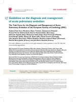
Most of thyroid cancer patients (75 %) presented low pathological tumor-node,-metastases
(stages I and II). Most papillary cancers presented with either stage I (63 %) or stage II (18
%). Stage III accounted for fewer than 12 % of cases. Few (1.2 %) patients presented with
distant metastases and had stage IV disease. For follicular and Hürthle cancers these figures
were 37, 28, 15, 6% and 25, 58, 8 and 0 % respectively. Most patients with medullary thyroid
cancer (43.5 %) had stage II; patients with stage I accounted for only 4 %; and stages III and
IV, 26 % and 13 % respectively. Figure 1, illustrates the distribution of pTNM stage and
histologic subgroup of thyroid cancer patients.
3.1.5 Trends in thyroid cancer presentation: Tumour size
Table 1 shows no significant change over time for sex distribution and age between the three
time periods (1978–1985, 1986–1993, and 1994–2001). The proportion of MPTC among total
papillary thyroid cancers cases increased significantly over time: 16.7% (1978 to 1985), 23%
(1986 to 1993), and 43% (1994 to 2001). The papillary to follicular ratio significantly increased
over time from 2.3 to 3.6 and 11.5. When MPTC was excluded, the papillary to follicular
ratios were 1.9, 2.7, and 6.6, respectively. Besides MPTC cases, no significant variations were
observed with respect to tumour size (pT) at presentation, in papillary and follicular over
time. For some patients there was no precise pathological description about tumour size
(pTx), (Table 5).
An Epidemiological Analysis of Thyroid Cancer in a
Spanish Population: Presentation, Incidence and Survival
11
Fig. 1. Thyroid cancer pTNM stages and histologic distribution at diagnosis (1978-2001).
1º Period
1978-1985
2ºPeriod
1986-1993
3ºPeriod
1994-2001
p
Female/Male
4.3 3.6 3.4 0.854
Mean age ± DT (years)
42.2±16.5 46.8±17.2 47.6±16.7 0.601(1º vs·3º)
Papillary/Follicular
2.3 3.6 11.5 0.000*
Papillary (no-MPTC)/Follicular
1.9 2.7 6.6 0.013*
(%) MPTC/ Total Papillary
16.7 % 23 % 43 % 0.010*
Papillary
(no- MPTC)
T
2 (n=81)
47.8 % 45.8 % 60 %
0.360
T
3(n=20)
8.7 % 20.8 % 10 %
T
4(n=27)
17.4 % 18.8 % 17.5 %
T
x (n=22)
26.1 % 14.6 % 12.5 %
Follicular T
1 (n=4)
7.7 % 16.7 % 0 %
0.213
T
2 (n=20)
38.5% 38.9% 61.5 %
T
3 (n=4)
0 % 11.1 % 15.4 %
T
4 (n=8)
15.4 % 16.7 % 23.1 %
T
x (n=8)
38.5 % 16.7 % 0 %
Table 5. Time trend of thyroid cancer presentation (1978-2001).
3.2 Trends in thyroid surgery
A total of 2345 thyroidectomies were performed during the studied period. During this
period the percentage of the population undergoing a thyroid surgery significantly
increased from 13.76 per 100,000 each year (95% CI 12.35–14.56) to 23.83 (95% CI 22.17–
I
II
III
IV
Unknown
Stage
100,00%
ANAPLASTIC FOLLICULAR HÜRTHLE
MEDULLARY PAPILLARY
37,50%
28,13%
6,25%
15,63%
12,50%
25,00%
58,33%
8,33%
8,33%
4,35%
43,48%
26,09%
13,04%
13,04%
63,27%
18,37%
11,84%
1,22%
5,31%
Updates in the Understanding and Management of Thyroid Cancer
12
24.73) and 45.01 (95% CI 42.45–46.39) in 1978–1985, 1986–1993, and 1994–2001, respectively.
The proportion of thyroid carcinomas among operated patients rose from 9.92% in 1978–
1985 to 12.31% in 1986–1993 and to 15.35% in 1994–2001, respectively (p <0.015). Total
thyroidectomy accounted for 48% of initial surgical procedures (1978–1985) and 74% during
1994–2001.
3.3 Trends in thyroid cancer incidence
As shown in Fig. 2 and Table 6, incidence rates were considerably lower for males than for
females. Overall crude incidence of thyroid cancer in women increased significantly from
1.61 per 100,000 each year (1978 to 1985) to 4.43 (1986 to 1993) and 10.29 (1994 to 2001). These
figures in men were 0.35, 1.31, and 3.24, respectively. Age-standardized incidence rates
(ASR) over this period show the same tendency, with a significant increase in females: 1.56
per 100,000 each year (1978 to 1985) to 3.83 (1986 to 1993) and 8.23 (1994 to 2001); and males:
0.33, 1.19, and 2.65, respectively (Table 6).
0
2
4
6
8
10
12
14
16
Incidence/100 000-year
Years
Fig. 2. Annual crude incidence of thyroid cancer, by sex (1978-2001); females (circles) and
males (squares).
Females Males
Period (years)
Crude
Incidence
ASR * IC (95 %)
Crude
Incidence
ASR * IC (95 %)
1978-1985 1.61 1.56 1.03-2.08 0.35 0.33 0.08-0.58
1986-1993 4.43 3.83 2.93-4.71 1.31 1.19 0.67-1.70
1994-2001 10.29 8.23 6.82-9.63 3.24 .65 1.82-3.46
Table 6. Time trend of crude and age-standardized incidence rates of thyroid cancer, by sex.
(*) Age-standardized incidence rate (ASR).
An Epidemiological Analysis of Thyroid Cancer in a
Spanish Population: Presentation, Incidence and Survival
13
3.3.1 Trends in thyroid cancer incidence by histopathology: Incidence of MPTC
Figure 3 displays the overall (males and females) crude incidence rates of thyroid cancer in
relation to the histological types; the increase in the incidence of thyroid cancer over the
three periods of time was primarily due to an increase in papillary cancer incidence. After
the second period, the incidence of follicular cancer decreased and there was no significant
change in the incidence of MTC and anaplastic cancer. Table 7 shows that the increase in the
incidence of PTC was the result of an increased incidence of both MPTC and papillary
measuring more than 1 cm (Papillary non-MPTC). This occurred both in males and females.
0
1
2
3
4
5
6
1978-1985 1986-1993 1994-2001
Incidence/100 000-year
Years
Papillary Follicular Medullary Anaplastic
Fig. 3. Time trend of crude incidence rates of thyroid cancer, by histology.
Females Males
Period Papillary
No-
MPTC
Incidence
CI (95%)
MPTC
Incidence
CI (95%)
Papillary
No-
MPTC
Incidence
CI (95%)
MPTC
Incidence
CI
(95%)
1978-
1985
0.97 0.55-1.38 0.14 -0.02-0.29 0.15 -0.02-0.32 0.10 -0.04-
0.24
1986-
1993
2.19 1.49-2.88 0.81 0.38-1.23 0.75 0.32-1.17 0.12 -0.05-
0.30
1994-
2001
4.82 3.65-5.98 3.94 2.89-4.99 1.58 0.89-2.27 0.79 0.30-
1.28
Table 7. Time trend of papillary thyroid cancer crude incidence rates, by sex
(CI: Confidence Interval)
Updates in the Understanding and Management of Thyroid Cancer
14
3.4 Trends in thyroid cancer prevalence
Table 8 shows that prevalence of thyroid cancer increased substantially between 1985 and
2001 in both sexes. Thyroid cancer was significantly more prevalent in female than in male
subjects.
Year Sex Prevalence CI (95%)
1985
Female
12.53
8.38-16.68
1993
Female
65.89
53.23-78.56
2001
Female
128.34
111.75-144.92
1985
Male
2.72
0.70-4.73
1993
Male
17.85
10.99-24.71
2001
Male
35.66
26.56-44.77
Table 8. Time trend of thyroid cancer prevalence, by sex.
3.5 Thyroid cancer survival
We followed a total of 321 cases of thyroid cancer. The median follow-up was 7.7 years,
ranging between 4 and 27.8 years. We recorded a total of 43 deaths, of which 30 (70%) were
directly related to thyroid cancer, yielding a cancer- specific mortality rate of 9. 3 % for the
whole cohort. Over 4 %( 4.3) of cancer -specific deaths was represented by patients with
differentiated thyroid carcinomas. Among the remaining 13 deaths not attributable to thyroid
cancer, 9 (69%) were due to second malignancies (three breast cancer case, 1 prostate cancer
case, 1 case of sigmoid colon cancer, 1 case of liver cancer, 1 case of glioblastoma multiform, 1
case of pancreatic cancer , 1 case of multiple myeloma) and 4 (31%) were attributed to other
causes. Overall survival of patients diagnosed with thyroid cancer in the period 1978-2001 was
88 % at 25 years, being 90 % for women and 80% for men; although survival was higher in
women, there were no significant differences between both genders (p = 0, 097), (Table 9).
When excluding MPTC, we observed a decrease in thyroid cancer survival. Thus, the overall
survival of thyroid cancer was 84% at 25 years, being 87% in women and 76% in men, again
without significant differences between genders (p = 0.15), (Table 10).
Gender Patients Survival
1 year 5 years 10 years 20 years 25 years
Female
251 97% 93% 91% 90% 90%
Male
75 95% 91% 84% 80% 80%
Total
321 96% 93% 89% 88% 88%
Table 9. Overall cause-specific survival of thyroid cancer (1978-2001).
Gender Patients Survival
1 year 5 years 10 years 20 years 25 years
Female
180 96% 91% 89% 87% 87%
Male
56 94% 89% 81% 76% 76%
Total
236 95% 90% 86% 84% 84%
Table 10. Overall cause-specific survival of thyroid cancer (1978-2001), excluding MPTC.
An Epidemiological Analysis of Thyroid Cancer in a
Spanish Population: Presentation, Incidence and Survival
15
3.5.1 Cause –specific survival according to age
Table 11 and Figure 4, reflect the cause-specific survival by age group (excluding MPTC)
and emphasizes the influence of age on the prognosis of patients with thyroid carcinoma. As
can be seen there is one more striking decline in survival after 55 years of age.
Age Patients Survival
1 year 5 years 10 years 20 years 25 years
ago-44
117 100% 98% 96% 94% 94%
45-54
45 97% 95% 95% 95% 95%
55-64
34 90% 84% 74% 63% -
65-74
22 86% 77% 69% 57%,18 years -
75-91
18 83% 59% 47% 47%,18 years -
Table 11. Cause-specific survival of thyroid cancer by age group, excluding MPTC (1978-2001).
0 5 10 15 20 25 30
Years after diagnosis
0,0
0,2
0,4
0,6
0,8
1,0
Cancer-specific Survival
8-44 years of age
45-54 years of age
55-64 years of age
65-74 years of age
75-95 years of age
Fig. 4. Cause-specific survival of thyroid cancer by age group, excluding MPTC (1978-2001).
3.5.2 Cause -specific survival according to histological type
As known, histologic type is a strong determinant of thyroid cancer survival. In our series,
papillary thyroid cancer patients had 25-year specific-survival greater than 93 %, even when
excluding MPTC. The survival of MPTC was 100% at 25 years in the present study.
Follicular and medullary carcinoma patients had lower survivals (83% at 25 years and %at
20 years, respectively). However, the prognosis was is ominous for anaplastic thyroid
carcinoma (Table 12 and Figure 5).
Updates in the Understanding and Management of Thyroid Cancer
16
0 5 10 15 20 25 30
Years after diagnosis
0,0
0,2
0,4
0,6
0,8
1,0
Cancer-specific survival
ANAPLASTIC
FOLLICULAR
MEDULLARY
PAPILLARY
Fig. 5. Cause–specific survival of thyroid cancer according to histological type (1978-2001).
Histologic
type
Patients Survival
1 year 5 years 10 years 20 years 25 years
Papillary
(total)
245 99% 97% 96% 95% 95%
Papilar
(no MPTC)
160 98% 96% 95% 93% 93%
Follicular
(including
Hürthle)
43 97% 90% 87% 83% 83%
Medullary
23 95% 86% 70% 63% -
Anaplastic
10 40% 10% - - -
Table 12. Cause-specific survival of thyroid cancer according to histological type (1978-2001).
3.5.3 Cause –specific survival according to p TNM stage distribution
Stage at diagnosis is a strong prognostic factor for thyroid cancer survival. Thus, cause
specific-survival vas 100% at 25 years of follow- up in stage I. At more advanced stages
survival decreases progressively (Table 13).
An Epidemiological Analysis of Thyroid Cancer in a
Spanish Population: Presentation, Incidence and Survival
17
Stage Patients Survival
1 year 5 years 10 years 20 years 25 years
I
171 100% 100% 100% 100% 100%
II
71 100% 100% 97% 94% -
III
38 97% 88% 69% 69% 69%
IV
21 37% 15% 15% 0% -
Unknown
20 100% 100% 94% 94% 94%
Table 13. Cause-specific survival of thyroid cancer by pTNM stage (1978-2001).
3.5.4 Prognostic analysis in differentiated thyroid carcinoma
Risk factors associated with differentiated thyroid cancer mortality were identified by Cox
regression analysis. Univariate and multivariate analysis results for thyroid cancer mortality
are illustrated in Table 14. In the univariate analysis, the following factors were significantly
associated with mortality for differentiated thyroid cancer: age, follicular histology, local
tumor extension and distant metastases at presentation. Neither sex nor the presence of
lymph node metastases contributed to mortality risk. Multivariate analysis confirmed as
independent predictor variables of increased risk of cancer mortality-only age and presence
of distant metastases.
Variables
Variables
Univariate
Analysis
Multivariate
Analysis.
RR (CI 95 %) RR (CI 95 %)
Sex
Female 1
Male 1,5 (0,48-4,95)
8 – 44 1
45 – 54 2,3 (0,14-36,7) 3,17 ( 0,2-51,6)
Age (years)
55 – 64 20,7 (2,42-178) 17,8 (2,12-150)
65 – 74 30,5 (3,4-274) 15,6 (1,6-147)
> 75 38,5 (3,90-377) 38,5 (3,30-338)
Histology
Papillary 1
Follicular 4,07 (1,41-11,76)
T1 1
Tumoral size
T2 2,75 (0,26-24,5)
T3 3,33 (0,20-53,5)
T4 24,80 (3,1-198)
Regional extension
N0 1
N1 2,5 (0,78-8,40)
Distance extension.
M0 1
M1 29,9 (10,4-85) 17,68 (6,11-51,1)
Table 14. Univariate and Multivariate survival analysis of prognostic factors of
differentiated thyroid cancer (1978-2001).