Báo cáo khoa học: Structural elucidation of polysaccharide part of glycoconjugate from Treponema medium ATCC 700293 doc
Bạn đang xem bản rút gọn của tài liệu. Xem và tải ngay bản đầy đủ của tài liệu tại đây (357.15 KB, 9 trang )
Structural elucidation of polysaccharide part of glycoconjugate
from
Treponema medium
ATCC 700293
Masahito Hashimoto
1
, Yasuyuki Asai
1
, Takayoshi Jinno
1
, Seiji Adachi
2
, Shoichi Kusumoto
2
and Tomohiko Ogawa
1
1
Department of Oral Microbiology, Asahi University School of Dentistry, Gifu, Japan;
2
Graduate School of Science,
Osaka University, Osaka, Japan
Glycoconjugates are distributed on the cell surfaces of some
small-sized treponemes and have been reported to be com-
pletely different from lipopolysaccharides. We separated a
glycoconjugate fraction from Treponema medium ATCC
700293, a medium-sized oral spirochete, to assess its immu-
nobiological activities and elucidate the chemical structure
of its polysaccharide part using phenol/water extraction,
hydrophobic chromatography, and gel filtration. The
glycoconjugate showed negligible or weak endotoxic and
immunobiological properties. The chemical structure of the
polysaccharide part was shown by two-dimensional NMR
and MALDI-TOF-MS to be a tetrasaccharide backbone
with two amino acids:
[ fi 4)b-
D
-GlcpNAc3NAcA(1fi 4)b-
D
-ManpNAc3NA
Orn(1fi 3)b-
D
-GlcpNAc(1fi 3)a-
D
-Fucp4NAsp(1fi ]
where GlcNAc3NAcA is 2,3-diacetamido-2,3-dideoxy-
glucuronic acid, ManNAc3NAOrn is N
d
-(2-acetamido-
3-amino-2,3-dideoxymannuronyl)ornithine, and Fuc4NAsp
is 4-(a-aspartyl)amino-4,6-dideoxygalactose.
Keywords: glycoconjugate; MALDI-TOF-MS; NMR;
Treponema medium.
Treponemal species are anaerobic bacteria with a typical
helical shape that have been implicated in the induction of
chronic human diseases, such as syphilis caused by Trepo-
nema pallidum [1] and periodontal diseases caused by
Treponema denticola [2]. Such chronic diseases are charac-
terized by an inflammatory reaction induced by pathogens
followed by extensive tissue loss. Cell surface components
released by treponemal species have been shown to mediate
the synthesis of inflammatory mediators by host cells, such
as macrophages. In a review of reports regarding the lipo-
protein of Escherichia coli [3], treponemal species lipo-
proteins were shown to have potent virulence, i.e. the
lipoprotein of T. pallidum [4] induced NF-jB translocation
in monocytes [5] and that of T. denticola [6] activated
macrophages [7]. Although some researchers have suggested
that lipopolysaccharide (LPS) of treponemal species is
responsible for the activation of host cells, the presence of
LPS in spirochetes remains controversial, as a genome
analysis of T. pallidum revealed the absence of LPS
synthesis genes [8]. Recently, novel glycolipids were extrac-
ted from the small-sized spirochetes T.denticola, Treponema
maltophilum,andTreponema brennaborense [9,10], and
characterized as activators of mitogen-activated protein
kinase and NF-jB [11,12], which may be potent virulent
factors of treponemal species. Further, though the chemical
structures of glycolipids have not been fully elucidated, they
are known to be different from that of LPS [9,10].
Treponema medium is a medium-sized oral spirochete
found in subgingival plaque from patients with adult
periodontitis [13]. As T.medium seems to be associated
with chronic inflammation [14], its cell surface components
are expected to possess immunostimulating activities like
other treponemal species. We previously demonstrated that
the outer membrane extract of T.mediumactivates epithelial
cells [15] and may be responsible for periodontal diseases,
however, it has not been shown that a carbohydrate-
containing component of T.medium acts as a virulence
factor. In the present study, we separated a glycoconjugate
from T.medium, and then elucidated the chemical structure
as well as its endotoxic and immunobiological properties.
Materials and methods
Bacteria and preparation of glycoconjugate
T.medium ATCC 700293 was grown anaerobically in
tripticase-yeast extract-gelatin-volatile fatty acids-rabbit
serum broth containing 5% rabbit serum, as described
previously [16]. The cells were subjected to phenol/water
extraction [17], and then the extract was subjected to
enzymatic digestion with DNase and RNase followed by
proteinase K. To remove contaminated proteins, the diges-
ted portion was further subjected to phenol/water extraction
to yield a glycoconjugate preparation, which was designated
as Tm-Gp. Tm-Gp was then subjected to hydrophobic
interaction chromatography. Tm-Gp was dissolved in 0.1
M
acetate buffer (pH 4.5) containing 15% 1-propanol and
applied to an Octyl Sepharose CL-4B column (36 · 2.5 cm)
(Amersham Bioscience, Piscataway, NJ, USA). The column
Correspondence to T. Ogawa, Department of Oral Microbiology,
Asahi University School of Dentistry, 1851-1 Hozumi, Mizuho,
Gifu 501-0296, Japan. Fax/Tel.: + 81 58 329 1421,
E-mail:
Abbreviations: CID, collision induced dissociation; HMBC, hetero-
nuclear multiple bond connectivity; LPS, lipopolysaccharide;
LTA, lipoteichoic acid; MS/MS, tandem MS.
(Received 4 March 2003, revised 23 April 2003,
accepted 30 April 2003)
Eur. J. Biochem. 270, 2671–2679 (2003) Ó FEBS 2003 doi:10.1046/j.1432-1033.2003.03644.x
was eluted using the buffer with the linear gradient of
1-propanol (15–60%). Six mililiter fractions
1
were collected,
after which they were monitored by measuring phos-
phorous, hexose, and amino group contents. The eluates
were then combined, dialyzed using a Spectra/Por 7
(MWCO 3500; Spectrum Laboratories Inc., Dominguez,
CA, USA), and lyophilized to yield pass-through (OS-P)
and retained fractions. The retained fraction was designated
as Tm-GC and used as a glycoconjugate fraction, while the
OS-P portion was dissolved in water, applied to a Sephacryl
S-200 HR column (81 · 1.5 cm) (Amersham Bioscience),
eluted with water, and 2.5 mL fractions were collected and
monitored as above. Finally, the eluates were combined,
dialyzed, and lyophilized to obtain a polysaccharide frac-
tion, which was designated as Tm-PS.
Analytical procedures
Phosphorous contents were determined according to the
method of Bartlett [18] and hexose contents were measured
using the anthrone/sulfuric acid method [19]. Amino group
contents were analyzed using ninhydrin. Samples (100 lL)
wereaddedto500lL of ninhydrin reagent (ninhydrin/
collidine/acetic acid/ethanol ¼ 0.6 : 10 : 75 : 250, w/v/v/v)
andheatedat100°C for 5 min, after which the absorbance
at 570 nm was measured.
Analysis of the sugar constituents in the sample was
performed using the alditol acetate method [20] and the
absolute configurations of sugars were determined using
R-(+)-2-butanol [21]. Estimation of 3-deoxy-
D
-manno-oct-
2-ulosonic acid (Kdo) was performed by the thiobarbiturate
method [22], and fatty acids were analyzed according to
the method of Ikemoto et al.[23].
For amino acids analysis, the samples were hydrolyzed
with 6
M
hydrochloric acid for 24 h at 110 °C.The
hydrolysate was analyzed by the o-phthalaldehyde/
N-acetylcysteine method [24] using an LC-10AD amino
acids analysis system (Shimadzu, Kyoto, Japan). The
absolute configurations of amino acids were determined
using 1-fluoro-2,4-dinitrophenyl-5-
L
-leucinamide [25].
SDS/PAGE was performed with 10% polyacrylamide
gels according to the method of Laemmli [26] and visualized
using the silver staining method [27].
Endotoxic and immunobiological assays
Lethal toxicity in
D
-GalN-sensitized C57BL/6 mice, cyto-
kine production in human peripheral blood mononuclear
cells, and mitogenicity of mononuclear cells from C3H/
HeN and C3H/HeJ mice were assayed as described
previously
2
[28]. Animals received humane care in accor-
dance with our institutional guidelines and the legal
requirements of Japan.
Chemical degradation and separation
Tm-GC was treated with 0.6% AcOH at 100 °C for 2 h,
0.1
M
NaOH at 37 °C for 2 h, or 48% aqueous HF at 4 °C
for 24 h, and the liberated hydrophobic products were
extracted using chloroform/methanol (2 : 1, v/v). Tm-PS
was hydrolyzed with anhydrous trifluoromethanesulfonic
acid at 0 °C for 1 h and the reaction mixture was neutralized
with ammonia solution [29]. The hydrolysate was subjected
to gel filtration chromatography using a Toyopearl HW-
40S (Tosoh, Tokyo, Japan) to give oligosaccharides.
NMR spectroscopy and mass spectrometry
1
Hand
13
C NMR spectra were measured at 500 and
126 MHz, respectively, using a JMN-LA500 spectrometer
(JEOL, Tokyo, Japan) equipped with an indirect detection
gradient probe, IDG500-5VJ (Nanorac Cryogenics, Marti-
nez, CA, USA). Spectra were obtained at 310 K in D
2
Oor
10% D
2
O/H
2
O. The chemical shifts were expressed as d
values with water (d 4.7) as the internal standard for
1
H NMR spectra and with dioxane (d 67.4) as the external
standard for
13
C NMR spectra.
MALDI-TOF-MS was measured using an Ultraflex
instrument (Bruker Daltnics, Bremen, Germany). Samples
were dissolved in water, combined with 2,5-dihydroxy-
benzoic acid as a matrix, and placed on a sample plate, then
spectra were obtained in positive ion reflector mode, and
tandem MS (MS/MS) spectra were obtained in positive ion
TOF/TOF mode. ESI-MS was measured using a Q-TOF
instrument (Micromass. Manchester, UK). Spectra were
obtained in negative reflector and collision induced disso-
ciation (CID) modes.
Results and discussion
Separation of glycoconjugate from
T. medium
Extraction of a glycoconjugate from T.medium was
performed using standard hot phenol/water extraction
followed by chromatographic separations. The bacteria
were subjected to phenol/water extraction, nuclease and
proteinase digestion, and re-extraction with phenol/water
to yield a glycoconjugate preparation, Tm-Gp (5.3%). As
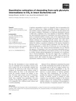
shown in Fig. 1, a ladder-like pattern was observed in the
SDS/PAGE profile of Tm-Gp, indicating the presence of
a glycoconjugate with repeating units. During phenol/
water extraction of bacteria, capsular and extracellular
polysaccharides as well as glycoconjugates are simulta-
neously extracted in the aqueous phase [30], thus, we
further separated the glycoconjugate fraction using hydro-
phobic interaction chromatography [31,32]. Tm-Gp was
subjected to hydrophobic interaction chromatography to
yield a pass-through fraction, OS-P (50% based on
Tm-Gp), and a retained fraction, Tm-GC (14%). The
glycoconjugate in Tm-GC was confirmed by SDS/PAGE
(Fig. 1). OS-P was further subjected to gel filtration
Fig. 1. SDS/PAGE profiles of Tm-Gp and Tm-GC. SDS/PAGE was
performed using a 10% gel and visualized by silver staining.
2672 M. Hashimoto et al. (Eur. J. Biochem. 270) Ó FEBS 2003
chromatography to yield a polysaccharide fraction,
Tm-PS (65% based on OS-P), that contained no material
visualized by SDS/PAGE, demonstrating the absence of
the glycoconjugate.
Tm-Gp lacked lethal toxicity in mice at a dose of up to
100 lg per mouse, and stimulated no or very weak IL-1b
and IL-6 production in human peripheral blood mono-
nuclear cells at concentrations up to 100 lg per well. Fur-
ther, Tm-Gp also exhibited no mitogenic activity towards
spleen cells from C3H/HeN and C3H/HeJ mice at the same
concentrations. These results indicate that the glycoconju-
gate from T.mediumpossessed a negligible or very weak
endotoxin-like acute phase activity. Schro
¨
der et al. [11] and
Opitz et al. [12] have reported that glycolipids extracted
from small-sized spirochetes stimulated activation in mito-
gen-activated protein kinases and NF-jB in monocytic cells
at similar concentrations. The different immunobiological
activity seen in the present study may have been caused by
differences in the structure of the glycoconjugate.
Structural elucidation of polysaccharide part
Because treponemal species are associated with chronic
diseases in humans, they have a mechanism that allows
escape from the host defense system. With many bacteria,
capsular polysaccharide renders the organisms resistant to
phagocytosis, the major clearance system in hosts lacking
specific antibodies [33]. Although the location of Tm-GC/
Tm-PS on the cell has not been elucidated, this component
may act as a protective factor. Thus, we examined the
structure of the polysaccharide part.
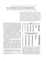
The chemical composition (Table 1) and
1
H-NMR
spectrum (Fig. 2) of Tm-PS was found to be similar to
those of Tm-GC, except for the fatty acids, suggesting that
the fundamental structure of the polysaccharide part in both
is the same. Further, it is reasonable to assume that Tm-PS
is a dephosphorylated or deacylated component of Tm-GC
therefore we elucidated the structure of the polysaccharide
part using Tm-PS.
Compositional analysis showed that Tm-PS contained
mainly glucosamine (GlcN), aspartic acid (Asp), and
ornithine (Orn) (Table 1). The absolute configuration of
GlcN was determined as
D
, while the absolute configura-
tions of the amino acids were determined as
D
for Asp and
L
for Orn.
The
1
Hand
13
C NMR spectra of Tm-PS are shown in
Figs 2 and 3. Four anomeric signals (at d 4.63, d4.78, d 5.05,
and d 5.13 for
1
H, and d 98.8, d 99.8, d 100.4, and d 102.0 for
13
C) were mainly observed, and the corresponding sugars
were designated as a to d in order of
13
C NMR chemical
shift. Eight signals at d 50.3, d 51.0, d 53.7, d 53.8, d 54.0,
d 54.2, d 54.5, and d 54.7 in the
13
C NMR spectrum were
assigned to carbons substituted by the amino group. Six
amide proton signals at d 7.92, d8.14, d 8.22, d 8.27, d 8.47,
and d 8.71 were observed in the
1
H NMR spectra measured
in 10% D
2
O. The
1
H NMR signals were assigned using the
DQF-COSY, TOCSY, and HSQC-TOCSY spectra, while
the
13
C NMR signals were assigned using the HMQC,
HSQC-TOCSY, and HMBC spectra (Table 2). The coup-
ling constants were estimated from a one-dimensional
spectrum and DQF-COSY. Based on the assignment, one
4-amino-4,6-deoxyhexose for residue a, two 2,3-diamino-
2,3-dideoxyhexuronic acids each for residues b and c, one
2-amino-2-deoxyhexose for residue d,Asp,andOrnwere
found.
Residue d was assigned to 2-amino-2-deoxy-b-glucopyra-
nose (b-GlcpN). The coupling constants for the anomeric
protons,
3
J
1,2
of 8.0 Hz, showed a b-configuration.
Although the coupling constant
3
J
2,3
could not be deter-
mined,
3
J
3,4
and
3
J
4,5
of 10 Hz indicated the glucopyra-
nosyl configuration. The chemical shift of d 54.7 for C2-d
was indicative of a 2-amino-2-deoxy structure. Intraresidual
ROESY couplings from H1-d to H3-d and H1-d to H5-d
(Fig.4)aswellasthe
1
J
C,H
value for the anomeric atoms
(160.7 Hz) determined from the nondecoupling DEPT
spectrum [34] supported a b-configuration.
As judged by the coupling constant, residue a was
4-amino-4,6-dideoxy-a-galactopyranose (a-Fucp4N). The
coupling constants,
3
J
2,3
of 11 Hz,
3
J
3,4
of 5Hz,and
a characteristically small
3
J
4,5
determined from the TOCSY
spectrum showed the galactopyranosyl configuration. The
coupling constant for the anomeric proton,
3
J
1,2
of 4.4 Hz,
and the
1
J
C,H
value of the anomeric atoms (172.3 Hz)
indicated an a-configuration. The chemical shift of d 54.0
for C4-a was indicative of a 4-amino-4-deoxy structure,
while d 15.7 for C6-a showed a 6-deoxy structure.
Residue b was assigned to 2,3-diamino-2,3-dideoxy-
b-mannopyranuronic acid (b-ManpN3NA). The coupling
constants of residue b, characteristically small ( 3Hz)
3
J
2,3
,andlarge( 11 Hz)
3
J
3,4
and
3
J
4,5
,aswellasasinglet
like signal of H-1b were typical for a mannopyranosyl
configuration. Intraresidual ROESY couplings from H1-b
to H3-b and H1-b to H5-b along with the
1
J
C,H
value
(163.0 Hz) indicated a b-configuration. The chemical shifts
of d50.3 for C2-b and d53.7 for C3-b were indicative of a
2,3-diamino-2,3-dideoxy structure, and d 168.7 for C6-b
showed a uronic acid structure.
Residue c was determined to be 2,3-diamino-2,3-dideoxy-
b-glucopyranuronic acid (b-GlcpN3NA). The coupling
constants for the anomeric proton,
3
J
1,2
of 8.0 Hz, indicated
a b-linkage, while the coupling constants
3
J
2,3
of 10 Hz,
3
J
3,4
of 8Hz, and
3
J
4,5
of 11 Hz indicated a gluco-
pyranosyl configuration. The chemical shifts of d 53.8 for
C2-c and d54.2 for C3-c were indicative of a 2,3-diamino-
2,3-dideoxy structure, and d 174.5 for C6-c showed a uronic
acid structure.
The sequences of the sugar and amino acid residues were
determined using ROESY, NOESY, and HMBC spectra.
Four glycosidic linkages were established from results of
Table 1. Chemical composition (lmolÆmg
)1
)ofTm-GCandTm-PS.
Tm-GC Tm-PS
D
-GlcN 1.06 0.71
D
-Asp 0.83 0.64
L
-Orn 1.06 0.92
Phosphate 0.02 0.08
Fatty acids
14:0 0.02 0
13-Me 14:0 0.02 0
16:0 0.06 0
17:0
D9,10
0.02 0
18:1
c9
0.02 0
Ó FEBS 2003 Polysaccharide of T. medium (Eur. J. Biochem. 270) 2673
the ROESY (Fig. 4) and HMBC experiments. Interresidual
couplings from H1-a to H4-c indicated that residue a was
linked to O4 of residue c, while those from H1-b to H3-d
showed that residue b was linked to O3 of residue d, those
from H1-d to H3-a indicated that residue d waslinkedtoO3
of residue a, and those from H1-c to H4-b showed that
residue c was linked to O4 of residue b. Long-range HMBC
couplings from H1-c to C4-b,H1-b to C3-d,H1-d to C3-a,
and H1-a to C4-c were observed (data not shown), which
supported the above linkages.
Two amide linkages were established by the NOESY
(Fig. 5) and HMBC experiments. Couplings from the amide
proton at the 4-position of residue a (NH4-a)toH2-Asp
indicated that the carboxyl group of the 1-position in Asp
was attached to N4 of residue a by an amide linkage.
Couplings from NH5-Orn to C5-b indicated that the
amino group at the 5-position of Orn was attached to the
6-position of residue b by an amide linkage. Long-range
HMBC couplings from H4-a to C1-Asp and H5-Orn to
C6-b (data not shown) supported the amide linkages. These
results showed the sequence, [fi4)b-GlcN3NA(1fi4)b-
ManN3NA6Orn(1fi3)b-GlcN(1fi3)a-Fuc4NAsp(1fi].
To elucidate the amidation pattern of Tm-PS, the pD
dependence of
1
H NMR chemical shifts was studied. The
signals for H5-c and H5-b were shifted downfield by +0.15
and +0.02, respectively, with a decrease of pD from 7 to 2,
indicating a free carboxyl group in residue c, however, not in
residue b. The signals for H3-Asp and H2-Orn were shifted
downfield by +0.23–0.28 and +0.19, respectively, with a
decrease of pD from 7 to 2, indicating a free carboxyl group
of the 4-position in Asp and the 1-position in Orn. The
signals for H2-b,H3-b,andH4-b were shifted upfield by
)0.34, )0.69, and )0.38, respectively, with an increase of pD
from 7 to 13, indicating a free amino group at the 3-position
of residue b. The signals for H2-Asp and H2-Orn were
shifted upfield by )0.55 and )0.54, respectively, with an
increase of pD from 7 to 13, indicating a free amino group
at the 2-position of Asp and the 2-position of Orn.
To determine the relative absolute configuration of the
constituent monosaccharids, glycosylation effects [35] in the
Fig. 2. 500 MHz
1
H NMR spectra of Tm-GC
(A) and Tm-PS (B). Spectra were obtained
at 310 K in D
2
O. The spectrum in (B) was
recorded using a DANTE sequence for water
suppression.
Fig. 3. 126 MHz
13
CNMRspectraofTm-PS.
Spectra were obtained at 310 K in D
2
O.
2674 M. Hashimoto et al. (Eur. J. Biochem. 270) Ó FEBS 2003
13
C NMR spectrum of Tm-PS were analyzed. For the
b-linked disaccharide d fi a, b-GlcN(1 fi 3)Fuc4N, the
observed values were +6.9 for the a-effect of C1-d,+8.4
for that of C3-a,and)0.4 for the b-effect of C4-a,as
compared with those for standard GlcNAc and Fuc4NAc
[36]. The expected effects on C1-d,C3-a,andC4-a
were +8.0 ± 0.4, +9.1 ± 1.3, and )0.4 ± 0.4, respec-
tively, for the same and +2.9 ± 0.9, +5.4 ± 1.6, and
Fig. 4. ROESY spectra of Tm-PS. Spectra were obtained in phase sensitive mode with DANTE water suppression at 310 K in D
2
O. The mixing
time was 200 ms. Solid and dashed squares represent interresidual and intraresidual couplings, respectively.
Table 2.
1
H and
13
C-NMR data for T. medium glycoconjugate. Spectra were measured at 310K. Chemical shifts are expressed as d values.
Atoms
Residues
a-Fucp4N (a) b-ManpN3NA (b) b-GlcpN3NA (c) b-GlcpN(d) Asp Orn
H1 5.13 5.05 4.63 4.78
H2 4.23 4.58 3.84 3.74 4.33 3.81
H3 4.08 3.73 4.22 3.78 2.77, 2.86 1.95
H4 4.43 4.12 3.94 3.55 1.66, 1.76
H5 4.23 4.04 3.96 3.49 3.24, 3.53
H6 1.06 3.78, 3.93
C1 98.8 99.8 100.4 102.0 170.5 175.2
C2 67.3 50.3 53.8 54.7 51.0 54.5
C3 77.0 53.7 54.2 83.4 37.8 27.8
C4 54.0 72.8 74.3 68.7 176.1 24.1
C5 65.9 75.8 77.9 75.3 39.2
C6 15.7 168.7 174.5 60.5
NH2 8.22 7.92 8.16
NH3 8.27
NH4 8.47
NH5 8.71
Ó FEBS 2003 Polysaccharide of T. medium (Eur. J. Biochem. 270) 2675
)3.3 ± 0.6, respectively, for different absolute configura-
tions. These results showed that residues d and a had the
same absolute configuration. For the b-linked disaccharide
b fi d, b-ManN3NA(1 fi 3)GlcN, the observed value was
)1.4 for the b-effect of C4-d, as compared with standard
GlcNAc. The expected effects were )1.4 ± 0.2 for the same
and )0.2 ± 0.4 for different absolute configurations. This
finding also suggested that residues d and a had the same
absolute configuration. For the a-linked disaccharide
a fi c, a-Fuc4N(1 fi 4)GlcN3NA, the observed value
was )0.1 for the b-effect of C3-c as compared with the
nonreducing terminal GlcNAc3NAc (54.3 p.p.m. for C3) of
the oligosaccharides described below. The expected effect
was +0.3 ± 0.6 for the same and )1.3 ± 0.4 for different
absolute configurations. Thus, residues a and c were found
to have the same absolute configuration. Therefore, all the
sugars had the same absolute configuration as
D
.
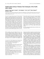
To confirm its structure, Tm-PS was subjected to
solvolysis with trifluoromethanesulfonic acid [29]. The
MALDI-TOF-MS of the resulting oligosaccharides showed
pseudomolecular ions [M + H]
+
at m/z 1070.5 and 810.3,
and their dehydrated ions at m/z 1052.4 and 792.2. The
ions at m/z 1070.5 represented a tetrasaccharide com-
posed of amino-deoxyhexose (HexN), amino-dideoxy-
hexose (dHexN), and two diamino-dideoxyhexuronic
acids (HexNNA), with Asp, Orn, and four acetyl groups.
The tetrasaccharide could be obtained by the selective
cleavage of the 4-aminofucosidic linkage of Tm-PS and the
MS/MS spectrum of the ion at m/z 1070 (Fig. 6) indicated
the sequence (HexNNA + 2Ac) fi (HexNNA + Ac +
Orn) fi (HexN + Ac) fi (dHex + Asp). The ions at
m/z 810.3 represented a trisaccharide composed of a single
HexN and two HexNNA, with Orn and four acetyl groups.
The trisaccharide could be yielded by a double cleavage
of the 2-aminoglucosidic and 4-aminofucosidic linkages of
Tm-PS. The results of solvolysis agreed with the structure
established by NMR analysis.
From our results, we proposed a chemical structure of
Tm-PS and the polysaccharide part of Tm-GC, as shown
in Fig. 7, which is the first known structural elucidation
of a polysaccharide obtained from a treponemal species.
Although the structure is novel, the constituents, inclu-
ding the unusual aminosugar and diaminouronic acids,
have been previously found in O-specific, capsular, and
Fig. 5. NOESY spectra of Tm-PS. Spectra were obtained in phase sensitive mode with DANTE water suppression at 310 K in 10% D
2
O. The
mixing time was 490 ms. Solid and dashed squares represent interresidual and intraresidual couplings, respectively.
2676 M. Hashimoto et al. (Eur. J. Biochem. 270) Ó FEBS 2003
exo-polysaccharides from other bacteria [37,38]. As many
of these kind of polysaccharides seem to have antigeni-
city, the unusual sugars seen in the present study may be
involved with antigen epitopes. Therefore, vaccination
usingpartofthestructureofTm-GCmaybeusefulfor
prevention of periodontal diseases caused by T.medium.
As many oral treponemal species contribute to perio-
dontal diseases [39], the structures of glycoconjugates
obtained from other treponemal species should also be
elucidated.
Structural features of the glycoconjugate
LPS is composed of a polysaccharide part, O-antigen and
core regions, and a lipid anchor termed lipid A. Because the
structure of the polysaccharide part of Tm-GC is similar to
that of O-antigen of LPS, we attempted to determine
whether Tm-GC is a kind of LPS. LPS usually contains
characteristic components, such as heptose, b-hydroxy fatty
acid, and Kdo, however, no heptose or b-hydroxy fatty acid
was observed in the compositional analysis of Tm-GC,
while the content of Kdo was under the detection limit of
the thiobarbiturate assay. Kdo in LPS from some species
does not respond in a thiobarbiturate assay, due to
phosphorylation [22]. However, as the HF-treated products
of Tm-GC were negative in the thiobarbiturate assay, Kdo
was considered to be absent from Tm-GC.
We also characterized the hydrophobic region of
Tm-GC. Alkaline treated products of Tm-GC contained
no fatty acid, which showed that all fatty acids in Tm-GC
are attached by ester linkages. HF-treatment liberated
hydrophobic products from Tm-GC. ESI-MS of the
Fig. 6. MALDI-TOF-MS/MS spectrum of the parent ion at m/z 1070 in TfOH-hydrolysate of Tm-PS. The spectrum was obtained in positive ion
TOF/TOF mode using 2,5-dihydroxy benzoic acid as a matrix.
Fig. 7. Proposed chemical structure of Tm-PS.
Ó FEBS 2003 Polysaccharide of T. medium (Eur. J. Biochem. 270) 2677
products showed two series of ions [M-H]
–
at m/z 383.2,
397.2, and 411.2 (relative intensity ratio, 0.57 : 0.40 : 1.00),
and m/z 455.2, 469.2, and 483.2 (0.22 : 0.18 : 0.51), respect-
ively. CID-MS/MS of the ion at m/z 411.2 showed daughter
ions at m/z 255.2 and 155.0, and that at m/z 483.2 showed
daughter ions at m/z 255.2, 227.0, and 153.0. The fragmen-
tation patterns indicated that the former represents a
lysophosphatidic acid deivative, acylglycero-fluorophos-
phoric acid, and the latter was lysophosphatidylglycerol.
As a fluorophosphoric acid structure can be produced by
the cleavage of phosphodiester linkage with HF, lysophos-
phatidylglycerol was considered to be the hydrophobic
region of Tm-GC. Further, as lysophosphatidylglycero-
phosphate or its derivative were not observed, the linkage
between the polysaccharide part and the hydrophobic
region may be an acid labile glycosyl bond. Lipid A, which
generally consists of a diglucosamine backbone with amide
and ester bound fatty acids, and occasionally monoester
linked phosphate, is attached to Kdo located at the reducing
end of the polysaccharide of LPS, via a glycosyl bond.
Therefore, the glycoconjugate from T.mediumis likely to be
different from LPS.
The present observations agreed with earlier analyses of
glycolipids in the small-sized treponemal species T. denticola
[9], T. maltophilum,andT. brennaborense [10], in which it
was proposed that the structure of the glycolipids resemble
lipoteichoic acid (LTA). Although the structure of the
polymeric hydrophilic part is quite different, Tm-GC may
be this kind of glycolipid, though the LTA-like treponemal
glycolipids are known to have immunostimulating activities,
whereas Tm-GC possessed no such activity. In biologically
active glycoconjugates, the detailed structure of the lipo-
philic part is closely related to their activity, such as the
acylation and phosphorylation patterns in lipid A of LPS
[28], and the glycosylation position of the glycolipid in LTA
[40,41]. To determine the structure–activity relationship, a
detailed structural elucidation of the lipophilic part of
immunobiologically active glycoconjugates from trepone-
mal species is required.
Acknowledgements
This study was supported in part by a Grant-in-Aid for Scientific
Research (B) (13470390 to T. O.) from the Japan Society for the
Promotion of Science and a Grant-in-Aid for Encouragement of
Young Scientists (147701014 to M. H.) from the Ministry of
Education, Culture, Sports, Science and Technology. We are grateful
to Mr Nirasawa at Bruker Daltonics for his skilful measurements of
MALDI-TOF-MS. We thank Mr M. Benton for his critical reading of
the manuscript.
References
1. Singh, A.E. & Romanowski, B. (1999) Syphilis: review with
emphasis on clinical, epidemiologic, and some biologic features.
Clin. Microbiol. Rev. 12, 187–209.
2. Sela, M.N. (2001) Role of Treponema denticola in periodontal
diseases. Crit.Rev.Oral.Biol.Medical12, 399–413.
3. Hauschildt, S., Hoffmann, P., Beuscher, H.U., Dufhues, G.,
Heinrich, P., Wiesmuller, K.H., Jung, G. & Bessler, W.G. (1990)
Activation of bone marrow-derived mouse macrophages by
bacterial lipopeptide: cytokine production, phagocytosis and Ia
expression. Eur. J. Immunol. 20, 63–68.
4. Radolf, J.D., Chamberlain, N.R., Clausell, A. & Norgard, M.V.
(1988) Identification and localization of integral membrane pro-
teins of virulent Treponema pallidum subsp. pallidum by phase
partitioning with the nonionic detergent triton X-114. Infect.
Immun. 56, 490–498.
5. Norgard, M.V., Arndt, L.L., Akins, D.R., Curetty, L.L., Harrich,
D.A. & Radolf, J.D. (1996) Activation of human monocytic cells
by Treponema pallidum and Borrelia burgdorferi lipoproteins and
synthetic lipopeptides proceeds via a pathway distinct from that
of lipopolysaccharide but involves the transcriptional activator
NF-jB. Infect. Immun. 64, 3845–3852.
6. Sela, M.N., Bolotin, A., Naor, R., Weinberg, A. & Rosen, G.
(1997) Lipoproteins of Treponema denticola: their effect on human
polymorphonuclear neutrophils. J. Periodontal Res. 32, 455–466.
7. Rosen, G., Sela, M.N., Naor, R., Halabi, A., Barak, V. & Shapira,
L. (1999) Activation of murine macrophages by lipoprotein and
lipooligosaccharide of Treponema denticola. Infect. Immun. 67,
1180–1186.
8. Fraser, C.M., Norris, S.J., Weinstock, G.M., White, O., Sut-
ton, G.G., Dodson, R., Gwinn, M., Hickey, E.K., Clayton, R.,
Ketchum, K.A., Sodergren, E., Hardham, J.M., McLeod,
M.P.,Salzberg,S.,Peterson,J.,Khalak,H.,Richardson,D.,
Howell, J.K., Chidambaram, M., Utterback, T., McDonald, L.,
Artiach, P., Bowman, C., Cotton, M.D., Fujii, C., Garland, S.,
Hatch, B., Horst, K., Roberts, K., Sandusky, M., Weidoman,
J.,Smith,H.O.&Venter,J.C.(1998)Completegenome
sequence of Treponema pallidum, the syphilis spirochete. Science
281, 375–388.
9. Schultz, C.P., Wolf, V., Lange, R., Mertens, E., Wecke, J., Nau-
mann,D.&Za
¨
hringer, U. (1998) Evidence for a new type of outer
membrane lipid in oral spirochete Treponema denticola. Func-
tioning permeation barrier without lipopolysaccharides. J. Biol.
Chem. 273, 15661–15666.
10. Schro
¨
der, N.W., Opitz, B., Lamping, N., Michelsen, K.S., Za
¨
h-
ringer, U., Go
¨
bel, U.B. & Schumann, R.R. (2000) Involvement of
lipopolysaccharide binding protein, CD14, and Toll-like receptors
in the initiation of innate immune responses by Treponema
glycolipids. J. Immunol. 165, 2683–2693.
11. Schro
¨
der, N.W., Pfeil, D., Opitz, B., Michelsen, K.S., Amberger,
J., Za
¨
hringer, U., Go
¨
bel, U.B. & Schumann, R.R. (2001) Acti-
vation of mitogen-activated protein kinases p42/44, 38, and stress-
activated protein kinases in myelo-monocytic cells by Treponema
lipoteichoic acid. J. Biol. Chem. 276, 9713–9719.
12. Opitz, B., Schro
¨
der, N.W., Spreitzer, I., Michelsen, K.S., Kirsch-
ning, C.J., Hallatschek, W., Za
¨
hringer, U., Hartung, T., Go
¨
bel,
U.B. & Schumann, R.R. (2001) Toll-like receptor-2 mediates
Treponema glycolipid and lipoteichoic acid-induced NF-jB
translocation. J. Biol. Chem. 276, 22041–22047.
13. Asai, Y., Jinno, T., Igarashi, H., Ohyama, Y. & Ogawa, T. (2002)
Detection and quantification of oral treponemes in subgingival
plaque by real-time PCR. J. Clin. Microbiol. 40, 3334–3340.
14. Paster, B.J., Boches, S.K., Galvin, J.L., Ericson, R.E., Lau, C.N.,
Levanos,V.A.,Sahasrabudhe,A.&Dewhirst,F.E.(2001)
Bacterial diversity in human subgingival plaque. J. Bacteriol. 183,
3770–3783.
15. Asai, Y., Jinno, T. & Ogawa, T. (2003) Oral treponemes and their
outer membrane extracts activate human gingival epithelial cells
through Toll-like receptor 2. Infect. Immun. 71, 717–725.
16. Ohta, K., Makinen, K.K. & Loesche, W.J. (1986) Purification and
characterization of an enzyme produced by Treponema denticola
capable of hydrolyzing synthetic trypsin substrates. Infect. Immun.
53, 213–220.
17. Westphal, O. & Jann, K. (1965) Bacterial lipopolysaccharides:
extraction with phenol-water and further applications to the
procedure. In Methods in Carbohydrate Chemistry,Vol.5(Whis-
tler, R.L., ed.), pp. 83–91. Academic Press, NewYork.
2678 M. Hashimoto et al. (Eur. J. Biochem. 270) Ó FEBS 2003
18. Bartlett, G.R. (1959) Phosphorus assay in column chromato-
graphy. J. Biol. Chem. 234, 466–468.
19. Ashwell, G. (1957) Colorimetric analysis of sugars. Methods
Enzymol. 3, 73–105.
20. Torello, L.A., Yates, A.J. & Thompson, D.K. (1980) Critical
study of the alditol acetate method for quantitating small quan-
tities of hexoses and hexosamines in gangliosides. J. Chromatogr.
202, 195–209.
21. Baumann, H., Tzianabos, A.O., Brisson, J.R., Kasper, D.L. &
Jennings, H.J. (1992) Structural elucidation of two capsular
polysaccharides from one strain of Bacteroides fragilis using high-
resolution NMR spectroscopy. Biochemistry 31, 4081–4089.
22. Chaby, R., Charon, D., Caroff, M., Sarfati, S.R. & Trigalo, F.
(1993) Estimation of 3-deoxy-
D
-manno-2-octurosonic acid in
lipopolysaccharides: an unsolved problem. In Methods in Carbo-
hydrate Chemistry (BeMiller, J.N. & Whistler, R.L., eds),
pp. 33–46. John Wiley & Sons, New York.
23. Ikemoto,S.,Katoh,K.&Komagata,K.(1978)Cellularfattyacid
composition in methanol-utilizing bacteria. J. General Appl.
Microbiol. 24, 41–49.
24. Nimura, N. & Kinoshita, T. (1986) o-Phthalaldehyde-N-acetyl-
L-cysteine as a chiral derivatization reagent for liquid chromato-
graphic optical resolution of amino acid enantiomers and its
application to conventional amino acid analysis. J. Chromatogr.
352, 169–177.
25. Fujii, K., Ikai, Y., Oka, H., Suzuki, M. & Harada, K. (1997) A
nonempirical method using LC/MS for determination of the
absolute configuration of constituent amino acids in a peptide:
combination of Marfey’s method with mass spectrometry and its
practical application. Anal. Chem. 69, 5146–5151.
26. Laemmli, U.K. (1970) Cleavage of structural proteins during the
assembly of the head of bacteriophage T4. Nature 227, 680–685.
27. Tsai, C.M. & Frasch, C.E. (1982) A sensitive silver stain for
detecting lipopolysaccharides in polyacrylamide gels. Anal. Bio-
chem. 119, 115–119.
28. Ogawa, T., Asai, Y., Yamamoto, H., Taiji, Y., Jinno, T., Kod-
ama, T., Niwata, S., Shimauchi, H. & Ochiai, K. (2000)
Immunobiological activities of a chemically synthesized lipid A of
Porphyromonas gingivalis. FEMS Immunol. Med. Microbiol. 28,
273–281.
29. Perepelov, A.V., Senchenkova, A.N., Shashkov, A.S.,
Komandrova, N.A., Tomshich, S.V., Shevchenko, L.S., Knirel,
Y.A. & Kochetkov, N.K. (2000) First application of triflic acid for
selective cleavage of glycosidic linkages in structural studies of a
bacterial polysaccharide from Pseudoalteromonas sp. KMM 634.
J. Chem. Soc., Perkin Trans. 1, 363–366.
30. Pantosti, A., Tzianabos, A.O., Onderdonk, A.B. & Kasper, D.L.
(1991) Immunochemical characterization of two surface
polysaccharides of Bacteroides fragilis. Infect. Immun. 59, 2075–
2082.
31. Fischer, W., Koch, H.U. & Haas, R. (1983) Improved preparation
of lipoteichoic acids. Eur. J. Biochem. 133, 523–530.
32. Fischer, W. (1990) Purification and fractionation of lipopolysac-
charide from gram-negative bacteria by hydrophobic interaction
chromatography. Eur. J. Biochem. 194, 655–661.
33. Lindberg, A.A. (1999) Glycoprotein conjugate vaccines. Vaccine.
17, S28–S36.
34. Tvaroska, I. & Taravel, F.R. (1995) Carbon-proton coupling
constants in the conformational analysis of sugar molecules. Adv.
Carbohydr. Chem. Biochem. 51, 15–61.
35. Shashkov, A.S., Lipkind, G.M., Knirel, Y.A. & Kochetkov, N.K.
(1988) Stereochemical factors determining the effects of glycosy-
lation on the
13
C chemical shifts in carbohydrates. Magn. Reson.
Chem. 26, 735–747.
36. Knirel, Y.A., Paramonov, N.A., Shashkov, A.S., Kochetkov,
N.K., Zdorovenko, G.M., Veremeychenko, S.N. & Zakharova,
I.Y. (1993) Somatic antigens of pseudomonads: structure of the
O-specific polysaccharide of Pseudomonas fluorescens biovar A
strain IMV 1152. Carbohydr. Res. 243, 205–210.
37. Lindberg, B. (1990) Components of bacterial polysaccharides.
Adv. Carbohydr. Chem. Biochem. 48, 279–318.
38. Knirel, Y.A. & Kochetkov, N.K. (1994) The structure of lipo-
polysaccharides of gram-negative bacteria. III. The structure of
O-antigens (a review). Biochemistry (Moscow) 59, 1325–1383.
39. Chan, E.C. & McLaughlin, R. (2000) Taxonomy and virulence of
oral spirochetes. Oral Microbiol. Immunol. 15, 1–9.
40. Takada, H., Kawabata, Y., Arakaki, R., Kusumoto, S., Fukase,
K., Suda, Y., Yoshimura, T., Kokeguchi, S., Kato, K., Komuro,
T., Tanaka, N., Saito, M., Yoshida, T., Sato, M. & Kotani, S.
(1995) Molecular and structural requirements of a lipoteichoic
acid from Enterococcus hirae ATCC 9790 for cytokine-inducing,
antitumor, and antigenic activities. Infect Immun. 63, 57–65.
41. Morath, S., Stadelmaier, A., Geyer, A., Schmidt, R.R. & Hartung,
T. (2002) Synthetic lipoteichoic acid from Staphylococcus aureus
is a potent stimulus of cytokine release. J. Exp. Med. 195,
1635–1640.
Ó FEBS 2003 Polysaccharide of T. medium (Eur. J. Biochem. 270) 2679