Báo cáo khoa học: Functional characterization of the maltose ATP-binding-cassette transporter of Salmonella typhimurium by means of monoclonal antibodies directed against the MalK subunit pot
Bạn đang xem bản rút gọn của tài liệu. Xem và tải ngay bản đầy đủ của tài liệu tại đây (467.45 KB, 12 trang )
Functional characterization of the maltose ATP-binding-cassette
transporter of
Salmonella typhimurium
by means of monoclonal
antibodies directed against the MalK subunit
Anke Stein
1
, Martina Seifert
2
, Rudolf Volkmer-Engert
2
,Jo¨ rg Siepelmeyer
3
, Knut Jahreis
3
and Erwin Schneider
1
1
Humboldt Universita
¨
t zu Berlin, Institut fu
¨
r Biologie, Berlin, Germany;
2
Humboldt Universita
¨
t zu Berlin, Institut fu
¨
r Medizinische
Immunologie, Berlin;
3
Universita
¨
t Osnabru
¨
ck, Fachbereich Biologie/Chemie, Germany
The maltose ATP-binding cassette transporter of Salmonella
typhimurium is composed of a membrane-associated com-
plex (MalFGK
2
) and a periplasmic receptor (MalE). In
addition to its role in transport, the complex acts as a
repressor of maltose-regulated gene expression and is subject
to inhibition in the process of inducer exclusion. These
activities are thought to be mediated by interactions of the
ATPase subunit, MalK, with the transcriptional activator,
MalT, and nonphosphorylated enzyme IIA of the glucose
phosphotransferase system, respectively. To gain further
insight in protein regions that are critical for these functions,
we have generated nine MalK-specific monoclonal anti-
bodies. These bind to four nonoverlapping linear epitopes:
60-LFig-63 (5B5), 113-RVNQVAEVLQL-123 (represented
by 4H12), 309-GHETQI-314 (2F9) and 352-LFREDG
SACR-361 (represented by 4B3). All mAbs recognize their
epitopes in soluble MalK and in the MalFGK
2
complex with
K
d
values ranging from 10
)6
to 10
)8
M
. ATP reduced the
affinity of the mAbs for soluble MalK, indicating a confor-
mational change that renders the epitopes less accessible.
4H12 and 5B5 inhibit the ATPase activity of MalK and the
MalE/maltose-stimulated ATPase activity of proteolipo-
somes, while their Fab fragments displayed no significant
effect. The results suggest a similar solvent-exposed position
of helix 3 in the MalK dimer and in the intact complex and
might argue against a direct role in the catalytic process. 4B3
and 2F9 exhibit reduced binding to the MalFGK
2
complex
in the presence of MalT and enzyme IIA
Glc
, respectively,
thereby providing the first direct evidence for the C-terminal
domain of MalK being the site of interaction with the reg-
ulatory proteins.
Keywords: ABC transporter; MalFGK
2
; enzyme IIA
Glc
;
MalT; monoclonal antibodies.
The family of ATP-binding-cassette (ABC) transport sys-
tems comprises an extremely diverse class of membrane
proteins that couple the energy of ATP hydrolysis to the
translocation of solutes across biological membranes [1,2].
A prototype ABC transporter is composed of four
entities: two membrane-integral domains, which presuma-
bly constitute a translocation pore, and two ATPase
domains (also referred to as ABC subunits/domains), that
provide the energy for the transport process. The ABC
domains are characterized by a set of canonical Walker A
and B motifs, required for nucleotide binding and by a
unique signature sequence (LSGGQ motif) of still unknown
function [3]. The crystal structures of several prokaryotic
ABC domains have been solved in recent years that agree
largely on the overall folds. Accordingly, the structures can
be subdivided in an F
1
-type ATP-binding domain, encom-
passing both Walker sites, a specific a-helical subdomain,
containing the LSGGQ motif and a specific antiparallel-b-
subdomain [4–7].
The binding protein-dependent maltose/maltodextrin
transporter of enterobacteria, such as Escherichia coli and
Salmonella typhimurium, is a well-characterized model
system for studying the mechanism of action of the ABC
transport family [8]. Based on computational analysis, it
belongs to a subclass of ABC importers designated CUT1
(carbohydrate uptake transporter) [9] or OSP (oligosaccha-
rides and polyols) [10], respectively. Members of this
subclass transport a variety of di- and oligosaccharides,
glycerol phosphate and polyols and are recognized by their
common subunit composition (two individual membrane-
spanning subunits and two copies of a single ABC protein)
and by an extension of approximately a hundred amino acid
residues at the C-terminus of the ABC protein [11].
The maltose transporter of E. coli/S. typhimurium is
composed of the periplasmic maltose binding protein, MalE,
and of the membrane-associated complex, MalFGK
2
,con-
sisting of one copy each of the hydrophobic subunits MalF
and MalG and two copies of the nucleotide-binding subunit
MalK [12]. Crystals of Salmonella MalK are available [13]
but their structure could not be solved yet. However, the
tertiary structure of a MalK homolog, isolated from the
Correspondence to E. Schneider, Humboldt Universita
¨
t zu Berlin,
Mathematisch-Naturwissenschaftliche Fakulta
¨
tI,
Institut fu
¨
r Biologie, Bakterienphysiologie, Chausseestr. 117,
D-10115 Berlin, Germany.
Tel.: + 49 (0)30 2093 8121, Fax: + 49 (0)30 2093 8126,
E-mail:
Abbreviations: IF-medium, Iscove’s DMEM/NUT MIX F12.
(Received 27 March 2002, revised 6 June 2002, accepted 8 July 2002)
Eur. J. Biochem. 269, 4074–4085 (2002) Ó FEBS 2002 doi:10.1046/j.1432-1033.2002.03099.x
hyperthermophilic archaeon Thermococcus litoralis,was
recently determined [5]. Two molecules are present per
asymmetric unit that contact each other through the ATPase
domains with the C-terminal domains attached at opposite
poles. Based on these data, a 3D model of the E. coli MalK
protein was recently presented [14].
Enterobacterial MalK can be purified in fairly large
amounts [15] and displays a spontaneous ATPase activity
that is insensitive to inhibition by vanadate, a typical
inhibitor of ABC transporters [16]. The purified MalFGK
2
complex, when incorporated into liposomes, also exhibits a
low intrinsic ATPase activity that, however, is stimulated
severalfold in the presence of substrate-loaded MalE and is
vanadate-sensitive [12,17–19].
According to a current transport model, the presence of
substrate in the medium is thought to be signalled by
liganded MalE via interaction with externally exposed
peptide loops of MalF and MalG [20]. As a consequence,
conformational changes of the latter are transmitted to the
MalK subunits which, in turn, become activated. Hydro-
lysis of ATP would then trigger subsequent conformational
changes that eventually lead to the translocation of the
substrate molecule. Recent findings suggested that these
steps occur rather simultaneously [21].
Interaction of MalK with the hydrophobic subunits
involves contact of residues in the helical subdomain
with conserved cytoplasmic loops (EAA motifs) in MalF
and MalG [22–24]. This view, based on suppressor
mutational analyses and cross-linking studies, is largely
consistent with the recently solved crystal structure of
MsbA, an ABC transporter mediating the export of
the lipid A component of the E. coli outer membrane
[25].
Besides acting as an import system for maltose/malto-
dextrins, the MalFGK
2
complex is involved in the regula-
tion of genes belonging to the maltose regulon [8]. In the
absence of substrate, the idle transporter is thought to
interact with the positive transcriptional regulator, MalT,
via the MalK subunits, thereby preventing MalT from
binding to its target sequences upstream of maltose-
regulated promotors. When the transporter becomes
engaged in translocating maltose across the membrane,
MalT is released and transcription of maltose-regulated
genes can occur [26].
In addition, the maltose transporter is subject to inhibi-
tion by binding of dephosphorylated enzyme IIA of the
glucose transporter (phosphoenolpyruvate phosphotrans-
ferase system) to the MalK subunits in a process called
inducer exclusion in the context of global carbon regulation
in enteric bacteria [27].
Both regulatory activities of MalK are largely mediated
by the C-terminal domain of the protein [5,28–30].
Obviously, specific protein–protein interactions within
the MalFGK
2
complex as well as between the transporter
and regulatory proteins are crucial for its role in intact cells.
However, in the absence of tertiary structural information
on the complete transporter, these interactions are still
poorly understood at the molecular level. Here, we describe
the use of monoclonal antibodies raised against the MalK
subunit as tools to gain further insights in the structural
basis of transporter functions.
EXPERIMENTAL PROCEDURES
Preparative procedures
MalK [15], MalFGK
2
[18], and MalE [31] were purified
as described. MalE/maltose-loaded proteoliposomes con-
taining the MalFGK
2
complex were prepared by a
detergent dilution procedure as published elsewhere
[18,32].
Enzyme IIA
Glc
was purified from the cytosolic fraction of
E. coli strain BL21 D (pts43crr::kan
R
) harbouring plasmid
pCRL13 (crr on pET23A) [33] by Ni-NTA affinity chro-
matography.
Crude extract containing MalT was prepared according
to [34] from E. coli strain JM109 (Stratagene), carrying
plasmid pAS8 (malT
E.c.
on pSE380, p
trc
,amp
R
) (this study).
For competitive inhibition ELISA, N-terminally his-tagged
MalT was partially purified from strain JM109, harbouring
plasmid pAS9 (malT
E.c.
on pQE9, p
T5
,amp
R
)byNi-NTA
chromatography.
Preparation of mAbs
Ten-week-old-femaleBalb/cmicewereimmunizedintra-
peritoneally either with native MalK or with an
N-terminal fragment (encompassing residues 1–179 [30])
(100 lg each), dissolved in NaCl/P
i
[35]. On day 12, 25
and 62 the animals were boosted with 50 lgofprotein
each. The final boost was given 4 days prior to the fusion.
For hybridoma production spleen cells were isolated and
fused with myeloma cells SP2/0 as described [36] using
poly(ethylene glycol) 1500 as fusion agent. Selection of
hybridoma cells was performed in hypoxanthine, aminop-
terin and thymidine selection medium supplement. Grow-
ing hybridomas were screened by ELISA using MalK as
bound antigen. Selected hybrid cell lines were cloned at
least three times by limiting dilution. Cloned hybridoma
cells were maintained in 20% IF-medium, supplemented
with 70% fetal bovine serum and 10% dimethylsulfoxide
for several days at )80 °C and subsequently stored in
liquid nitrogen. For the production of mAbs, cells were
grown in IF or RPMI 1690 medium (Bichrom KG,
Berlin) in 2 L culture flasks.
mAbs were purified by loading concentrated culture
supernatant on a Protein G-Sepharose 4 fast flow matrix
equilibrated with 20 m
M
sodium phosphate buffer, pH 7.
After washing off unbound material, mAbs were eluted with
0.1
M
glycine/HCl, pH 2.7, and immediately dialyzed
against NaCl/P
i
overnight at 4 °C.
Isolation of Fab fragments
One millilitre of mAbs (1–3 mg) were mixed with 0.5 mL of
papain-agarose beads in 20 m
M
phosphate buffer, pH 7.5,
supplemented with 20 m
ML
-cysteine and 1 m
M
EDTA, and
incubated overnight at 37 °C. Subsequently, Fab fragments
were separated from uncleaved mAbs by incubating the
mixture with protein A–Sepharose for 1 h at 4 °C.
Unbound material (Fab fragments) was collected, dialyzed
overnight against 3 L of 50 m
M
Tris/HCl, pH 7.5, and
stored at 4 °C until use.
Ó FEBS 2002 Protein–protein interactions of MalFGK
2
(Eur. J. Biochem. 269) 4075
Determination of isotypes
Isotopes of the mAbs were determined by using Roche’s
ISO STRIP-mouse isotyping kit according to the manufac-
turer’s instructions.
Peptide synthesis on cellulose membranes – SPOT
synthesis
Cellulose-bound peptide libraries were automatically pre-
pared on Whatman 50 paper (Whatman, Maidstone, UK)
according to standard SPOT synthesis protocols [37] using a
SPOT synthesizer (Abimed GmbH, Langenfeld, Germany)
as described elsewhere [38–41]. The sequence files were
generated with the software
DIGEN
(Jerini AG, Berlin,
Germany). The peptides were derived from S. typhimurium
MalK. Libraries consisting of 10meric peptides (overlapping
by 9 amino acids) and peptide-substitutional analyses were
synthesized. All peptides are C-terminally attached to
cellulose via a (a-Ala)
2
spacer.
Epitope mapping
The screening of cellulose-bound peptides followed a
protocol published elsewhere [39,40]. Peptide libraries
were incubated with mAbs overnight at 4 °C in blocking
buffer (10% blocking reagent, Roche, in TNT, 10%
sucrose) and binding was detected with peroxidase-
conjugated goat anti(mouse IgG) antibody on hyperfilm
(Amersham/Pharmacia, Braunschweig, Germany) using the
Western Blot Chemiluminescence Reagent Plus System of
NEN (Boston, MA, USA).
Peptide synthesis
Peptides, structurally derived from the epitopes identified
after screening of the peptide libraries as described above
were synthesized on solid phase (50 lmol scale) on Tentagel
SRam resin (Rapp Polymere, Tu
¨
bingen, Germany) by using
PyBOP activation and a standard Fmoc-chemistry-based
protocol of an AMS 422 Peptide Synthesizer (Abimed,
Langenfeld, Germany). Side-chain protections of amino
acids are as follows: Glu, Asp (OtBu); Ser, Thr, Tyr,
Trp (tBu); His, Lys (Boc); Asn, Gln (Trt); Arg (Pbf).
Trifluoracetic acid /phenole/triisopropylsilane/H
2
O(9.4:
0.1 : 0.3 : 0.2) was used for resin cleavage and side-chain
deblocking. The crude peptides were purified to homogen-
eity by RP-HPLC using the linear solvent gradient 5–60% B
in A for 30 min, with A ¼ 0.05% trifluoracetic acid in
water, and B ¼ 0.05% trifluoracetic acid in acetonitrile.
The HPLC had the UV detector at 214 nm, a Vydac C
18
column of 20 · 250 mm, and a flow rate 10 mLÆmin
)1
.The
MS were performed on a matrix-assisted laser desorption
ionization-time of flight mass spectrometer (Laser Bench-
TopII, Applied Biosystems). The purity of the product was
characterized by analytical HPLC.
ELISA
For ELISA, microtiter plates were coated with purified
MalK (2.5 pmol) diluted in 100 m
M
sodium carbonate
buffer, pH 9.6, and incubated overnight at 4 °C. Remaining
binding sites were blocked with 2% BSA in NaCl/P
i
150 m
M
NaCl, 3 m
M
KCl, 8 m
M
Na
2
HPO
4
· 2H
2
O, 1 m
M
KH
2
PO
4
) for 2 h at room temperature. Subsequently, the
wells were incubated overnight at 4 °C with mAb diluted in
2% BSA in NaCl/P
i
/Tween (NaCl/P
i
containing 0.5%
Tween 80). Incubation with the second antibody (HRP-
conjugated goat anti(mouse IgG); 5 · 10
)2
-fold dilution]
occurred for 2 h at room temperature. After each step the
excess protein was removed by fourfold washing with NaCl/
P
i
/Tween. Antibody binding was detected by adding 100 lL
of 62.5 lgmL
)1
3,3¢,5,4¢-tetramethylbenzidine, 0.0026%
(v/v) H
2
O
2
in 0.1
M
sodium acetate/0.1
M
citric acid, pH 6.
After 10 min at room temperature, the reaction was stopped
by addition of 100 lLH
2
SO
4
and the color development
was measured at 450 nm.
Binding constants of mAbs were measured by com-
petitive inhibition ELISA according to [42]. Suitable
concentrations of mAbs were first determined by adding
various amounts of antibody to microtiter wells, coated
with various amounts of MalK, under experimental
conditions identical to those used in binding experiments
(ELISA).
The competitive inhibition ELISA was essentially carried
out as described above. In order to allow competition
between bound antigen (MalK) and free antigen (MalK,
MalFGK
2
-containing proteoliposomes, synthetic peptides)
the mAbs and an equal volume of free antigen in different
concentrations were incubated overnight at 4 °C in the wells
coated with MalK (2.5 pmol). The mAbs were used in the
following concentrations: 2F9 and 4B3, 2 · 10
)9
M
;4H12,
6.5 · 10
)9
M
;5B5,8· 10
)10
M
.
Analytical methods
Hydrolysis of ATP was assayed in microtiter plates
essentially as described in [43]. Protein was assayed using
the BCA kit from Bio-Rad. SDS/PAGE and immunoblot
analyses were performed as described in [44].
RESULTS
Monoclonal antibodies recognize epitopes
in the N-terminal (ATPase) domain and in the C-terminal
domain of MalK, respectively
Monoclonal antibodies were prepared against the MalK
subunit of the S. typhimurium maltose ABC transporter
using purified, nondenatured MalK or an N-terminal MalK
fragment (MalKN1, encompassing residues 1–179 [30]), as
antigen for immunization. Nine individual hybridoma cell
lines producing antibodies of the Ig subclass IgG1 were
obtained and immunoblot analyses revealed a specific
reaction with the corresponding antigen in each case when
purified MalK or MalFGK
2
complex were separated by
SDS/PAGE.
Immunoblots using truncated MalK proteins [30]
suggested that five mAbs, obtained with MalKN1,
recognize epitopes in the N-terminal (ATPase) domain
while the remaining four mAbs, obtained with intact
MalK, bind to the C-terminal (regulatory) domain. For
precise determination of the epitopes overlapping deca-
peptides corresponding to the entire MalK sequence were
synthesized on cellulose membranes by SPOT-synthesis
[37–41]. The results for the binding analyses of the
4076 A. Stein et al.(Eur. J. Biochem. 269) Ó FEBS 2002
different mAbs are shown in Fig. 2. Four peptide epitopes
were identified. One mAb (5B5) recognizes the peptide
53-ETITSGDLTRM-67, located close to the Walker A
motif, four (4H12, 6E6, 3A12, 4D8) bind to
111-NQRVNQVAEVLQL-123, located within the helical
subdomain (helices 2–4, Fig. 1A), three (2F9, 1D8, 2G4)
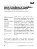
Fig. 1. Location of epitopes in the amino acid sequence of S. typhimurium MalK (A) and in the modelled 3D structure of E. coli MalK (B). (A) The
Walker A and B motifs and the ABC signature are highlighted in yellow. The epitopes recognized by the mAbs are highlighted in red. Residues that
when mutated render E. coli MalK insensitive to inducer exclusion are underlined while residues that cause a regulatory phenotype when mutated
are doubly underlined [14]. a-Helices and b-strands that have been identified in the structure of T. litoralis MalK [5] are indicated above the
sequences as broken and dotted lines, respectively. Please note that the primary structures of S. typhimurium (acc. no X54292) and E. coli MalK
(acc. no. J01648) differ only by 16 amino acid changes and by the lack of the dipeptide PM in S. typhimurium MalK after residue L258.
Furthermore, A320 (underlined) corresponds to S322 in E. coli MalK. (B) Stereo representation of the model of monomeric E. coli MalK [14]. The
epitopes recognized by the mAbs are indicated in red. The figure was drawn with RasMol 2.6 ( using the
coordinates provided by W. Welte (Universita
¨
tKonstanz).
Ó FEBS 2002 Protein–protein interactions of MalFGK
2
(Eur. J. Biochem. 269) 4077
recognize 304-VVEQLGHETQIHIQIP-319 and one (4B3)
binds to 352-LFREDGSACR-361, both located in the
C-terminal domain (Fig. 1A and B). The fact that in each
case strong signals with successive overlapping peptides
were obtained argues in favour of linear rather than
discontinuous epitopes. Only mAbs 5B5, 4H12, 2F9, and
4B3 were further characterized.
In order to identify those amino acid residues that are
indispensable for binding within each epitope substitu-
tional analyses of the peptides were performed. In these
experiments every position was substituted one-at-a-time
by all other genetically encoded amino acids. Thus, all
possible single site substitution analogs were synthesized
and screened. Discrete substitution patterns were
identified (Fig. 3) and the results are summarized in
Table 1.
In the case of 4H12, four residues at the N-terminus
(N111–V114) are not essential for binding. However, the
third and fourth position of the peptide are nonetheless
required as revealed by an additional analyses using
peptides that varied in length at the N- or C-terminal end
or both (not shown). Thus, the minimum epitope encom-
passes residues R113 to L123. Furthermore, E119 and Q122
can be replaced by various amino acids without loss of
binding.
Binding of mAb 5B5 is strongly dependent on the
residues L60–G63 which are either indispensable or can be
substituted only by chemically related amino acids
(Fig. 3B). This result was confirmed by length analysis
(not shown).
Similarly, the data clearly revealed that the peptide G309-
I314 is absolutely essential for binding of mAb 2F9
(Fig. 3C). The observation that 4B3 bound only to one
spot (out of 150) in the peptide scan (Fig. 2D) already
suggested that the peptide L352–R361 would be the
minimum epitope. This notion was basically confirmed by
mutational analyses (Fig. 3D) and by the failure of the mAb
to recognize peptides lacking residues at either the N- or
C-terminus (not shown). Interestingly, substitution of
several residues, in particular L352C, R354N, E355M,
A359I/V and C360F/L, resulted in significantly increased
binding of 4B3.
ATP affects binding of mAbs to soluble MalK
but not to MalFGK
2
-containing proteoliposomes
The affinities of the mAbs for their respective antigens were
determined by competitive inhibition ELISA according to
Friguet et al. (1987) [42], using soluble MalK, proteolipo-
somes containing the MalFGK
2
complex or synthetic
soluble peptides as free antigen. The resulting dissociation
constants are summarized in Table 2.
All mAbs have largely similar affinities for their respective
epitopes in both MalK and the MalFGK
2
complex with K
d
values ranging from 0.1 l
M
(4H12) to 10 l
M
(4B3). This
finding is not only consistent with the surface-exposed
localization of the epitopes in the tertiary structure of MalK
[5] (Fig. 1B) but also suggests that complex assembly is not
accompanied by a significant change in accessibility. None-
theless, the use of synthetic peptides as free antigen resulted
Fig. 2. Binding of mAbs to MalK-derived peptide scans (10-mers). The MalK fragments given below were scanned with cellulose-bound peptides
shifted by one amino acid. The numbers of spots in each row and the total number of rows are indicated above and at the right-hand side of each
blot, respectively. Blots were incubated with mAbs and developed as described in Experimental procedures. (A) mAbs 4H12, 3A12, 4D8, 6E6:
fragment G104–L134, elongated at the N-terminal end by the tripeptide QAA (42 spots in total); peptide sequences read as follows: row 1/spot 1,
empty; 1/2, QAAG104AKKEVM-110; 1/3, AAG104AKKEVMN-111; 1/4, AG104AKKEVMNQ-112 and so forth. (B) 5B5: fragment G51–F98,
elongated at the C-terminal end by the dipeptide RP (41 spots in total); peptide sequences read as follows: row 1/spot 1, 51-GLETITSGDL60; 1/2,
52-LETITSGDLF-61; 1/3, 53-ETITSGDLFI-62 and so forth. (C) 2F9, 1D8, 2G4: fragment R211-V369 (150 spots in total); peptide sequences read
as follows: row 1/spot 1, 211-RVAQVGKPLE220; 1/2, 212-VAQVGKPLEL221; 1/3, 213-AQVGKPLELY222 and so forth. D. 4B3: fragment
R211-V369 (150 spots in total); peptide sequences read as in C. See Fig. 1 A for sequence information.
4078 A. Stein et al.(Eur. J. Biochem. 269) Ó FEBS 2002
in lower K
d
values, except for 4H12, indicating that the
epitopes are not fully exposed when part of the folded
polypeptide chain.
Remarkably however, when the binding assays were
performed in the presence of ATP, the K
d
values determined
with soluble MalK increased for all mAbs between two-
(2F9) and sevenfold (4H12) (Table 2). These data suggest
that the epitopes become less accessible in the ATP-bound
form of the subunit, thereby probably reflecting
ATP-induced structural alterations previously observed by
limited proteolysis [45]. In this study, ATP was found to
render the peptide fragment between residues R66 and R146
more resistant to protease [45]. Our finding that both mAbs
for which the strongest reduction in affinity was observed
(5B5, 4H12) recognize epitopes located within this fragment
is consistent with this result. In addition, the ATP-induced
global conformational change apparently also affects the
C-terminal domain as the K
d
values of 4B3 and, to a lesser
extent, 2F9, were increased too. In contrast, ATP did not
change the affinity of the mAbs for their epitopes in
complex-associated MalK, although ATP-induced confor-
mational changes were also observed with the transport
complex [24,49]. Thus, this finding gives rise to the
speculation that these changes must differ from those of
soluble MalK.
Fab fragments of 5B5 and 4H12 only slightly inhibit
ATPase activity of MalK and of proteoliposomes
Having established the binding properties of the mAbs, we
analyzed their possible effects on transporter functions.
While the spontaneous ATPase activity exhibited by
purified MalK can be taken as a measure for the catalytic
Fig. 3. Substitutional analyses of the peptide epitopes recognized by the mAbs. Each amino acid of the four peptide epitopes (indicated at the left-
hand side of each blot) which are recognized by the mAbs (identified by the analysis shown in Fig. 2) is substituted by all other 20
L
-amino acids
(rows) in alphabetical order (shown on top of each blot) and tested for binding to the respective mAb. All spots in the left column comprise the wild
type (wt) sequence of the epitopes. (A) mAb 4H12, peptide analyzed: 111-NQRVNQVAEVLQL123; (B) mAb 5B5, peptide analyzed
53-ETITSGDLTRM67; (C) mAb 2F9, peptide analyzed: 304-VVEQLGHETQIHIQIP319; (D) mAb 4B3, peptide analyzed: 352-LFREDG
SACR361. See Fig. 1A for sequence information.
Table 1. Location of recognition sites of mAbs in MalK. See also Fig. 1
AandB.
mAbs Recognition sequence Location
5B5 60-LFIG-63 C-terminus of Walker A
(within b4)
4H12 113-RVNQVAEVLQL-123 Helical subdomain
(within a3)
2F9 309-GHETQI-314 C-terminal domain
(between b17 and b18)
4B3 352-LFREDGSACR-361 C-terminal domain
(end of b21 and beyond)
Ó FEBS 2002 Protein–protein interactions of MalFGK
2
(Eur. J. Biochem. 269) 4079
activity, the coupling between transport and ATP hydrolysis
is conveniently assayed by monitoring MalE-maltose-
stimulated ATPase activity of MalFGK
2
-containing
proteoliposomes. Thus, MalK and proteoliposomes were
incubated with excess mAbs and subsequently assayed for
ATP hydrolysis. The results are shown in Fig. 4. While
4H12 and 5B5 that bind to epitopes in the N-terminal
ATPase domain strongly reduced the enzymatic activity of
MalK, 4B3 and 2F9, both recognizing epitopes in the
C-terminal domain, did not (Fig. 4A). Similar results were
obtained with proteoliposomes, except that the inhibitory
effect of 5B5 was only moderate (Fig. 4B). These findings
are consistent with the assumed roles of the N- and
C-terminal domains of MalK in catalytic and regulatory
functions of the transporter, respectively.
However, when using intact (divalent) mAbs the possi-
bility that inhibition, where observed, resulted from steric
effects caused by the Fc regions of these mAbs cannot be
excluded. To address this possibility, Fab fragments of
mAbs 5B5 and 4H12 were prepared and tested to determine
whether these monovalent fragments are also capable of
inhibiting the ATPase activity of MalK and MalFGK
2
.
Indeed, the hydrolytic activity of MalK was only moder-
ately affected by the Fabs whereas in proteoliposomes, the
observed effect was negligible (Fig. 4C). In control experi-
ments, we assured that this result was not due to a loss in
binding affinity of the Fab fragments for their epitopes. As
shown in Table 2, both Fab fragments have similar or even
better affinities for the protein antigen than the correspond-
ing mAbs. Moreover, the K
d
values also changed in the
Fig. 4. Effects of mAbs and Fab fragments on ATPase activity of MalK
and MalFGK
2
containing proteoliposomes. ATPase activities were
monitored after incubation of mAbs with MalK (A) or MalE-maltose-
loaded proteoliposomes (B) in 50 m
M
Tris/HCl, pH 7.5, for 1 h at
room temperature at molar ratios of 1.5 : 1 and 3 : 1, respectively
(molecular mass of MalK, 40 kDa; molecular mass of complex,
171 kDa). In the case of proteoliposomes, this actually corresponds to
a12-foldmolarexcessofmAbsoverMalKproteinscontributingto
enzymatic activity. This calculation is based on the finding that only
25% of the added complex protein becomes incorporated into the
liposomes with the MalK subunits facing the medium [18]. (C) ATP
hydrolysis was assayed after incubation of Fab fragments of 4H12 and
5B5 with MalK or proteoliposomes as above at molar ratios of 1.5 : 1
and 10 : 1, respectively (corresponding to a 40-fold excess over
medium-exposed MalK in proteoliposomes). The data represent the
average of at least three independent experiments. Control activities:
MalK, 0.12 lmol P
i
Æmin
)1
Æmg
)1
;MalFGK
2
,0.75lmol P
i
Æmin
)1
Æmg
)1
(these values correspond to approximately half of the routinely meas-
ured activities due to the removal of dithiothreitol from the buffer by
dialysis in order to avoid dissociation of the antibodies). PLS,
MalFGK
2
-containing proteoliposomes.
Table 2. Binding constants of mAbs and Fab fragments. K
d
values (
M
) were determined as described in [42]. Values given are means ± SEM from at
least three different experiments. ND, not determined.
Antigen 5B5 5B5-Fab 4H12 4H12-Fab 2F9 4B3
MalK 4.9 ± 3.7 · 10
)7
5.4 ± 1.3 · 10
)8
1.1 ± 0.5 · 10
)7
3.8 ± 1.2 · 10
)8
2.6 ± 0.2 · 10
)6
1.1 ± 0.1 · 10
)5
MalK + ATP
(2 m
M
)
2.7 ± 1.1 · 10
)6
2.6 ± 0.5 · 10
)7
7.6 ± 1.6 · 10
)7
3.4 ± 1.3 · 10
)7
5.5 ± 0.5 · 10
)6
5.5 ± 2.6 · 10
)5
MalFGK
2
4.9 ± 1.2 · 10
)7
3.8 ± 0.5 · 10
)7
1.7 ± 0.2 · 10
)7
4.2 ± 0.8 · 10
)7
1.0 ± 0.8 · 10
)6
4.8 ± 1.4 · 10
)6
MalFGK
2
+ ATP
(2 m
M
)
4.8 ± 0.2 · 10
)7
2.3 ± 0.3 · 10
)6
1.7 ± 0.7 · 10
)7
4.2 ± 0.8 · 10
)7
1.2 ± 0.2 · 10
)6
7.1 ± 0.5 · 10
)6
Peptide 5.7 ± 0.2 · 10
)8
ND 4.7 ± 4.4 · 10
)7
ND 3.4 ± 1.2 · 10
)7
8.0 ± 1.2 · 10
)7
TSGDLFIG NQVAELQLAH VVEQLGHETQ HLFREDGSACR
4080 A. Stein et al.(Eur. J. Biochem. 269) Ó FEBS 2002
presence of ATP when soluble MalK was used as the free
antigen. Thus, neither of the epitopes is likely to be directly
involved in the enzymatic reaction.
MalT interferes with binding of mAbs 4B3 and 2F9
to their epitopes
The maltose transporter of E. coli and S. typhimurium is
involved in the regulation of transcription of genes belong-
ing to the maltose regulon [26]. This notion is based on
mutational analyses [28,30,44,46] and supported by dem-
onstrating physical interaction of MalK and MalT in
coelution experiments [47]. However, evidence that MalT
also binds to the intact transporter is lacking. Thus, before
studying the possible effects of mAbs on MalT binding, we
set out to directly demonstrate complex–MalT interaction
by using a coelution approach similar to that in [47]. To this
end, purified MalFGK
2
was reloaded on a Ni-NTA matrix,
incubated with a cytosolic fraction of strain JM109 (pAS8)
containing MalT, and, after extensive washing, eluted with
250 m
M
imidazole. Subsequent analysis by SDS/PAGE and
immunoblotting then clearly revealed that MalT coeluted
with MalFGK
2
(data not shown), thereby indicating a
specific interaction of MalT with the complex.
Then, we studied whether binding of MalT to the
reconstituted transport complex would prevent any of the
mAbs from getting access to their epitopes by competitive
inhibition ELISA. To this end, proteoliposomes containing
the MalFGK
2
complex were incubated with partially
purified MalT for 4 h at 4 °C prior to the addition of
mAbs. After overnight incubation at 4 °C, binding assays
were performed in microtiter plates coated with purified
MalK as described in Experimental Procedures. The results
are shown in Fig. 5. Preincubation with MalT reduced the
interaction of 4B3 (Fig. 5B), and to a lesser extent, 2F9
(Fig. 5A), with the transport complex, while no such
reaction was observed with 4H12 (Fig. 5C) and 5B5
(Fig. 5D), recognizing epitopes in the N-terminal domain.
Thus, these findings clearly suggest that MalT binds to the
transport complex by interaction with the C-terminal
domains of the MalK subunits.
Enzyme IIA
Glc
eliminates binding of mAbs 2F9 and 4B3
to the MalFGK
2
complex
The nonphosphorylated form of enzyme IIA
Glc
of the
phosphoenolpyruvate phosphotransferase system blocks
maltose transport by inhibition of ATPase activity in the
process of inducer exclusion [18,29,48]. The majority of
missense mutations that render maltose uptake insensitive
to inducer exclusion is clustered in the C-terminal domain of
MalK [14,28,29], indicating an interaction of enzyme IIA
Glc
with the MalK subunits. Again, to obtain direct evidence in
favour of this notion, we investigated a possible overlap of
binding sites for mAbs and enzyme IIA
Glc
by competitive
inhibition ELISA. The results are shown in Fig. 6. Clearly,
preincubation with enzyme IIA
Glc
resulted in similarly
reduced binding of mAbs 2F9 and 4B3 that both recognize
epitopes in the C-terminal domain (Fig. 6A,B), while
binding of 4H12 and 5B5 remained unaffected (Fig. 6C,D).
DISCUSSION
In this communication we describe the isolation and
characterization of nine monoclonal antibodies raised
against the MalK subunit of the maltose ABC transporter
of S. typhimurium that bind to four nonoverlapping linear
epitopes. Two epitopes, recognized representatively by
mAbs 5B5 and 4H12 are located in the N-terminal
ATPase domain of MalK in between the Walker A and B
motifs while those recognized representatively by mAbs
2F9 and 4B3 are located in the C-terminal regulatory
Fig. 5. Effect of MalT on competitive inhibition ELISA with proteoliposomes as free antigen. Proteoliposomes (containing 15 lg of complex protein)
were incubated with partially purified MalT (12 lg) in 50 m
M
Tris/HCl, pH 7.5, containing 100 m
M
KCl, 10% (v/v) glycerol, 5 m
M
MgCl
2
and
1m
M
ATP for 4 h at 4 °C in a total volume of 150 lL. Subsequently, aliquots were removed, further incubated with mAbs overnight at 4 °Cand
assayed for binding by competitive inhibition ELISA as described in Experimental procedures. In control experiments, the mAbs were replaced with
an equal volume of buffer. (A) 2F9; (B) 4B3; (C) 4H12; (D) 5B5. Symbols: squares, + MalT; triangles, control. Representative data are shown.
Ó FEBS 2002 Protein–protein interactions of MalFGK
2
(Eur. J. Biochem. 269) 4081
domain (Fig. 1A). All mAbs bind their epitopes in soluble
MalKandintheMalFGK
2
complex both in the
denatured and native states, suggesting a surface exposure
of the respective peptide fragments. This notion is
consistent with the three-dimensional structure of the
close homolog MalK of T. litoralis [5], which is used as a
model for the E. coli/S. typhimurium MalK protein [14]
(Fig. 1B).
The peptide 60-LFig-63 (mAb 5B5) is located carboxy-
terminal of the Walker A motif in a region that consists of
antiparallel b sheets (Fig. 1A,B). Binding of the intact mAb
strongly inhibited the ATPase activity of the MalK subunit
but had only a moderate effect on ATP hydrolysis catalyzed
by the reconstituted MalFGK
2
complex. In contrast, Fab
fragments displayed the same minor effect on the catalytic
activity of both systems, indicating that the epitope is not
essential for transport function per se. This conclusion is in
agreement with the lack of reports on mutations in this
region that cause a defect in maltose uptake.
The peptide recognized by mAb 4H12 is located in the
helical subdomain of MalK (encompassing helices 2–4),
covering most of the C-terminal part of a3 (Fig. 1A). The
helical subdomain is supposedly next to the membrane
components as suggested from suppressor analyses [22,23]
and crosslinking experiments [24]. In particular, residues
V114, V117, and L123, all part of the epitope to which
4H12 binds, were proposed to participate in interaction
with MalFG. Interestingly, V114 was found to be located
in close proximity to MalF only in the presence of ATP,
suggesting an ATP-induced conformational change that
affects the relative positions of helix 3 and the EAA loop.
These data gave rise to the speculation that during
transport residues in helix 3 might participate in trans-
mitting signals to the membrane-integral subunits via the
conserved EAA loops or vice versa [24]. The results
presented here provide further insight into the putative
role of helix 3.
At first glance, the finding that MalE-maltose stimulated
ATPase activity of the reconstituted transport complex was
only slightly inhibited by Fab fragments of 4H12 might be
taken as evidence against a direct role of the peptide
fragment and thus, of helix 3, in transport function.
However, this does not exclude that the epitope is nonethe-
less affected by (ATP-induced) conformational changes
during substrate translocation. The observation that bind-
ing of the intact mAb was not tolerated with respect to
ATPase activity in both MalK and the MalFGK
2
complex
is consistent with this notion. Moreover, residues V114,
V117 and L123 are largely buried within the MalK dimer
[14] despite the fact that helix 3 as such is located at the
surface of the protein. Thus, in the assembled complex,
interactions with MalFG may take place within a hydro-
phobic pocket inaccessible to the antibody. This view would
be in line with the observation that V114 is dispensable for
antibody binding but is in contrast to the result from
substitutional analysis that V117 and L123 are almost fully
essential for antibody recognition (Fig. 3A). However, the
accessibility of these residues in the folded polypeptide and
in the context of the assembled and reconstituted complex is
likely to differ from that in synthetic peptides. Thus, binding
of complete 4H12 or of its Fab fragments to the epitope in
the native environment might preferentially occur via those
residues that, according to the MalK structure [14], are
clearly surface-exposed. These include N115, Q116, E119,
V120 and Q122 (Fig. 7A or B). Then, binding of Fab
fragments would not interfere with subunit–subunit inter-
actions.
None of the mAbs recognizing epitopes in the C-terminal
domain inhibited the catalytic activity of MalK or the
transport cycle. This finding is consistent with the
C-terminal extension being a unique structural feature of
the MalK-subfamily of ABC proteins [11] and with its
presumed role in regulatory functions. However, two highly
conserved residues have been identified in the C-terminal
domain that, when mutated, abolish transport. Substituting
lysine for glutamate at position 306 in S. typhimurium
MalK (E308 in E. coli MalK), caused a substantially
reduced ATPase activity of the protein [50], while the
F355Y mutation in E. coli MalK resulted in a defect in
maltose utilization [14]. E306 is located close to the epitope
of mAb 2F9 while F355 (F353 in S. typhimurium MalK)
constitutes part of the recognition site of 4B3 (Fig. 1A).
Fig.6. EffectofenzymeIIA
Glc
on competitive
inhibition ELISA with proteoliposomes as free
antigen. Proteoliposomes (containing 25 lgof
complex protein) were incubated with purified
enzyme IIA
Glc
(230 lg) in 50 m
M
Tris/HCl,
pH 7.5, containing 100 m
M
NaCl, 12.5%
(v/v) glycerol, for 5 min at 37 °C in a total
volume of 150 lL. Subsequently, aliquots
were removed, further incubated with mAbs
(final concentration: 0.16 lgÆmL
)1
)for5hat
4 °C and analyzed by competitive inhibition
ELISA as described in Experimental proce-
dures. In control experiments, the mAbs were
replaced with an equal volume of buffer. (A)
2F9; (B) 4B3; (C) 4H12; (D) 5B5. Symbols:
diamonds, + enzyme IIA
Glc
;triangles,–con-
trol. Representative data are shown.
4082 A. Stein et al.(Eur. J. Biochem. 269) Ó FEBS 2002
Because a direct role for both residues in the enzymatic
reaction is highly unlikely, structural disorders caused by the
different chemical nature of the replacing residue may
account for the observed phenotypes. Our finding that both
mAbs failed to inhibit the ATPase activity of MalK is at
least not contradictory to this notion.
Based on extensive mutational analyses, the activities of
the maltose transporter in transcriptional regulation and as
target for enzyme IIA
Glc
in the process of inducer exclusion
have been largely attributed to the C-terminal domain of
MalK [14,28–30]. The observation that MalK phenotypi-
cally acts as a repressor of maltose-regulated genes was
interpreted in favour of a direct interaction with the positive
regulator, MalT, of the mal regulon, for which biochemical
evidence was presented [47]. Our results confirm and extend
the current knowledge on MalT-transporter interplay by
demonstrating binding of MalT to the purified MalFGK
2
complex in a coelution experiment. Furthermore, the
finding that MalT reduced binding of mAb 4B3, and to a
lesser extent, 2F9 to their respective epitopes in proteo-
liposomes provides the first biochemical evidence for the
C-terminal domain of MalK being the site of interaction
with MalT.
Bo
¨
hm et al. [14] recently showed that in E. coli MalK,
residues that when mutated diminish or abolish the
repressing effect on the mal regulon (Fig. 1A) are exposed
on one surface of the C-terminal domain only. They mark
the contours of a putative MalT binding site that may cover
the top and central part (Fig. 7A, residues highlighted in
yellow). None of these residues are included in the epitopes
recognized by mAbs 2F9 and 4B3, respectively (Fig. 7A,
residues highlighted in red). Most of the residues constitu-
ting the epitope recognized by 2F9 are located on the same
site but at the bottom part of the C-terminal domain. The
only moderate effect of MalT on binding of 2F9 argues
against these residues being part of the MalT–MalK
interaction face. Rather, the epitope may be located at its
periphery. The peptide fragment to which 4B3 binds is
largely exposed on the opposite surface but significantly
protrudes into the cavity between the N- and C-terminal
domain. Thus, it is very well possible that MalT when
bound to the MalK subunits sterically hinders 4B3 from
gaining access to its epitope.
Our results also indicate that both C-terminal epitopes
are likely to overlap with a putative binding site of enzyme
IIA
Glc
. Mutations known to render maltose transport
insensitive to inducer exclusion are all but two located in
the C-terminal domain [14,28,29] (Fig. 1A). However,
compared to the residues constituting a putative MalT
binding site they are exposed on the opposite surface of the
protein (Fig. 7B). Clearly, the epitope recognized by mAb
4B3 is in such close contact to R228 and F241 that a
competition with enzyme IIA
Glc
for binding appears likely.
Inhibition of binding of mAb 2F9 by enzyme IIA
Glc
is less
obvious. This epitope is mostly exposed on the opposite
surface with only one residue, H310 (N312 in E.coli.)
protruding at the bottom of the C-terminal domain
(Fig. 7B). Thus, one may speculate that enzyme IIA
Glc
,
when associated with MalFGK
2
, is expanding into this
region, thereby interfering with antibody binding.
Interestingly enough, two mutations that restore maltose
transport in the presence of enzyme IIA
Glc
in vivo affect
residues in the N-terminal domain of MalK (E119, A124)
(Figs 1 and 7B). This location makes it highly unlikely that
both residues are taking part in an enzyme IIA
Glc
binding
site. Rather, as already discussed by Bo
¨
hm et al.[14],the
residues may be involved in the signalling pathway that
upon binding of enzyme IIA
Glc
results in the inhibition of
ATP hydrolysis. Our finding that binding of mAb 4H12 to
the MalFGK
2
complex was unaffected by enzyme IIA
Glc
,
Fig. 7. Location of epitopes and of residues putatively involved in MalT and enzyme IIA
Glc
. Space-fill representation of the model of monomeric
E. coli MalK. Residues that correspond to epitopes recognized by the mAbs and to amino acid residues identified by mutational analyses in E. coli
MalK [14,31,32] (see also Fig. 1A) are highlighted in different colors: Epitopes of mAbs are colored red and residues putatively involved in MalT
(A) and enzyme IIA
Glc
(B) binding are shown in yellow and orange, respectively. Numbers correspond to the position of the indicated residue in the
E. coli MalK protein (see Fig. 1A). Please note that in Fig. 7B, the epitope of 5B5 is located on the back side of the protein and therefore not visible.
This is indicated by a broken line.
Ó FEBS 2002 Protein–protein interactions of MalFGK
2
(Eur. J. Biochem. 269) 4083
although both residues are located within or close to the
epitope (Figs 1A and 7B), may be interpreted in favour of
this notion.
In summary, the monoclonal antibodies described
here have proven to be useful tools in further elucida-
ting protein–protein interactions of the maltose ABC
transporter. Possible future applications, especially of
4H12 and 5B5, will include studies on the assembly of the
MalFGK
2
complex in vitro which are in progress in this
laboratory.
ACKNOWLEDGEMENTS
The authors thank Heidi Landmesser for excellent technical assistance
and Wolfram Welte (Universita
¨
t Konstanz) for providing the modelled
coordinates of the E. coli MalK structure. This work was supported by
the Deutsche Forschungsgemeinschaft (SCHN 274/6-3, 6-4; SFB 431,
TP K1, K2; FOR 299, ZP), and by the Fonds der Chemischen Industrie
(to E. S.).
REFERENCES
1. Holland, I.B. & Blight, M.A. (1999) ABC-ATPases, adaptable
energy generators fuelling transmembrane movement of a variety
of molecules in organisms from bacteria to humans. J. Mol. Biol.
293, 381–399.
2. Young, J. & Holland, I.B. (1999) ABC transporters: bacterial
exporters-revisited five years on. Biochim. Biophys. Acta 1461,
177–200.
3. Schneider, E. & Hunke, S. (1998) ATP-binding-cassette (ABC)
transport systems: functional and structural aspects of the ATP-
hydrolyzing subunits/domains, FEMS Microbiol. Rev. 22, 1–20.
4. Hung, L W., Wang, I.X., Nikaido, K., Liu, P Q., Ames, G.F L.
& Kim, S H. (1998) Crystal structure of the ATP-binding subunit
of an ABC transporter. Nature 396, 703–707.
5. Diederichs, K., Diez, J., Greller, G., Mu
¨
ller, C., Breed, J., Schnell,
C.,Vonrhein,C.,Boos,W.&Welte,W.(2000)Crystalstructure
of MalK, the ATPase subunit of the trehalose/maltose ABC
transporter of the archaeon Thermococcus litoralis. EMBO J. 19,
5951–5961.
6. Yuan, Y R., Blecker, S., Martsinkevich, O., Millen, L., Thomas,
P.J. & Hunt, J.F. (2001) The crystal structure of the MJ0796 ATP-
binding cassette: implications for the structural consequences of
ATP hydrolysis in the active site of an ABC-transporter. J. Biol.
Chem. 276, 32313–32321.
7. Karpowich, N., Martsinkevich, O., Millen, L., Yuan, Y R., Dai,
P.L., MacVey, K., Thomas, P.J. & Hunt, J.F. (2001) Crystal
structures of the MJ1267 ATP binding cassette reveal an induced-
fit effect at the ATPase active site of an ABC transporter. Structure
9, 571–586.
8. Boos, W. & Shuman. H. (1998) Maltose/Maltodextrin system of
Escherichia coli: transport, metabolism, and regulation. Microbiol.
Mol. Biol. Rev. 62, 204–229.
9. Saier, M.H. Jr (2000) Families of transmembrane sugar transport
proteins. Mol. Microbiol. 35, 699–710.
10. Dassa, E. & Bouige, P. (2001) The ABC of ABCs: a phylogenetic
and functional classification of ABC systems in living organisms.
Res. Microbiol. 152, 211–229.
11. Schneider, E. (2001) ABC transporter catalyzing carbohydrate
uptake. Res. Microbiol. 152, 303–310.
12. Davidson, A.L. & Nikaido, H. (1991) Purification and char-
acterization of the membrane-associated components of the mal-
tose transport system from Escherichia coli. J. Biol. Chem. 266,
8946–8951.
13. Schmees, G., Ho
¨
ner zu Bentrup, K., Schneider, E., Vinzenz, D. &
Ermler, U. (1999) Crystallization and preliminary X-ray analysis
of the bacterial ATP-binding-cassette (ABC) -protein MalK. Acta
Crystallogr. D-Biol. Cryst. 55, 285–286.
14. Bo
¨
hm, A., Diez, J., Diederichs, K., Welte, W. & Boos, W. (2001)
Structural model of MalK, the ABC subunit of the maltose
transporter of Escherichia coli:Implicationsformal gene regula-
tion, inducer exclusion and subunit assembly. J. Biol. Chem. 277,
3708–3717.
15. Schneider, E., Linde, M. & Tebbe, S. (1995) Functional purifica-
tion of a bacterial ATP-binding cassette transporter protein
(MalK) from the cytoplasmic fraction of an overproducing strain.
Protein Expression Purif. 6, 10–14.
16. Morbach, S., Tebbe, S. & Schneider, E. (1993) The ATP-binding
cassette (ABC) transporter for maltose/maltodextrins of Salmon-
ella typhimurium. Characterization of the ATPase activity associ-
ated with the purified MalK subunit. J. Biol. Chem. 268, 18617–
18621.
17. Hunke, S., Dro
¨
se, S. & Schneider, E. (1995) Vanadate and bafilo-
mycin A1 are potent inhibitors of the ATPase activity of the
reconstituted bacterial ATP-binding cassette transporter for malt-
ose (MalFGK
2
). Biochem. Biophys. Res. Commun. 216, 589–594.
18. Landmesser, H., Stein, A., Blu
¨
schke, B., Brinkmann, M., Hunke,
S. & Schneider, E. (2002) Large-scale purification, dissociation
and functional reassembly of the maltose ATP-binding cassette
transporter (MalFGK
2
)ofSalmonella typhimurium. Biochim.
Biophys. Acta in press.
19. Sharma, S. & Davidson, A.L. (2000) Vanadate-induced trapping
of nucleotides by purified maltose transport complex requires ATP
hydrolysis. J. Bacteriol. 182, 6570–6576.
20. Davidson, A.L., Shuman, H.A. & Nikaido, H. (1992) Mechanism
of maltose transport in Escherichia coli: transmembrane signaling
by periplasmic binding proteins. Proc. Natl Acad. Sci. USA 89,
2360–2364.
21. Chen, J., Sharma, S., Quiocho, F.A. & Davidson, A.L. (2001)
Trapping the transition state of an ATP-binding cassette trans-
porter: evidence for a concerted mechanism of maltose transport.
Proc. Natl Acad. Sci. USA 98, 1525–1530.
22. Mourez, M., Hofnung, M. & Dassa, E. (1997) Subunit interaction
in ABC transporters. A conserved sequence in hydrophobic
membrane proteins of periplasmic permeases define sites of
interaction with the helical domain of ABC subunits. EMBO-J.
16, 3066–3077.
23. Wilken, S., Schmees, G. & Schneider, E. (1996) A putative helical
domain in the MalK subunit of the ATP-binding-cassette trans-
port system for maltose of Salmonella typhimurium (MalFGK
2
)is
crucial for interaction with MalF and MalG. A study using the
LacK protein of Agrobacterium radiobacter as a tool. Mol.
Microbiol. 22, 555–566.
24. Hunke, S. Mourez, M. Je
´
hanno, Dassa, E. & Schneider, E. (2000)
ATP modulates subunit–subunit interactions in an ATP-binding-
cassette transporter (MalFGK
2
) determined by site-directed
chemical cross-linking. J. Biol. Chem. 275, 15526–15534.
25. Chang, G. & Roth, C.B. (2001) Structure of MsbA from E. coli:a
homolog of the multidrug resistance ATP binding casette (ABC)
transporters. Science 293, 1793–1800.
26. Boos,W.&Bo
¨
hm, A. (2000) Learning new tricks from an old dog.
MalT of the Escherichia coli maltose system is part of a complex
regulatory network. Trends Genet. 16, 404–409.
27. Postma, P.W. Lengeler, J.W. & Jacobson, G.R. (1996) Phos-
phoenolpyruvate: carbohydrate phosphotransferase systems. In:
Escherichia Coli and Salmonella: Cellular and Molecular Biology
(F.C.Neidhardt,R.Curtiss,M.IIIRiley,M.Schaechter&H.E.
Umbarger eds), pp. 1149–1174. American Society for Microbio-
logy, Washington, D.C.
28. Ku
¨
hnau, S., Reyes, M., Sievertsen, A., Shuman, H.A. & Boos, W.
(1991) The activities of the Escherichia coli MalK protein in
maltose transport, regulation, and inducer exclusion can be
separated by mutations. J. Bacteriol. 173, 2180–2186.
4084 A. Stein et al.(Eur. J. Biochem. 269) Ó FEBS 2002
29. Dean, D.A., Reizer, J., Nikaido, H. & Saier, M.H. (1990) Reg-
ulation of the maltose transport system of Escherichia coli by the
glucose-specific enzyme III of the phosphoenolpyruvate-sugar
phosphotransferase system. Characterization of inducer exclusion-
resistant mutants and reconstitution of inducer exclusion in pro-
teoliposomes. J. Biol. Chem. 265, 21005–21010.
30. Schmees, G. & Schneider, E. (1998) Domain structure of the ATP-
binding-cassette (ABC) protein MalK of Salmonella typhimurium
as assessed by coexpressed half molecules and LacK¢-¢MalK chi-
meras. J. Bacteriol. 180, 5299–5305.
31. Ho
¨
ner zu Bentrup, K., Schmid, R. & Schneider, E. (1994) Maltose
transport in Aeromonas hydrophila: purification, biochemical
characterization and partial protein sequence analysis of a peri-
plasmic maltose-binding protein. Microbiology 140, 945–951.
32. Hall, J.A., Davidson, A.L. & Nikaido, H. (1998) Preparation and
reconstitution of the membrane-asociated maltose transporter
complex of Escherichia coli. Meth. Enzymol. 292, 20–29.
33. Kamionka,A.,Parche,S.,Nothaft,H.,Siepelmeyer,J.&Titge-
meyer, F. (2002) The phosphotransferase system of Streptomyces
coelicolor: IIA
Crr
exhibits properties that resemble transport and
inducer exclusion functions of enzyme IIA
Glucose
of Escherichia
coli. Eur. J. Biochem. 269, 2143–2150.
34. Raibaud, O. & Richet, E. (1987) Maltotriose is the inducer of the
maltose regulon. J. Bacteriol. 169, 3059–3061.
35. Peters, J.H., Baumgarten, H. & Schulze, M. (1985) Monoklonale
Antiko
¨
rper Herstellung und Charakterisierung. Springer Verlag,
Berlin,Heidelberg,N.Y.,Tokyo.
36.Seifert,M.,Eichler,W.&Fiebig,H.(1990)Characterisierung
muriner monoklonaler Antiko
¨
rper gegen das Common Acute
Leukimia Antigen (CALLA). Allerg. Immun. Leipz. 36, 267–276.
37. Frank, R. (1992) Spot synthesis: an easy technique for the posi-
tionally addressable, parallel chemical synthesis on a membrane
support. Tetrahedron 48, 9217–9232.
38. Kramer, A., Schuster, A., Reineke, U., Malin, R., Volkmer-
Engert, R., Landgraf, C. & Schneider- Mergener, J. (1994) Com-
binatorial cellulose-bound peptide libraries: screening tool for the
identification of peptides that bind ligands with predefined spe-
cifity. Methods 6, 388–395.
39. Kramer, A. & Schneider- Mergener, J. (1998) Synthesis and
screening of peptide libraries on continuous cellulose membrane
supports. Methods Mol. Biol. 87, 25–39.
40. Frank, R. & Overwin, H. (1996) SPOT synthesis: epitope analysis
with arrays of synthetic peptides prepared on cellulose
membranes. In Methods in Molecular Biology: Epitope Mapping
Protocols Vol. 66. (Morris, G.E. ed) p. 149. Humana Press,
Totowa, NJ.
41. Wenschuh, H., Volkmer-Engert, R., Schmidt, M., Schulz, M.,
Schneider-Mergener, J. & Reineke, U. (2000) Coherent membrane
supports for parallel microsynthesis and screening of bioactive
peptides. Biopolymers (Peptide Sci.) 55, 188–206.
42. Friguet, B., Chaffotte, A.F., Djaradi-Ohaniance, L. & Goldberg,
M.E. (1985) Measurement of the true affinity constant in solution
of antigen-antibody complexes by enzyme-linked immunosorbent
assay. J. Immunol. Methods 77, 305–319.
43. Nikaido, K., Liu, P Q. & Ames, G.F L. (1997) Purification and
characterization of HisP, the ATP-binding subunit of a traffic
ATPase (ABC transporter), the histidine permease of Salmonella
typhimurium. J. Biol. Chem. 272, 27745–27752.
44. Schneider, E. & Walter, C. (1991) A chimeric nucleotide-binding
protein, encoded by a hisP-malK hybrid gene, is functional in
maltose transport in Salmonella typhimurium. Mol. Microbiol. 5,
1375–1383.
45. Schneider,E.,Wilken,S.&Schmid,R.(1994)Nucleotide-induced
conformational changes of MalK, a bacterial ATP binding cas-
sette transporter protein. J. Biol. Chem. 269, 20456–20461.
46. Reyes, M. & Shuman, H.A. (1988) Overproduction of the MalK
protein prevents expression of the Escherichia coli mal regulon.
J. Bacteriol. 170, 4598–4602.
47. Panagiotidis, C.H., Boos, W. & Shuman, H.A. (1998) The ATP-
binding cassette subunit of the maltose transporter MalK antag-
onizes MalT, the activator of the Escherichia coli mal regulon.
Mol. Microbiol. 30, 535–546.
48. VanderVlag, J., van Dam, K. & Postma, P.W. (1994) Quantifi-
cation of the regulation of glycerol and maltose metabolism by
IIA
Glc
of the phosphoenolpyruvate-dependent glucose phospho-
transferase system in Salmonella typhimurium. J. Bacteriol. 176,
3518–3526.
49. Mannering, D.E., Sharma, S. & Davidson, A.L. (2001)
Demonstration of conformational changes associated with acti-
vation of the maltose transport complex. J. Biol. Chem. 276,
12362–12368.
50. Hunke, S., Landmesser, H. & Schneider, E. (2000) Novel mis-
sense mutations that affect the transport function of MalK, the
ATP-binding-cassette subunit of the Salmonella enterica serovar
typhimurium maltose transport system. J. Bacteriol. 182, 1432–
1436.
Ó FEBS 2002 Protein–protein interactions of MalFGK
2
(Eur. J. Biochem. 269) 4085