Direct bone marrow injection of human bone marrow derived stromal cells into mouse femurs results in greater prostate cancer pc 3 cell proliferation, but not specifically proliferation within the injected femurs
Bạn đang xem bản rút gọn của tài liệu. Xem và tải ngay bản đầy đủ của tài liệu tại đây (4.26 MB, 7 trang )
(2022) 22:554
Nowlan et al. BMC Cancer
/>
Open Access
RESEARCH
Direct bone marrow injection of human
bone marrow-derived stromal cells into mouse
femurs results in greater prostate cancer
PC-3 cell proliferation, but not specifically
proliferation within the injected femurs
Bianca Nowlan1,2, Elizabeth D. Williams1,2 and Michael Robert Doran1,2,3,4,5*
Abstract
Background: While prostate cancer (PCa) cells most often metastasize to bone in men, species-specific differences
between human and mouse bone marrow mean that this pattern is not faithfully replicated in mice. Herein we evaluated the impact of partially humanizing mouse bone marrow with human bone marrow-derived stromal cells (BMSC,
also known as "mesenchymal stem cells") on human PCa cell behaviour.
Methods: BMSC are key cellular constituents of marrow. We used intrafemoral injection to transplant 5 × 10⁵ luciferase (Luc) and green fluorescence protein (GFP) expressing human BMSC (hBMSC-Luc/GFP) into the right femur of
non-obese diabetic (NOD)-severe combined immunodeficiency (scid) interleukin (IL)-2γ−/− (NSG) mice. Two weeks
later, 2.5 × 10⁶ PC-3 prostate cancer cells expressing DsRed (PC-3-DsRed) were delivered into the mice via intracardiac
injection. PC-3-DsRed cells were tracked over time using an In Vivo Imaging System (IVIS) live animal imaging system,
X-ray and IVIS imaging performed on harvested organs, and PC-3 cell numbers in femurs quantified using flow cytometry and histology.
Results: Flow cytometry analysis revealed greater PC-3-DsRed cell numbers within femurs of the mice that received
hBMSC-Luc/GFP. However, while there were overall greater PC-3-DsRed cell numbers in these animals, there were not
more PC-3-DsRed in the femurs injected with hBMSC-Luc/GFP than in contralateral femurs. A similar proportion of
mice in with or without hBMSC-Luc/GFP had bone lessions, but the absolute number of bone lesions was greater in
mice that had received hBMSC-Luc/GFP.
Conclusion: PC-3-DsRed cells preferentially populated bones in mice that had received hBMSC-Luc/GFP, although
PC-3-DsRed cells not specifically localize in the bone marrow cavity where hBMSC-Luc/GFP had been transplanted.
hBMSC-Luc/GFP appear to modify mouse biology in a manner that supports PC-3-DsRed tumor development, rather
than specifically influencing PC-3-DsRed cell homing. This study provides useful insights into the role of humanizing
murine bone marrow with hBMSC to study human PCa cell biology.
*Correspondence: ;
5
Skeletal Biology Section, National Institute of Dental and Craniofacial
Research, National Institutes of Health, Bethesda, USA
Full list of author information is available at the end of the article
© The Author(s) 2022. Open Access This article is licensed under a Creative Commons Attribution 4.0 International License, which
permits use, sharing, adaptation, distribution and reproduction in any medium or format, as long as you give appropriate credit to the
original author(s) and the source, provide a link to the Creative Commons licence, and indicate if changes were made. The images or
other third party material in this article are included in the article’s Creative Commons licence, unless indicated otherwise in a credit line
to the material. If material is not included in the article’s Creative Commons licence and your intended use is not permitted by statutory
regulation or exceeds the permitted use, you will need to obtain permission directly from the copyright holder. To view a copy of this
licence, visit http://creativecommons.org/licenses/by/4.0/. The Creative Commons Public Domain Dedication waiver (http://creativeco
mmons.org/publicdomain/zero/1.0/) applies to the data made available in this article, unless otherwise stated in a credit line to the data.
Nowlan et al. BMC Cancer
(2022) 22:554
Page 2 of 13
Keywords: Prostate cancer, Bone marrow, Bone marrow mesenchymal stem cell, Bone marrow stromal cell, Mouse
models, Humanization, Metastasize
Background
Prostate cancer (PCa) is the second most common cancer
in men [1]. While the 5-year survival rate for men with
localized PCa is 99%, for patients with metastatic disease this decreases to 28% [1]. Of those who suffer metastatic disease, most (90.3%) will have bone metastasis [2].
When human PCa cells are transplanted into immunecompromised mice, metastasis to mouse bone does not
occur with the same propensity as observed in humans
[3, 4]. This disconnect is thought to reflect species-species differences between human and mouse bone marrow
[5, 6]. The notion that the bone marrow is fundamentally different is supported by the observation that many
human leukemias fail to engraft into mouse bone marrow, and that healthy human hematopoietic stem progenitor cells (HSPC) behave abnormally when engrafted into
mouse marrow [7–9].
Bone marrow-derived stromal cells (BMSC, also
known as “mesenchymal stem cells”) are viewed as a
critical component of the bone marrow microenvironment [10]. BMSC are known to have a direct impact on
HSPC engraftment and PCa cell metastasis [10–12].
Mouse and human BMSC have known species differences [13–15]. As BMSC play a critical role in the bone
marrow microenvironment, BMSC species differences
are likely to contribute to the different behaviour of PCa
cells with respect to human and mouse marrow. In studies where ectopic bone marrows were established from
human stromal cells, PCa cells populated the humanized
marrows preferentially over mouse marrow [3, 6]. These
data suggest that partially humanized marrow functions as a superior model for studying human disease,
relative to native mouse marrow. In a variation on this
theme, researchers have populated mouse marrow cavities with human stromal cells, and observed that human
HSPC preferentially populated the humanized femurs
[16–18]. For example, in a study reported by Carrancio
et al., human BMSC (hBMSC) were directly transplanted
into the femurs of NOD/SCID mice, and human HSPC
transplanted either by co-injection into the femurs or via
intravenously [19]. Greater human HSPC engraftment
was observed in femurs populated by hBMSC. hBMSC
were found only in the femurs that they had been directly
injected into, suggesting that this was a viable method for
establishing hBMSC population localized within a mouse
bone marrow cavity. We reasoned that a similar model
of direct injection of hBMSC into the marrow cavities of
mice could be used to facilitate the study of human PCa
cells.
Herein we partially humanized mouse bone marrow
cavities, as previously described [20], by injecting 5 × 10⁵
luciferase (Luc) and green fluorescence protein (GFP)
expressing hBMSC (hBMSC-Luc/GFP) into the right
femur of NOD/scid IL2γ−/− (NSG) mice. After allowing
animals to recover for 2 weeks, 2.5 × 10⁶ DsRed labelled
PC-3 human PCa (PC-3-DsRed) cells were delivered into
mice via intracardiac injection. We tracked hBMSC-Luc/
GFP and PC-3-DsRed location and number in live animals with an In Vivo Imaging System (IVIS) system for
4 weeks. Animals were sacrificed, and PC-3-DsRed tumor
formation was characterized by X-ray, harvested organs
characterized using IVIS, and cell number in femurs estimated using flow cytometry and histology.
Methods
hBMSC‑Luc/GFP cells
The collection and use of human bone marrow was
approved by the Mater Hospital Human Research Ethics Committee and by the Queensland University of
Technology Human Research Ethics Committee (Ethics
No.: 1000000938). Volunteer donors provided informed
written consent, and all processes followed the National
Health and Medical Research Council of Australia guidelines. hBMSC from two donors were used to optimize
direct bone marrow injection. Finally, hBMSC from a
22-year-old male donor were used in the PCa cell studies described here. hBMSC were isolated and cultured as
previously described by our team [21]. Unless specified,
all cell culture reagents were sourced from Thermo Fisher
Scientific (Massachusetts, USA). hBMSC were enriched
for by plastic adherence and expanded in medium formulated from low glucose Dulbecco’s Modified Eagle’s
Medium (LG-DMEM), 10% fetal bovine serum (FBS),
1% penicillin/streptomycin (P/S) and 10 ng/mL fibroblast
growth factor-1 (FGF-1, Peprotech, Rehovot, Israel). Cultures were maintained in a humidified 2% O2 and 5% CO2
incubator.
hBMSC were transduced to express GFP and luciferase
(hBMSC-Luc/GFP) as previously described [20]. In brief,
a third-generation lentiviral system was used to integrate the Luc/GFP genes, where expression was driven
by a Murine Stem Cell Virus promotor (MSCV, System
Bioscience, pBLIV301PA-1, California, USA). Viral particles were produced using HEK293T cells, with the Luc/
GFP construct delivered in combination with the TGEN
Nowlan et al. BMC Cancer
(2022) 22:554
packaging plasmid mix at a ratio of 1:3 (μg DNA: μL reagent) in Lipofectamine 2000 (Thermo Fisher Scientific).
Medium containing viral particles was collected and used
to transduce hBMSC. Three days later, G
FP+ hBMSCLuc/GFP were enriched for by flow cytometry sorting
(Beckman Coulter Astrios, California, USA), and these
cells further expanded in culture. Experiments were performed using passage 4–6 hBMSC-Luc/GFP.
PC‑3‑DsRed cells
PC-3 expressing pDsRed2-N1 cells (PC-3-DsRed, Supplementary Fig. 1) were transduced as described previously [22]. In brief, parental PC-3 cells were transduced
with pDsRed2-N1 (BD Biosciences, cat no. 632406, New
Jersey, USA). PC-3-DsRed were cultured in high glucose
DMEM (HG-DMEM, Gibco) supplemented with 10%
FBS and 1% P/S. Cells were tested for stability without
selective vector pressure by culturing with or without
800 μg/mL G418 (Merck). Cells were characterized on a
Beckman Coulter Cytoflex to measure the relative fluorescent signal from PC-3-DsRed, with or without selection pressure, and from a control (non-transduced) PC-3
cell population. Analysis of data was performed with
FlowJo v10 software (BD Biosciences). Cell fluorescence
was validated using microscopy, and titrations of cells in
a 96 well plate used to demonstrate that a linear signal,
relative to cell number, could be acquired with an IVIS.
Animal handling and ethics
All animal work was designed and approved as per the
National Health and Medical Research Council of Australia guidelines. Animal breeding and procedures
were approved by the University of Queensland Animal Ethics Committee and by the Queensland University of Technology (QUT) Ethics Committee. NOD-scid
IL2γ−/− (NSG) mice breeding pairs were purchased from
Jackson Laboratories (Stock No. 001976, Maine, USA),
and animals bred at the Translational Research Institute
Biological Research Facility (Brisbane, Australia). Mice
were maintained on ad-lib standard chow and water in
standard conditions with a 12-h light/dark cycle. Male
mice, 6–8 weeks old, were used in these studies. Mice
were average weight of 28.3 g (22.1–34.5 g) at the start of
experiment.
Transplant of hBMSC‑Luc/GFP and injection of PC‑3‑DsRed
Mice were conditioned with 2 Gy γ-total body irradiation (137Cs, Gammacell 40 Exactor, Best Theratronics). On the following day, mice were allocated
to groups and administrated anesthesia of Ketamine
(75 mg/Kg) and Xylazine (15 mg/Kg). hBMSC-Luc/GFP
(5 × 10⁵) were resuspended in X-VIVO 10 (Lonza, Basel,
Page 3 of 13
Switzerland). Cells were injected into the right femur of
mice using a previously described protocol [23]. Mice
were given analgesia (Buprenorphine, 0.03 mg/kg) the day
of injection and the next day. Two weeks after hBMSCLuc/GFP transplant, saline or 2.5 × 10⁶ PC-3-DsRed were
delivered via intracardiac injection. Mice were assigned a
group using a random number generator to assign injection order. Four animal groups were established: (1) no
cells, (2) PC-3-DsRed only, (3) hBMSC-Luc/GFP only,
and (4) hBMSC-Luc/GFP + PC-3-DsRed as outlined in
Supplementary Fig. 2. Intracardiac injection was performed with animals anesthetised with isoflurane. Mice
were monitored for health and weight.
IVIS imaging of animals
Animals were imaged immediately following injection
of hBMSC-Luc/GFP, and at weekly intervals afterwards.
Bioluminescence was used to detect hBMSC-Luc/GFP,
and fluorescence signal used to detect PC-3-DsRed.
Bioluminescence signal was acquired while the animals
were sedated following hBMSC-Luc/GFP and D-luciferin
injection (imaging 10
min post-D-luciferin injection,
150 mg/Kg, Perkin Elmer, New Jersey, USA). Bioluminescence data required a region of interest (ROI) to be
drawn around the injected femur. In some mice (9/19,
47.4%) we observed a bioluminescence signal in the lungs
immediately following transplant. These animals were
initially analyzed separately (Supplementary Fig. 3) to
determine if this influenced results, and subsequently all
data sets were combined in the final analysis.
DsRed fluorescence signal was captured used the IVIS
dual filter method (excitation background 500 nm or
DsRed 570 nm, emission filter 620 nm, Supplementary
Fig. 4) at injection and each week following. Mice that
displayed an elevated DsRed signal in the heart at week
zero were excluded from further analysis. The relative
DsRed fluorescent signal was estimated using the Live
Image Math algorithms (Perkin Elmer), subtracting the
background signal from a no cell control animal with
each image. To quantify the fluorescence signal, we utilized the auto-threshold determination of ROI set at 15%
to non-bias detection of fluorescence (Supplementary
Fig. 4). Where multiple ROIs were measured per mouse,
these values were combined during analysis.
Tissue harvest
Mice were euthanized (carbon dioxide), and imaged using
X-ray (Faxitron, Hologic, Arizona, USA). Legs, liver, lung,
and spleens were harvested, laid out in petri-dishes, and
PC-3-DsRed signal captured with the IVIS. Tissue cell
content was subsequently further characterized by flow
Nowlan et al. BMC Cancer
(2022) 22:554
cytometry, or tissues fixed in 4% paraformaldehyde (PFA,
Sigma-Aldrich) overnight for histological analysis.
Histology
All antibodies used in this project are listed in Supplementary Table 1. Bones were decalcified with 15% ethylenediaminetetraacetic acid (EDTA, Merck) plus 0.5%
paraformaldehyde in phosphate-buffered saline (PBS).
Decalcified tissues were then dehydrated in ethanol
(16 h) and embedded in paraffin. Paraffin sections (5 μm)
adhered to a Super Frost slide, and slides were set at 50 °C
for 1 h to assist in adhesion. Slides were de-paraffined
with exchanges of xylene, and then rehydrated in dilutions of ethanol into PBS. Tissue slices were stained with
hematoxylin and eosin (H&E) or with antibodies.
In preparation for antibody staining, antibody retrieval
was performed by treating tissue slices in citrate buffer
(10 mM Sodium Citrate, 0.05% Tween 20, pH 6.0, Merck)
for 20 min in a 95 °C water bath. Samples were then
blocked with Background Sniper (Biocare Medical, Cat
no. BS966, California, USA) reagent according to manufacturer instructions and stained overnight with chicken
anti-GFP or primary antibody omitted as a control. Samples were then washed with Tris-buffered saline with
0.05% Tween-20 and stained with donkey anti-chicken
Alexa Fluor 647. Samples were then washed and stained
for 10 min with 1 μg/mL 4′, 6-diamidino-2-phenylindole
(DAPI, Thermo Fisher Scientific, Cat no. D1306) for
nuclei identification, and coverslipped using Prolong
Gold (Thermo Fisher Scientific, Cat no. P36934).
Slides were imaged on a 3DHISTECH Slide Scanner (Budapest, Hungary) at 20X magnification. Resultant images were analyzed on the Case Viewer (V2.2,
3DHISTECH) and staining quantified using ImageJ [24].
Slides were imaged using autofocus and the auto acquisition protocol. Background fluorescence was quantified
by scanning an unused channel, and these data were used
to threshold the sample. The number of hBMSC-Luc/
GFP was estimated by acquiring three random images
of the bone marrow and counting the number of events
that were G
FP+ and DAPI+, relative to the total DAPI+
events.
Page 4 of 13
Flow cytometry analysis
Injected and contralateral femurs were analyzed separately. Femurs were gently crushed, and treated with
3 mg/mL Collagenase Type I (Worthington, New Jersey, USA) for 40 min at 37 °C. Cells were separated from
debris by passing through a 40 μm strainer. Cells were
stained with anti-mouse CD45 and the live-dead discriminator 7-amino-actinomycin D ((7-AAD) Merck,
20 μg/mL, Cat no. A1310), and analyzed on a Beckman
Coulter Cytoflex to detect and quantify the relative number of PC-3-DsRed. Analysis of data was performed with
FlowJo v10 software.
Statistics
Mice were masked with the mouse number during
image selection and processing. Mice groups were only
unmasked after analysis. All statistics were completed
using GraphPad Prism 8 (La Jolla, CA) after column statistics were used to select the correct test. The ROUT test
was used to identify outliers in analysis. Reported numbers are group average ± one standard deviation. Linear
regression was used on repeated measurements to determine group differences with fit-test completed using
Alkaines Information Criterion (AICc). Paired comparisons were completed with Mann-Whitney t-tests.
Results
hBMSC‑Luc/GFP and PC‑3‑DsRed imaging in live animals
Mice were injected with media or hBMSC-Luc/GFP 24 h
after 2 Gy total body irradiation. hBMSC-Luc/GFP signal
from the injected femurs tapered with time but remained
visible at 6 weeks post-transplant (Fig. 1a-b, Supplementary Fig. 5). At the time of hBMSC-Luc/GFP transplant,
a bioluminescence signal could be detected in the lungs
of some animals, however, by the time of PC-3-DsRed
injection; bioluminescence signal could only be detected
as emanating from the injected femurs. Previous studies
demonstrate that hBMSC entrapped in the lungs of mice
are rapidly cleared [25], and this is consistent with our
IVIS imaging. The analysis was completed with and without animals that had a transient bioluminescence signal
from the lungs (Supplementary Fig. 3), and based on the
similarity of results, data from all animals was pooled
for the primary analysis in this paper. PC-3-DsRed cells
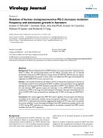
(See figure on next page.)
Fig. 1 Live animal IVIS imaging. (a) Bioluminescence signal from representative mice that received hBMSC-Luc/GFP (image time point was two
weeks after transplant). (b) Graphical representation of bioluminescence hBMSC-Luc/GFP signal overtime for animals that did or did not receive
PC-3-DsRed injections (8 mice with hBMSC-Luc/GFP (green), and 18 mice with hBMSC-Luc/GFP + PC-3-DsRed (red)). (c) Fluorescence signal from
PC-3-DsRed, minus background fluorescence, for select mice from each group at 4 weeks (14 mice with PC-3-DsRed and 18 mice with hBMSC-Luc/
GFP + PC-3-DsRed). (d) Graphical representation of PC-3-DsRed fluorescence signal from mice overtime after PC-3-DsRed injection. Pooled
experiments of three biological repeats. All IVIS images are found in Supplementary Figs. 5 and 7. Statistics were not significant between curves
after using linear-regression calculation and fit determined by Alkaines Information Criterion (AICc) or multiple t-tests with the Holm-Sidak method
(Supplementary Fig. 6).
Nowlan et al. BMC Cancer
(2022) 22:554
Fig. 1 (See legend on previous page.)
Page 5 of 13
Nowlan et al. BMC Cancer
(2022) 22:554
were injected into mice at 2 weeks post-hBMSC-Luc/GFP
transplant.
Analysis of IVIS images indicated no difference in
hBMSC-Luc/GFP bioluminescence signal between animals that received PC-3-DsRed or those that did not
(Fig. 1b and Supplementary Fig. 6a). In Supplementary
Fig. 6a, AICc fit-test was used to estimate the probability that a single curve fit bioluminescence data from mice
with or without PC-3-DsRed. This analysis suggested
that the presence of PC-3-DsRed cells did not influence
the growth of hBMSC-Luc/GFP in mice.
PC-3-DsRed fluorescence signal was also monitored
with IVIS (Fig. 1c-d, Supplementary Fig. 7). Signal was
variable between animals, likely due to the exponential
expansion of PC-3-DsRed in some animals, although
greater signal was derived from animals that had received
hBMSC-Luc/GFP. AICc fit-test was used to estimate the
probability that a single curve fit PC-3-DsRed fluorescence signal data from animals with or without hBMSCLuc/GFP, and this was found to be unlikely suggesting
that the presence of hBMSC-Luc/GFP did influence PC3-DsRed numbers (Linear regression, AiCc
= 55.06%,
Supplementary Fig. 6b).
Spatial quantification of hBMSC‑Luc/GFP and PC‑3‑DsRed
We used histology to identify and quantify hBMSC-Luc/
GFP within the femurs of mice at harvest. As previously reported [20], we detected the GFP+ cells in both
in the injected femurs and in the contralateral femurs,
indicating that hBMSC-Luc/GFP had disseminated
to other marrow cavities (Fig. 2a, b). Previous studies reported that intravenously transplanted hBMSC
home and engraft within the bone the marrow of mice
[26, 27]. Immediately following hBMSC-Luc/GFP transplant, a bioluminescence signal emanating from the
lungs could be seen in some mice, demonstrating that
detectable numbers of cells had escaped from the bone
marrow cavity into the general circulation, and we presume that some of these cells homed to distal bone marrow cavities. In histological sections of injected and
contralateral femurs, 6 week after initial transplant, the
difference between the hBMSC-Luc/GFP numbers in
these marrow cavities was insignificant (injected femur
Page 6 of 13
2.2 ± 0.5% versus contralateral femur 1.4 ± 1.4%, MannWhitney t-test, p = 0.1797). We did not detect a change
in cellularity of femurs that were injected with hBMSCLuc/GFP compared to either the contralateral femur or
femurs from mice that did not receive hBMSC at all (student t-test, p = 0.5898). This indicated that the hBMSC
transplant did not cause a detectable long-term impact
on marrow cellularity (Supplementary Fig. 8).
The number of PC-3-DsRed in each femur was quantified using flow cytometry. PC-3-DsRed were identified
as viable cells (7-AAD−), negative for mouse CD45, and
positive for DsRed (see Gating strategy in Supplementary Fig. 8). PC-3-DsRed were detected (higher than
0.01% of live C
D45− cells) in 1 out of 10 mice in the PC3-DsRed only group (12.5%), compared to 6 out of 11 in
the hBMSC-Luc/GFP + PC-3-DsRed group (54.5%). The
hBMSC-Luc/GFP + PC-3-DsRed group had an additional
mouse that had 5-fold greater PC-3-DsRed burden. This
animal was considered an outlier and excluded from subsequent analysis. hBMSC-Luc/GFP + PC-3-DsRed mice
had an overall higher PC-3-DsRed burden in femurs
(Fig. 2e, 0.018 ± 0.018% vs 0.002 ± 0.003%, Mann-Whitney t-test with a 95% confidence p = 0.0445, individual flow plots Supplementary Fig. 10). There was not a
greater frequency of PC3-DsRed in the specific humanized femur relative to the contralateral femur in the same
animal that had not been injected with hBMSC-Luc/
GFP (Fig. 2f, Mann-Whitney t-test, p = 0.5223). In summary, the presence of hBMSC-Luc/GFP in the animal
increased the frequency of PC-3-DsRed detected in the
femurs, but PC-3-DsRed cells did not specifically localize
in the femur where hBMSC-Luc/GFP had been initially
transplanted.
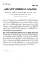
PC‑3‑DsRed tumor burden in the bone marrow and visceral
tissue
Tissue sections were stained with H&E. Regions containing PC-3-DsRed cells were selected for analysis in samples from mice injected with tumour cells. Characteristic
irregular cell morphology was visible in the bone marrow
(Fig. 3a, b, normal versus tumor-bearing) and in the liver
(Fig. 3c, d, normal versus tumor-bearing).
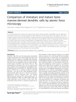
(See figure on next page.)
Fig. 2 Analysis of hBMSC and PC-3 by histology and flow cytometry. (a, b) Quantification of hBMSC-Luc/GFP in femur histology slices. (a) Histology
40x magnification image of marrow with anti-GFP (green) and DAPI (blue) to detect hBMSC-Luc/GFP. Scale bar = 20 μm. (b) Comparison of relative
hBMSC-Luc/GFP numbers in histology slices at 6 weeks (PC-3-DsRed n = 4, hBMSC-Luc/GFP + PC-3-DsRed n = 6). Flow cytometry quantification of
PC-3-DsRed numbers in (c) mouse contralateral and (d) injected femurs. Gating identified live singlet cells, which were negative for mouse CD45,
but positive for a DsRed signal (Supplementary Fig. 9). (e) Quantification of total PC-3-DsRed numbers taking the average of both femurs, in animals
that either did or did not receive hBMSC-Luc/GFP. Statistics determined by the Mann-Whitney t-test detected a significant difference (p = 0.0445) in
the number of PC-3-DsRed in animals that had been transplanted with hBMSC-LUC/GFP. (f) Comparison of the distribution of PC-3-DsRed between
femurs in individual mice femurs. Individual flow images are found in Supplementary Fig. 10. Mann Whitney t-test did not identify difference
between injected vs non-injected femur (PC-3-DsRed, p = 0.6589; hBMSC-Luc/GFP + PC-3-DsRed, p = 0.5223). Two flow experiments pooled, (no
cells n = 2, PC-3-DsRed only n = 8, hBMSC-Luc/GFP only n = 7, hBMSC-Luc/GFP + PC-3-DsRed n = 11).
Nowlan et al. BMC Cancer
(2022) 22:554
Fig. 2 (See legend on previous page.)
Page 7 of 13