schumann - dynamics of the bacterial chromosome - structure and function (wiley, 2006)
Bạn đang xem bản rút gọn của tài liệu. Xem và tải ngay bản đầy đủ của tài liệu tại đây (6.06 MB, 443 trang )
Wolfgang Schumann
Dynamics of the Bacterial
Chromosome
Dynamics of the Bacterial Chromosome. Structure and Function. W. Schumann
Copyright
g 2006 Wiley-VCH Verlag GmbH & Co. KGaA
ISBN 3-527-30496-7
Related Titles
Sensen, C. W. (ed.)
Handbook of Genome Research
Genomics, Proteomics, Metabolomics, Bioinformatics, Ethical and Legal Issues
2005
ISBN 3-527-31348-6
Dale, J. W., Park, S.
Molecular Genetics of Bacteria
2004
ISBN 0-470-85084-1
Reece, R. J.
Analysis of Genes and Genomes
2004
ISBN 0-470-84379-9
Gellissen, G. (ed.)
Hansenula polymorpha
Biology and Applications
2002
ISBN 3-527-30341-3
Wolfgang Schumann
Dynamics of the Bacterial
Chromosome
Structure and Function
The Author
Dr. Wolfgang Schumann
Institute of Genetics
University of Bayreuth
95440 Bayreuth
Germany
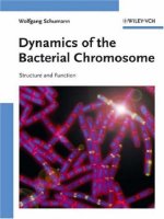
Cover illustration:
The picture shows DAPI (blue) and
FM4-64-stained (red) pre-divisional
sporangia of Bacillus subtilis of a
RacA-GFP-producing strain at h 1.5
of sporulation. RacA-GFP (green) is
seen as foci at the poles and as a haze
over the nucleoids. From the work of
S. Ben-Yehuda, D. Rudner and
R. Losick 2003, Science 299, 532.
All books published by Wiley-VCH are carefully
produced. Nevertheless, authors, editors, and
publisher do not warrant the information
contained in these books, including this book, to
be free of errors. Readers are advised to keep in
mind that statements, data, illustrations,
procedural details or other items may
inadvertently be inaccurate.
Library of Congress Card No.:
applied for
British Library Cataloguing-in-Publication
Data
A catalogue record for this book is available from
the British Library.
Bibliographic information published by
Die Deutsche Bibliothek
Die Deutsche Bibliothek lists this publication in
the Deutsche Nationalbibliografie; detailed biblio-
graphic data is available in the Internet at
<>.
g 2006 WILEY-VCH Verlag GmbH & Co. KGaA,
Weinheim
All rights reserved (including those of translation
into other languages). No part of this book may
be reproduced in any form ± by photoprinting,
microfilm, or any other means ± nor transmitted
or translated into a machine language without
written permission from the publishers. Regis-
tered names, trademarks, etc. used in this book,
even when not specifically marked as such, are
not to be considered unprotected by law.
Typesetting Fotosatz Detzner, Speyer
Printing Strauss GmbH, Mörlenbach
Bookbinding J. Schäffer GmbH, Grünstadt
Printed in the Federal Republic of Germany
Printed on acid-free paper
ISBN-13: 978-3-527-30496-7
ISBN-10: 3-527-30496-7
L
Contents
Foreword XI
Bacterial Species and their Abbreviation XIII
Color Plates XV
1 Structure of the Bacterial Cell 1
1.1 The Cytoplasm Compartment 1
1.2 The Cytoplasmic Membrane Compartment 3
1.3 The Cell Wall Compartment 8
1.4 The Outer Membrane Compartment 9
1.5 The Periplasmic Compartment 15
1.6 Extracellular Matrices 19
1.7 Appendages 21
2 Organization of the Bacterial Chromosome 29
2.1 Structure of the Chromosomes 29
2.2 Principles to Compact the Bacterial Chromosome 31
2.2.1 Superhelicity 31
2.2.2 Histone-like Proteins in E. coli 32
2.3 Organization of the Bacterial Chromosome into Genes and Repetitive
Sequences 36
2.3.1 Genes 36
2.3.2 Repetitive Sequences 37
2.4 Large Rearrangements Within the Chromosome 44
2.4.1 Duplications 44
2.4.2 Deletions 45
2.4.3 Inversions 49
3 The Bacterial Cell Cycle: Replication of the Chromosome,
Partitioning and Cell Division 51
3.1 Replication 51
3.1.1 Replication of Circular Chromosomes 51
V
Dynamics of the Bacterial Chromosome. Structure and Function. W. Schumann
Copyright
g 2006 Wiley-VCH Verlag GmbH & Co. KGaA
ISBN 3-527-30496-7
3.1.2 Replication of Linear Chromosomes and Plasmids 67
3.2 Partitioning (Segregation) of the Daughter Chromosomes 68
3.3 Cell Division 73
3.4 Plasmid- and Chromosome-encoded Toxin±Antitoxin Modules 79
3.4.1 Postsegregational Killing of Plasmid-free Cells 80
3.4.2 Chromosomal Toxin±Antitoxins 81
4 Recombination 83
4.1 Homologous Recombination 83
4.1.1 The RecBCD Pathway of Homologous Recombination 85
4.1.2 The RecF Pathway of Homologous Recombination 90
4.1.3 Additional Homologous Recombination Functions 91
4.1.4 Recovery of Replication at a Blocking DNA Lesion 92
4.2 Sequence-specific Recombination 97
4.2.1 Integration/Excision Systems 99
4.2.2 Resolvase Systems 100
4.2.3 Inversion Systems 103
4.2.4 Shufflons 107
4.2.5 Integrons 107
4.2.6 Homing Endonucleases 110
4.3 Illegitimate Recombination 112
4.3.1 Insertion Sequence Elements 113
4.3.2 Transposons 114
4.3.3 Transposing Bacteriophages 115
4.3.4 Mechanisms of Transposition 116
4.3.5 Conjugative and Integrative Elements 118
4.3.6 Nonhomologous Endjoining (NHEJ) 119
5 Origin of Mutations and Repair of DNA Lesions 121
5.1 Classes of Mutations 121
5.2 Origin of Mutations 124
5.2.1 Spontaneous Mutations 124
5.2.2 Induced Mutations 126
5.2.3 Detection Systems for Mutations 128
5.2.4 Adaptive Mutations 129
5.3 Repair of DNA Lesions 131
5.3.1 The Methyl-mediated Mismatch Repair System 132
5.3.2 Very Short Patch Repair 133
5.3.3 The Nucleotide Excision Repair Systems 134
5.3.4 Pathways of Base Excision Repair 136
5.3.5 Repair of Oxidized Nucleotides 138
5.3.6 Photoreactivation of Cyclobutane Dimers 140
5.3.7 Transcription-coupled Nucleotide Excision Repair 141
5.3.8 The Adaptive Response 141
5.3.9 The SOS Response 143
5.3.10 Replication and Repair 148
Contents
VI
5.3.11 Repair of Interstrand Crosslinks in DNA 150
5.3.12 Deinococcus radiodurans 151
6 Principles of Gene Regulation 153
6.1 Regulation at the Level of DNA 153
6.1.1 Alterations of the Structure of the DNA 154
6.1.2 GATC Methylation and Gene Expression 157
6.1.3 Programmed DNA Rearrangements Within the Chromosome 159
6.1.4 Transcriptional Silencing and Cryptic Genes 159
6.2 Regulation at the Level of Transcription 162
6.2.1 The DNA-dependent RNA Polymerase 162
6.2.2 Regions of the Promoter 168
6.2.3 Initiation of Transcription 170
6.2.4 Elongation of Transcription 173
6.2.5 Termination of Transcription 175
6.2.6 Anti-sigma Factors 177
6.2.7 Anti-termination/Attenuation of Transcription 179
6.2.8 Regulators of the DNA-dependent RNA Polymerase 188
6.3 Post-transcriptional Regulation 220
6.3.1 Protein-mediated Regulation 220
6.3.2 Riboregulation 222
6.3.3 Riboswitches 227
6.3.4 mRNA Decay 230
6.3.5 Introns 239
6.3.6 RNA-binding Proteins 242
6.4 Regulation at the Level of Translation 244
6.4.1 Initiation of Translation 247
6.4.2 Elongation of Translation 248
6.4.3 Termination of Translation 252
6.4.4 Ribosome Recycling 253
6.4.5 Translational Control 255
6.4.6 The 21. and 22. Amino Acid Selenocysteine and Pyrrolysine 258
6.4.7 Mechanisms to Create Multiple Gene Products From a Single
Cistron 259
6.5 Post-translational Regulation 266
6.5.1 Proteolysis 266
6.5.2 Protein Splicing 266
6.5.3 Protein Glycosylation 266
6.5.4 Circular Polypeptides 268
7 Protein Quality Control Through Molecular Chaperones, Protein Folding
Catalysts and ATP-dependent Proteases 271
7.1 Introduction 271
7.2 Molecular Chaperones 271
7.2.1 Folder Chaperones 272
VII
Contents
7.2.2 Holder Chaperones 279
7.2.3 Disaggregating Chaperones 287
7.2.4 Intramolecular Chaperones 289
7.2.5 Folding of Nascent Polypeptide Chains 290
7.3 Protein-folding Catalysts 292
7.3.1 Peptidyl-prolyl Isomerases 292
7.4 Disulfide Bond Formation 293
7.5 Proteases 297
7.5.1 ATP-dependent Proteases 298
7.5.2 ATP-independent Proteases 305
8 Secretion of Proteins 309
8.1 Introduction 309
8.2 Protein Sorting Signals for Subcellular Localization 310
8.3 Cytosolic Targeting Factors 315
8.4 Translocases Move Newly Made Proteins Into or Across
Membranes 316
8.4.1 The Sec Pathway of Protein Translocation Through the Inner
Membrane 317
8.4.2 The Tat Pathway of Secretion Proteins Through the Inner
Membrane 321
8.4.3 Insertion Proteins Into the Inner Membrane 323
8.5 Anchoring of Lipoproteins Into the Outer Membrane 324
8.6 Protein Translocation Pathways Through Both Membranes of
Gram-negative Bacteria 326
8.6.1 The Type I Secretion Pathway 326
8.6.2 The Type II Secretion Pathway 327
8.6.3 The Type III Secretion Pathway 330
8.6.4 The Type IV Secretion Pathway 333
8.6.5 Type V Secretion Pathway (Autotransporter) 335
8.6.6 Vesicle-mediated Export 336
8.7 Cell Wall-anchored Surface Proteins 337
8.8 S-Layers 339
9 Stress Genes and Their Regulation 341
9.1 The Stress Response Pathway 341
9.2 The Heat Shock Response 342
9.3 The Cold Shock Response 349
9.4 The Osmotic Shock Response 353
9.4.1 The Hyperosmotic Shock Response 354
9.4.2 The Hypoosmotic Shock Response 359
9.5 The Oxidative Shock Response 361
9.5.1 The OxyR Regulon 361
9.5.2 The SoxRS Regulon 362
9.6 pH-regulated Genes 364
Contents
VIII
9.6.1 Acid Stress 367
9.6.2 Alkaline Stress 369
9.7 Metabolic Stress: the Stringent Response 371
9.8 Nutrient Limitations (Starvation Stress) 373
9.9 Envelope Stress Response 376
10 Exchange of Genes: Transformation, Conjugation and
Transduction 381
10.1 Transformation 381
10.1.1 Transformation of Gram-positive Bacteria 384
10.1.2 Transformation of Gram-negative Bacteria 387
10.2 Conjugation 388
10.2.1 Gram-negative Bacteria 389
10.2.2 Gram-positive Bacteria 396
10.2.3 Conjugative Transposons 398
10.3 Transduction 398
10.3.1 Generalized Transduction 400
10.3.2 Specialized Transduction 401
10.4 Horizontal Gene Transfer 402
Glossary 405
Subject Index 413
Contents
IX
Foreword
Over the past 50 years, molecular microbiology has provided fundamental contri-
butions not only to the understanding of microbiology as a unique discipline but
also to the understanding of what makes a living cell. In 1945 Schrödinger in his
famous lecture
,,
What is life?
,,
addressed the challenging task of defining the
properties of a necessary (e.g. minimal/required) information-containing system.
More than half a century later, we can state that many aspects of this fundamental
question
,,
What is life?
,,
have been answered. This series of great discoveries
started with the elucidation of the DNA structure by F. Crick and J. Watson more
than 50 years ago and was subsequently expanded by the exploration of the basic
mechanisms of DNA replication and gene expression. These scientific achieve-
ments were complemented and fostered by the development of crucial techni-
ques, such as gene cloning, DNA sequencing and PCR. The discoveries culmi-
nated in an event that will certainly have a fixed position in future text books of
science history: the first complete elucidation of a genome sequence of a living or-
ganism, Haemophilus influenzae, in 1995. At the beginning of 2006, the complete
genomes of nearly 300 bacterial species are available. Knowledge of the complete
genome sequence is an essential prerequisite in order to gain a comprehensive
understanding of the molecular mechanisms of life. The genome sequence, how-
ever, only provides the
,,
blueprint
,,
of life, not life itself. Now, functional genomics
such as transcriptomics, proteomics, metabolomics and bioinformatics are re-
quired to bring the blueprint of life to the real life of living organisms. The combi-
nation of the expertise and accumulated knowledge in the traditional disciplines
such as microbiology, biochemistry, molecular genetics and molecular biology
with this panoramic view of the recent
,,
omics technologies
,,
on the cell as an
entity will enable/facilitate a new quality in the understanding of what makes a
cell viable. Because of their low complexity, single-cell bacterial systems such as
Escherichia coli, Bacillus subtilis and others constitute perfect model systems for
tackling such an ambitious goal as understanding life as an entity. The unexpected
finding that, even in the best analyzed model organisms such as Escherichia coli,
one-third of all genes codes for proteins with still unknown functions emphasizes
the challenge and indicates that many pages of the
,,
bible of life
,,
are still empty.
There is a comprehensive literature on the entire field of molecular microbiology,
from the structure, flexibility and stability of bacterial genomes to the various facets
of the regulation of gene expression, a field that even in bacteria includes mechan-
XI
Dynamics of the Bacterial Chromosome. Structure and Function. W. Schumann
Copyright
g 2006 Wiley-VCH Verlag GmbH & Co. KGaA
ISBN 3-527-30496-7
isms of not only transcriptional initiation but also transcriptional elongation and ter-
mination and regulation at the posttranscriptional, translational and posttranslational
level. Even if the first description of the regulation of a bacterial operon, the lac-oper-
on in E. coli by Jacob and Monod, was a milestone in the history of microbiology, ho-
nored by the Nobel Price in 1965, we had however to realize that this regulation me-
chanism represents only one of hundreds of mechanisms evolved during the three
billion years that bacteria have populated our planet. For those involved in teaching
students, it is difficult to keep up with the pace of the development of all these differ-
ent fields of molecular microbiology. A textbook covering all these aspects of molecu-
lar microbiology is equally useful for teachers and students in microbiology at univer-
sities, because this very genuine, innovative and essential new field of microbiology
needs more space in the teaching programs for life science students.
Wolfgang Schumann, professor for genetics at the University Bayreuth, accepted
the challenge of writing a textbook on molecular bacteriology. For many years, he
has lectured students of biology and biochemistry on the genetics and molecular
biology of bacteria. He is appreciated as an expert in molecular genetics and bacter-
ial gene regulation and his contributions to many fields of molecular genetics, e.g.
the mechanisms of heat induction in Gram-positive bacteria and the discovery of
the CIRCE element (to mention only some of them), are accepted worldwide.
I have always been impressed by his detailed knowledge of the scientific litera-
ture on quite different fields of molecular microbiology. This is one reason which
convinced me that Wolfgang Schumann is the right person to take over this ambi-
tious task. I have already used the chance to read some of the chapters of this book
in preparing my lectures at the University of Greifswald this year. I enjoyed this
reading, very conveniently providing me with an excellent survey. Assessing the
recent literature critically and in detail an essential part of the training of young
students has been done in an excellent way. All essential aspects of molecular
bacteriology (which might be extended by a similar textbook by other experts on
the molecular microbiology of Archaea or eukaryotic microorganisms) have been
addressed in a very authentic, comprehensive manner and have been illustrated
by a lot of impressive and useful figures.
This book covers the most essential chapters in molecular microbiology, start-
ing with the structure of the bacterial cell via the organization of bacterial chromo-
somes to the bacterial cell cycle, followed by chapters on molecular genetics, re-
combination and mutations and their repair. A comprehensive chapter deals with
the various mechanisms of gene regulation, followed by a description of the role
of chaperones in protein quality control and protein secretion mechanisms. An ex-
tra chapter on stress genes and their regulation is a reflection of the specific re-
search interests of the author. A final chapter on gene transfer makes the book
complete. I just missed a chapter on the prokaryotic development of bacteria, in-
cluding endospore-forming bacteria such as B. subtilis, heterocyst formation in
Anabaena and the dimorphic life cycle of Caulobacter crescentus, to mention some.
All in all, this new book offers an actual, comprehensive view of the present
state of the art in the field of molecular bacteriology. It is a very valuable source of
information not only for students, but also for scientists who wish to become ac-
quainted with one of the most exciting fields of current microbiology.
Greifswald, 04.04.2006 Michael Hecker
Foreword
XII
Bacterial Species and their Abbreviations
A. actinomycetemcomitans Actinobacillus actinomycetemcomitans
A. salmonicida Aeromonas salmonicida
A. tumefaciens Agrobacterium tumefaciens
A. vinelandii Azotobacter vinelandii
B. bronchiseptica Bordetella bronchiseptica
B. burgdorferi Borrelia burgdorferi
B. japonicum Bradyrhizobium japonicum
B. megaterium Bacillus megaterium
B. pertussis Bordetella pertussis
B. polymyxa Bacillus polymyxa
B. pseudomallei Burkholderia pseudomallei
B. hyodysenteriae Brachyspira hyodysenteriae
B. subtilis Bacillus subtilis
C. acetobutylicum Clostridium acetobutylicum
C. crescentus Caulobacter crescentus
C. difficile Clostridium difficile
C. diphtheriae Corynebacterium diphtheria
C. freundii Citrobacter freundii
C. jejuni Campylobacter jejuni
C. perfingens Clostridium perfringens
C. psittacii Chlamydia psittacii
C. trachomatis Chlamydia trachomatis
C. violaceum Chromobacterium violaceum
D. radiodurans Deinococcus radiodurans
E. amylovora Erwinia amylovora
E. carotovora Erwinia carotovora
E. cloacae Enterobacter cloacae
E. coli Escherichia coli
E. crysanthemie Erwinia crysanthemie
E. faecalis Enterococcus faecalis
H. ducreyi Haemophilus ducreyi
H. halobium Halobacterium halobium
H. influenzae Haemophilus influenzae
H. pylori Helicobacter pylori
H. salinarium Halobacterium salinarium
K. oxytoca Klebsiella oxytoca
XIII
Dynamics of the Bacterial Chromosome. Structure and Function. W. Schumann
Copyright
g 2006 Wiley-VCH Verlag GmbH & Co. KGaA
ISBN 3-527-30496-7
K. pneumoniae Klebsiella pneumoniae
L. lactis Lactococcus lactis
L. monocytogenes Listeria monocytogenes
M. genitalium Mycoplasma genitalium
M. janashii Methanococcus janashii
M. leprae Mycobacterium leprae
M. loti Mesorhizobium loti
M. pulmonis Mycoplasma pulmonis
M. tuberculosis Mycobacterium tuberculosis
M. xanthus Myxococcos xanthus
N. gonorrhoeae Neisseria gonorrhoeae
N. meningitis Neisseria meningitis
P. aeruginosa Pseudomonas aeruginosa
P. fluorescens Pseudomonas fluorescens
P. gingivalis Porphyromonas gingivalis
P. putida Pseudomonas putida
P. syringae Pseudomonas syringae
R. capsulatus Rhodobacter capsulatus
R. etli Rhizobium etli
R. solanacearum Ralstonia solanacearum
S. agalactiae Streptococcus agalactiae
S. albus Streptomyces albus
S. ambofaciens Streptomyces ambofaciens
S. aurantiaca Stigmatella aurantiaca
S. aureus Staphylococcus aureus
S. avermitilis Streptomyces avermitilis
S. coelicor Streptomyces coelicor
S. enterica Salmonella enterica
S. flexneri Shigella flexneri
S. hygroscopius Streptomyces hygroscopius
S. meliloti Sinrhizobium meliloti
S. pneumoniae Streptococcus pneumoniae
S. pyogenes Streptococcus pyogenes
S. sonnei Shigella sonnei
S. tsukubaensis Streptomyces tsukubaensis
S. typhi Salmonella typhi
S. typhimurium Salmonella typhimurium
T. aquaticus Thermus aquaticus
T. maritima Thermotoga maritima
T. thermophilus Thermus thermophilus
V. alginolyticus Vibrio alginolyticus
V. cholerae Vibrio cholerae
V. fischeri Vibrio fischeri
V. harveyi Vibrio harveyi
X. campestris Xanthomonas campestris
Y. enterocolitica Yersinia enterocolitica
Y. pestis Yersinia pestis
Bacterial Species and their Abbreviations
XIV
Color Plates
XV
Dynamics of the Bacterial Chromosome. Structure and Function. W. Schumann
Copyright
g 2006 Wiley-VCH Verlag GmbH & Co. KGaA
ISBN 3-527-30496-7
Fig. 1.2 Structure of the OmpF porin. View of the monomer from the side
(left) and view of the trimer from the top. The loop inside the opening narrows
the channel. H. Nikaido 2003, Microbiol. Mol. Biol. Rev. 67, 593±556; Fig. 2.
Color Plates
XVI
Fig. 1.3 Shuttle model for TonB-dependent
energy transduction. Uncharged TonB (1) is
energized by uptake of a proton (2) and shut-
tles to the outer membrane where is docks to
OmpA/Lpp waiting for a transporter such as
FepA with a bound ligand. Upon interaction
with the Ton box of the the transporter the
conformational energy is transduced to the
transporter (3) triggering uptake of the ligand
into the periplasm (4). The discharged TonB
shuttles back to the ExbBD complex to be-
come recharged. K. Postle, R.J. Kadner 2003,
Mol. Microbiol. 49, 869±882; Fig. 2.
Fig. 3.1 Organization of the oriC region of E. coli. The oriC region of E. coli
consists of five DnaA boxes called R1 through R4 and M, binding sites for the
histone-like proteins FIS and IHF, three AT-rich regions and 11 GATC methyla-
tion sites. W. Messer 2002, FEMS Microbiol. Rev. 26, 355; Fig. 1.
Color Plates
XVII
Fig. 3.10 Schematic representation of the toxin±
antitoxin mechanisms. The genes coding for the
toxin and antitoxin form a bicistronic operon. The
toxin (red) and antitoxin (blue) proteins form a tight
complex thereby keeping the toxin inactive. The anti-
toxin is more rapidly degraded by an ATP-dependent
protease (green) than the toxin, but the latter is con-
tinually bound by newly synthesized antitoxin. Cells
carrying the plasmid tolerate the TA complex (right),
but daughter cells without the plasmid (left) are
unable to replenish the antitoxin. The released toxin
attacks an intracellular target to cause cell death or
growth arrest. In this scheme, the toxin attacks the
chromosomal DNA. F. Hayes 2003, Science 301,
1496; Fig. 1.
Color Plates
XVIII
Fig. 5.2 Methyl-mediated mismatch repair.
MutS recognizes and binds to the mismatch
followed by binding of ATP. This complex re-
cruits MutL which recognizes the nearest
hemimethylated GATC and binds MutH.
MutH cuts 5¢ or 3¢ to the unmodified GATC
sequence, UvrD binds to the nick and un-
winds the double-strand DNA followed by re-
synthesis and ligation. T.A. Kunkel, D.A. Erie
2005, Annu. Rev. Biochem. 74, 681; Fig. 2C.
Fig. 6.3 Turning pap pilus synthesis on and off. See text for explanation who a
cell can shift from the OFF- to the ON-state and vice versa. A. Lùbner-Olesen,
et al. 2005, Curr. Opin. Microbiol. 8, 154±160.
Color Plates
XIX
Fig. 6.27 Signalling pathways in bacterial che-
motaxis. Dimeric MCPs form a-helical coiled-
coil structures spanning the inner membrane.
Chemical compounds (attractants and repel-
lents) enter the periplasm through porins and
interact either directly with the periplasmic
domain of the appropriate chemoreceptor or
indirectly through a periplasmatic binding pro-
tein (PBP; MBP, RBP, GBP and DPP are mal-
tose, ribose, galactose and dipeptide binding
protein, respectively). The CheW coupling mo-
lecule transduces the signals to the CheA sen-
sor kinase which, after trans-phosphorylation,
transfers the phosphate first to CheY and then
to methylesterase CheB. CheY~P interacts with
the flagellar motor to bring about a change in
the direction. CheB~P competes with a consti-
tutive methyltransferase, CheR, to control the
degree of methylation of specific glutamates in
the MCPs. This resets the signaling state of the
chemoreceptors and allows them to adapt to
the present concentration of attractant and to
sense subsequent changes. Dephosphorylation
of CheY~P is accelerated by the phosphatese
CheZ. G.H. Wadhams 2004, Nat. Rev. Mol. Cell
Biol. 5, 1024±1037; Fig. 2.
Color Plates
XX
Fig. 6.37 Proposed structure of the CsrB
RNA. This structure contains 18 repeated
structures numbered 1 to 18 which may
facilitate binding of CsrA. T. Romeo 1998,
Mol. Microbiol. 29, 1321±1330; Fig. 1.
Color Plates
XXI
Fig. 6.38 The B. subtilis gcvT riboswitch. The
intergenic region between yqhH and the
gcvPA-gcvPB operon designated gcvT acts as a
riboswitch and contains the two glycine sen-
sing aptamers I and II. If the glycine concen-
tration in the cell is low, the two aptamers re-
main unoccupied favoring formation of the
terminator structure resulting in transcription
attenuation. If the glycine concentration is
high, it will bind to the two aptamers prevent-
ing formation of the transcription terminator,
and the two structural genes will be ex-
pressed. M. Mandal, et al. 2004, Science 306,
275; Fig. 4, modified.
Color Plates
XXII
Fig. 7.1 Domain organization of the DnaK
chaperone. DnaK consists of an N-terminal
ATPase and a C-terminal peptide-binding
domain. The ATPase domain consists of two
lobes which form a deep cleft serving as a
binding pocket for ATP. The peptide-binding
domain (here shown with a heptapeptide
substrate) forms a â sandwich composed of
two sheets of four strands each, followed by
a helices spanning back over the sandwich.
Further indicated by open arrows are inter-
action sites for GrpE and DnaJ. F.U. Hartl,
M. Hayer-Hartl 2002, Science 295, 1852±1858;
Fig. 3A.
Fig. 7.2 Reaction cycle of the DnaK chaper-
one machine. DnaK-ATP with the open lid
constitutes the active form able to accept non-
native substrate polypeptide chains. These are
either targeted to DnaK by DnaJ (as shown
here) or bind directly to the peptide-binding
domain of DnaK. The initial binding of the
substrate protein is characterized by low affi-
nity allowing fast dissociation. Binding of
DnaJ to DnaK stimulates its ATPase activity
resulting in the release of inorganic phosphate
followed by closing of the lid, as shown in the
cartoon. Now, the substrate protein is trapped
and can start to refold. Later, GrpE binds to
the complex, stimulates release of ADP fol-
lowed by opening of the lid with concomitant
dissociation of the substrate protein. After
binding of ATP, a new reaction cycle can be
started. F.U. Hartl, M. Hayer-Hartl 2002,
Science 295, 1852±1858; Fig. 3B.
Color Plates
XXIII
Fig. 7.3 The GroE chaperonin complex. (a) The three functional domains of the
GroEL monomer. (b) Structure of two GroEL rings (14-mer) with three subunits
shown at the front. (c) The GroEL-ADP-GroES complex with one GroEL and
GroES subunit each. H.R. Saibil, N.A. Ranson 2002, Trends Biochem. Sci. 27,
627±632; Fig. 2.
Fig. 7.4 The GroEL-GroES reaction cycle.
(A) A substrate protein diffuses into the upper
ring where it will bind to hydrophobic side chains
exposed on the inner surface of the apical do-
main. (B) Next, each equatorial domain of the
upper ring binds one molecule of ATP with posi-
tive cooperativity. (C) This in turn triggers bind-
ing of a GroES ring to the upper ring causing an
upward movement of the apical domain, retrac-
tion of the hydrophobic side-chains and release
of the substrate protein into the cavity of the
upper GroEL ring where it starts to fold. (D) ATP
is hydrolyzed and (E) another substrate molecule
diffuses into the lower ring, followed by binding
of ATP. (F) GroES diffuses away from the upper
ring, followed by the substrate protein; and the
substrate molecule bound to the lower ring gets
encapsulated. H.R. Saibil, N.A. Ranson 2002,
Trends Biochem. Sci. 27, 627±632; Fig. 1.
Color Plates
XXIV
Fig. 7.8 Model of Hsp33 activation. Under
reducing conditions, Hsp33 occurs in a mono-
meric form and four cysteine residues are in-
volved in zinc coordination. After addition of
H
2
O
2
, zinc is released and two intramolecular
disulfide bonds are formed. Next, two oxi-
dized Hsp33 monomers form a highly active
dimer. P.C.F. Graf, U. Jacob 2002, Cell. Mol.
Life Sci. 59, 1624±1631; Fig. 1.
Color Plates
XXV
Fig. 7.9 Biogenesis of the P pilus. (i) The
components of the P pilus and those involved
in its biogenesis are synthesized in the cyto-
plasm and secreted into the periplasm using
the Sec pathway. (ii) If components of the P
pilus do not interact with the PapD chaperone,
they fold inappropriately and are degraded by
periplasmic proteases. (iii) Most P pilus com-
ponents are bound by the PapD chaperone
and guided to the PapC protein, an integral
outer membrane protein acting as an usher
(iv). (v) PapC monitors the correct assembly
of the different components which form the
active P pilus (vi). F.G Sauer, et al. 2000, Curr.
Opin. Struct. Biol. 10, 548±556; Fig. 1.