molecular analysis of cancer - carrie fidler
Bạn đang xem bản rút gọn của tài liệu. Xem và tải ngay bản đầy đủ của tài liệu tại đây (2.54 MB, 302 trang )
M E T H O D S I N M O L E C U L A R M E D I C I N E
TM
Edited by
Jacqueline Boultwood
Carrie Fidler
Molecular
Analysis of
Cancer
Humana Press
Humana Press
Edited by
Jacqueline Boultwood
Carrie Fidler
Molecular
Analysis of
Cancer
Molecular Analysis of Cancer
M E T H O D S I N M O L E C U L A R M E D I C I N E
TM
John M. Walker, Series Editor
70. Cystic Fibrosis Methods and Protocols,
edited by William R. Skach, 2002
69. Gene Therapy Protocols, 2nd ed., edited
by Jeffrey R. Morgan, 2002
68. Molecular Analysis of Cancer, edited by
Jacqueline Boultwood and Carrie Fidler, 2002
67. Meningococcal Disease: Methods and
Protocols, edited by Andrew J. Pollard and
Martin C. J. Maiden, 2001
66. Meningococcal Vaccines: Methods and
Protocols, edited by Andrew J. Pollard and
Martin C. J. Maiden, 2001
65. Nonviral Vectors for Gene Therapy:
Methods and Protocols, edited by Mark A.
Findeis, 2001
64. Dendritic Cell Protocols, edited by Stephen
P. Robinson and Andrew J. Stagg, 2001
63. Hematopoietic Stem Cell Protocols,
edited by Christopher A. Klug and Craig T.
Jordan, 2001
62. Parkinson’s Disease: Methods and Protocols,
edited by M. Maral Mouradian, 2001
61. Melanoma Techniques and Protocols:
Molecular Diagnosis, Treatment, and
Monitoring, edited by Brian J. Nickoloff, 2001
60. Interleukin Protocols, edited by Luke A. J.
O’Neill and Andrew Bowie, 2001
59. Molecular Pathology of the Prions, edited
by Harry F. Baker, 2001
58. Metastasis Research Protocols: Volume 2,
Cell Behavior In Vitro and In Vivo, edited
by Susan A. Brooks and Udo Schumacher,
2001
57. Metastasis Research Protocols: Volume 1,
Analysis of Cells and Tissues, edited by Susan
A. Brooks and Udo Schumacher, 2001
56. Human Airway Inflammation: Sampling
Techniques and Analytical Protocols, edited
by Duncan F. Rogers and Louise E. Donnelly,
2001
55. Hematologic Malignancies: Methods and
Protocols, edited by Guy B. Faguet, 2001
54. Mycobacterium tuberculosis Protocols, edited
by Tanya Parish and Neil G. Stoker, 2001
53. Renal Cancer: Methods and Protocols,
edited by Jack H. Mydlo, 2001
52. Atherosclerosis: Experimental Methods and
Protocols, edited by Angela F. Drew, 2001
51. Angiotensin Protocols, edited by Donna
H. Wang, 2001
50. Colorectal Cancer: Methods and Protocols,
edited by Steven M. Powell, 2001
49. Molecular Pathology Protocols, edited by
Anthony A. Killeen, 2001
48. Antibiotic Resistance Methods and
Protocols, edited by Stephen H. Gillespie,
2001
47. Vision Research Protocols, edited by P.
Elizabeth Rakoczy, 2001
46. Angiogenesis Protocols, edited by J.
Clifford Murray, 2001
45. Hepatocellular Carcinoma: Methods and
Protocols, edited by Nagy A. Habib, 2000
44. Asthma: Mechanisms and Protocols, edited by
K. Fan Chung and Ian Adcock, 2001
43. Muscular Dystrophy: Methods and
Protocols, edited by Katherine B. Bushby
and Louise Anderson, 2001
42. Vaccine Adjuvants: Preparation Methods
and Research Protocols, edited by Derek T.
O’Hagan, 2000
41. Celiac Disease: Methods and Protocols,
edited by Michael N. Marsh, 2000
40. Diagnostic and Therapeutic Antibodies,
edited by Andrew J. T. George and
Catherine E. Urch, 2000
39. Ovarian Cancer: Methods and Protocols, edited
by John M. S. Bartlett, 2000
38. Aging Methods and Protocols, edited by
Yvonne A. Barnett and Christopher R.
Barnett, 2000
37. Electrochemotherapy, Electrogenetherapy,
and Transdermal Drug Delivery:
Electrically Mediated Delivery of Molecules to
Cells, edited by Mark J. Jaroszeski, Richard
Heller, and Richard Gilbert, 2000
36. Septic Shock Methods and Protocols,
edited by Thomas J. Evans, 2000
Humana Press Totowa, New Jersey
M e t h o d s i n M o l e c u l a r M e d i c i n e
TM
Molecular Analysis
of Cancer
Edited by
Jacqueline Boultwood
and
Carrie Fidler
Leukaemia Research Fund Molecular Haematology Unit,
University of Oxford, NDCLS, John Radcliffe Hospital, Oxford, UK
© 2002 Humana Press Inc.
999 Riverview Drive, Suite 208
Totowa, New Jersey 07512
www.humanapress.com
All rights reserved. No part of this book may be reproduced, stored in a retrieval system, or transmitted in
any form or by any means, electronic, mechanical, photocopying, microfilming, recording, or otherwise
without written permission from the Publisher. Methods in Molecular Medicine
™
is a trademark of The
Humana Press Inc.
The content and opinions expressed in this book are the sole work of the authors and editors, who have
warranted due diligence in the creation and issuance of their work. The publisher, editors, and authors are
not responsible for errors or omissions or for any consequences arising from the information or opinions
presented in this book and make no warranty, express or implied, with respect to its contents.
This publication is printed on acid-free paper. '
ANSI Z39.48-1984 (American Standards Institute) Permanence of Paper for Printed Library Materials.
Cover Illustration: Fig. 1A from Chapter 3: “Spectral Karyotyping in Cancer Cytogenetics” by E. Hilgenfeld, et al.
Production Editor: Jessica Jannicelli.
Cover design by Patricia F. Cleary.
For additional copies, pricing for bulk purchases, and/or information about other Humana titles, contact
Humana at the above address or at any of the following numbers: Tel.: 973-256-1699; Fax: 973-256-8341;
E-mail: ; Website: humanapress.com
Photocopy Authorization Policy:
Authorization to photocopy items for internal or personal use, or the internal or personal use of specific
clients, is granted by Humana Press Inc., provided that the base fee of US $10.00 per copy, plus US $00.25
per page, is paid directly to the Copyright Clearance Center at 222 Rosewood Drive, Danvers, MA 01923.
For those organizations that have been granted a photocopy license from the CCC, a separate system of
payment has been arranged and is acceptable to Humana Press Inc. The fee code for users of the Transactional
Reporting Service is: [0-89603-622-7/02 $10.00 + $00.25].
Printed in the United States of America. 10 9 8 7 6 5 4 3 2 1
Library of Congress Cataloging in Publication Data
Main entry under title:
Methods in molecular medicine
™
.
Molecular analysis of cancer/edited by Jacqueline Boultwood and Carrie Fidler.
p. ; cm. (Methods in molecular medicine ; 68)
Includes bibliographical references and index.
ISBN 0-89603-622-7 (alk. paper)
1. Cancer Genetic aspects Research Methodology. 2. Cancer Molecular
aspects Research Methodology. I. Boultwood, Jacqueline. II. Fidler, Carrie. III. Series.
[DNLM: 1. Neoplasms genetics. 2. Cell Transformation, Neoplastic genetics. 3.
Gene Expression Regulation, Neoplastic. 4. Genetic Techniques. QZ 200 M7175 2001]
RC268.4 .M627 2001
616.99'4042 dc21
2001024306
v
Over the past 20 years, technological advances in molecular biology have
proven invaluable to the understanding of the pathogenesis of human cancer.
The application of molecular technology to the study of cancer has not only
led to advances in tumor diagnosis, but has also provided markers for the
assessment of prognosis and disease progression. The aim of Molecular Analy-
sis of Cancer is to provide a comprehensive collection of the most up-to-date
techniques for the detection of molecular changes in human cancer. Leading
researchers in the field have contributed chapters detailing practical proce-
dures for a wide range of state-of-the-art techniques.
Molecular Analysis of Cancer includes chapters describing techniques
for the identification of chromosomal abnormalities and comprising: fluores-
cent in situ hybridization (FISH), spectral karyotyping (SKY), comparative
genomic hybridization (CGH), and microsatellite analysis. FISH has a promi-
nent role in the molecular analysis of cancer and can be used for the detection
of numerical and structural chromosomal abnormalities. The recently described
SKY, in which all human metaphase chromosomes are visualized in specific
colors, allows for the definition of all chromosomal rearrangements and marker
chromosomes in a tumor cell. Protocols for the detection of chromosomal rear-
rangements by PCR and RT-PCR are described, as well as the technique of
DNA fingerprinting, a powerful tool for studying somatic genetic alterations
in tumorigenesis. A number of approaches to identify mutations are detailed,
and include SSCP, DGGE, the nonisotopic RNase cleavage assay, the protein
truncation assay, and DNA sequencing. A change in DNA methylation status
is commonly observed in cancer, and specific methodology for methylation
analysis is also provided by this volume.
The analysis of gene expression represents a key area of research in the
study of human cancer and a number of chapters in Molecular Analysis of
Cancer address this subject. Global RNA expression analysis using microarray
technology allows the identification of genes that are differentially expressed
in tumor versus normal tissues. This is a powerful approach for identifying
genes that are central to disease development or progression and can also iden-
tify new prognostic markers.
Preface
vi Preface
A reduction in telomere length, together with expression of the telomere
maintenance enzyme, telomerase, has been described in a wide range of
human cancers. To complete the volume, we include chapters describing the
measurement of telomere length and telomerase levels, an area of extensive
study in the field of cancer research.
We wish to thank the authors of the various chapters of Molecular Analysis
of Cancer for their excellent contributions. Clearly, they share our hope that
this volume will assist other researchers in the analysis and detection of
genetic abnormalities occurring in human malignancy, and lead to a better
understanding of the molecular pathogenesis of cancer.
Jackie Boultwood
Carrie Fidler
Contents
Preface v
Contributors ix
1Molecular Analysis of Cancer:
An Overview
Ken Mills 1
2Detection of Chromosome Abnormalities in Leukemia Using
Fluorescence
In Situ
Hybridization
Lyndal Kearney, Sabrina Tosi, and Rina J. Jaju 7
3 Spectral Karyotyping in Cancer Cytogenetics
Eva Hilgenfeld, Cristina Montagna, Hesed Padilla-Nash,
Linda Stapleton, Kerstin Heselmeyer-Haddad,
and Thomas Ried 29
4Comparative Genomic Hybridization Analysis
Binaifer R. Balsara, Jianming Pei, and Joseph R. Testa 45
5Detection of Chromosomal Deletions by Microsatellite Analysis
Rachel E. Ibbotson and Martin M. Corcoran 59
6Detection and Quantification of Leukemia-Specific Rearrangements
Andreas Hochhaus 67
7Detection of t(2;5)(p23;q35) Translocation by Long-Range PCR
of Genomic DNA
Yunfang Jiang, L. Jeffrey Medeiros, and Andreas H. Sarris 97
8Use of DNA Fingerprinting to Detect Genetic Rearrangements
in Human Cancer
Vorapan Sirivatanauksorn, Yongyut Sirivatanauksorn,
Arthur B. McKie, and Nicholas R. Lemoine 107
9Mutation Analysis of Large Genomic Regions in Tumor DNA Using
Single-Strand Conformation Polymorphism:
Lessons from
the
ATM
Gene
Igor Vorechovsky 115
10 Mutational Analysis of Oncogenes and Tumor Suppressor Genes
in Human Cancer Using Denaturing Gradient Gel Electrophoresis
Per Guldberg, Kirsten Grønbæk, Jesper Worm, Per thor Straten,
and Jesper Zeuthen 125
vii
viii Contents
11 Detection of Mutations in Human Cancer Using Nonisotopic
RNase Cleavage Assay
Marianna Goldrick and James Prescott 141
12 Mutational Analysis of the Neurofibromatosis Type 1 Gene
in Childhood Myelodysplastic Syndromes Using a Protein
Truncation Assay
Lucy Side 157
13 Mutation Analysis of Cancer Using Automated Sequencing
Amanda Strickson and Carrie Fidler 171
14 Detection of Differentially Expressed Genes in Cancer Using
Differential Display
Yineng Fu 179
15 Genomewide Gene Expression Analysis Using cDNA Microarrays
Chuang Fong Kong and David Bowtell 195
16 Gene Expression Profiling in Cancer Using cDNA Microarrays
Javed Khan, Lao H. Saal, Michael L. Bittner, Yuan Jiang,
Gerald C. Gooden, Arthur A. Glatfelter, and Paul S. Meltzer 205
17 Wilms Tumor Gene
WT1
as a Tumor Marker for Leukemic Blast Cells
and Its Role in Leukemogenesis
Haruo Sugiyama 223
18 Detection of Aberrant Methylation of the
p15
INK4B
Gene Promoter
Toshiki Uchida 239
19 Clonality Studies in Cancer Based on X Chromosome Inactivation
Phenomenon
John T. Phelan II and Josef T. Prchal 251
20 Telomere Length Changes in Human Cancer
Dominique Broccoli and Andrew K. Godwin 271
21 Measurement of Telomerase Activity in Human Hematopoietic Cells
and Neoplastic Disorders
Kazuma Ohyashiki and Junko H. Ohyashiki 279
Index
301
Contributors
BINAIFER R. BALSARA • Human Genetics Program, Division of Population
Sciences, Fox Chase Cancer Center, Philadelphia, PA
M
ICHAEL L. BITTNER • Cancer Genetics Branch, National Human Genome
Research Institute, National Institutes of Health, Bethesda, MD
J
ACQUELINE BOULTWOOD • Leukaemia Research Fund Molecular Haematology
Unit, University of Oxford, NDCLS, John Radcliffe Hospital, Oxford, UK
D
AVID BOWTELL • Research Division, Peter MacCallum Cancer Institute,
Melbourne, Australia
D
OMINIQUE BROCCOLI • Medical Sciences Division, Department of Medical
Oncology, Fox Chase Cancer Center, Philadelphia, PA
M
ARTIN M. CORCORAN • Molecular Biology Laboratory, Royal Bournemouth
Hospital, Bournemouth, UK
C
ARRIE FIDLER • Leukaemia Research Fund Molecular Haematology Unit at
the University of Oxford, NDCLS, John Radcliffe Hospital, Oxford, UK
Y
INENG FU • Department of Pathology, Beth Israel-Deaconess Medical Center
and Harvard Medical School, Boston; and Department of Pathology,
Ardais Corporation, Lexington, MA
A
RTHUR A. GLATFELTER • Cancer Genetics Branch, National Human Genome
Research Institute, National Institutes of Health, Bethesda, MD
A
NDREW K. GODWIN • Medical Sciences Division, Department of Medical
Oncology, Fox Chase Cancer Center, Philadelphia, PA
M
ARIANNA GOLDRICK • Ambion RNA Diagnostics, Austin, TX
G
ERALD C. GOODEN • Cancer Genetics Branch, National Human Genome
Research Institute, National Institutes of Health, Bethesda, MD
K
IRSTEN GRØNBÆK • Department of Tumour Cell Biology, Institute of Cancer
Biology, Danish Cancer Society, Copenhagen, Denmark
P
ER GULDBERG • Department of Tumour Cell Biology, Institute of Cancer
Biology, Danish Cancer Society, Copenhagen, Denmark
K
ERSTIN HESELMEYER-HADDAD • Genetics Department, Center for Cancer
Research, National Cancer Institute, National Institutes of Health,
Bethesda, MD
E
VA HILGENFELD • Genetics Department, Center for Cancer Research,
National Cancer Institute, National Institutes of Health, Bethesda, MD
ix
x Contributors
ANDREAS HOCHHAUS • III. Medizinische Universitätsklinik, Klinikum
Mannheim der Universität Heidelberg, Mannheim, Germany
R
ACHEL E. IBBOTSON • Molecular Biology Laboratory, Royal Bournemouth
Hospital, Bournemouth, UK
R
INA J. JAJU • Leukaemia Research Fund Molecular Haematology Unit,
University of Oxford, NDCLS, John Radcliffe Hospital, Oxford, UK
Y
UAN JIANG • Cancer Genetics Branch, National Human Genome Research
Institute, National Institutes of Health, Bethesda, MD
Y
UNFANG JIANG • Laboratory of Lymphoma Biology, Department of
Lymphoma and Myeloma, University of Texas MD Anderson Cancer
Center, Houston, TX
L
YNDAL KEARNEY • MRC Molecular Haematology Unit, Weatherall Institute
of Molecular Medicine, Oxford, UK
J
AVED KHAN • Oncogenomics Section, Pediatric Oncology Branch, Center
for Cancer Research, National Cancer Institute, National Institutes of
Health, Bethesda, MD
C
HUANG FONG KONG • Research Division, Peter MacCallum Cancer Institute,
Melbourne, Australia
N
ICHOLAS R. LEMOINE • Imperial Cancer Research Fund Oncology Unit,
Imperial College School of Medicine, Hammersmith Hospital, London, UK
A
RTHUR B. MCKIE • Imperial Cancer Research Fund Oncology Unit, Imperial
College School of Medicine, Hammersmith Hospital, London, UK
L. J
EFFREY MEDEIROS • Department of Hematopathology, University of Texas
MD Anderson Cancer Center, Houston, TX
P
AUL S. MELTZER • Cancer Genetics Branch, National Human Genome
Research Institute, National Institutes of Health, Bethesda, MD
K
EN MILLS • Department of Haematology, University of Wales College of
Medicine, Heath Park, Cardiff, Wales, UK
C
RISTINA MONTAGNA • Genetics Department, Center for Cancer Research,
National Cancer Institute, National Institutes of Health, Bethesda, MD
K
AZUMA OHYASHIKI • First Department of Internal Medicine, Tokyo Medical
University, Tokyo, Japan
J
UNKO H. OHYASHIKI • First Department of Internal Medicine, Tokyo
Medical University, Tokyo, Japan and the Division of Virology, Medical
Research Institute, Tokyo Medical and Dental University, Tokyo, Japan
H
ESED PADILLA-NASH • Genetics Department, Center for Cancer Research,
National Cancer Institute, National Institutes of Health, Bethesda, MD
J
IANMING PEI • Human Genetics Program, Division of Population Sciences,
Fox Chase Cancer Center, Philadelphia, PA
J
OHN T. PHELAN II • Rochester General Hospital, Rochester, NY
Contributors xi
JOSEF T. PRCHAL • Department of Medicine Hematology and Oncology,
Baylor College of Medicine, Houston, TX
J
AMES PRESCOTT • UroCor, Inc., Oklahoma City, OK
THOMAS RIED • Genetics Department, Center for Cancer Research, National
Cancer Institute, National Institutes of Health, Bethesda, MD
L
AO H. SAAL • Cancer Genetics Branch, National Human Genome Research
Institute, National Institutes of Health, Bethesda, MD
A
NDREAS H. SARRIS • Laboratory of Lymphoma Biology, Department of
Lymphoma and Myeloma, University of Texas MD Anderson Cancer
Center, Houston, TX
L
UCY SIDE • Leukaemia Research Fund Molecular Haematology Unit at the
University of Oxford, NDCLS, John Radcliffe Hospital, Oxford, UK
V
ORAPAN SIRIVATANAUKSORN • Imperial Cancer Research Fund Oncology
Unit, Imperial College School of Medicine, Hammersmith Hospital,
London, UK
Y
ONGYUT SIRIVATANAUKSORN • Department of Surgery, Anaesthetics and
Intensive Care, Imperial College School of Medicine, Hammersmith
Hospital, London, UK
L
INDA STAPLETON • Genetics Department, Center for Cancer Research,
National Cancer Institute, National Institutes of Health, Bethesda, MD
A
MANDA STRICKSON • Leukaemia Research Fund Molecular Haematology
Unit at the University of Oxford, NDCLS, John Radcliffe Hospital,
Oxford, UK
P
ER THOR STRATEN • Department of Tumour Cell Biology, Institute of Cancer
Biology, Danish Cancer Society, Copenhagen, Denmark
H
ARUO SUGIYAMA • Department of Clinical Laboratory Science, Osaka
University Medical School, Yamada-Oka, Suita City
J
OSEPH R. TESTA • Human Genetics Program, Division of Population
Sciences, Fox Chase Cancer Center, Philadelphia, PA
S
ABRINA TOSI • MRC Molecular Haematology Unit, Weatherall Institute of
Molecular Medicine, Oxford, UK
T
OSHIKI UCHIDA • First Department of Internal Medicine, Nagoya University
School of Medicine, Showa-ku, Nagoya, Japan
I
GOR VORECHOVSKY • Department of Biosciences at NOVUM, Karolinska
Institute, Huddinge, Sweden
JESPER WORM • Department of Tumour Cell Biology, Institute of Cancer
Biology, Danish Cancer Society, Copenhagen, Denmark
J
ESPER ZEUTHEN • Department of Tumour Cell Biology, Institute of Cancer
Biology, Danish Cancer Society, Copenhagen, Denmark
Overview of Molecular Cancer Genetics 1
1
From:
Methods in Molecular Medicine, vol. 68: Molecular Analysis of Cancer
Edited by: J. Boultwood and C. Fidler © Humana Press Inc., Totowa, NJ
1
Molecular Analysis of Cancer
An Overview
Ken Mills
1. Introduction
Cancer is a complex disease occurring as a result of a progressive accumula-
tion of genetic aberrations and epigenetic changes that enable escape from nor-
mal cellular and environmental controls (1). Neoplastic cells may have
numerous acquired genetic abnormalities including aneuploidy, chromosomal
rearrangements, amplifications, deletions, gene rearrangements, and loss-of-
function or gain-of-function mutations. Recent studies have also highlighted
the importance of epigenetic alterations of certain genes that result in the
inactivation of their functions in some human cancers. These aberrations
lead to the abnormal behavior common to all neoplastic cells: dysregulated
growth, lack of contact inhibition, genomic instability, and propensity for
metastasis.
The genes affected by mutations in cancer may be divided into two main
classes: genes that have gain-of-function (activating) mutations, which are
known as oncogenes; and genes for which both alleles have loss-of-function
(inactivating) mutations, which are known as tumor suppressor genes. Close to
100 genes have been shown to play a role in the development or progression of
human cancers, some of which have been implicated in a broad spectrum of
malignancies, whereas others are unique to a specific type. Cancers can arise
via the aberration of different combinations of genes, which in turn may be
mutated, overexpressed, or deleted. The order in which these events occur has
also proved to be important. For example, in breast cancer it has been proposed
that at least 10 distinct gene alterations may be involved in disease initiation
and progression (2). The study of colon cancer has shown that carcinogenesis
2 Mills
is a multistage process involving the activation of cellular oncogenes, the dele-
tion of multiple chromosomal regions, and the loss of function of tumor sup-
pressor genes (3).
Technologic advances in molecular biology over the past 20–25 yr have led
to a dramatic increase in the identification of the molecular processes involved
in tumorigenesis. Over this period, the molecular basis of cancer no longer
holds the mystery that it once did (1). It is, however, also clear that the knowl-
edge that has been accumulated is insufficient to claim a total understanding of
the mechanism of cancer development. This volume has brought together a
number of relevant techniques by which genetic abnormalities occurring in
cancer can be detected and analyzed. This, in turn, will give rise to other
avenues of study, such as: how mutations affect function, how these genes are
regulated, and how they interact with each other.
The mutational analysis of oncogenes and tumor suppressor genes can pro-
vide evidence for a specific association between these genes and tumor type.
These genes can be altered during carcinogenesis by different mechanisms such
as point mutations, chromosomal translocations, gene amplification, or dele-
tion. Furthermore, these genes may be analyzed at different levels—DNA,
RNA, or expressed proteins.
2. DNA Analysis
Mutational analysis can be performed using a variety of techniques, and the
majority of these are highlighted in this volume. The amplification of specific
regions of DNA or RNA (Chapters 5–8) by the polymerase chain reaction (PCR)
has opened endless possibilities that can be used for the rapid and efficient detec-
tion of alterations, even single nucleotide changes. These PCR-based techniques
rely on changes in electrophoretic mobility induced by altered single-stranded
secondary structure (single-strand conformation polymorphism) (Chapter 9), by
altered dissociation rates of the DNA fragments (denaturing gradient gel electro-
phoresis) (Chapter 10), or by RNase cleavage assays (Chapter 11). PCR can also
be used for the rapid and quantitative detection of chromosomal rearrangements,
such as commonly observed in leukemia (Chapter 7). PCR is designed to specifi-
cally amplify genomic fragments that are not normally contiguous and are, there-
fore, unique to that type of gene rearrangement. Converting the RNA to DNA
with reverse transcriptase (RT) prior to the PCR stage is usually required for this
assay. However, in some cases, genomic DNA can be used for the direct ampli-
fication of translocation break points (Chapter 7). A variation on the PCR theme
involves the use of DNA fingerprints to detect genetic rearrangements in cancer
(Chapter 8). The primers are often arbitrary or repeat (e.g., ALU) sequences,
which will give, after electrophoresis, a DNA fingerprint that can be used for the
detection of genetic abnormalities. Microsatellite repeats occur throughout the
Overview of Molecular Cancer Genetics 3
genome and can be used as markers for genetic alterations, usually for the loss of
heterozygosity, which will indicate that a deletion has occurred that overlaps that
specific marker (Chapter 5). For specific genes involved in certain cancers, the
mutational analysis can be carried out using a protein truncation assay (Chapter 12).
This assay involves the identification of abnormal polypeptides synthesized in
vitro from RT-PCR products, and the truncating mutations are usually confirmed
by sequence analysis.
3. RNA Expression Analysis
DNA microarray technology, which makes use of high-density two-dimen-
sional oligonucleotide probe arrays containing hundreds or thousands of oli-
gonucleotide probes, represents a powerful new DNA sequence analysis tool
to test for a variety of genetic mutations (Chapters 15 and 16). Hybridization to
cDNA microarrays allows the simultaneous parallel expression analysis of
thousands of genes. High-throughput gene expression profiling increasingly is
becoming a valuable method for identifying genes differentially expressed in
tumor vs normal tissues. Gene expression microarrays hold great promise for
studies of human tumorigenesis, and the large gene expression data sets pro-
duced have the potential to provide novel insights into fundamental cancer
biology at the molecular level (Chapter 16). Indeed, cDNA microarray tech-
nology has already begun to aid in the elucidation of the genetic events under-
lying the initiation and progression of some human cancers. Differentially
expressed genes can also be detected by other techniques such as differential
display (Chapter 14), which involves a random primed RT-PCR display or
fingerprint of subsets of expressed RNA, or subtractive hybridization, which
involves the enrichment of genes preferentially expressed in one tissue com-
pared with a second.
4. Chromosomal Analysis
Fluorescence in situ hybridization (FISH) is one of the techniques with an
expanding role in the molecular analysis of cancer (Chapter 2). It can be used
for the simple detection of numerical and structural chromosomal abnormali-
ties that may occur in cancer cells and is particularly useful as a tool for the
diagnosis of nonrandom translocations in leukemia and numerous other can-
cers. To date, most FISH studies have involved the use of single whole-chro-
mosome or gene probes. This has been taken to new levels by the development
of spectral karyotyping, which involves the hybridization of 24 fluorescently
labeled chromosome painting probes to metaphase spreads in such a manner
that simultaneous visualization of each of the chromosomes in a different color
is accomplished (Chapter 3). Using this method, it is possible to define all
chromosomal rearrangements and identify all of the marker chromosomes in
4 Mills
tumor cells. Comparative genomic hybridization (CGH) is a FISH-based tech-
nique that can detect gains and losses of whole chromosomes and
subchromosomal regions (Chapter 4). CGH is based on a two-color, competi-
tive FISH of differentially labeled tumor and reference DNA to normal
metaphase chromosomes and can scan the whole genome without prior knowl-
edge of specific chromosomal abnormalities.
5. Analysis of Methylation Status
Some molecular methods will analyze specific changes to the DNA struc-
ture or genomic modifications. Changes in the DNA methylation status are one
of the most common detectable abnormalities in human cancer. Hypermethy-
lation within the promoters of selected genes is especially common and is usu-
ally associated with inactivation of the involved gene or genes and may be an
early event in the pathogenesis of some cancers, whereas other genes become
methylated during disease progression (Chapter 18).
6. Telomere and Telomerase Activity
Telomeres are repetitive DNA sequences at chromosome ends, which are
necessary for maintaining chromosomal integrity. A reduction in telomere
length has been described in a wide range of human cancers, including both
solid tumors and leukemias. The enzyme telomerase synthesizes de novo
telomeric repeats and incorporates them onto the DNA 3' ends of chromosomes.
Telomere shortening in normal cells is a result of DNA replication events, and
reduction beyond a critical length is a signal for cellular senescence. However,
the maintenance of telomere length, by the activation of the enzyme telomerase,
is thought to be essential for immortalization of human cancer cells to compen-
sate for the loss of DNA from the ends of chromosomes. Therefore, the mea-
surement of telomere length (Chapter 20) and telomerase enzyme activity
levels (Chapter 21) are important in monitoring disease progression or response
to therapy. Recently, the possible manipulation of telomerase has generated
some excitement as an anticancer strategy.
7. Clonal Origin of Cancer
The methods I have described allow the investigator to study the myriad of
genetic alterations that can occur during the initiation, development, and pro-
gression of cancer. However, it is also possible to provide insight into the
transition from somatic cell mutation to neoplasia. The clonal origin of cells
can be assessed in patients with X chromosome-linked polymorphisms, taking
advantage of the random inactivation of the X chromosome (Chapter 19). The
inactivation is related to the differentially methylated patterns on the active
and inactive X chromosomes.
Overview of Molecular Cancer Genetics 5
8. Summary
Human cancers are generally characterized by acquisition of a series of
somatic mutations. Molecular techniques, such as those described in this vol-
ume, have been used to identify a plethora of chromosomal translocations and
mutations associated with carcinogenesis. The analysis and comparison of the
array of genetic changes occurring in malignancy will enable a move toward a
better understanding of cancer development. This will eventually lead to the
development of improved therapies tailored to take into account the cytoge-
netic and molecular characteristics of specific human cancers.
References
1. Weinberg, R. A. (1996) How cancer arises. Sci. Am. 275, 62–70.
2. Devilee, P., Schuuring, E., van de Vijver, M. J., and Cornelisse, C. J. (1994)
Recent developments in the molecular genetic understanding of breast cancer.
Crit. Rev. Oncogen. 5, 247–270.
3. Goyette, M. C., Cho, K., Fasching, C. L., Levy, D. B., Kinzler, K. W., Paraskeva, C.,
et al. (1992) Progression of colorectal cancer is associated with multiple tumor
suppressor gene defects but inhibition of tumorigenicity is accomplished by correc-
tion of any single defect via chromosome transfer. Mol. Cell. Biol. 12, 1387–1395.
FISH to Detect Abnormalities in Leukemia 7
7
From:
Methods in Molecular Medicine, vol. 68: Molecular Analysis of Cancer
Edited by: J. Boultwood and C. Fidler © Humana Press Inc., Totowa, NJ
2
Detection of Chromosome Abnormalities
in Leukemia Using Fluorescence
In Situ
Hybridization
Lyndal Kearney, Sabrina Tosi, and Rina J. Jaju
1. Introduction
Cytogenetic analysis plays a pivotal role in the diagnosis and management of
patients with hematologic malignancies. In research, the identification of specific
chromosomal rearrangements associated with defined clinical groups has led to an
explosion in the knowledge of basic mechanisms contributing to leukemogenesis.
The strength of cytogenetic analysis is as a direct method for screening the whole
genome. However, the interpretation of the banding pattern of highly rearranged
chromosomes is often unreliable. Since the advent of molecular cytogenetic tech-
nologies based around fluorescence in situ hybridization (FISH), the accuracy of
cytogenetic diagnosis has been considerably enhanced. Specific problems hamper-
ing the accurate analysis of leukemic karyotypes such as the low mitotic index,
heterogeneity of the sample, and often poor morphology of chromosomes are also
largely overcome by FISH. One of the most significant advances is the use of
interphase FISH, which permits the use of nondividing cells as DNA targets and
enables a large number of cells to be evaluated (1–4). This has advantages for
monitoring disease progression, response to treatment, and success of bone mar-
row transplantation. The simultaneous identification of cell type (by morphology
or immunophenotype) and chromosome abnormality (by FISH) is also possible,
allowing the identification of cell lineages involved in the neoplastic clone (5).
The application of FISH to metaphase chromosomes provides unequivocal
evidence of chromosome rearrangements. Whole-chromosome painting
probes, derived from chromosome-specific libraries, or polymerase chain
reaction (PCR) amplification of flow-sorted or microdissected chromosomes
can be used to identify accurately the components of complex rearrangements
and marker chromosomes (6–10). Chromosome-specific centromeric probes,
8 Kearney et al.
targeting the tandemly repeated alpha (or beta) satellite sequences present in
the heterochromatin of chromosome centromeres, are invaluable for the rapid
visualization of numerical chromosome abnormalities. Specific gene probes
for the detection of leukemia-associated translocations and inversions (11–13)
allow accurate detection of these rearrangements, especially in complex or
masked versions of the translocation, and are particularly useful for interphase
analysis. A significant advance in the resolution of FISH for the visualization
of translocations is provided by hybridization to extended DNA fibers, so-called
fiber-FISH. This is particularly valuable for the analysis of chromosome rear-
rangements with highly variable breakpoints, provided there is a well-charac-
terized contig of the region (14,15).
One of the most appealing aspects of FISH is the ability to identify several
targets simultaneously using different colors (so-called multicolor FISH) (see
Fig. 1A). The most recent developments in this area are those that enable “color
karyotyping,” using whole-chromosome painting probes that delineate each of
the 22 pairs of autosomes and the sex chromosomes in a different color. The
related techniques of multiplex-FISH (M-FISH) and spectral karyotyping
(SKY) (16,17) provide the prospect of a molecular analysis of karyotype. One
of these, SKY, is detailed in Chapter 3. Herein we outline the basic FISH meth-
odologies, as well as some of the more advanced techniques, with particular
reference to specific applications in hematologic malignancy. Further special-
ized in situ hybridization methods are given in ref. 18.
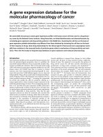
Fig. 1. Examples of applications for FISH in leukemia. (A) Dual-color FISH using
whole chromosome painting probes for chromosomes 7 (green) and 12 (red) to a leu-
kemic metaphase from an acute myeloid leukemia patient. This identified a cryptic
translocation between chromosomes 7 and 12. In this metaphase chromosome 12
material is visible on the der(7) (arrow); however, the reciprocal chromosome 7 mate-
rial is not visible on the der(12). (B) Dual-color FISH to map the extent of the 5q
deletion in a leukemic metaphase from a patient with 5q- syndrome. A 5p subtelomeric
probe (red) was cohybridized to tag both chromosomes 5, to ensure that only
metaphases containing the del(5q) were evaluated. Green fluorescent signal corre-
sponding to a probe containing the CSF1R gene is present on the normal homolog, but
absent from the del(5q) (arrow). (C,D) Combined immunophenotyping and FISH
(using a YAC probe containing the CSF1R gene) to bone marrow cells from a patient
with a del(5q) clonal chromosome marker. In each case, the APAAP positive cells
show bright red fluorescence. In (C) the CD3 antibody identifies a T cell with two
green fluorescent FISH signals corresponding to the YAC. In (D) the glycophorin A
antibody identifies an erythroid precursor with only one fluorescent signal. This con-
firms that the erythroid, but not the T-lymphocyte, lineage is involved in the malignant
FISH to Detect Abnormalities in Leukemia 9
clone in this patient. (E) M-FISH karyotype of a metaphase from the leukemia-de-
rived cell line GF-D8. Metaphases were hybridized with a set of combinatorially labeled
whole chromosome painting probes, and chromosomes assigned a pseudocolor according
to their unique fluorochrome composition using Powergene M-FISH software (Applied
Imaging, Newcastle, UK). Structurally abnormal chromosomes thus identified are indicated
by arrows.
10 Kearney et al.
2. Materials
2.1. Preparation of Bone Marrow Metaphase Chromosomes
1. Bone marrow aspirate collected into sterile bottles containing transport medium
(RPMI 1640 plus 50 U/mL of penicillin, 50 µg/mL of streptomycin, and 10 U/mL
of preservative-free lithium heparin).
2. Thymidine, crystalline (Sigma, St. Louis, MO): 100 µM stock.
3. 5-Fluorodeoxyuridine (Sigma): 100 µM stock.
4. Uridine (Sigma): 400 µM stock.
5. Colcemid (10 µg/mL) (Gibco).
6. Culture medium: RPMI 1640, 50 U/mL of penicillin, 50 µg/mL of streptomycin,
2 mM L-glutamine, 20% fetal calf serum (FCS) (all from Gibco-BRL).
7. Hypotonic solution: 0.075 M KCl.
8. Fixative: 3:1 AnalaR methanol:glacial acetic acid, at 4°C.
9. Precleaned microscope slides (Superfrost, BDH).
2.2. Pretreatment of Chromosomes and Nuclei
1. Pepsin (100 mg/mL) (Sigma).
2. Phosphate-buffered saline (PBS)/50 mM MgCl
2
: 50 mL of 1 M MgCl
2
+ 950 mL
of 1X PBS.
3. PBS/50 mM MgCl
2
/1% formaldehyde (make up fresh each time): 2.7 mL of form-
aldehyde in 100 mL of PBS/MgCl
2.
4. PBS (1X): 8 g of NaCl, 0.2 g of KCl, 1.44 g of Na
2
HPO
4
, 0.2 g of KH
2
PO
4
in 800 mL
of H
2
O, pH to 7.4 with HCl. Add H
2
O to 1 L.
5. RNase A (10 mg/mL) (Sigma) (boiled for 10 min to remove contaminating
DNase).
6. Formaldehyde (40% [w/v]).
2.3. Preparation of Probe DNA
2.3.1. Cosmids, P1 Artificial Chromosomes (PACs)
1. 2X TY medium (1 L): 16 g of Bacto tryptone, 10 g of yeast extract, 5 g of NaCl.
2. Glucose/EDTA/Tris (GET): 0.9% glucose, 10 mM EDTA, 25 mM Tris-HCl, pH 7.0.
3. NaOH/sodium dodecyl sulfate (SDS): 0.2 M NaOH, 1% SDS.
4. 3 M KOAc, pH 5.5.
5. RNase A (DNase free) (10 mg/mL) (Sigma).
2.3.2. Yeast Artificial Chromosomes (YACs)
1. YEPD medium (1 L): 10 g of Bacto yeast extract, 20 g of Bactopeptone, 20 g of
dextrose, 10 mL of adenine sulfate (0.5% in 0.5 M of HCl).
2. GDIS: 2% Triton X-100, 1% SDS, 100 mM NaCl, 10 mM Tris-HCl, pH 7.4,
1 mM EDTA.
3. Phenol:chloroform:isoamyl alcohol (25:24:1).
4. RNase A (DNase free) (10 mg/mL) (Sigma).
5. Glass beads, 710–1180 µm, acid washed (Sigma).
FISH to Detect Abnormalities in Leukemia 11
2.4. Nick Translation Labeling
1. Purified probe DNA (1 µg).
2. 10X Nick translation buffer: 0.5 M Tris-HCl, pH 7.5, 50 mM MgCl
2
, 0.5 mg/mL
of nuclease-free bovine serum albumin (BSA).
3. 1 mM Biotin-16-dUTP (bio-16-dUTP), 1 mM digoxigenin-11-dUTP (dig-11-
dUTP) (Roche Diagnostics).
4. 100 mM Dithiothreitol (DTT) (Sigma).
5. dNTP mix: 0.5 mM each dATP, dCTP, dGTP, and 0.1 mM dTTP (Roche Diag-
nostics).
6. DNase 1 (200,000 U) (Roche Diagnostics).
7. DNase 1 dilution buffer: 50% glycerol, 0.15 M NaCl, 20 mM sodium acetate, pH 5.0.
8. DNA polymerase 1 (10 U/µL) (New England Biolabs).
9. MicroSpin G50 columns (Amersham Pharmacia Biotech).
10. Escherichia coli tRNA (10 mg/mL) (Roche Diagnostics).
11. Salmon sperm DNA (5 mg/mL, sonicated to 200–500 bp) (Sigma).
12. TE: 10 mM Tris-HCl, pH 7.5, 1 mM EDTA.
13. Gel-loading buffer (5X bromophenol blue): 10% (w/v) Ficoll, 0.1 M Na
2
EDTA,
0.5% (w/v) SDS, 0.1% (w/v) bromophenol blue.
14. Electrophoresis buffer (10X TBE): 108 g of Tris base (89 mM), 55 g of boric acid
(89 mM), 40 mL of 0.5 M EDTA, pH 8.0 (2 mM) per liter.
15. PhiX174 HaeIII size marker (BRL Life Technologies).
2.5. Competitive
In Situ
Suppression Hybridization
1. Human Cot-1 DNA (BRL Life Technologies).
2. 3 M Sodium acetate.
3. Denaturing solution: 70% (v/v) formamide, 2X saline sodium citrate (SSC),
0.1 mM EDTA, pH 7.0.
4. Hybridization buffer: 50% (v/v) formamide, 10% (w/v) dextran sulfate, 1% (v/v)
Triton X-100, 2X SSC, pH 7.0.
5. Formamide (purified) (Fluka).
6. 50% Dextran sulfate.
7. 20X SSC: 1X SSC = 150 mM sodium chloride, 15 mM sodium citrate, pH 7.0.
8. Blocking solution: 3% (w/v) BSA in 4X SSC, 0.05% (v/v) Triton X-100 (make
up fresh).
9. Wash solution: 4X SSC, 0.05% (v/v) Triton X-100.
2.6. Detection of Bound, Labeled Probe
1. Fluorescence microscope (epifluorescence illumination), with suitable fluores-
cence objectives and filter sets (usually need separate filter sets for fluorescein
isothiocyanate [FITC], Texas red/rhodamine and 4,6-diamidino-2-phenylindole
[DAPI]/AMCA, as well as a double or triple filter block).
2. Avidin-DCS-FITC (1 mg/mL) (Vector).
3. Biotinylated anti-avidin D (0.5 mg/mL) (Vector).
4. Propidium iodide (Sigma).
12 Kearney et al.
5. DAPI (Sigma).
6. Vectashield mountant (Vector).
7. Avidin DCS-Texas red (2.5 mg/mL stock) (Vector).
8. Diluent for antibodies: blocking solution, filtered through a 0.45-µm syringe fil-
ter. Stock antibody solutions are stored at –20°C.
9. Monoclonal antidigoxigenin (Sigma).
10. Rabbit antimouse Ig-FITC (Sigma).
11. Monoclonal antirabbit-FITC (Sigma).
2.7. Degenerate Oligonucleotide Primer-PCR Amplification
of Flow-Sorted Chromosomes
1. Flow-sorted chromosomes (approximate concentration: 500/µL).
2. 2X PCR buffer: 10 mM MgCl
2
, 100 mM KCl, 20 mM Tris-HCl, pH 8.4,
0.2 mg/mL of gelatin.
3. dNTP mix: 2 mM each dATP, dCTP, dGTP, dTTP.
4. 6-MW primer: 5' CCGACTCGAGNNNNNNATGTGG 3' (30 µM).
5. Taq 1 (2.5 U/µL) polymerase (Roche Diagnostics).
6. 1 mM Biotin-16-dUTP or 1 mM dig-11-dUTP (Roche Diagnostics).
2.8. Alkaline Phosphatase Antialkaline Phosphatase Staining
1. Thin bone marrow smears (store unfixed wrapped in foil at –20°C).
2. Tris-buffered saline (TBS): 1 M Tris, 0.5 M NaCl.
3. Appropriate primary monoclonal antibody.
4. Rabbit antimouse antibody (Z259; Dako, Cambridge, UK) diluted 1:500 in TBS.
5. Monoclonal alkaline phosphatase antialkaline phosphatase (APAAP) complex
(1:500 dilution) (Roche Diagnostics).
6. Alkaline phosphatase substrate: Dissolve 2 mg of naphthol AS mix (Sigma) into
10 mL of 0.1 M Tris buffer (pH 8.2). To this add 10 mg of Fast Red TR mix
(Sigma) and dissolve. Then add levamisole (0.1 M) (Sigma) to block endogenous
alkaline phosphatase. Filter before use.
3. Methods
3.1. Preparation of Target Material
3.1.1 Culture and Harvesting of Mitotic Chromosomes from Leukemic
Bone Marrow (
see
ref.
19
)
1. Set up between one and four cultures, depending on the white cell count. Each culture
should contain approx 1 × 10
6
cells/mL. In most cases, the following will suffice:
a. Direct harvesting after 1 h exposed to colcemid (0.1 µg/mL).
b. A 24-h incubation with the addition of colcemid for the last hour.
c. Twenty-four hour synchronized cultures. For these, add fluorodeoxyuridine
(0.1 µM) and uridine (4 µM) after 24 h and reincubate the cultures overnight
(16–18 h). Finally, add thymidine (10 µM), and reincubate for 5 to 6 h before
adding of colcemid for 10 min before harvesting.
FISH to Detect Abnormalities in Leukemia 13
2. Centrifuge at 100g for 5 min. Discard the supernatant and resuspend the pellet in
hypotonic solution (prewarmed to 37°C). Incubate at 37°C for 20 min.
3. Centrifuge, discard the supernatant, and mix the pellet in the small volume of
hypotonic solution remaining. Add freshly made fixative dropwise, with mixing.
Add the first milliliter of fixative slowly, and then make up to 10 mL.
4. Leave in fixative for 30 min at 4°C. Centrifuge at 100g for 5 min, then wash in
three to five changes of fixative before making slides.
5. Wipe Superfrost slides clean with absolute ethanol just before use.
6. Place a drop of cell suspension on each slide and air-dry. Monitor the quality of
chromosome spreading under phase contrast. Chromosomes should be well
spread without visible cytoplasm and should appear dark gray under phase con-
trast (not black and refractile or light gray and almost invisible).
The “direct” culture can be replaced by overnight incubation with colcemid
(0.5 µg/mL). For cell lines, culture according to their specified growth require-
ments, then harvest when growing logarithmically, usually 24–48 h after a
change of medium. Add colcemid for the final 1 h before harvesting.
3.1.2. Preparation of Interphase Nuclei
Interphase nuclei are present in large numbers on slides from leukemic bone
marrow or blood prepared as in Subheading 3.1.1. Interphase nuclei can also
be prepared from fresh bone marrow after Ficoll separation of mononuclear
cells. After washing pellets in culture medium (RPMI, without FCS), fix the
cell pellet in several changes of methanol:acetic acid (3:1). Drop onto clean
slides. Nuclei from a variety of tissues and culture types can be prepared by
cytospin, then fixed in methanol (10–20 min). Bone marrow smears are pre-
pared in the usual way and stored unfixed, wrapped in foil at –20°C until
required.
3.2. Pretreatment of Chromosomes and Nuclei
The methanol/acetic acid fixation of metaphase chromosomes removes some
basic proteins that might interfere with hybridization. However, there is still a
variable amount of other protein and cytoplasmic contaminants on metaphase
chromosome preparations that may block hybridization, or cause nonspecific
background. We routinely use an RNase treatment and postfixation with form-
aldehyde. For interphase FISH, it may be necessary to add a proteolytic diges-
tion (e.g., pepsin) treatment to this, to aid access of the probe and detection
reagents. However, overdigestion can cause loss of cells from slides, so use
only when absolutely necessary.
1. Place 100 µL of RNase A (100 µg/mL) on slides under a 24 × 50 mm coverslip
and incubate at 37°C for 30 min to 1 h.
2. Wash three times (3 min each) in 2X SSC (with agitation).
14 Kearney et al.
3. Pepsin treatment (optional): 50 µg/mL in 0.01 M HCl. Incubate for 10 min at RT.
4. Wash (two times for 5 min each) in 1X PBS.
5. Wash (once for 5 min) in 1X PBS/50 mM MgCl
2
.
6. Fix in PBS/50 mM MgCl
2
/1% formaldehyde (2.7 mL of formaldehyde in 100 mL
of 1X PBS/50 mM MgCl
2
[fresh solution]) for 10 min.
7. Wash in 1X PBS for 5 min (with agitation).
8. Dehydrate slides through an alcohol series (70%, 95%, absolute) and allow to air-
dry. Slides can be stored desiccated at 4°C for up to 1 mo before use (see Note 1).
3.3. Preparation of Probe DNA
3.3.1. Cosmid, P1, and PAC DNA
Any DNA purification method that produces DNA suitable for sequencing
will generally also work for FISH. The following medium-scale alkaline lysis
method gives a high yield of cosmid, PAC, or P1 DNA. However, this is rela-
tively impure and may require additional purification steps. As a guide, if the
DNA fails to cut with DNase I, purify with phenol/chloroform or CsCl gradi-
ent centrifugation.
1. Inoculate 250 mL of 2X YT medium plus antibiotic (final concentration: 30 µg/mL
of kanamycin, 50 µg/mL of ampicillin) in a 500-mL sterile plugged flask with a
single well-separated colony.
2. Grow at 37°C with shaking (300 rpm) until approaching saturation (approx 18 h)
3. Transfer to a 250-mL bottle. Centrifuge at 4000g for 10 min.
4. Discard the supernatant medium and drain briefly. Add 50 mL of cold glucose/
EDTA/Tris (GET). Resuspend by drawing up in a 10-mL pipet.
5. Add 50 mL of NaOH/SDS at room temperature. Mix by very gentle, minimal
inversions. Leave for 5 min (room temperature).
6. Add 50 mL of cold 3 M KoAc. Mix by very gentle, minimal inversions. Place on
ice for 20 min.
7. Centrifuge at 9000g for 20 min (4°C).
8. Carefully transfer the supernatant to a fresh 250-mL bottle through a mesh.
9. Add 90 mL of isopropanol (0.7X vol) and mix. Leave at room temperature for 5 min.
10. Centrifuge at 5000g for 15 min at room temperature. Discard supernatant.
11. Add 25 mL of 70% ethanol, and rotate the bottle to rinse the inner surface. Trans-
fer pellet to 50-mL Falcon tubes.
12. Centrifuge at 5000g for 5 min (4°C). Discard the supernatant.
13. Allow to stand for 1 min, and then remove final traces of 70% ethanol with a Gilson.
14. Air-dry. Resuspend in approx 200 µL of H
2
O (or TE).
15. Incubate with RNase A (final concentration: 30 µg/mL) at 37°C for 30 min.
3.3.2. Yeast Artificial Chromosome DNA
(20)
The following method yields high quantities of total yeast DNA suitable for
FISH (see Note 2). The average yield from a 10-mL culture is 10–20 µg.