antisense technology, part a
Bạn đang xem bản rút gọn của tài liệu. Xem và tải ngay bản đầy đủ của tài liệu tại đây (12.11 MB, 597 trang )
Preface
Antisense technology reached a watershed year in 1998 with the FDA
approval of the antisense-based therapy, Vitravene, developed by ISIS.
This is the first drug based on antisense technology to enter the marketplace
and makes antisense technology a reality for therapeutic applications. How-
ever, antisense technology still needs further development, and new applica-
tions need to be explored.
Contained in this Volume 313 (Part A) of
Methods in Enzymology and
its companion Volume 314 (Part B) are a wide range of methods and
applications of antisense technology in current use. We set out to put
together a single volume, but it became obvious that the variations in
methods and the numerous applications required at least two volumes, and
even these do not, by any means, cover the entire field. Nevertheless, the
articles included represent the work of active research groups in industry
and academia who have developed their own methods and techniques. This
volume, Part A: General Methods, Methods of Delivery, and RNA Studies,
includes several methods of antisense design and construction, general
methods of delivery, and antisense used in RNA studies. In Part B: Applica-
tions, chapters cover methods in which antisense is designed to target
membrane receptors and antisense application in the neurosciences, as
well as in nonneuronal tissues. The therapeutic applications of antisense
technology, the latest area of new interest, complete the volume.
Although
Methods in Enzymology is designed to emphasize methods,
rather than achievements, I congratulate all the authors on their achieve-
ments that have led them to make their methods available. In compiling
and editing these two volumes I could not have made much progress without
the excellent secretarial services of Ms. Gayle Butters of the University of
Florida, Department of Physiology.
M. IAN PHILLIPS
xiii
Contributors to Volume 313
Article numbers are in parentheses following the names of contributors.
Affiliations listed are current.
SURESH ALAHARI (19), Department of Phar-
macology, School of Medicine, University
of North Carolina, Chapel Hill, North Caro-
lina 27599
SIDNEY ALTMAN (26), Department of Molecu-
lar, Cellular and Developmental Biology,
Yale University, New Haven, Connecticut
06520
ANNA ASTRIAB (19), Department of Pharma-
cology, School of Medicine, University of
North Carolina, Chapel Hill, North Caro-
lina 27599
DAVID BELLIDO (14), Unitat de Biologia Cel-
lular, Departament de Bioqulmica i Fisio-
logia, Universitat de Barcelona, E-08028
Barcelona, Spain
LYUBA BENIMETSKAYA (16), Columbia Uni-
versity, New York, New York 10032
ECKHART BUDDECKE (15), Division of Molec-
ular Cardiology, Institute for Arteriosclero-
sis Research, University of Miinster, D-
48149 Miinster, Germany
JEFFREY S. BUZBY (22), Hematology Research
Laboratory, Children's Hospital of Orange
County, Orange, California 92868
ALAN CARLETON (7), Institut Alfred Fessard,
CNRS, 91198 Gif-sur-Yvette Cedex, France
DANIELA CASTANOTI'O (23), Department of
Molecular Biology, Beckman Research In-
stitute of the City of Hope, Duarte, Califor-
nia 91010
DOUGLAS L. COLE (12), Manufacturing Pro-
cess Department, ISIS Pharmaceuticals,
Inc., Carlsbad, California 92008
STANLEY T. CROOKE (1), ISIS Pharmaceuti-
cals, Inc., Carlsbad, California 92008
JOHN M. DAGLE (24), Department of Pediat-
rics, University of Iowa, Iowa City, Iowa
52242
CHARLOTI'E DARRAH (29), Department of
Human Anatomy and Genetics, Oxford
University, Oxford 051 3QX,, United
Kingdom
SCOTT F. DEAMOND (17), Department of BiD-
chemistry, The Johns Hopkins School of
Hygiene and Public Health, Baltimore,
Maryland 21225
RANJIT R. DESHMUKH (12), Manufacturing
Process Department, ISIS Pharmaceuticals,
Inc., Carlsbad, California 92008
DAVID DESMAISONS (7), Institut Alfred Fes-
sard, CNRS, 91198 Gif-sur-Yvette Cedex,
France
SONIA DHEUR (3), Laboratoire de Biophy-
sique, INSERM U201, CNRS URA481,
Museum National d'Histoire Naturelle,
75005 Paris, France
BEIHUA DONG (31), Department of Cancer,
Lerner Research Institute, The Cleveland
Clinic Foundation, Cleveland, Ohio 44195
ROBERT J. DUFF (17), Department of BiD-
chemistry, The Johns Hopkins School of
Hygiene and Public Health, Baltimore,
Maryland 21225
GEORGE L. ELICEIRI (25), Department of Pa-
thology, Saint Louis University School of
Medicine, St. Louis, Missouri 63104-1028
RAMON ERITJA (14), European Molecular Bi-
ology Laboratory, D-69012 Heidelberg,
Germany
LOUISE EVERATT (29), Department of Human
Anatomy and Genetics, Oxford University,
Oxford OS1 3QX, United Kingdom
JEAN-CHRISTOPHE FRANCOIS (4), Laboratoire
de Biophysique, INSERM U201, CNRS
UMR8646, Museum National d'Histoire
Naturelle, 75005 Paris, France
CHANDRAMALLIKA GHOSH (6), AVI Bio-
Pharma, Inc., Corvallis, Oregon 97333
x CONTRIBUTORS TO VOLUME
313
RICHARD V. GILES (5),
Department of Haem-
atology, The University of Liverpool, Royal
Liverpool University Hospital, Liverpool
L7 8XP, United Kingdom
LINDA GORMAN (30),
Lineberger Cancer Cen-
ter, University of North Carolina, Chapel
Hill, North Carolina 27599
VLADIMIR V. GORN (9),
Epoch Pharmaceuti-
cals, Inc., Redmond, Washington 98052
CECILIA GUERRIER-TAKADA (26),
Depart-
ment of Molecular, Cellular and Develop-
mental Biology, Yale University, New Ha-
ven, Connecticut 06520
TROY O. HARASYM (18),
Inex Pharmaceuti-
cals Corporation, Burnaby, British Colum-
bia, Canada V5J 5J8
KAIZHANG HE (13),
Department of Chemis-
try, Duke University, Durham, North Caro-
lina 27708-0346
MICHAEL J. HOPE (18),
Inex Pharmaceuticals
Corporation, Burnaby, British Columbia,
Canada V5J 5J8
JEFF HUGHES (19),
Department of Pharma-
ceutics, University of Florida, Gainesville,
Florida 32610
MASAYORI INOUYE (28),
Department of Bio-
chemistry, Robert Wood Johnson Medical
School, Piscataway, New Jersey 08854
PATRICK IVERSEN (6),
A VI BioPharma, Inc.,
Corvallis, Oregon 97333
EMMA R. JAKOX (27),
Department of Physiol-
ogy, Medical College of Virginia~Virginia
Commonwealth University, Richmond, Vir-
ginia 23298
R. L. JULIANO (19),
Department of Pharma-
cology, School of Medicine, University of
North Carolina, Chapel Hill, North Caro-
lina 27599
SHIN-HONG KANG (30),
Lineberger Cancer
Center, University of North Carolina,
Chapel Hill, North Carolina 27599
HARUKO KATAYAMA (20),
Department of
Neurosurgery, Teikyo University Ichihara
Hospital, lchihara City, Chiba 299-0111,
Japan
MICHAEL W. KILPATRICK (29),
Department of
Pediatrics, University of Connecticut Health
Center, Farmington, Connecticut 06030
SANDRA K. KLIMUK (18),
Department of Bio-
chemistry and Molecular Biology, The Uni-
versity of British Columbia, Vancouver,
British Columbia, Canada V6T 1Z3
RYSZARD KOLE (30),
Lineberger Cancer Cen-
ter and Department of Pharmacology, Uni-
versity of North Carolina, Chapel Hill,
North Carolina 27599
IGOR KUTYAVlN (9),
Epoch Pharmaceuticals,
Inc., Redmond, Washington 98052
JI~ROME LACOSTE (4),
Plasticit~ et expression
des g~nomes microbiens, CNRS EP2029,
CEA LRC12, CERMO, Universit~ Joseph
Fourier, 38041 Grenoble, France
LAURENT LACROIX (4),
Laboratoire de Bio-
physique, INSERM U201, CNRS
UMR8646, Museum National d'Histoire
Naturelle, 75005 Paris, France
BERNARD LEBLEU (11),
Institut de G~n~tique
Mol~culaire de Montpellier, UMR5535,
CNRS, F-34293 Montpellier, France
EARVIN LIANG (19),
Department of Pharma-
ceutics, University of Florida, Gainesville,
Florida 32610
PIERRE-MARIE LLEDO (7),
InstitutAlfred Fes-
sard, CNRS, 91198 Gif-sur-Yvette Cedex,
France
EUGENE A. LUKHTANOV (9),
Epoch Pharma-
ceuticals, Inc., Redmond, Washington 98052
RATAN K. MAITRA (31),
HIV Core Facility,
Lerner Research Institute, The Cleveland
Clinic Foundation, Cleveland, Ohio 44195
DESPINA MANIOTIS (29),
Department of Hu-
man Anatomy and Genetics, Oxford Uni-
versity, Oxford OS1 3QX, United Kingdom
AKIRA MATSUNO (20),
Department of Neuro-
surgery, Teikyo University lchihara Hospi-
tal, Ichihara City, Chiba 299-0111, Japan
JEAN-LOuiS MERGNY (4),
Laboratoire de Bio-
physique, 1NSERM U201, CNRS
UMR8646, Museum National d'Histoire
Naturelle, 75005 Paris, France
DAVID MILESI (9),
Epoch Pharmaceuticals,
Inc., Redmond, Washington 98052
CONTRIBUTORS TO VOLUME 313 xi
OLEG MIROCHNITCHENKO (28), Department
of Biochemistry, Robert Wood Johnson
Medical School, Piscataway, New Jersey
08854
PAUL A. MORCOS (10), Gene Tools, LLC,
Corvallis, Oregon 97333
TADASHI NAGASHIMA (20), Department of
Neurosurgery, Teikyo University Ichihara
Hospital, Ichihara City, Chiba 299-0111,
Japan
PETER E. NIELSEN (8), Department of Medical
Biochemistry and Genetics, The Panum In-
stitute, University of Copenhagen, D K-2200
Copenhagen N, Denmark
ANVSCH PEYMAN (15), Chemical Research G
838, Hoechst Marion Roussel Deutschland
GmbH, D-65926 Frankfurt am Main,
Germany
M. IAn PHILLIPS (2), Department of Physiol-
ogy, University of Florida College of Medi-
cine, Gainesville, Florida 32610
LEONIDAS A. PHYLACTOU (29), Cyprus Insti-
tute of Neurology and Genetics, 1683 Ni-
cosia, Cyprus
JAUME PIULATS (14), Laboratorio de Bioin-
vestigaci6n, Merck Farma y Qufmica, S.A.,
E-08010 Barcelona, Spain
MARX( R. PLACER (31), 3-Dimensional Phar-
maceutical, Inc., Extort, Pennsylvania 19341
KEN PORXER (13), Department of Chemistry,
Duke University, Durham, North Carolina
27708-0346
VLADIMIR RAIT (13), Department of Chemis-
try, Duke University, Durham, North Caro-
lina 27708-0346
MICHAEL W. REED (9), Epoch Pharmaceuti-
cals, Inc., Redmond, Washington 98052
IAN ROBBINS (11), Institut de G~n~tique Mo-
l~culaire de MontpeUier, UMR5535, CNRS,
F-34293 Montpellier, France
CLINTON ROBV (17), Department of Biochem-
istry, The Johns Hopkins School of Hygiene
and Public Health, Baltimore, Maryland
21225
JoNN J. RossI (23), Department of Molecular
Biology, Beckman Research Institute of the
City of Hope, Duarte, California 91010
ANTONINA RYTE (15), Lombardi Cancer Cen-
ter, Georgetown University Medical Center,
Washington, DC 20007-2197
E.
TULA SAISON-BEHMOARAS (3), Labora-
toire de Biophysique, INSERM U201,
CNRS URA481, Museum National d'His-
toire NatureUe, 75005 Paris, France
YOGESH S. SANGHVI (12), Manufacturing
Process Department, ISIS Pharmaceuticals,
Inc., Carlsbad, California 92008
MICHAELA SCHERR (23), Abteilung Haemato-
logie und Onkologie, Medizinische Hoch-
schule Hannover, D-30625 Hannover,
Germany
ANNETTE SCHMIDT (15), Division of Molecu-
lar Cardiology, Institute for Arteriosclerosis
Research, University of Manster, D-48149
Miinster, Germany
lEAN C. SEMPLE (18), Inex Pharmaceuticals
Corporation, Burnaby, British Columbia,
Canada VIJ 5J8
DMITRI SERGUEEV (13, 19), Department of
Chemistry, Duke University, Durham,
North Carolina 27708-0346
ZINAIDA SERGUEEVA (13), Department of
Chemistry, Duke University, Durham,
North Carolina 27708-0346
W. L. SEVERT (27), Department of Physiology,
Medical College of Virginia~Virginia Com-
monwealth University, Richmond, Vir-
ginia 23298
BARBARA RAMSAY SHAW (13, 19), Depart-
ment of Chemistry, Duke University, Dur-
ham, North Carolina 27708-0346
HALINA SIERAKOWSKA (30), Lineberger Can-
cer Center, University of North Carolina,
Chapel Hill, North Carolina 27599
ROBERT H. IlLVERMAN (31), Department of
Cancer, Lerner Research Institute, The
Cleveland Clinic Foundation, Cleveland,
Ohio 44195
DAVID G. SPILLER (5), School of Biological
Sciences, The University of Liverpool, Liv-
erpool L69 7ZB, United Kingdom
C. A. STEIN (16), Columbia University, New
York, New York 10032
xii CONTRIBUTORS TO VOLUME 313
DAVID STEIN (6),
AVI BioPharma, Inc., Cor-
vallis, Oregon 97333
JACK SUMMERS (13),
Department of Chemis-
try, Duke University, Durham, North Caro-
lina 27708-0346
AKIRA TAMURA (20),
Department of Neuro-
surgery, Tokyo University Hospital, Ita-
bashi-ku, Tokyo 173-0003, Japan
ANA M. TARI
(21),
Department of Bioimmu-
notherapy, University of Texas MD Ander-
son Cancer Center, Houston, Texas 77030
GEMMA TARRAS6N (14),
Laboratorio de Bio-
investigaci6n, Merck Farina y Qu[mica,
S.A., E-08010 Barcelona, Spain
DAVID M. TIDD (5),
School of Biological Sci-
ences, The University of Liverpool, Liv-
erpool L69 7ZB, United Kingdom
JOHN TONKINSON (16),
Columbia University,
New York, New York 10032
PAUL F. TORRENCE (31),
Section on Biomedi-
cal Chemistry, Laboratory of Medicinal
Chemistry, National Institute of Diabetes
and Digestive and Kidney Diseases, Na-
tional Institutes of Health, Bethesda, Mary-
land 20892-0805
PAUL O. P. Ts'o (17),
Department of Bio-
chemistry, The Johns Hopkins School of
Hygiene and Public Health, Baltimore,
Maryland 21225
EUGEN UHLMANN (15),
Chemical Research G
838, Hoechst Marion Roussel Deutschland
GmbH, D-65926 Frankfurt am Main,
Germany
SEN~N VILAR6 (14),
Unitat de Biologia Cellu-
lar, Departament de Bioqu[mica i Fisio-
logia, Universitat de Barcelona, E-08028
Barcelona, Spain
JEAN-DIDIER VINCENT (7),
Institut Alfred Fes-
sard, CNRS, 91198 Gif-sur-Yvette Cedex,
France
DANIEL L. WEEKS (24),
Department of Bio-
chemistry, University of Iowa, Iowa City,
Iowa 52242
DWIGHT WELLER (6),
A VI BioPharma, Inc.,
Corvallis, Oregon 97333
SHIRLEY A. WILLIAMS
(22),
Hematology Re-
search Laboratory, Children's Hospital of
Orange County, Orange, California 92868
HooN Yoo (19),
Department of Pharmacol-
ogy, School of Medicine, University of
North Carolina, Chapel Hill, North Caro-
lina 27599
Y. CLARE ZHANG (2),
Department of Physiol-
ogy, University of Florida College of Medi-
cine, Gainesville, Florida 32610
YUANZHONG ZHOU (17),
Cell Works, Inc.,
Baltimore, Maryland 21227
[ 1] PROGRESS IN ANTISENSE TECHNOLOGY 3
[ I] Progress in Antisense Technology:
The End of the Beginning
By
STANLEY T. CROOKE
Introduction
During the past decade, intense efforts to develop and exploit antisense
technology have been mounted. With the recent FDA approval of Vitra-
vene, the first drug based on antisense technology to be commercialized,
the technology has achieved an important milestone. Nevertheless, the
technology is still in its infancy. Although the basic questions have been
answered, there are still many more unanswered than answered questions.
The objectives of this article are to provide an overview of the progress
in converting the antisense concept into broad therapeutic reality and to
provide advice about appropriate experimental design and interpretation
of data with regard to the therapeutic potential of the technology.
Proof of Mechanism
Factors That May Influence Experimental Interpretations
Clearly, the ultimate biological effect of an oligonucleotide will be influ-
enced by the local concentration of the oligonucleotide at the target RNA,
the concentration of the RNA, the rates of synthesis and degradation of
the RNA, the type of terminating mechanism, and the rates of the events
that result in termination of the activity of RNA. At present, we understand
essentially nothing about the interplay of these factors.
Oligonucleotide Purity.
Currently, phosphorothioate oligonucleotides
can be prepared consistently and with excellent purity. 1 However, this has
only been the case since the mid-1990s. Prior to that time, synthetic methods
were evolving and analytical methods were inadequate. In fact, our labora-
tory reported that different synthetic and purification procedures resulted
in oligonucleotides that varied in cellular toxicity 2 and that potency varied
from batch to batch. Although there are no longer synthetic problems with
phosphorothioates, they undoubtedly complicated earlier studies. More
1 S. T. Crooke and C. K. Mirabelli, "Antisense Resarch and Applications." CRC Press,
Boca Raton, FL, 1993.
2 R. M. Crooke,
Anti-Cancer Drug Design
6, 609 (1991).
Copyright © 1999 by Academic Press
All rights of reproduction in any form
reserved.
METHODS IN ENZYMOLOGY, VOL. 313 0076-6879/99 $30.00
4 GENERAL METHODS [ II
importantly, with each new analog class, new synthetic, purification, and
analytical challenges are encountered.
Oligonucleotide Structure.
Antisense oligonucleotides are designed to be
single stranded. We now understand that certain sequences, e.g., stretches of
guanosine residues, are prone to adopt more complex structures. 3 The
potential to form secondary and tertiary structures also varies as a function
of the chemical class. For example, higher affinity 2'-modified oligonucleo-
tides have a greater tendency to self-hybridize, resulting in more stable
oligonucleotide duplexes than would be expected based on rules derived
from oligodeoxynucleotides. 3a
RNA Structure.
RNA is structured. The structure of the RNA has a
profound influence on the affinity of the oligonucleotide and on the rate
of binding of the oligonucleotide to its RNA target. 4,5 Moreover, the RNA
structure produces asymmetrical binding sites that then result in very diver-
gent affinity constants depending on the position of oligonucleotide in that
structure:: This in turn influences the optimal length of an oligonucleotide
needed to achieve maximal affinity. We understand very little about how
RNA structure and RNA protein interactions influence antisense drug
action.
Variations in in Vitro Cellular Uptake and Distribution.
Studies in several
laboratories have clearly demonstrated that cells in tissue culture may take
up phosphorothioate oligonucleotides via an active process and that the
uptake of these oligonucleotides is highly variable depending on many
conditions. 2'8 Cell type has a dramatic effect on total uptake, kinetics of
uptake, and pattern of subcellular distribution. At present, there is no
unifying hypothesis to explain these differences. Tissue culture conditions,
such as the type of medium, degree of confluence, and the presence of
serum, can all have enormous effects on uptake. 8 The oligonucleotide chem-
ical class obviously influences the characteristics of uptake as well as the
mechanism of uptake. Within the phosphorothioate class of oligonucleo-
3 j. R. Wyatt, T. A. Vickers, J. L. Roberson, R. W. Buckheit, Jr., T. Klimkait, E. DeBaets,
P. W. Davis, B. Rayner, J. L. Imbach, and D, J. Ecker,
Proc. Natl. Acad. Sci. U.S.A.
91,
1356 (1994).
3a S. M. Freier, unpublished results.
4 S. M. Freier,
in
"Antisense Research and Applications" (S. T. Crooke and B. Lebleu,
eds0, p. 67. CRC Press, Boca Raton, FL, 1993.
5 D. J. Ecker,
in
"Antisense Research and Applications" (S. T. Crooke and R. Lebleu, eds.)
p. 387. CRC Press, Boca Raton, FL, 1993.
6 W. F. Lima, B. P. Monia, D. J. Ecker, and S. M. Freier,
Biochemistry
31, 12055 (1992).
7 D. J. Ecker, T. A. Vickers, T. W. Bruice, S. M. Freier, R. D. Jenison, M. Manoharan, and
M. Zounes,
Science
257, 958 (1992).
8 S. T. Crooke, L. R. Grillone, A. Tendolkar, A. Garrett, M. J. Fratkin, J. Leeds, and
W. H. Barr,
Clin. Pharmacol. Ther.
56, 641 (1994).
[ 1 ] PROGRESS IN ANTISENsE TECHNOLOGY 5
tides, uptake varies as a function of length, but not linearly. Uptake varies
as a function of sequence and stability in cells is also influenced by the se-
quence. 8,9
Given the foregoing, it is obvious that conclusions about in vitro uptake
must be made very carefully and generalizations are virtually impossible.
Thus, before an oligonucleotide could be said to be inactive in vitro, it
should be studied in several cell lines. Furthermore, while it may be abso-
lutely correct that receptor-mediated endocytosis is a mechanism of uptake
of phosphorothioate oligonucleotides, 1° it is obvious that a generalization
that all phosphorothioates are taken up by all cells in vitro primarily by
receptor-mediated endocytosis is simply unwarranted.
Finally, extrapolations from in vitro uptake studies to predictions about
in vivo pharmacokinetic behavior are entirely inappropriate and, in fact,
there are now several lines of evidence in animals and humans that demon-
strate that even after careful consideration of all in vitro uptake data, one
cannot predict in vivo pharmacokinetics of the compounds. 8,11-~3
Binding to and Effects of Binding to Nonnucleic Acid Targets. Phospho-
rothioate oligonucleotides tend to bind to many proteins and those interac-
tions are influenced by many factors. The effects of binding can influence
cell uptake, distribution, metabolism, and excretion. They may induce non-
antisense effects that can be mistakenly interpreted as antisense or compli-
cate the identification of an antisense mechanism. By inhibiting RNase H,
protein binding may inhibit the antisense activity of some oligonucleotides.
Finally, binding to proteins can certainly have toxicological consequences.
In addition to proteins, oligonucleotides may interact with other biologi-
cal molecules, such as lipids or carbohydrates, and such interactions, like
those with proteins, will be influenced by the chemical class of oligonucleo-
tide studied. Unfortunately, essentially no data bearing on such interactions
are currently available.
An especially complicated experimental situation is encountered in
many in vitro antiviral assays. In these assays, high concentrations of drugs,
viruses, and cells are often coincubated. The sensitivity of each virus to
9 S. T. Crooke, in "Burger's Medicinal Chemistry and Drug Discovery" (M. E. Wolff, ed.),
vol. 1, p. 863. Wiley, New York, 1995.
10 S. L. Loke, C. A. Stein, X. H. Zhang, K. Mori, M. Nakanishi, C. Subasinghe, J. S. Cohen,
and L. M. Neckers, Proc. Natl. Acad. Sci. U.S.A. 86, 3474 (1989).
alp. A. Cossum, H. Sasmor, D. Dellinger, L. Truong, L. Cummins, S. R. Owens, P. M.
Markham, J. P. Shea, and S. Crooke, J. Pharmacol. Exp. Ther. 267, 1181 (1993).
12 p. A. Cossum, L. Truong, S. R. Owens, P. M. Markham, J. P. Shea, and S. T. Crooke, J.
Pharmacol. Exp. Ther. 269, 89 (1994).
13 H. Sands, L. J. Gorey-Feret, S. P. Ho, Y. Bao, A. J. Cocuzza, D. Chidester, and F. W.
Hobbs, Mol. Pharmacol. 47, 636 (1995).
6 GENERAL METHODS [ 11
nonantisense effects of oligonucleotides varies depending on the nature of
the virion proteins and the characteristics of the oligonucleotides. 14,15 This
has resulted in considerable confusion. In particular for human immune
deficiency virus (HIV), herpes simplex viruses, cytomegaloviruses, and in-
fluenza virus, the nonantisense effects have been so dominant that identi-
fying oligonucleotides that work via an antisense mechanism has been
difficult. Given the artificial character of such assays, it is difficult to know
whether nonantisense mechanisms would be as dominant
in vivo
or result
in antiviral activity.
Terminating Mechanisms.
It has been amply demonstrated that oligonu-
cleotides may employ several terminating mechanisms. The dominant ter-
minating mechanism is influenced by RNA receptor site, oligonucleotide
chemical class, cell type, and probably many other factors. 16 Obviously, as
variations in terminating mechanism may result in significant changes in
antisense potency and studies have shown significant variations from cell
type to cell type
in vitro,
it is essential that the terminating mechanism be
well understood. Unfortunately, at present, our understanding of terminat-
ing mechanisms remains rudimentary.
Effects of "Control Oligonucleotides."
A number of types of control
oligonucleotides have been used, including randomized oligonucleotides.
Unfortunately, we know little to nothing about the potential biological
effects of such "controls," and the more complicated a biological system
and test the more likely that "control" oligonucleotides may have activities
that complicate interpretations. Thus, when a control oligonucleotide dis-
plays a surprising activity, the mechanism of that activity should be explored
carefully before concluding that the effects of the "control oligonucleotide"
prove that the activity of the putative antisense oligonucleotide is not due
to an antisense mechanism.
Kinetics of Effects.
Many rate constants may affect the activities of
antisense oligonucleotides, e.g., the rate of synthesis and degradation of
the target RNA and its protein, the rates of uptake into cells, the rates of
distribution, extrusion, and metabolism of an oligonucleotide in cells, and
similar pharmacokinetic considerations in animals. Despite this, relatively
few time courses have been reported and
in vitro
studies have been reported
that range from a few hours to several days. In animals, we have a growing
body of information on pharmacokinetics, but in most studies reported to
14 L. M. Cowsert,
in
"Antisense Research and Applications" (S. T. Crooke and B. Lebleu,
eds.), p. 521. CRC Press, Boca Raton, FL, 1993.
15 R. F. Azad, V. B. Driver, K. Tanaka, R. M. Crooke, and K. P. Anderson,
Antimicrob.
Agents Chemother.
37, 1945 (1993).
16 S. T. Crooke, "Therapeutic Applications of Oligonucleotides." R. G. Landes Company,
Austin, TX, 1995.
[ 1] PROGRESS IN ANTISENSE TECHNOLOGY 7
date, the doses and schedules were chosen arbitrarily and, again, little
information on duration of effect and onset of action has been presented.
Clearly, more careful kinetic studies are required and rational
in vitro
and
in vivo
dose schedules must be developed.
Recommendations
Positive Demonstration of Antisense Mechanism and Specificity.
Until
more is understood about how antisense drugs work, it is essential to
positively demonstrate effects consistent with an antisense mechanism. For
RNase H-activating oligonucleotides, Northern blot analysis showing selec-
tive loss of the target RNA is the best choice and many laboratories are
publishing reports
in vitro
and
in vivo
of such activities) 7-2° Ideally, a
demonstration that closely related isotypes are unaffected should be in-
cluded.
More recently, in our laboratories we have used RNA protection assays
and DNA chip arrays. 2°a These assays provide a great deal of information
about the levels of various RNA species. Coupled to careful kinetic analysis,
such approaches can help assure that the primary mechanism of action of
the drug is antisense and can identify events that are secondary to antisense
inhibition of a specific target. This can then support the assignment of a
target to a particular pathway, the analysis of the roles of a particular target,
and the factors that regulate its activity. We have adapted all these methods
for use in animals and will be determining their utility in clinical trials.
In brief, then, for proof of mechanism, the following steps are recom-
mended.
Perform careful dose-response curves
in vitro
using several cell lines
and methods of
in vitro
delivery.
Correlate the rank order potency
in vivo
with that observed
in vitro
after thorough dose-response curves are generated
in vivo.
Perform careful "gene walks" for all RNA species and oligonucleotide
chemical classes.
Perform careful time courses before drawing conclusions about po-
tency.
17 M. Y. Chiang, H. Chan, M. A. Zounes, S. M. Freier, W. F. Lima, and C. F. Bennett, J.
Biol. Chem.
266, 18162 (1991).
18 N. M. Dean and R. McKay,
Proc. Natl. Acad. Sci. U.S.A.
91, 11762 (1994).
19 T. Skorski, M. Nieborowska-Skorska, N. C. Nicolaides, C. Szczylik, P. Iversen, R. V. Iozzo,
G. Zon, and B. Calabretta,
Proc. Natl. Acad. Sci. U.S.A.
91, 4504 (1994).
20 N. Hijiya, J. Zhang, M. Z. Ratajezak, J. A. Kant, K. DeRiel, M. Hertyn, G. Zon, and
A. M. Gewirtz,
Proc. Natl. Acad. Sci. U.S.A.
91, 4499 (1994).
20a j. F. Taylor, Q. Q. Zhang, B. P. Monia, E. G. Marcusson, and N. M. Dean,
Oncogene,
in press.
8 GENERAL METHODS [
1]
Directly demonstrate the proposed mechanism of action by measuring
the target RNA and/or protein.
Evaluate specificity and therapeutic indices via studies on closely re-
lated isotypes and with appropriate toxicological studies.
Perform sufficient pharmacokinetics to define rational dosing schedules
for pharmacological studies.
When control oligonucleotides display surprising activities, determine
the mechanisms involved.
Molecular Mechanisms of Antisense Drugs
Occupancy-Only Mediated Mechanisms
Classic competitive antagonists are thought to alter biological activities
because they bind to receptors preventing natural agonists from binding
the inducing normal biological processes. Binding of oligonucleotides to
specific sequences may inhibit the interaction of the RNA with proteins,
other nucleic acids, or other factors required for essential steps in the
intermediary metabolism of the RNA or its utilization by the cell.
Inhibition of Splicing.
A key step in the intermediary metabolism of
most mRNA molecules is the excision of introns. These "splicing" reactions
are sequence specific and require the concerted action of spliceosomes.
Consequently, oligonucleotides that bind to sequences required for splicing
may prevent the binding of necessary factors or physically prevent the
required cleavage reactions. This would then result in inhibition of the
production of the mature mRNA. Although there are several examples of
oligonucleotides directed to splice junctions, none of the studies present
data showing inhibition of RNA processing, accumulation of splicing inter-
mediates, or a reduction in mature mRNA. Nor are there published data
in which the structure of the RNA at the splice junction was probed and
the oligonucleotides demonstrated to hybridize to the sequences for which
they were designed. 21-24 Activities have been reported for
anti-c-myc
and
antiviral oligonucleotides with phosphodiester, methylphosphonate, and
21 M. E. McManaway, L. M. Neckers, S. L. Loke, A. A. A1-Nasser, R. L. Redner, B. T.
Shiramizu, W. L. Goldschmidts, B. E. Huber, K. Bhatia, and I. T. Magrath,
Lancet
335,
808 (1990).
22 M. Kulka, C. C. Smith, L. Aurelian, R. Fishelevich, K. Meade, P. Miller, and P. O. P. Ts'o,
Proc. Natl. Acad. Sci. U.S.A.
86, 6868 (1989).
23 p. C. Zamecnik, J. Goodchild, Y. Taguchi, and P. S. Sarin,
Proc. Natl. Acad. Sci. U.S.A.
83, 4143 (1986).
24 C. C. Smith, L. Aurelian, M. P. Reddy, P. S. Miller, and P. O. P. Ts'o,
Proc. Natl. Acad.
Sci. U.S.A.
83, 2787 (1986).
[ 1]
PROGRESS IN ANTISENSE TECHNOLOGY
9
phosphorothioate backbones. An oligonucleotide has been reported to
induce alternative splicing in a cell-free splicing system and, in that system,
RNA analyses confirmed the putative mechanism. 25
In our laboratory, we have attempted to characterize the factors that
determine whether splicing inhibition is effected by an antisense
drug. 26
To this end, a number of luciferase-reporter plasmids containing various
introns were constructed and transfected into HeLa cells. The effects of
antisense drugs designed to bind to various sites were then characterized.
The effects of RNase H-competent oligonucleotides were compared to
those of oligonucleotides that do not serve as RNase H substrates. The
major conclusions from this study were, first, that most of the earlier studies
in which splicing inhibition was reported were probably due to nonspecific
effects. Second, less effectively spliced introns are better targets than those
with strong consensus splicing signals. Third, the 3'-splice site and branch-
point are usually the best sites to which to target to the oligonucleotide to
inhibit splicing. Fourth, RNase H-competent oligonucleotides are usually
more potent than even higher affinity oligonucleotides that inhibit by occu-
pancy only.
Translational Arrest.
A mechanism for which the many oligonucleotides
have been designed is to arrest the translation of targeted protein by binding
to the translation initiation codon. The positioning of the initiation codon
within the area of complementarily of the oligonucleotide and the length
of oligonucleotide used have varied considerably. Again, unfortunately,
only in relatively few studies have the oligonucleotides, in fact, been shown
to bind to the sites for which they were designed, and data that directly
support translation arrest as the mechanism have been lacking.
Target RNA species that have been reported to be inhibited by a
translational arrest mechanism include HIV, vesicular stomatitis virus
(VSV),
n-myc,
and a number of normal cellular
genes, z7-33
In our labora-
tories, we have shown that a significant number of targets may be inhibited
by binding to translation initiation codons. For example, ISIS 1082 hybrid-
25 Z. Dominski and R. Kole,
Proc. Natl. Acad. Sci. U.S.A.
90, 8673 (1993).
26 D. Hodges and S. T. Crooke,
Mol. Pharmacol.
48, 905 (1995).
27 S. Agrawal, J. Goodchild, M. P. Civeira, A. H. Thornton, P. S. Sarin, and P. C. Zameenik,
Proc. Natl. Acad. Sci. U.S.A.
85, 7079 (1988).
28 M. Lemaitre, B. Bayard, and B. Lebleu,
Proc. Natl. Acad. Sci. U.S.A.
84, 648 (1987).
29 A. Rosolen, L. Whitesell, N. Ikegaki, R. H. Kennett, and L. M. Neckers,
Cancer Res. $0,
6316 (1990).
3o G. Vasanthakumar and N. K. Ahmed,
Cancer Commun. 1,
225 (1989).
3x Sburlati, A. R., R. E. Manrow, and S. L. Berger,
Proc. Natl. Aead. Sci. U.S.A. 88,
253 (1991).
32 H. Zheng, B. M. Sahai, P. Kilgannon, A. Fotedar, and D. R. Green,
Proc. Natl. Acad. Sci.
U.S.A. 86,
3758 (1989).
33 j. A. Maier, P. Voulalas, D. Roeder, and T. Maciag,
Science
249, 1570 (1990).
10 GENERAL METHODS [ 1]
izes to the AUG codon for the UL13 gene of herpes virus types 1 and 2.
RNase H studies confirmed that it binds selectively in this area.
In vitro
protein synthesis studies confirmed that it inhibited the synthesis of the
UL13 protein, and studies in HeLa cells showed that it inhibited the growth
of herpes type 1 and type 2 with IC50 of 200-400 nM by translation
arrest. 34
Similarly, ISIS 1753, a 30-mer phosphorothioate complementary to the
translation initiation codon and surrounding sequences of the E2 gene of
bovine papilloma virus, was highly effective and its activity was shown to
be due to translation arrest. ISIS 2105, a 20-mer phosphorothioate comple-
mentary to the same region in human papilloma virus, was shown to be a
very potent inhibitor. Compounds complementary to the translation initia-
tion codon of the E2 gene were the most potent of the more than 50
compounds studied complementary to various other regions in the RNA. 35
We have shown inhibition of translation of a number of other mRNA
species by compounds designed to bind to the translation codon as well.
In conclusion, translation arrest represents an important mechanism of
action for antisense drugs. A number of examples purporting to employ
this mechanism have been reported, and studies on several compounds
have provided data that unambiguously demonstrate that this mechanism
can result in potent antisense drugs. However, very little is understood
about the precise events that lead to translation arrest.
Disruption of Necessary RNA Structure.
RNA adopts a variety of three-
dimensional structures induced by intramolecular hybridization, the most
common of which is the stem loop. These structures play crucial roles in
a variety of functions. They are used to provide additional stability for
RNA and as recognition motifs for a number of proteins, nucleic acids,
and ribonucleoproteins that participate in the intermediary metabolism and
activities of RNA species. Thus, given the potential general activity of the
mechanism, it is surprising that occupancy-based disruption RNA has not
been exploited more extensively.
As an example, we designed a series of oligonucleotides that bind to
the important stem-loop present in all RNA species in HIV, the TAR
element. We synthesized a number of oligonucleotides designed to disrupt
TAR and showed that several indeed did bind to TAR, disrupt the structure,
and inhibit TAR-mediated production of a reporter gene. 36 Furthermore,
34 C. K. Mirabelli, C. F. Bennett, K. Anderson, and S. T. Crooke Anti-Cancer Drug Design
6, 647 (1991).
35 L. M. Cowsert, M. C. Fox, G. Zon, and C. K. Mirabelli, Antimicrob. Agents Chemother.
37, 171 (1993).
36 T. Vickers, B. F. Baker, P. D. Cook, M. Zounes, R. W. Buckheit, Jr., J. Germany, and
D. J. Ecker, Nucleic Acids Res. 19, 3359 (1991).
[ 11 PROGRESS 1N ANTISENSE TECHNOLOGY 1 1
general rules useful in disrupting stem-loop structures were developed
as well. 7
Although designed to induce relatively nonspecific cytotoxic effects,
two other examples are noteworthy. Oligonucleotides designed to bind to
a 17 nucleotide loop in Xenopus 28 S RNA required for ribosome stability
and protein synthesis inhibited protein synthesis when injected into Xeno-
pus oocytes. 37 Similarly, oligonucleotides designed to bind to highly con-
served sequences in 5.8 S RNA inhibited protein synthesis in rabbit reticulo-
cyte and wheat germ systems, as
Occupancy-Activated Destabilization
RNA molecules regulate their own metabolism. A number of structural
features of RNA are known to influence stability, various processing events,
subcellular distribution, and transport. It is likely that as RNA intermediary
metabolism is better understood, many other regulatory features and mech-
anisms will be identified.
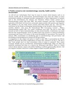
5'-Capping. A key early step in RNA processing is 5'-capping (Fig. 1).
This stabilizes pre-mRNA and is important for the stability of mature
mRNA. It also is important in binding to the nuclear matrix and transport
of mRNA out of the nucleus. As the structure of the cap is unique and
understood, it presents an interesting target.
Several oligonucleotides that bind near the cap site have been shown
to be active, presumably by inhibiting the binding of proteins required to
cap the RNA. For example, the synthesis of SV40 T-antigen was reported
to be most sensitive to an oligonucleotide linked to polylysine and targeted
to the 5'-cap site of RNA. 39 However, again, in no published study has this
putative mechanism been demonstrated rigorously. In fact, in no published
study have the oligonucleotides been shown to bind to the sequences for
which they were designed.
In our laboratory, we have designed oligonucleotides to bind to 5'-cap
structures and reagents to specifically cleave the unique 5'-cap structure. 4°
These studies demonstrate that 5'-cap-targeted oligonucleotides were capa-
ble of inhibiting the binding of the translation initiation factor eIF-4a. 41
Inhibition of 3'-Polyadenylation. In the 3'-untranslated region of pre-
mRNA molecules are sequences that result in the posttranscriptional
37 S. K. Saxena and E. J. Ackerman, J. Biol. Chem, 265, 3263 (1990).
3s K. Walker, S. A. Elela, and R. N. Nazar, J. Biol. Chem. 265, 2428 (1990).
39 p. Westermann, B. Gross, and G. Hoinkis, Biomed. Biochim Acta 48, 289 (1989).
40 B. F. Baker, J. Am. Chem. Soc. 115, 3378 (1993).
41B. F. Baker, L. Miraglia, and C. H. Hagedorn, J. Biol. Chem. 267, 11495 (1992).
12 GENERAL METHODS [ 11
Transcription
A~( g
Capping/ C
Splicing CA ~ AAAA
; .[
Nucleus :
",
CAP ~ "'
Transpod "' '~ I F ~AAAA + ~. ""
• ~. °"
Cytoplasm
"*"°°~' °° .° °
° °
CAP JAAAA CAP_
Degradation "~ r -~
CAP
Translation ~- t._~J AAAA 9
FIG. 1.
RNA processing.
Transcriptional
Arrest
Effects on
Anabollsm of
rnRNA
Effocts on
~.,91 Catabolism of
mRNA
Translational
Arrest
addition of long (hundreds of nucleotides) tracts of polyadenylate. Poly-
adenylation stabilizes mRNA and may play other roles in the intermediary
metabolism of RNA species. Theoretically, interactions in the 3'-terminal
region of pre-mRNA could inhibit polyadenylation and destabilize the
RNA species. Although there are a number of oligonucleotides that interact
in the 3'-untranslated region and display antisense activities, to date, no
study has reported evidence for alterations in polyadenylation. 17
Other Mechanisms
In addition to 5'-capping and 3'-adenylation, there are clearly other
sequences in the 5'- and 3'-untranslated regions of mRNA that affect the
stability of the molecules. Again, there are a number of antisense drugs
that may work by these mechanisms.
Zamecnik and Stephenson 42 reported that 13-mer targeted to untrans-
lated 3'- and 5'-terminal sequences in Rous sarcoma viruses was active•
Oligonucleotides conjugated to an acridine derivative and targeted to a 3'-
terminal sequence in type A influenza viruses were reported to be active•
Against several RNA targets, studies in our laboratories have shown that
42 p. C.
Zamecnik and
M. L. Stephenson,
Proc.
Natl. Acad.
Sci. U.S.A.
75, 289 (1978).
[1]
PROGRESS IN ANTISENSE TECHNOLOGY
13
sequences in the 3'-untranslated region of RNA molecules are often the
most sensitive. 43-45 For example, ISIS 1939, a 20-mer phosphorothioate that
binds to and appears to disrupt a predicted stem-loop structure in the 3'-
untranslated region of the mRNA for the intracellular adhesion molecule
(ICAM), is a potent antisense inhibitor. However, inasmuch a 2'-methoxy
analog of ISIS 1939 was much less active, it is likely that, in addition to
destabilization to cellular nucleolytic activity, the activation of RNase H
(see later) is also involved in the activity of ISIS 1939.17
Activation of RNase H
RNase H is an ubiquitous enzyme that degrades the RNA strand of an
RNA-DNA duplex. It has been identified in organisms as diverse as viruses
and human cells. 46 At least two classes of RNase H have been identified
in eukaryotic cells. Multiple enzymes with RNase H activity have been
observed in prokaryotes. ~
Although RNase H is involved in DNA replication, it may play other
roles in the cell and is found in the cytoplasm as well as the nucleus. 47
However, the concentration of the enzyme in the nucleus is thought to be
greater, and some of the enzyme found in cytoplasmic preparations may
be due to nuclear leakage.
RNase H activity is quite variable in cells. It is absent or minimal in
rabbit reticulocytes but is present in wheat germ
extracts. 46'48 In HL-60
ceils, for example, the level of activity in undifferentiated cells is greatest,
relatively high in dimethyl sulfoxide- and vitamin D-differentiated cells, and
much lower in phorbol ester-differentiated cells (Hoke, unpublished data).
The precise recognition elements for RNase H are not known. However,
it has been shown that oligonucleotides with DNA-like properties as short
as tetramers can activate RNase
H. 49
Changes in the sugar influence RNase
H activation as sugar modifications that result in RNA-like oligonucleo-
tides, e.g., 2'-fluoro or 2'-methoxy do not appear to serve as substrates for
43 A. Zerial, N. T. Thuong, and C. Helene,
Nucleic Acids Res.
15, 9909 (1987).
44 N. T. Thuong, U. Asseline, and T. Monteney-Garestier,
in
"Oligodeoxynucleotides: Anti-
sense Inhibitors of Gene Expression," p. 25. CRC Press, Baca Raton, FL, 1989.
45 C. Helene and J J. Toulme,
in
"Oligonucleotides: Antisense Inhibitors of Gene Expression"
(J. S. Cohen, ed.), p. 137. CRC Press, Boca Raton, FL, 1989.
46 R. J. Crouch and M L. Dirksen,
in
"Nucleases" (S. M. Linn and R. J. Roberts, eds.),
p. 211. Cold Spring Harbor Laboratory Press, Cold Spring Harbor, NY, 1985.
47 C. Crum, J. D. Johnson, A. Nelson, and D. Roth,
Nucleic Acids Res.
16, 4569 (1988).
48 M. T. Haeuptle, R. Frank, and B. Dobberstein,
Nucleic Acids Res.
14, 1427 (1986).
49 H. Donis-Keller,
Nucleic Acids Res.
7 (1979).
14 GENERAL METHODS
[ 11
RNase H. 5°'51 Alterations in the orientation of the sugar to the base can
also affect RNase H activation as ot-oligonucleotides are unable to induce
RNase H or may require parallel annealing. 52'53 Additionally, backbone
modifications influence the ability of oligonucleotides to activate RNase
H. Methylphosphonates do not activate RNase
H. 54'55
In contrast, phos-
phorothioates are excellent
substrates. 34'56'57
In addition, chimeric mole-
cules have been studied as oligonucleotides that bind to RNA and acti-
vate RNase
H. 58'59
For example, oligonucleotides composed of wings of
2'-methoxyphosphonates and a five-base gap of deoxyoligonucleotides bind
to their target RNA and activate RNase
H. 58'59
Furthermore, a single ribo-
nucleotide in a sequence of deoxyribonucleotides was shown to be sufficient
to serve as a substrate for RNase H when bound to its complementary
deoxyoligonucleotide.60
That it is possible to take advantage of chimeric oligonucleotides de-
signed to activate RNase H and have greater affinity for their RNA recep-
tors and to enhance specificity has also been demonstrated. 61,62 In a recent
study, RNase H-mediated cleavage of target transcript was much more
selective when deoxyoligonucleotides composed of methylphosphonate
deoxyoligonucleotide wings and phosphodiester gaps were compared to
full phosphodiester oligonucleotides. 62
Despite the information about RNase H and the demonstration that
many oligonucleotides may activate RNase H in lysate and purified enzyme
assays, relatively little is known yet about the role of structural features in
RNA targets in activating RNase
H. 63-65
In fact, direct proof that RNase
50 A. M. Kawasaki, M. D. Casper, S. M. Freier, E. A. Lesnik, M. C. Zounes, L. L. Cummins,
C. Gonzalez, and P. D. Cook, J.
Med. Chem. 36,
831 (1993).
51 B. S. Sproat, A. I. Lamond, B. Beijer, P. Neuner, and U. Ryder,
Nucleic Acids Res.
17,
3373 (1989).
52 F. Morvan, B. Rayner, and J. L. Irnbach,
Anticancer Drug Des.
6, 521 (1991).
53 C. Gagnor, B. Rayner, J. P. Leonetti, J. L. Imbach, and B. Lebleu,
Nucleic Acids Res.
17,
5107 (1989).
54 t. J. Maher, III, B. Wold, and P. B. Dervan,
Science
245, 725 (1989).
55 p. S. Miller,
in
"Oligodeoxynucleotides: Antisense Inhibitors of Gene Expression" (J. S.
Cohen, ed.), p. 79. CRC Press, Boca Raton, FL, 1989.
56 C. A. Stein and Y C. Cheng,
Science
261, 1004 (1993).
57 C. Cazenave, C. A. Stein, N. Loreau, N. T. Thuong, L. M. Neckers, C. Subasinghe,
C. Hel6ne, J. S. Cohen, and J J. Toulm,
Nucleic Acids Res.
17, 4255 (1989).
s8 R. S. Quartin, C. L. Brakel, and J. G. Wetmur,
Nucleic Acids Res.
17, 7253 (1989).
59 p. j. Furdon, Z. Dominski, and R. Kole,
Nucleic Acids Res.
17, 9193 (1989).
60 p. S. Eder and J. A. Walder, J.
Biol. Chem. 266,
6472 (1991).
61 B. P. Monia, E. A. Lesnik, C. Gonzalez, W. F. Lima, D. McGee, C. J. Guinosso, A. M.
Kawasaki, P. D. Cook, and S. M. Freier, J.
Biol. Chem. 268,
14514 (1993).
62 R. V. Giles and D. M. Tidd,
Nucleic Acids Res. 20,
763 (1992).
63 R. Y. Walder and J. A. Walder,
Proc. Natl. Acad. Sci. U.S.A.
85, 5011 (1988).
[ 1]
PROGRESS IN ANTISENSE TECHNOLOGY 15
H activation is, in fact, the mechanism of action of oligonucleotides in cells
is, to a large extent, lacking.
Studies in our laboratories provide additional, albeit indirect, insights
into these questions. ISIS 1939 is a 20-mer phosphorothioate complemen-
tary to a sequence in the 3'-untranslated region of ICAM-1 RNA. 17 It
inhibits ICAM production in human umbilical vein endothelial cells, and
Northern blots demonstrate that ICAM-1 mRNA is degraded rapidly. A
2'-methoxy analog of ISIS 1939 displays higher affinity for the RNA than the
phosphorothioate, is stable in cells, but inhibits ICAM-1 protein production
much less potently than ISIS 1939. It is likely that ISIS 1939 destabilizes
the RNA and activates RNase H. In contrast, ISIS 1570, an 18-mer phospho-
rothioate that is complementary to the translation initiation codon of the
ICAM-1 message, inhibited production of the protein, but caused no degra-
dation of the RNA. Thus, two oligonucleotides that are capable of activating
RNase H had different effects depending on the site in the mRNA at which
they bound. 17
A more direct demonstration that RNase H is likely a key factor in the
activity of many antisense oligonucleotides was provided by studies in which
reverse-ligation polymerase chain reaction (RT-PCR) was used to identify
cleavage products from bcr-abl mRNA in cells treated with phosphorothio-
ate oligonucleotides. 66
Given the emerging role of chimeric oligonucleotides with modifications
in the 3'- and 5'-wings designed to enhance affinity for the target RNA
and nuclease stability and a DNA-type gap to serve as a substrate for RNase
H, studies focused on understanding the effects of various modifications on
the efficiency of the enzyme(s) are also of considerable importance. In one
such study on
Escherichia coil
RNase H, we have reported that the enzyme
displays minimal sequence specificity and is processive. When a chimeric
oligonucleotide with 2'-modified sugars in the wings was hybridized to
the RNA, the initial site of cleavage was the nucleotide adjacent to the
methoxy-deoxy junction closest to the 3' end of the RNA substrate. The
initial rate of cleavage increased as the size of the DNA gap increased, and
the efficiency of the enzyme was considerably less against an RNA target
duplexed with a chimeric antisense oligonucleotide than a full DNA-type
oligonucleotide.67
64 j. MinshuU and T. Hunt,
Nucleic Acids Res.
14, 6433 (1986).
6s C. Gagnor, J. R. Bertrand, S. Thenet, M. Lemaitre, F. Morvan, B. Rayner, C. Malvy, B.
Lebleu, J. L. Imbach, and C. Paoletti,
Nucleic Acids Res.
15, 10419 (1987).
66 R. V. Giles, D. G. Spiller, and D. M. Tidd,
Antisense Res. Dev.
5, 23 (1995).
67 S. T. Crooke, K. M. Lemonidis, L. Neilson, R. Griffey, E. A. Lesnik, and B. P. Monia,
Biochem.
J. 312, 599 (1995).
16 GENERAL METHODS [ 11
In subsequent studies, we have evaluated the interactions of antisense
oligonucleotides with structured and unstructured targets and the impacts
of these interactions on RNase H in more detail. 68 Using a series of non-
cleavable substrates and Michaelis-Menten analyses, we were able to evalu-
ate both binding and cleavage. We showed that, in fact,
E. coli
RNase H1
is a double-strand RNA-binding protein. The Kd for RNA duplex was 1.6
/zM; the Kd for a DNA duplex was 176/.~M; and the Kd for single-strand
DNA was 942 ~M. In contrast, the enzyme could only cleave RNA in an
RNA-DNA duplex. Any 2' modification in the antisense drug at the cleav-
age site inhibited cleavage, but a significant charge reduction and 2' modifi-
cations were tolerated at the binding site. Finally, placing a positive charge
(e.g., 2'-propoxyamine) in the antisense drug reduced affinity and cleavage.
We have also examined the effects of antisense oligonucleotide-induced
RNA structures on the activity of
E. coli
RNase H1.69 Any structure in the
duplex substrate was found to have a significant negative effect on the
cleavage rate. Further, cleavage of selected sites was inhibited entirely, and
this was explained by steric hindrance imposed by the RNA loop traversing
either the minor or the major grooves or the heteroduplex.
We have succeeded in cloning, expressing, and characterizing a human
RNase H that is homologous to
E. coli
RNase H1 and has properties
comparable to the type 2 enzyme.
TM
Additionally, we have cloned and
expressed a second RNase H homologous to
E. coli
RNase H2.
TM
Given
these steps, we are now in position to evaluate the roles of each of these
enzymes in cellular activities and antisense pharmacology. We are also
characterizing these proteins and their enzymological properties.
Activation of Double-Strand RNase
By using phosphorothioate oligonucleotides with 2'-modified wings and
a ribonucleotide center, we have shown that mammalian cells contain en-
zymes that can cleave double-stranded RNAs.
TM
This is an important step
forward because it adds to the repertoire of intracellular enzymes that may
be used to cleave target RNAs and because chimeric oligonucleotide 2'-
modified wings and oligoribonucleotide gaps have a higher affinity for RNA
targets than chimeras with oligodeoxynucleotide gaps.
68 W. F. Lima and S. T. Crooke,
Biochemistry
36, 390 (1997).
69 W. F. Lima, M. Venkatraman, and S. T. Crooke,
J. Biol. Chem.
272, 18191 (1997).
70 H. Wu, W. F. Lima, and S. T. Crooke,
Antisense Nucleic Acid Drug Dev. 8,
53 (1998).
70a H. Wu and S. T. Crooke, unpublished observations.
[ 11 PROGRESS IN ANTISENSE TECHNOLOGY 17
Selection of Optimal RNA-Binding Site
It has been amply demonstrated that a significant fraction of every RNA
species is not accessible to phosphorothioate oligodeoxynucleotides in a
fashion that permits antisense effects (for a review, see Crooke71). Thus,
substantial efforts have been directed to the development of methods that
might predict optimal sites for binding within RNA species. Although a
number of screening methods have been proposed, 72-75 in our experience
the correlation between these antisense effects in cells is insufficient to
warrant their
use.
TM
Consequently, we have developed rapid throughput systems that use a
96-well format and a 96-channel oligonucleotide synthesizer coupled to an
automated RT-PCR instrument. This provides rapid screening of up to 80
sites in an RNA species for two chemistries under consistent highly con-
trolled experimental conditions. It is hoped that based on such a system
(we are currently evaluating two genes per week), we will be able to develop
improved methods that predict optimal sites.
Characteristics of Phosphorothioate Oligodeoxynucleotides
Introduction
Of the first-generation oligonucleotide analogs, the class that has re-
sulted in the broadest range of activities and about which the most is known
is the phosphorothioate class. Phosphorothioate oligonucleotides were first
synthesized in 1969 when a poly(rlrC)phosphorothioate was synthesized. 76
This modification clearly achieves the objective of increased nuclease stabil-
ity. In this class of oligonucleotides, one of the oxygen atoms in the phos-
phate group is replaced with a sulfur. The resulting compound is negatively
charged, is chiral at each phosphorothioate phosphodiester, and is much
more resistant to nucleases than the parent phosphorothioate. 77
71 S. T. Crooke, FASEB J. 7, 533 (1993).
72 T. W. Bruice and W. F. Lima, Biochemistry 36, 5004 (1997).
73 O. Matveeva, B. Felden, S. Audlin, R. F. Gesteland, and J. F. Atkins, Nucleic Acids Res.
25, 5010 (1997).
74 E. M. Southern, Ciba Foundation, Wiley, London, 1977.
75 S. P. HO, Y. Bao, T. Lesher, R. Malhotra, L. Y. Ma, S. J. Fluharty, and R. R. Sakai, Nat.
Biotechnol. 16, 59 (1998).
75a Wyatt, unpublished results.
76 E. De Clercq, F. Eckstein, and T. C. Merigan, Science 165, 1137 (1969).
77 j. S. Cohen, in "Antisense Research and Applications" (S. T. Crooke and B. Lebleu, eds.),
p. 205, CRC Press, Boca Raton, FL, 1993.
18 GENERAL METHODS [ 1 ]
Hybridization
The hybridization of phosphorothioate oligonucleotides to DNA and
RNA has been characterized thoroughly. 1'78-s° The Tm of a phosphorothio-
ate oligodeoxynucleotide for RNA is approximately 0.5 ° less per nucleotide
than for a corresponding phosphodiester oligodeoxynucleotide. This reduc-
tion in Tm per nucleotide is virtually independent of the number of phospho-
rothioate units substituted for phosphodiesters. However, the sequence
context has some influence as the A Tm can vary from -0.3 to 1.0 °, depending
on the sequence. Compared to RNA and RNA duplex formation, a phos-
phorothioate oligodeoxynucleotide has a Tm approximately -2.2 ° lower
per unit. 4 This means that to be effective in vitro, phosphorothioate oligo-
deoxynucleotides must typically be 17- to 20-mer in length and that invasion
of double-stranded regions in RNA is
difficult. 6'36'61'81
Association rates of phosphorothioate oligodeoxynucleotide to unstruc-
tured RNA targets are typically
106-107 M -1 sec -1
independent of oligonu-
cleotide length or sequence. 4,6 Association rates to structured RNA targets
can vary from 102 to 108 M 1 sec 1, depending on the structure of the
RNA, site of binding in the structure, and other factors. 4 Said another
way, association rates for oligonucleotides that display acceptable affinity
constants are sufficient to support biological activity at therapeutically
achievable concentrations. Interestingly, in a study using phosphodiester
oligonucleotides coupled to fluorescein, hybridization was detectable within
15 min after microinjection into K562 cells, s2
The specificity of hybridization of phosphorothioate oligonucleotides
is, in general, slightly greater than phosphodiester analogs. For example,
a T-C mismatch results in a 7.7 or 12.8 ° reduction in Tin, respectively, for
a phosphodiester or phosphorothioate oligodeoxynucleotide 18 nucleotides
in length with the mismatch centered. 4 Thus, from this perspective, the
phosphorothioate modification is quite attractive.
Interactions with Proteins
Phosphorothioate oligonucleotides bind to proteins. Interactions with
proteins can be divided into nonspecific, sequence specific, and structure-
78 S. T. Crooke,
Bio/Technology
10, 882 (1992).
79 R. M. Crooke,
in
"Antisense Research and Applications" (S. T. Crooke and B. Lebleu,
eds.), p. 427, CRC Press, Boca Raton, FL, 1993.
80 S. T. Crooke,
Annu. Rev. Pharmacol. Toxicol.
32, 329 (1992).
81 B. P. Monia, J. F. Johnston, D. J. Ecker, M. A. Zounes, W. F. Lima, and S. M. Freier, J.
Biol. Chem.
267, 19954 (1992).
82 D. L. Sokol, X. Zhang, P. Lu, and A. M. Gewirtz,
Proc. Natl. Acad. Sci. U.S.A.
95,
11538 (1998).
[11 PROGRESS IN ANTISENSE TECHNOLOGY 19
specific binding events, each of which may have different characteristics
and effects. Nonspecific binding to a wide variety of proteins has been
demonstrated. Exemplary of this type of binding is the interaction of phos-
phorothioate oligonucleotides with serum albumin. The affinity of such
interactions is low. The Kd for albumin is approximately 200/zM, thus, in
a similar range with aspirin or penicillin. 83'84 Furthermore, in this study, no
competition between phosphorothioate oligonucleotides and several drugs
that bind to bovine serum albumin was observed. In this study, binding
and competition were determined in an assay in which electrospray mass
spectrometry was used. In contrast, in a study in which an equilibrium
dissociation constant was derived from an assay using albumin loaded on
a CH-Sephadex column, the Km ranged from 1-5 × 10 -5 M for bovine
serum albumin to 2-3 × 10 -4 M for human serum albumin. Moreover,
warfarin and indomethacin were reported to compete for binding to serum
albumin. 85 Clearly, much more work is required before definitive conclu-
sions can be drawn.
Phosphorothioate oligonucleotides can interact with nucleic acid-bind-
ing proteins such as transcription factors and single-strand nucleic acid-
binding proteins. However, very little is known about these binding events.
Additionally, it has been reported that phosphorothioates bind to an 80-
kDa membrane protein that was suggested to be involved in cellular uptake
processes. 1° However, again, little is known about the affinities, sequence,
or structure specificities of these putative interactions. More recently, inter-
actions with 30- and 46-kDa surface proteins in T15 mouse fibroblasts
were reported. 86
Phosphorothioates interact with nucleases and DNA polymerases.
These compounds are metabolized slowly by both endo- and exonucleases
and inhibit these enzymes. 78'87 The inhibition of these enzymes appears to
be competitive, which may account for some early data suggesting that
phosphorothioates are almost infinitely stable to nucleases. In these studies,
the oligonucleotide to enzyme ratio was very high and, thus, the enzyme was
inhibited. Phosphorothioates also bind to RNase H when in an RNA-DNA
duplex and the duplex serves as a substrate for RNase H. 88 At higher
83 S. T. Crooke, M. J. Graham, J. E. Zuckerman, D. Brooks, B. S. Conklin, L. L. Cummins,
M. J. Greig, C. J. Guinosso, D. Kornburst, M. Manoharan, H. M. Sasmor, T. Schleich,
K. L. Tivel, R. H. Griffey et aL, J. PharmacoL Exp. Ther. 277, 923 (1996).
84 R. W. Joos and W. H. Hall, J. Pharmacol. Exp. Ther. 166, 113 (1969).
85 S. K. Srinivasan, H. K. Tewary, and P. L. Iversen, Antisense Res. Dev. 5, 131 (1995).
86 p. Hawley and I. Gibson, Antisense Nucleic Drug Dev. 6, 185 (1996).
87 R. M. Crooke, M. J. Graham, M. E. Cooke, and S. T. Crooke, J. Pharmacol. Exp. Ther.
275, 462 (1995).
88 W Y. Gao, F S. Han, C. Storm, W. Egan, and Y C. Cheng, Mol. Pharmacol. 41, 223
20 GENERAL METHODS
[ II
concentrations, presumably by binding as a single strand to RNase H,
phosphorothioates inhibit the enzyme. 67'78 Again, the oligonucleotides ap-
pear to be competitive antagonists for the DNA-RNA substrate.
Phosphorothioates have been shown to be competitive inhibitors of
DNA polymerase ot and/3 with respect to the DNA template and noncom-
petitive inhibitors of DNA polymerases "y and 8. 88 Despite this inhibition,
several studies have suggested that phosphorothioates might serve as prim-
ers for polymerases and be extended. 9'56'89 In our laboratories, we have
shown extensions 2-3 nucleotides only. At present, a full explanation as to
why longer extensions are not observed is not available.
Phosphorothioate oligonucleotides have been reported to be competi-
tive inhibitors for HIV-reverse transcriptase and inhibit RT-associated
RNase H activity. 9°'91 They have been reported to bind to the cell surface
protein, CD4, and to protein kinase C (PKC). 92 Various viral polymerases
have also been shown to be inhibited by phosphorothioates. 56 Additionally,
we have shown potent, nonsequence-specific inhibition of RNA splicing
by phosphorothioates. 26
Like other oligonucleotides, phosphorothioates can adopt a variety of
secondary structures. As a general rule, self-complementary oligonucleo-
tides are avoided, if possible, to avoid duplex formation between oligonucle-
otides. However, other structures that are less well understood can also
form. For example, oligonucleotides containing runs of guanosines can form
tetrameric structures called G-quartets, which appear to interact with a
number of proteins with relatively greater affinity than unstructured oligo-
nucleotides. 3
In conclusion, phosphorothioate oligonucleotides may interact with a
wide range of proteins via several types of mechanisms. These interactions
may influence the pharmacokinetic, pharmacologic, and toxicologic proper-
ties of these molecules. They may also complicate studies on the mechanism
of action of these drugs and may, in fact, obscure an antisense activity.
For example, phosphorothioate oligonucleotides were reported to enhance
lipopolysaccharide-stimulated synthesis or tumor necrosis factor. 93 This
would obviously obscure antisense effects on this target.
89S. Agrawal, J. Temsamani, and J. Y. Tang, Proc. Natl. Acad. Sci. U.S.A. 88, 7595
(1991).
90 C. Majumdar, C. A. Stein, J. S. Cohen, S. Broder, and S. H. Wilson, Biochemistry, 28,
1340 (1989).
91 y. Cheng, W. Gao, and F. Han, Nucleosides Nucleotides, 10, 155 (1991).
9z C. A. Stein, M. Neckers, B. C. Nair, S. Mumbauer, G. Hoke, and R. Pal, J. Acq. Immune
Deficiency Syndr. 4, 686 (1991).
93 G. Hartmann, A. Krug, K. Waller-Fontaine, and S. Endres, MoL Med. 2, 429 (1996).
[ 1]
PROGRESS IN ANTISENSE TECHNOLOGY
21
Pharmacokinetic Properties
To study the pharmacokinetics of phosphorothioate oligonucleotides,
a variety of labeling techniques have been used. In some cases, 3'- or 5'-
32p end-labeled or ftuorescently labeled oligonucleotides have been used
in in vitro or in vivo studies. These are probably less satisfactory than
internally labeled compounds because terminal phosphates are removed
rapidly by phosphatases and fluorescently labeled oligonucleotides have
physicochemical properties that differ from unmodified oligonucleotides.
Consequently, either uniformly 35S-labeled or base-labeled phosphorothio-
ates are preferable for pharmacokinetic studies. In our laboratories, a tri-
tium exchange method that labels a slowly exchanging proton at the C-8
position in purines was developed and proved to be quite
useful. 94
A method
that added radioactive methyl groups via S-adenosylmethionine has also
been used successfully. 95 Finally, advances in extraction, separation, and
detection methods have resulted in methods that provide excellent pharma;
cokinetic analyses without radiolabeling. 83
Nuclease Stability. The principle metabolic pathway for oligonucleotides
is cleavage via endo- and exonucleases. While quite stable to various
nucleases, phosphorothioate oligonucleotides are competitive inhibitors of
nucleases.16,88,96-98 Consequently, the stability of phosphorothioate oligonu-
cleotides to nucleases is probably a bit less than initially thought, as high
concentrations (that inhibited nucleases) of oligonucleotides were em-
ployed in early studies. Similarly, phosphorothioate oligonucleotides are
degraded slowly by cells in tissue culture with a half-life of 12-24 hr and
are metabolized slowly in animals, n'16'96 The pattern of metabolites sug-
gests primarily exonuclease activity with perhaps modest contributions
by endonucleases. However, a number of lines of evidence suggest that,
in many cells and tissues, endonucleases play an important role in the
metabolism of oligonucleotides. For example, 3'- and 5'-modified oligonu-
cleotides with phosphodiester backbones have been shown to be degraded
relatively rapidly in ceils and after administration to animals. 13'99 Thus,
94 M. J. Graham, S. M. Freier, R. M. Crooke, D. J. Ecker, R. N. Maslova, and E. A. Lesnik,
Nucleic Acid. Res. 21, 3737 (1993).
95 H. Sands, L. J. Gorey-Feret, A. J. Cocuzza, F. W. Hobbs, D. Chidester, and G. L. Trainor,
Mol. Pharmacol. 45, 932 (1994).
96 G. D. Hoke, K. Draper, S. M. Freier, C. Gonzalez, V. B. Driver, M. C. Zounes, and
D. J. Ecker, Nucleic Acids Res. 19, 5743 (1991).
97 E. Wickstrom, J. Biochem. Biophys. Methods 13, 97 (1986).
9s j. M. Campbell, T. A. Bacon, and E. Wickstrom, J. Biochem. Biophys. Methods 20, 259
(1990).
99 T. Miyao, Y, Takakura, T. Akiyama, F. Yoneda, H. Sezaki, and M. Hashida, Antisense
Res Dev. 5, 115 (1995).
22 GENERAL METHODS [ 1 ]
strategies in which oligonucleotides are modified at only the 3' and 5'
terminus as a means of enhancing stability have not proven to be suc-
cessful.
In Vitro Cellular Uptake. Phosphorothioate oligonucleotides are taken
up by a wide range of cells in vitro. 2A6'88'1°°'1°1 In fact, the uptake of phospho-
rothioate oligonucleotides into a prokaryote, Vibrio parahaemolyticus, has
been reported, as has uptake into Schistosoma rnansoni. 1°2'1°3 Uptake is
time and temperature dependent. It is also influenced by cell type, cell
culture conditions, media and sequence, and length of the oligonucleotide J 6
No obvious correlation between the lineage of cells, whether the cells are
transformed or whether the cells are infected virally, and uptake has been
identified. 16 Nor are the factors that result in differences in the uptake of
different sequences of oligonucleotide understood. Although several studies
have suggested that receptor-mediated endocytosis may be a significant
mechanism of cellular uptake, data are not yet compelling enough to con-
clude that receptor-mediated endocytosis accounts for a significant portion
of the uptake in most cells, m
Numerous studies have shown that phosphorothioate oligonucleotides
distribute broadly in most cells once taken
up. 16'79
Again, however, signifi-
cant differences in the subcellular distribution between various types of
cells have been noted.
Cationic lipids and other approaches have been used to enhance the
uptake of phosphorothioate oligonucleotides in cells that take up little
oligonucleotide in vitro. 1°4-1°6 Again, however, there are substantial varia-
tions from cell type to cell type. Other approaches to enhanced intracellular
uptake in vitro have included streptolysin D treatment of cells and the use
of dextran sulfate and other liposome formulations as well as physical
means such as microinjectionsJ 6'66,1°7
10o R. M. Crooke, in "Antisense Research and Applications" (S. T. Crooke and B. Lebleu,
eds.), p. 471. CRC Press, Boca Raton, FL, 1993.
101 L. M. Neckers, in "Antisense Research and Applications" (S. T. Crooke and B. Lebleu,
eds.), p. 451. CRC Press, Boca Raton, FL, 1993.
lo2 L. A. Chrisey, S. E. Walz, M. Pazirandeh, and J. R. Campbell, Antisense Res. Dev. 3,
367 (1993).
lo3 L. F. Tao, K. A. Marx, W. Wongwit, Z. Jiang, S. Agrawal, and R. M. Coleman, Antisense
Res. Dev. 5, 123 (1995).
104 C. F. Bennett, M. Y. Chiang, H. Chan, and S. Grimm, J. Liposome Res. 3, 85 (1993).
lo5 C. F. Bennett, M. Y. Chiang, H. Chan, J. E. E. Shoemaker, and C. K. Mirabelli, Mol.
Pharmacol. 41, 1023 (1992).
lO6 A. Quattrone, L. Papucci, N. Schiavone, E. Mini, and S. Capaccioli, Anti-Cancer Drug
Design 9, 549 (1994).
107 S. Wang, R. J. Lee, G. Cauchon, D. G. Gorenstein, and P. S. Low, Proc. Natl. Acad. Sci.
U.S.A. 92, 3318 (1995).