basic dna and rna protocols
Bạn đang xem bản rút gọn của tài liệu. Xem và tải ngay bản đầy đủ của tài liệu tại đây (30.71 MB, 493 trang )
CHAPTER
1
The Simultaneous Isolation
of RNA and DNA
from Tissues and Cultured Cells
Frank Merante, Sandeep Raha,
Juta K. Reed, and Gerald Proteau
1. Introduction
Many techniques are currently available that allow the isolation of
DNA (I-7) or RNA (8-231, but such methods allow only the purification
of one type of nucleic acid at the expense of the other. Frequently, when
cellular material is limiting, it is desirable to isolate both RNA and DNA
from the same source. Such is the case for biopsy specimens, primary
cell lines, or manipulated embryonic stem cells.
Although several procedures have been published that address the need
to simultaneously purify both RNA and DNA from the same source
(2&31), most methods are simply a modification of the original proce-
dure of Chirgwin et al. (8). Such procedures utilize strong chaotropic
agents, such as guanidinium thiocyanate and cesium trifluoroacetate
(25,2 7), to simultaneously disrupt cellular membranes and inactivate
potent intracellular RNases (26,29,30). The limitations of such tech-
niques are the need for ultracentrifugation (26-28,30) and long process-
ing times (ranging 1644 h).
Methods for isolating both RNA and DNA that circumvent the
ultracentrifugation step take advantage of the fact that phenol (1,32) can
act as an efficient deproteinization agent quickly disrupting cellular
integrity and denaturing proteins (24,31). The method presented here
From Methods m Molecular Brology, Vol 58 Basic DNA and RNA Protocols
Edlted by A Harwood Humana Press Inc , Totowa, NJ
3
4 Merante et al.
takes advantage of the qualities offered by phenol extraction when it is
coupled with a suitable extraction buffer and a means for selectively sepa-
rating high-mol-wt DNA from RNA (31).
The method utilizes an initial phenol extraction coupled with two
pheno1:chlorofor-m extractions to simultaneously remove proteins and
lipids from nucleic acid containing solutions. In addition, the constitu-
ents of the aqueous extraction buffer are optimized to increase nucleic
acid recovery, as discussed by Wallace (33). For example, the pH of the
buffer (pH 7.9, the presence of detergent (0.2% SDS), and relatively
low salt concentration (100 mA4 LiCl) allow the efficient partitioning
of nucleic acids into the aqueous phase and the dissociation of proteins.
In addition, the presence of 10 mM EDTA discourages the formation of
protein aggregates (33) and chelates Mg
2+, thereby inhibiting the action
of magnesium dependent nucleases (34).
This method differs from that presented by Krieg et al. (24) in that the
lysis and extraction procedure is gentle enough to allow the selective removal
of high-mol-wt DNA by spooling onto a hooked glass rod (2,34,35) follow-
ing ethanol precipitation. This avoids additional LiCl precipitation steps
following the recovery of total nucleic acids. Finally, the procedure can be
scaled up or down to accommodate various sample sizes, hence allowing
the processing of multiple samples at one time. The approximate time required
for the isolation of total cellular RNA and DNA is 2 h. Using this method
nucleic acids have been isolated from PC12 cells and analyzed by South-
em and Northern blotting techniques (31).
2. Materials
Molecular biology grade reagents should be utilized whenever pos-
sible. Manipulations were performed in disposable, sterile polypropy-
lene tubes whenever possible, otherwise glassware that had been
previously baked at 280°C for at least 3 h was used.
2.1. Nucleic Extraction
from Nonadherent Tissue Culture Cells
1. PBS: 0.137MNaC1,2.68 mMKCl,7.98 mA4Na2HP04, 1.47 mMKH2P04,
pH 7.2.
2. DEPC-treated water: Diethylpyrocarbonate (DEPC)-treated water is pre-
pared by adding 1 mL DEPC to 1 L of double-distilled water (0.1% DEPC
v/v) and stirring overnight. The DEPC is inactlvated by autoclaving at 20
psi for 20 mm (see Note 1).
Isolation of RNA and DNA
5
3. STEL buffer: 0.2% SDS, 10 mMTris-HCl, pH 7.5,lO mMEDTA, and 100
mM LiCl. The buffer is prepared in DEPC-treated water by adding the
Tris-HCl, EDTA, and LiCl components first, autoclaving,
and then adding
an appropriate volume of 10% SDS. The 10% SDS stock solution 1s pre-
pared by dissolving 10 g SDS in DEPC-treated water and Incubating at
65OC for 2 h prior to use.
4. Phenol: Phenol is equilibrated as described previously (34). Ultrapure,
redistilled phenol, contaimng 0.1% hydroxyquinoline (as an antioxidant),
is initially extracted with 0.5M Tris-HCl, pH
8.0, and then repeatedly
extracted with 0. 1M Tns-HCl, pH 8.0, until the pH of the aqueous phase is
8.0. Then equilibrate with STEL extraction buffer twice prior to use. This
can be stored at 4°C for at least 2 mo.
5. Phenol:chloroform mixture: A 1: 1 mixture was made by adding an equal
volume of chloroform to STEL-equilibrated phenol. Can be stored at 4°C
for at least 2 mo. Phenol should be handled with gloves in a fume hood.
6. 5A4 LiCl: Prepare in DEPC-treated water and autoclave.
7. TE: 10 mM Tris-HCl, pH 8.0, 1 mM EDTA, pH 8.0. Prepare
in DEPC-
treated water and autoclave.
8. RNA guard, such as RNasin (Promega; Madison, WI).
2.2. Variations for Adherent Cell Cultures and Tissues
9. Trypsm: A 0.125% solution in PBS. For short term store at 4°C; for long
term freeze.
3. Methods
3.1.
Nucleic Extraction
from Nonadherent Tissue Culture Cells
In this section we detail nucleic extraction from nonadherent tissue
culture cells. Section 3.2. describes variations of this protocol for adher-
ent cell cultures and tissue.
To
prevent RNase contamination from skin, disposable gloves should
be worn throughout the RNA isolation procedure. In addition, it is advis-
able to set aside equipment solely for RNA analysis; for example, glass-
ware, pipets, and an electrophoresis apparatus.
1. Cultured cells (1 x 10⁷) should be cooled on ice (see Note 2). Transfer to
15-mL polypropylene tubes and pellet by centrifugation at 1 OOg for 5 min.
Wash the cells once with 10 mL of ice-cold PBS and repellet. The pelleted
cells may be left on ice to allow processmg of other samples.
2. Simultaneously add 5 mL of STEL-equilibrated phenol and 5 mL of ice-
cold STEL buffer to the pelleted cells, Gently mix the solution by mver-
6
Merante et al.
slon for 3-5 min, ensuring the cellular pellet is thoroughly dissolved (see
Notes 3 and 4).
3. Centrifuge the mixture at 10,OOOg for 5 min at 20°C to separate the phases.
Transfer the aqueous (upper) phase to a new tube using a sterile polypropy-
lene pipet and re-extract twice with an equal volume of phenol:chloroform
(see Note 5).
4. Transfer the aqueous phase to a 50-mL Falcon tube. Differentially precipi-
tate high-mol-wt DNA from the RNA component by addition of 0.1 vol of
ice-cold 5M LiCl and 2 vol of ice-cold absolute ethanol. The DNA will
precipitate immediately as a threaded mass.
5. Gently compact the mass by mixing and remove the DNA by spoolmg
onto a hooked glass rod Remove excess ethanol from the DNA by touch-
mg onto the side of the tube. Remove excess salts by rmsmg the DNA with
1 mL of ice-cold 70% ethanol while still coiled on the rod. Excess ethanol
can be removed by carefully washing the DNA with 1 mL of ice-cold TE,
pH 8.0 (see Note 6).
6. The DNA is then resolublhzed by transferrmg the glass rod mto an appro-
priate volume of TE, pH 8.0 and storing at 4OC.
7. The RNA IS precipitated by placing the tube with the remaimng solution at
-7O”C, or m an ethanol/dry ice bath for 30 mm.
8. Collect the RNA by centritiging at 10,OOOg for 15 min. Gently aspirate the
supernatant and rmse the pellet with ice-cold 70% ethanol. Recentrlfuge
for 5 min and remove the supernatant. Dry the RNA pellet under vacuum
and resuspend m DEPC-treated water.
9. For storage as aqueous samples add 5-10 U of RNasm (RNase inhlb-
itor) according to manufacturer’s instructions. Alternatively, the
RNA can be safely stored as an ethanol/LiCl suspension (see Notes 7
and 8).
3.2. Variations for Adherent Cell Cultures and Tissues
3.2.1. Adherent Cells
1. Remove the culture medium from the equivalent of 1 x 1 O7 cells by aspira-
tion and wash the cells once with 10 mL of PBS at 37°C.
2. Add 1 mL of trypsm solution and incubate plates at 37°C until the cells
have been dislodged. This should take approx 10 mm. Dilute the trypsm
solution by addition of ice-cold PBS.
3. Follow Section 3.1.) steps 2-9.
3.2.2. Procedure for Tissue
1, Rinse approx 500 mg of tissue free of blood with ice-cold PBS. Cool and
mince into 3-5-cm cubes with a sterile blade.
Isolation of RNA and DNA
2. Gradually add the tissue to a mortar containing hquid nitrogen and ground
to a tine powder.
3. Slowly add the powdered tissue to an evenly dispersed mixture of 5 mL of
phenol and 5 mL of STEL. This is best accomplished by gradually stirring
the powdered tissue into the phenol:STEL emulsion with a baked glass
rod. Mix the tissue until the components are thoroughly dispersed. Con-
tinue mixmg by gentle inversion for 5 min.
4. Follow Section 3.1.) steps 3-9.
4. Notes
1. DEPC is a suspected carcinogen and should be handled with gloves in a
fume hood. Because it acts by acylating hrstidine and tyrosme residues on
proteins, susceptible reagents, such as Tris solutions, should not be directly
treated with DEPC. Sensitive reagents should simply be made up in DEPC-
treated water as outlined.
2. The integrity of the nucleic acids will be improved by maintaining har-
vested cells or tissues cold.
3. The success of this procedure hinges on the ability to gently disrupt cellu-
lar integrity while maintaining DNA in an intact, high-mol-wt form. Thus,
mixing of the STEL:phenol should be performed by gentle inversion,
which minimizes shearing forces on the DNA.
4. The proteinaceous interface that partitions between the aqueous
(upper) and phenol phase following the nntial phenol extraction (Sec-
tion 3.1.) step 3) can be re-extracted with phenol:chloroform to improve
DNA recovery.
5. Chloroform is commonly prepared as a 24:l (v/v) mixture with isoamyl
alcohol, which acts as a defoaming agent. We have found that foaming is
not a problem if extractions are performed by gentle inversion or on a
rotating wheel and routinely omit isoamyl alcohol from the mixture.
6. The DNA may be air dried, but will then take longer to resuspend.
7. Followmg the selective removal of high-mol-wt DNA, the remaining RNA
is sufficiently free of DNA contamination such that DNA is not detected
by ethidmm bromide stammg (31). If the purified RNA is to be used for
PCR procedures it 1s strongly recommended that a RNase-treated control
be performed to ensure the absence of contaminatmg DNA. This recom-
mendatron extends to virtually any RNA purification procedure, particu-
larly those involvmg an initial step in which the DNA is sheared.
8. Typical yields of total cellular RNA range between 60-170 ,ug when using
approx 1.5-2 x 1 O7 cells with Az6,jA2s0 values of approx 1.86 (31). These
values compare favorably with those obtained using guanidinium thiocy-
anate CsCl centnfugatron methods (8).
8 Merante et al.
References
1. Graham, D E (1978) The isolatton of high molecular weight DNA from whole
organisms of large ttssue masses. Anal Biochem 85,609-6 13.
2. Bowtell, D D. (1987) Rapid isolation of eukaryotic DNA Anal Bzochem 162,
463-465
3 Longmire, J L , Albright, K. L , Meincke, L. J , and Hildebrand, C E. (1987) A
rapid and simple method for the isolation of high molecular weight cellular and
chromosome-specific DNA m solutton without the use of organic solvents. Nuclezc
Acids Res 15,859.
4 Owen, R. J. and Borman, P. (1987) A rapid biochemical method for punfymg htgh
molecular weight bacterial chromosomal DNA for restriction enzyme analysts.
Nuclex Acids Res. 15,363 1
5. Reymond, C. D. (1987) A rapid method for the preparation of multiple samples of
eukaryotic DNA. Nuclezc Aczds Res. 15, 8 118
6. Miller, S A. and Polesky, H F (1988) A simple salting out procedure for extract-
ing DNA from human nucleated cells Nuclezc Acids Res. 16, 12 15.
7. Grimberg, J , Nawoschik, S , Belluscio, L., McKee, R., Turck, A, and Etsenberg,
A. (1989) A simple and efficient non-orgamc procedure for the isolation of genomtc
DNA from blood Nuclerc Acids Res 17,839O.
8. Chtrgwm, J. M., Przybyla, A. E., MacDonald, R. J., and Rutter, W. J. (1979) Isola-
tion of biologically active ribonucleic acid from sources enriched m ribonuclease.
Biochemzstry 18,5294-5299
9. Auffray, C and Rougeon, F. (1980) Puriflcatton of mouse rmmunoglobulm heavy-
chain messenger RNAs from total myeloma tumour RNA. Eur J Bzochem 107,
303-3 14.
10. Elion, E A. and Warner, J. R. (1984) The major promoter element of rRNA tran-
scription in yeast lies 2 Kb upstream Cell 39,663-673.
11 Chomczynski, P and Sacchi, N (1987) Single-step method of RNA extraction by
acid guamdinium thiocyanate-phenol-chloroform extraction. Anal. Blochem. 162,
156-159.
12. Hatch, C L. and Bonner, W M. (1987) Direct analysis of RNA m whole cell and
cytoplasmic extracts by gel electrophoresis. Anal. Blochem 162,283-290
13. Emmett, M. and Petrack, B. (1988) Rapid isolation of total RNA from mammalian
tissues. Anal. Blochem. 174,658-661.
14. Gough, N M. (1988) Rapid and quantitative preparation of cytoplasmic RNA from
small numbers of cells. Anal Blochem 173,93-95.
15. Meter, R. (1988) A universal and efficient protocol for the tsolatton of RNA from
tissues and cultured cells Nucleic Acrds Res. 16,234O.
16. Wilkinson, M. (1988) RNA isolation a mini-prep method. Nuclezc Aczds Res. 16,
10,933
17. Wilkinson, M (1988) A rapid and convenient method for isolation of nuclear, cyto-
plasmic and total cellular RNA Nucleic Acids Res 16, 10,934.
18. Ferre, F and Garduno, F (1989) Preparation of crude cell extract suitable for
amplification of RNA by the polymerase chain reaction. Nuclerc Acids Res 17,2 141
19. McEntee, C. M. and Hudson, A. P. (1989) Preparation of RNA from unsphero-
plasted yeast cells (Saccharomyces cerevwae). Anal Blochem. 176,303-306
Isolation of RNA and DNA 9
20. Nemeth, G. G., Heydemann, A., and Bolander, M. E. (1989) Isolation and
analysis of ribonucleic acids from skeletal tissues. Anal Blochem 183,
30 l-304
21. Verwoerd, T. C., Dekker, B M. M., and Hoekema, A. (1989) A small-scale proce-
dure for the rapid isolation of plant RNAs Nuclezc Acrds Res 17,2362
22. Schnntt, M E., Brown, T A., and Trumpower, B L. (1990) A rapid and simple
method for preparatron of RNA from Saccharomyces cerevwae Nuclerc Acids
Res l&3091
23 Tavangar, K , Hoffman, A R., and Kraemer, F. B (1990) A micromethod for the
isolation of total RNA from adipose tissue Anal Bzochem. 186,60-63
24. Krieg, P., Amtmann, E., and Sauer, G. (1983) The simultaneous extraction of high-
molecular-weight DNA and of RNA from sohd tumours Anal Biochem 134,
288-294.
25. Mirkes, P E (1985) Simultaneous banding of rat embryo DNA, RNA and protein
m cesium trifluroracetate gradients. Anal Blochem 148,37&383
26. Meese, E. and Blin, N. (1987) Simultaneous isolation of high molecular weight
RNA and DNA from limited amounts of tissues and cells Gene Anal Tech. 4,
4549.
27. Zarlenga, D. S. and Gamble, H. R. (1987) Simultaneous isolatton of preparative
amounts of RNA and DNA from Trichinella spirahsby cesium trrfluoroacetate
isopycnic centrifugation. Anal. Blochem. 162,569-574
28. Chan, V T W , Fleming, K A , and McGee, J. 0 D (1988) Simultaneous extrac-
tion from clinical biopsies of high-molecular-weight DNA and RNA: comparattve
characterization by biotmylated and 32P-labeled probes on Southern and Northern
blots. Anal Blochem 168, 16-24
29. Karlinsey, J., Stamatoyannopoulos, G., and Enver, T. (1989) Simultaneous purifi-
cation of DNA and RNA from small numbers of eukaryotic cells. Anal Bzochem
180,303-306.
30. Coombs, L. M , Pigott, D , Proctor, A , Eydmann, M., Denner, J., and Knowles, M
A. (1990) Simultaneous isolation of DNA, RNA and antrgenic protem exhibiting
kinase activity from small tumour samples using guanidine tsothiocyanate. Anal.
Bzochem. 188,338-343
3 1 Raha, S., Merante, F , Proteau, G , and Reed, J. K (1990) Simultaneous tsolation
of total cellular RNA and DNA from tissue culture cells using phenol and hthmm
chloride. Gene Anal Tech. 7, 173-177
32. Kirby, K. S. (1957) A new method for the isolatron of deoxyribonucleic acids:
evidence on the nature of bonds between deoxyribonucleic acid and protein.
Blochem J 66,495-504
33. Wallace, D. M. (1987) Large and small scale phenol extractions, m Methods ln
Enzymology, vol 152 Guide to Molecular Clonmg Techniques (Berger, S. L. and
Kimmel, A R., eds.), Academic, Orlando, FL, pp. 334 1
34. Sambrook, J , Fritsch, E F., and Maniatis, T. (1989) Molecular Clonzng. A
Laboratory Manual, 2nd ed. Cold Spring Harbor Laboratory, Cold Spring,
Harbor, NY.
35. Davis, L G., Dibner, M. D., and Battey, J. F (1986) Basic Methods in MoZecuZur
Brology Elsevter, New York.
Restriction Endonuclease Digestion
of DNA
Duncan R. Smith
1. Introduction
The ability to cleave DNA at specific sites is one of the cornerstones
of today’s methods of DNA manipulation. Restriction endonucleases are
bacterial enzymes that cleave duplex DNA at specific target sequences
with the production of defined fragments. These enzymes can be pur-
chased from the many manufacturers of biotechnology products. The
nomenclature of enzymes is based on a simple system, proposed by Smith
and Nathans (I). The name of the enzyme (such as BarnHI, ,%&I, and
so on) tells us about the origin of the enzyme, but does not give us any
information about the specificity of cleavage (see Note 1). This has to be
determined for each individual enzyme. The recognition site for most of
the commonly used enzymes is a short palindromic sequence, usually
either 4, 5, or 6 bp in length, such as AGCT (for A&I), GAATTC (for
EC&I), and so on. Each enzyme cuts the palindrome at a particular site,
and two different enzymes may have the same recognition sequence, but
cleave the DNA at different points within that sequence. The cleavage
sites fall into three different categories, either flush (or blunt) in which
the recognition site is cut in the middle, or with either 5’- or 3’-over-
hangs, in which case unpaired bases will be produced on both ends of the
fragment. For a comprehensive review of restriction endonucleases, see
Fuchs and Blakesley (2).
From Methods fn Molecular Biology, Vol 58 i3a.m DNA and RNA Protocols
Edlted by A Harwood Humana Press Inc , Totowa, NJ
11
12
Smith
2. Materials
1. A 1 OX stock of the appropriate restriction enzyme buffer (see Note 2).
2. DNA to be digested (see Notes 3 and 4) in either water or TE (10 mM Tris-
HCl, pH 8.3, 1 rnMEDTA).
3. Bovine serum albumin (BSA) at a concentration of 1 mg/ mL (see Note 5).
4. Sterile distilled water (see Note 6).
5. The correct enzyme for the digest (see Note 7).
6. 5X loading buffer: 50% (v/v) glycerol, 100 mM Na2EDTA, pH 8,0.125%
(w/v) bromophenol blue (6pb), 0.125% (w/v) xylene cyanol.
7. 100 mM Sperrmdme (see Note 8).
3. Methods
1. Thaw all solutions, with the exception of the enzyme, and then place on ice.
2. Decide on a final volume for the digest, usually between 10 and 50 PL (see
Note 9), and then into a sterile Eppendorf tube, add l/10 vol of reaction
buffer, l/10 vol of BSA, between 0.5 and 1 pg of the DNA to be digested
(see Note 3), and sterile distilled water to the final volume.
3. Take the restriction enzyme stock directly from the -2OOC freezer, and
remove the desired units of enzyme (see Notes 7 and 10) with a clean
sterile pipet tip. Immediately add the enzyme to the reaction and mix (see
Note 11).
4. Incubate the tube at the correct temperature (see Note 12) for approx 1 h.
Genomic DNA can be digested overnight.
5. An aliquot of the reaction (usually l-2 pL) may be mixed with a 5X
concentrated loading buffer and analyzed by gel electrophorests (see
Chapter 3).
4. Notes
1. Enzymes are named according to the system proposed by Smith and
Nathans (1) m which enzymes are named according to the bacteria from
which they are first purified. Therefore, for example, a restriction enzyme
purified from Providencia stuartii, would be identified by the first letter of
the genus name (m this case Provzdencia and hence P) and the first two
letters of the specific epithet (m this case stuartiz and hence st) joined
together to form a three-letter abbreviation-Pst. The first restriction
enzyme isolated from this source of bacteria would therefore be called
PstI (with the number m Roman numerals), and the second P&II, and so
on. Note, however, that the name of the enzyme gives no mformation about
the speciflctty of cleavage, which must be determined from one of the
numerous lists of enzymes and cleavage specificities (the catalog of most
suppliers of restriction enzymes will provide extensive information about
Restriction Endonuclease Digestion
13
restriction enzymes, such as specificity of cleavage, optimal reaction con-
ditions, number of cleavage sites in common DNA templates, and so on,
and these catalogs should be treated as valuable sources of mformation).
2. Each enzyme has an optimal reaction buffer. The recommended reaction
conditions are normally to be found on the manufacturer’s assay sheet. In
practice, many enzymes share common conditions, and it is possible to
make up reaction buffers that are suitable for a number of enzymes. The
vast majority of enzymes will work in one of three buffers, either a high-,
low-, or medium-salt buffer, recipes for which are given below. These buf-
fers are normally made as a 10X stock and then l/10 final vol is added to
each digest. Great care must be taken in matching the buffer to the
enzyme, since the wrong buffer can give either a dramatically reduced
activity, altered specificity, or no activity at all. Several manufacturers
of restriction enzymes now provide the correct buffer with their enzymes
as an added benefit, and it is recommended that where these buffers are
provided, they should be used.
a. High-salt buffer (1X): 100 mMNaC1,50 mMTris-HCl, pH 7.5, 10 mM
MgC12, 1 mMDTT.
b. Medium-salt buffer (1X): 50 r&4 NaCl, 10 mM Tris-HCl, pH 7.5, 10
rniVMgC12, 1 mA4DTT.
c. Low-saltbuffer( lOmMTris-HC1,pH 7.5,lO mMMgC12, 1 mMDTT.
In addition, two “unrversal buffers” are occasionally used, which are buf-
fers in which all restriction enzymes have activity, although m some cases,
activity can be reduced to only 20% of optimal activity. These are the
potassium-glutamate (3) and potassium-acetate (4) buffers. These buffers
can be particularly useful when a piece of DNA must be digested by two
enzymes having very different optimal buffers.
3. The amount of DNA to be digested depends on subsequent steps. A rea-
sonable amount for a plasmid digestion to confirm the presence of an
msertion would be 500 ng to 1 pg, depending on the size of the msert. The
smaller the insert, the more DNA should be digested to enable visuahza-
tion of the insert after agarose gel analysis.
4. The DNA to be digested should be relatively pure and free from reagents,
such as phenol, chloroform, alcohols, salts, detergents, and chelating
agents. Any trace amounts of these chemicals will inhibit or inactivate the
restriction endonuclease.
5. BSA is routinely included in restriction digests to stabilize low protein
concentrations and to protect against factors that cause denaturation.
6. Good-quality sterile distilled water should be used in restriction digests.
Water should be free of ions and organic compounds, and must be deter-
gent-free.
14
7.
8.
9.
10.
11.
12.
13.
Smith
An enzyme unit is defined as the amount of enzyme required to digest 1 p.g
of a standard DNA in 1 h under optimal temperature and buffer conditions.
The standard DNA used is normally h DNA. Hence, for EcoRI (for
example), there are five sites for this enzyme m h. If one is digestmg
pBR322, which has one site with 1 U of enzyme for 1 h, this is actually a
fivefold overdigestion.
Digests of genomic DNA are dramatically improved by the mclusion of
spermidine in the digest mixture to a final concentration of 1 mM, since the
polycatiomc spermidine binds negatively charged contaminants. Note that
spermidine can cause precipitation of DNA at low temperatures, so it
should not be added while the reaction is kept on ice.
The smallest practical volume m which to undertake a restrictton digest is
10 pL. Below this, pipetmg errors can introduce significant errors m the
reaction condmons. This volume also allows the entire digest to be loaded
onto a small agarose gel after the addition of the stop/loading buffer. If
the stock DNA concentration IS too dilute to give 0.5-l pg in 5-6 pL, then
the reaction can be scaled up to 20-50 yL. If double digestion is to be
undertaken (i.e., digestion with two different enzymes), then 20 pL is the
recommended minimum volume, 1 l tL of each enzyme can be added, and
the glycerol concentration is kept low (see Note 10).
Many enzymes are susceptible to the presence of glycerol. The majority
of stock enzymes are provided m approx 50% (v/v) glycerol. A reaction
digest in which more than approx 10% (v/v) glycerol is present can give
cleavage at different sites from the normal (the so-called star activity).
For this reason, it is advisable to keep the enzyme total reaction volume
ratio at 1: 10 or lower. Similar star activity can result from incorrect salt
concentrations.
Stock restriction enzymes are very heat-labile and so should be removed
from-20°C storage for as short a time as possible and placed on ice.
Note that the incubation temperature for the vast majority of restriction
endonucleases is 37”C, but that this is not true for all enzymes. Other
enzymes, such as Tag1 and
SmaI,
require different optimal temperatures
(m this case 65 and 2YC, respectively). It is wise, therefore, to check new
or unfamiliar enzymes before use.
If large-scale preparative digests are to be undertaken (100-500 pL reac-
tion mixes), then the reaction is scaled up accordingly. However, care must
be taken to ensure that the reaction components are fully mixed, especially
with regard to the viscous constituents, such as DNA solutions and stock
restriction enzymes. For all volume digests, vortexing should be avoided,
since this can significantly reduce the activity of the enzyme. For small
volumes, mixing can be achieved by tapping or gently flicking the tube
Restriction Endonuclease Digestion
15
with a finger (often followed by a brief l-5 s spin m an Eppendorf centri-
fuge to deposit the reaction at the bottom of the tube). For larger volumes,
mixing can be achieved by gentle pipeting, taking liquid from the bottom
of the reaction volume and mixing at the top of the reaction volume until a
homogenous solution is obtained.
References
1. Smith, H. 0. and Nathans, D. (1973) A suggested nomenclature for bacterial host
modification and restriction systems and then enzymes, J A401 Bzol 81,4 19-423
2. Fuchs, R. and Blakesley, R. (1983) Guide to the use of Type II restriction endonu-
cleases. Methods Enzymol 100,3-38.
3. McClelland, M., Hatnsh, J., Nelson, M , and Patel, Y (1988) KGB* a single buffer
for all restriction endonucleases. Nucleic Aczds Res. 16,364.
4. O’Farrell, P H., Kutter, E., and Nakanishe, M (1980) A restriction map of the
bacteriophage T4 genome. MoZ Gen. Genet. 170,41 l-435
Agarose Gel Electrophoresis
Duncan R. Smith
1. Introduction
After digestion of DNA with a restriction enzyme (see Chapter 2), it is
usually necessary, for both preparative and analytical purposes, to sepa-
rate and visualize the products. In most cases, where the products
are between 200 and 20,000 bp long, this is achieved by agarose gel
electrophoresis. Agarose is a linear polymer that is extracted from sea-
weed and sold as a white powder that is melted in buffer and allowed to
cool, whereby the agarose forms a gel by hydrogen bonding. The hard-
ened matrix contains pores, the size of which depends on the concentra-
tion of agarose. The concentration of agarose is referred to as a
percentage of agarose to volume of buffer (w/v), and agarose gels are
normally in the range of 0.3-3%. Many different apparatus arrangements
have been devised to run agarose gels. For example, they can be run
horizontally or vertically, and the current can be conducted by wicks or
the buffer solution. However, today, the “submarine” gel system is al-
most universally used. In this method, the agarose gel is formed on a
supporting plate, and then the plate is submerged into a tank containing a
suitable electrophoresis buffer. Wells are preformed in the agarose gel
with the aid of a “comb” that is inserted into the cooling agarose before it
has gelled. Into these wells is loaded the sample to be analyzed, which
has been mixed with a dense solution (a loading buffer) to ensure that the
sample sinks into the wells.
Electrophoresis apparatus is arguably one of the most vital pieces of
equipment in the laboratory. It consists of four main parts: a power sup-
From Methods m Molecular Bology, Vol 58 Bas/c DNA and RNA Protocols
Edlted by A Harwood Humana Press Inc , Totowa, NJ
17
18 Smith
ply (capable of at least 100 V and currents of up to 100 mA), an elec-
trophoresis tank, a casting plate, and a well-forming comb. Apparatus
is available from many commercial suppliers, but tends to be fairly
expensive. Alternatively, apparatus can be “home made” with access
to a few sheets of perspex and minor electrical fittings. The construc-
tion of such apparatus is outside the scope of this chapter, but can be
found in refs. 1-3.
The essence of electrophoresis is that when DNA molecules within
an agarose gel matrix are subjected to a steady electric field, they first
orient in an end-on position (4,5) and then migrate through the gel at
rates that are inversely proportional to the log,, of the number of base
pairs
(6). This is because larger molecules migrate more slowly than
smaller molecules because of their higher frictional drag and greater dif-
ficulty in “worming” through the pores of the gel (I). This relationship
only applies to linear molecules. Circular molecules, such as plasmids,
migrate much more quickly than their molecular weight would imply
because of their smaller apparent size with respect to the gel matrix. The
migration rate also depends on other factors, such as the composition and
ionic strength of the electrophoresis buffer as well as the percentage of
agarose in the gel. The gel percentage presents the best way to control
the resolution of agarose gel electrophoresis (see Table 1). An excellent
treatment of the theory of gel electrophoresis can be found in Sam-
brook et al. (I).
2. Materials
1. Molecular-biology grade agarose (high melting point, see Table 1).
2. Running buffer at 1X and 10X concentrations (see Table 2 for choice).
3. Sterile distilled water.
4. A heating plate or microwave oven.
5. Suitable gel apparatus and power pack: see Section 1.
6. Ethidmm bromide: Dissolve m water at 10 mg/mL (see Note 1). Ethidium
bromide is both carcinogenic and mutagenic and therefore must be handled
with extreme caution.
7. An ultraviolet (UV) light transllluminator (long wave, 365 nm).
8. 5X loading buffer (see Note 2): Many variations exist, but this one is fairly
standard: 50% (v/v) glycerol, 50 mM EDTA, pH 8.0, 0.125% (w/v)
bromophenol blue, 0.125% (w/v) xylene cyanol.
9. A size marker: a predigested DNA sample for which the product band sizes
are known. Many such markers are commercially available.
Agarose Gel Electrophoresis 19
Table 1
Resolution of Agarose Gels
Agarose,
%
0.2
Mol wt range, kb
5-40
0.4 5-30
0.6 3-10
0.8 l-7
1 OS-5
1.5 0 3-3
2 0.2-l .5
3 0. l-l
Comments
Gel very weak; separation m 20-40 kb
range improved by increase in ionic
strength of running buffer (i.e.,
Loenings E); only use high-melting
pomt agarose
With care can use low-melting
point agarose
Essentially as above, but with greater
mechanical strength
General-purpose gel separation not
greatly affected by choice of running
buffer, bromophenol blue runs at
about 1 kb
As for 0.8%
As for 0.8%, bromophenol blue runs at
about 500 bp
Do not allow to cool to 50°C before
pouring
Can separate small fragments differmg
from each other by a small amount;
must be poured rapidly onto a
prewarmed glass plate
3. Methods
1. An appropriate amount of powdered agarose (Table 1) is weighed care-
fully into a conical flask.
2. One-tenth of the final volume of 1 OX concentrated running buffer is added
(Table 2), followed by distilled water to the final volume.
3. The contents of the flask are mixed by swirling, and placed on a hot plate
or in a microwave until the contents just start to boil and all the powdered
agarose is melted.
4. The contents are cooled to approx
50°C and ethidmm bromide solution
added to give a final concentration of 5 pg/mL. The gel mixture can then
be poured into the gel apparatus.
6. A “comb” is inserted into the apparatus to form the wells, and the gel is left
to solidify.
7. When the gel has set, the comb can be carefully removed and the solidified
gel, still on its gel plate support, placed into the running apparatus. Fill
with 1X running buffer to just cover the wells.
20 Smith
Table 2
Buffer
Commonly Used Agarose Gel Electrophoresrs Running Buffers
Descrrptron Solution
Loenmgs E High tonic strength,
and not recommended
for preparative gels
Glycme Low tonic strength,
very good for
preparative gels,
but can also be used
for analyttcal gels
Trts-borate EDTA (TBE)
Low ionic strength
can be used for both
preparative and
analytical gels
Tris-acetate (TAE) Good for analytrcal gels
and preparative gels
when the DNA IS to be
purified by glass beads
(see Chapter 28)
8. DNA samples (see Note 3) are prepared by the addition of 5X loading and
loaded into the wells of the gel. All samples are loaded at the same time. It
1s usual to mclude a size marker m one of the lanes.
9. The lid to the apparatus is closed and the current applied (see Note 4).
The gel 1s usually run between 1 and 3 h, depending on the percentage of
the gel and length.
10. After electrophoresis the gel is removed from the apparatus, and the prod-
ucts of the digestion can be viewed on a UV transilluminator.
4. Notes
1. Many workers do not like to include ethrdium bromide m their gels and
then running buffers, preferring instead to stain their gels after electro-
phoresis because ethidium mtercalation can affect the mobility of the DNA,
espectally where circular plasmids (either supercoiled or nicked circle) are
concerned. However, if the presence or absence of ethidium is kept con-
stant, then no dtfficulty is encountered.
A second problem that may be encountered when using ethidium bro-
mide is that tt promotes DNA damage under UV illumination (by
photonicking). For this reason, if the gel is run with ethidium bromide in
the gel and the running buffer, it 1s best to keep the viewing time to a
For 5 L of 10X 218 g of Tns base,
234 g of NaH2P04 * 2H20, and
18.6 g of Na,EDTA * 2H20
For 2 L 10X: 300 g of glycme,
300 mL of 1MNaOH (or 12 g
pellets), and 80 mL of 0 5M
EDTA, pH 8.0
For 5 L of 10X: 545 g of Tns, 278 g
boric acid, and 46.5 g of EDTA
For 1 L of 50X* 242 g of Tns base,
57.1 mL of glacial acetic acid,
and 100 rnL of 0 5M EDTA,
pH80
Agarose Gel Electrophoresis 21
minimum to prevent damage to the DNA molecule and subsequent
smearing on re-electrophoresis.
2. Loading buffer is a dense solution (usually containing either glycerol or
Ficoll) that when mixed with a DNA solution (or restriction digest) gives
the sample sufficient density to fall to the bottom of the sample well that
has already been filled with running buffer. Loading buffers normally con-
tain either one or two marker dyes that migrate in the electric field in the
same direction as the DNA Two commonly used dyes are bromophenol
blue and xylene cyanol. These dyes migrate at different rates from each
other and are useful for momtoring the progress of an electrophoretic run,
ensuring that the DNA does not pass out of the bottom of the gel. In a 0.8%
(w/v) agarose gel, bromophenol blue migrates with DNA of approx 1 kb.
Xylene cyan01 m the same gel migrates at approx 4 kb.
3. The amount of DNA that can be visualized m a single band with ethidium
bromide staining can, m ideal circumstances, be as low as 10 ng. In general
circumstances, a single fragment of approx 100 ng can be easily seen. If a
restriction enzyme digest produces a large number of bands, then relatively
more DNA will have to be loaded so all bands will be seen.
4. When running an analytical gel, the optimal resolution is obtained at about
10 V/cm of gel. When fragments of 5 kb and above are to be analyzed,
better resolution is obtained at about 5 V/cm. Fragments smaller than 1 kb
are normally resolved better at higher V/cm.
References
1 Sambrook, J , Fritsch, E. F., and Mamatts, T. (1989) Molecular Clonwg A Labo-
ratory Manual
Cold Spring
Harbor Laboratory, Cold Spring Harbor, NY
2. Sealey, P. G. and Southern, E. M. (1982) Electrophoresis of DNA, in Gel Electro-
phoresls ofNuclezc Acrds. A PractwalApproach (Rickwood, D. and Hames, B. D ,
eds
), IRL, Oxford and Washington, pp 3976.
3. Boffey, S. A. (1984) Agarose gel electrophoresis of DNA, m Methods in Molecu-
lar Biology Vol. 2 Nucleic Acids (Walker, J. M., ed ), Humana, Clifton, NJ,
pp. 43-50.
4. Fisher, M. P and Dingman, C. W. (1971) Role of molecular conformation in deter-
mining the electrophoretic properties of polynucleotides in agarose-acrylamtde
composite gels. Blochemrstry 10, 895.
5. AaiJ, C. and Borst, P (1972) The gel electrophoresls of DNA.
Blochem Blophys.
Acta 269, 192
6 Helling, R. B., Goodman, H. M., and Boyer, H W. (1974) Analysts of R.EcoRI
fragments of DNA from lambdoid bacteriophages and other viruses by agarose gel
electrophoresis. J Vzrol 14, 1235-1244.
Capillary Blotting of Agarose Gels
Duncan R. Smith and David Murphy
1. Introduction
Southern blotting is a well-known technique (I). DNA is cleaved with
restriction enzymes (Chapter 2)
to
produce fragments that are fractionated
according to their size in an agarose gel (Chapter 3). The DNA is then
partially cleaved by depurination (to facilitate the transfer of larger DNA
fragments) and alkali denatured by sequential soaking of the gel in solutions
containing HCl and NaOH, respectively. The denatured DNA fragments
are then transferred to a solid matrix or filter (usually a nylon membrane)
for subsequent hybridization to a specific labeled probe (Chapter 6).
2. Materials
1. Nylon hybridization membrane (e.g., Amersham [UK] Hybond-N; see
Note 1).
2. Capillary transfer system (see Note 2).
3. Depurination buffer: 0.25M HCl.
4. Denaturation buffer: 1.5M NaCl, 0.5M NaOH.
5. Transfer buffer: 1.5M NaCl, 0.25M NaOH.
6. 20X WC: 3MNaC1, 0.3M sodium citrate, pH 7.0.
7. 3 12-nm
W
light transilluminator.
3. Methods
1. Digest DNA and separate by electrophoresis (see Chapters 2 and 3; Notes
3
and
4).
2.
Remove unused portions of the gel with a clean scalpel blade.
3. Incubate the gel in approx 3 gel volumes of depurination buffer with gentle
agitation at room temperature for 30 mm, or until the bromophenol blue m
the loading dye turns yellow.
From
Methods m
Molecular
Biology, Vol 58 Basrc DNA and RNA Protocols
Edlted by A Harwood Humana Press Inc , Totowa, NJ
23
24
Smith and Murphy
3MM paper
Hybrldisation Membrane
Gel
Transfer Buffer
Reservoir
\
Plastic Stand
Wick - 4 sheets
3 MM paper
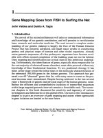
Fig. 1, A typical capillary action Southern transfer system.
4. Decant the depurmation buffer, and replace with 3 gel volumes of denatur-
ation buffer. Incubate with gentle agitation at room temperature for 30 mm.
5. Decant the denaturation buffer, and replace with 3 gel volumes of transfer
buffer. Equilibriate the gel with gentle agitation at room temperature for
30 min.
6. Place the gel on the platform of a capillary transfer system filled with trans-
fer buffer. A diagram of a transfer system is shown m Fig. 1. The system 1s
made up of a platform that sits m a reservoir containing transfer buffer. A
wick, made up of four thtcknesses of 3MM paper, is placed over the plat-
form, soaked m transfer buffer. All air bubbles must be removed from
the wick. The width and length of the platform correspond to the size of
the gel, and the wick is cut to the same width. The platform and reservoir
are made to the same height.
7. Cut a piece of nylon membrane to the same size as the gel. Wet this by
floating it on distilled water and then rinse m transfer buffer. Place the
filter on the gel, and smooth out any air bubbles.
8. Cut four pieces of 3MM paper to the same size as the gel. Soak two m
transfer buffer, and place them over the filter. Smooth out any bubbles.
Place two dry filters onto the sandwich, and then a stack of dry paper tow-
els. Place a l-kg weight on top, and allow the transfer to proceed for at
least 12 h.
9. Disassemble the transfer system. Prior to separating the gel from the filter,
the position of the gel slots can be marked. If this IS done with a pencil, the
marks will appear on the resulting autoradiograph. Rinse the filter m 2X
SSC, and bake the filter at 80°C for 20-60 mm.
Capillary Blotting
25
10. Covalently crosslmk the DNA to the matrix by exposure to a 3 12 nm UV
hght transillummator. Place the filter, DNA side down, on a piece of Saran
WrapTM, and expose for 2-3 min (see Note 5).
The filter can be used immediately for hybridization (see Chapter 6)
or stored dry until required.
4. Notes
1. Note that these methodologies have been developed for neutral nylon mem-
branes (e.g., Amersham Hybond-N) and have not been tested on positively
charged membranes (e.g., Amersham Hybond-N+, Bio-Rad [Richmond,
CA] Zetaprobe, NEN- Du Pont [Boston, MA] GeneScreenTM-Plus).
2. The capillary transfer system described here is efficient, but time-consum-
ing. A number of companies now market other systems (e.g., Vacuum blot-
ting: Pharmacia-LKB [Uppsala, Sweden], Hybaid [Twickenham, UK],
positive pressure blotting: Stratagene [La Jolla, CA]) that reduce the trans-
fer process to as little as 1 h.
3. The size of a hybndrzmg band is determined relattve to DNA standards of a
known size (e.g., bacteriophage h cut wrth EcoRI and HzndIII, or the conve-
nient 1 kb ladder marketed by Life Technologtes [Garthersburg, MD]). It is
best to en&label (see Chapter 15) DNA standards radioactively, such that
an image of their position is produced on the final autoradiograph
4. Gene copy number can be determined by Southern blotting by comparing
the level of hybridization to copy number standards (prepared by dilutmg a
known quantity of the unlabeled cloned DNA fragments) to the level of
hybridization to genomic DNAs. The latter figure should be corrected with
respect to the hybridization of a probe to an endogenous standard host gene.
5. Efficient crosslinking of DNA to nylon filters is achieved with an optimal
amount of exposure to UV light. After a certain value, the efficiency decreases
with increasing exposure. For this reason, it is best to calibrate a UV source
before usage. This can be done by taking filters with an identical amount
of DNA on each, exposing them to UV for different lengths of time, and
then hybridizing them to the same probe. The strongest signal will estab-
lish the optimal time for exposure. Note that the energy output of a stan-
dard UV transilluminator varies with the age of the bulb, thus necessitatmg
regular recalibration. Some manufacturers (e.g., Stratagene) produce UV
crosslinkers that automatically expose the filter to the radiation for the
optimal time, delivering a fixed dose of energy.
References
1. Southern, E. M. (1975) Detection of specific sequences among DNA fragments
separated by gel electrophoresls. J,
Mol Blol 98, 503-5
17
Random Primed 32P-Labeling of DNA
Duncan R. Smith
1. Introduction
Random primed labeling of DNA has now almost superseded the
method of nick translation of DNA. Random primed labeling, based on
the method of Feinberg and Vogelstein (‘I), is a method of incorporating
radioactive nucleotides along the length of a fragment of DNA. Random
primed labeling can give specific activities of between 2 x lo9 and 5 x
1 Og dpm/pg (see Note 1). The method below is essentially that described
by Feinberg and Vogelstein (2) in which a DNA fragment is denatured
by heating in a boiling water bath. Then, random sequence oligonucle-
otides are annealed to both strands. Klenow fragment polymerase is then
used to extend the oligonucleotides, using three cold nucleotides and one
radioactively labeled nucleotide provided in the reaction mixture to pro-
duce a uniformly labeled double-stranded probe. Each batch of random
oligonucleotides contains all possible sequences (for hexamers, which
are most commonly employed, this would be 4096 different oligonucle-
otides), so any DNA template can be used with this method.
2. Materials (see Note 2)
1. DNA fragment to be labeled in water or 1 X TE (10 n&I Tris-HCI, 1 mM
EDTA, pH 8.0, see Note 3).
2. OLB buffer. Make up the following solutions:
Solution 0: 1.25M Tris-HCl, pH 8.0, 0. 125MMgC12 (store at 4°C).
From Methods m Molecular Bology, Vol 58 Basic DNA and RNA Protocols
Edlted by A Harwood Humana Press Inc , Totowa, NJ
27