capillary electrophoresis of carbohydrates
Bạn đang xem bản rút gọn của tài liệu. Xem và tải ngay bản đầy đủ của tài liệu tại đây (1.78 MB, 297 trang )
HUMANA PRESS
Methods in Molecular Biology
TM
Edited by
Pierre Thibault
Susumu Honda
Capillary
Electrophoresis
of Carbohydrates
HUMANA PRESS
Methods in Molecular Biology
TM
VOLUME 213
Edited by
Pierre Thibault
Susumu Honda
Capillary
Electrophoresis
of Carbohydrates
Saccharide Diversity 3
1
Structural and Functional Diversity
of Glycoconjugates
A Formidable Challenge to the Glycoanalyst
Gerald W. Hart
1. Overview of Glycosylation
Glycoconjugates represent the most structurally and functionally diverse
molecules in nature. They range in complexity from relatively simple
glycosphingolipids and nuclear or cytosolic glycoproteins with dynamic
monosaccharide modifications to extraordinarily complex mucins and
proteoglycans (for review, see refs. 1,2). Some of the proteoglycans are per-
haps the most complex molecules in biology, with more than 100 different
saccharide side chains on a single polypeptide. We now realize that most
proteins, even those within intracellular compartments, are co- and/or post-
translationally modified by covalent attachment of saccharides (3).
1.1. The Glycocalyx and Extracellular Matrix
Many early electron microscopic studies using cationic stains, such as
ruthenium red or alcian blue, documented that virtually all cells are surrounded
by thick carbohydrate coats (4,5), termed the “glycocalyx.” The glycocalyx is
comprised of protein- and lipid-bound oligosaccharides and polysaccharides
attached to membrane-associated proteins and lipids. Although electron
micrographs visualize the glycocalyx as a distinct boundary many times the
thickness of the lipid bilayer of the plasma membrane, in reality, the glycocalyx
is probably even larger and is contiguous with the extrinsically associated
extracellular matrix glycoconjugates, which are washed away during sample
preparation for microscopy. Even the simplest eukaryotic cell, the erythrocyte,
has a large and complex glycocalyx (Fig. 1A) about which we have consider-
3
From:
Methods in Molecular Biology, Vol. 213: Capillary Electrophoresis of Carbohydrates
Edited by: P. Thibault and S. Honda © Humana Press Inc., Totowa, NJ
4Hart
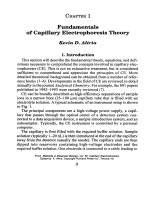
Fig. 1. (A) Electron micrograph of a human erythrocyte stained to illustrate the
large size of the Glycocalyx with respect to the lipid bilayer of the plasma membrane.
(From Voet and Voet, Biochemistry, 2nd ed., with permission). (B) An axonometric
projection of area 350 × 350 Å of the erythrocyte surface, representing approx 10
–5
of
the erythrocyte surface. (Both figures reproduced from ref. 6 with permission from
Elsevier Science).
able structural information (Fig. 1B) (6). The glycocalyx of all cells is com-
prised of an astonishingly complex array of glycoconjugates. This saccharide
“barrier” is critical to the biology of the cell by specifically mediating/modu-
lating its interactions with small molecules, macromolecules, other cells, and
with the extracellular matrix. In many respects, the glycocalyx has the physical
properties of both gel filtration and ion-exhange resins, but is much more com-
Saccharide Diversity 5
plex and selective in its molecular interactions. The protein- and lipid-bound
saccharides of the glycocalyx serve not only as recognition molecules in
multicellular interactions, but also as binding sites for viral and bacterial patho-
gens. The saccharides play a crucial role in the concentration and activation of
ligands for cell-surface receptors and in the lateral organization of membrane-
associated proteins and lipids (for review, see ref. 7).
The spaces between cells of eukaryotic multicellular organisms are filled with
secreted glycoproteins, such as collagens, laminins, fibronectin, and many oth-
ers. In addition, the proteoglycans and glycosaminoglycans play an important
role in fibrillogenesis and organization of the extracellular matrix. All of these
secreted macromolecules self assemble to form highly organized structures such
as basement membranes and lattices that define the elasticity and resiliency of
various tissues. For example, the collagens and proteoglycans secreted by the
three cell types of the cornea of the eye are highly organized to develop and
maintain the transparency of this tissue (8–10). Similarly, the elasticity of carti-
lage is largely defined by the structural organization of water by the collagens
and highly negatively charged proteoglycans that are synthesized in large quan-
tities by chondrocytes (11–13). The glycoconjugates of the extracellular matri-
ces are not only important for their physical properties, but they are also
informational molecules regulating development and cellular trafficking. For
example, we have only recently appreciated the enormous, almost DNA-like,
information content encoded by the specific saccharide modifications along the
sequence of the glycosaminoglycans, such as heparin (14–17). All of these
glycoconjugates display cell-type specific glycoforms, termed “glycotypes,”
whose structures are also developmentally dependent. Not only do these
glycotypes differ in saccharide linkages and chain lengths, but also in minor
saccharide substituents, and nonsaccharide components such as sulfation.
Clearly, elucidation of the structure/function of these macromolecules will
require separation technologies of extraordinary resolution and sensitivities.
1.2. Extracellular Glycoconjugates Have Incredible
Structural Diversity
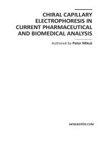
Glycosylation of proteins can be thought of as a spectrum (Fig. 2). At one
end of the spectrum are the collagens, which contain only a few mono- or
disaccharide side chains, and nuclear or cytosolic glycoproteins that contain
clusters of the monosaccharide, N-acetylglucosamine. In the middle of the
spectrum are the mucins, which typically contain many shorter side chains of-
ten terminating in sialic acids (18,19), but may contain so many sugar chains
that they can be mostly carbohydrate by weight. Next are the N-linked glyco-
proteins, which typically have only a few but longer, highly branched complex
saccharide side chains, all having a common inner core structure added en bloc
6Hart
during polypeptide synthesis (20). At the far end of the spectrum are the
proteoglycans, which can contain more than 100 large polysaccharide side
chains, many N-linked and “mucin-type” O-linked saccharide chains attached
to very large protein cores (21,22). For example, the cartilage proteoglycans
are among the most complicated molecules known.
Even though in higher eukaryotes, saccharide side chains are comprised of
only a few common monosaccharide components, including N-acetylglu-
cosamine, N-acetylgalactosamine, mannose, galactose, fucose, glucose, and sialic
acids, the structural diversity possible is much larger than that for proteins or
nucleic acids. This diversity results from the chirality about the glycosidic bond
(anomericity) and the ability of monosaccharides to branch. For example, as
illustrated in Table 1 even a small oligosaccharide with relatively small chain
length (N) has an enormous relative number of structural isomers possible. As
discussed below, extracellular glycoproteins and glycolipids typically have com-
plex glycans attached. The site-specific glycosylation of polypeptides is cell type
and developmental stage specific, as well as being controlled by the environment
surrounding the cell synthesizing the glycoprotein. Indeed, site-specific oligosac-
charide heterogeneity is one of the most important biological features of cell
surface and extracellular glycoproteins (23–26). In general, the outer glycans of
glycosphingolipids, which typically are comprised of saccharides covalently
attached to the lipid ceramide, resemble those of glycoproteins, and sometimes
share similar recognition functions (27–29).
1.3. Intracellular Glyconjugates Have Simpler Glycans
Until recently, dogma in textbooks dictated that nuclear and cytosolic pro-
teins were not glycosylated. However, we now realize that many (perhaps
most?) of these intracellular proteins are dynamically modified by single
Fig. 2. A model depicting the “spectrum” of glycosylated proteins.
Saccharide Diversity 7
N-acetylglucosamine moieties at specific serine or threonine hydroxyls (termed
O-GlcNAc, see Fig. 3) (30). O-GlcNAc is not elongated to more complex struc-
tures, but is simply rapidly added and removed to proteins in a manner similar
to protein phosphorylation. Stoichiometry of protein modification by O-GlcNAc
ranges from less than one sugar per mole of polypeptide to proteins with more
than 15 mol of sugar per mole of protein. Many O-GlcNAc proteins are modi-
fied at numerous sites, each of which is substoichiometrically occupied at any
point in time, making separation of glycoforms and subsequent structural analy-
ses very difficult. Recent data suggest that O-GlcNAc is as abundant as protein
phosphorylation, and may be important to numerous cellular processes. Genetic
knockouts have shown that O-GlcNAc is essential to the life of single cells and
to mammalian ontogeny. Despite its potential biological importance, O-GlcNAc
presents a formidable challenge to the analyst, as addition of the sugar gener-
ally does not affect polypeptide behavior in most of the commonly used sepa-
ration methods such as, sodium dodecyl sulfate-polyacrylamide gel
electrophoresis (SDS-PAGE), reverse-phase high-performance liquid chroma-
tography or other chromatographic techniques, and the current methods of
detection of the saccharide are insensitive (31). In contrast, capillary electro-
phoresis is readily capable of resolving unmodified and O-GlcNAcylated pep-
tides, and with laser-induced fluorescent detection methods, may provide the
sensitivity needed to study the glycosylation of low-abundance regulatory
molecules (32).
Evidence is emerging for the presence of more complex glycoconjugates
within the nucleoplasm and cytoplasm. For example, glycogenin is a glycopro-
tein glucosyltransferase that serves to prime glycogen synthesis by self-
glucosylation of a tyrosine hydroxyl (33,34). Marchase and colleagues have
shown that a key enzyme in energy metabolism, phosphoglucomutase, is
O-mannosylated by a saccharide that is further modified by the attachment of
Table 1
Branching and Anomericity of Saccharides Generates
Enormous Structural Diversity
Number of linear oligomers of length N
Oligosaccharides
N DNA Proteins N = 4 N = 8
14204 8
216400 128 800
3648000 4096 6.4 × 10
4
6 4096 6.4 × 10
7
1.34 × 10
8
3.27 × 10
10
10 1.04 × 10
6
1.28 × 10
13
1.4 × 10
14
1.34 × 10
18
8Hart
α-glucose-1-phosphate (35–37). West and co-workers have shown that a cyto-
solic Dictyostelium protein that is involved in cell-cycle regulation is modified
at hydroxy proline residues by complex oligosaccharides of the type Galα1-6-
Galα1-Fucα1-2Galβ1-3GlcNAc-(HyPro) (38). Raikhel and co-workers have
detected O-GlcNAc oligosaccharides attached to plant nuclear pore proteins
(39 40), and recently sialic acid containing oligosaccharides were suggested to
be on some mammalian nuclear pore proteins. Many studies, even as early as
1964, presented data supporting the presence of glycosaminoglycans within
the nucleus and cytoplasm (41–43). However, these findings remain contro-
versial in the mainstream proteoglycan community. Clearly, researchers study-
ing intracellular processes, such as the cell cycle, transcription, nuclear
transport, or cytoskeletal assembly, can no longer afford to be blissfully igno-
rant of protein glycosylation.
1.4. Classification of Glycolipids and Glycoproteins
The major glycoconjugates in higher eukaryotes are classified as shown in
Table 2 (see ref. 1 for review). This classification is somewhat arbitrary
because many glycoconjugates may contain more than one type of saccharide
component covalently attached. For example, many glycoproteins contain
N-linked saccharides, O-linked saccharides, and a glycosylphosphatidylinositol
(GPI) anchor. Glycoproteins are classified further based on the major type of
linkage between the saccharide and the polypeptide backbone.
Fig. 3. O-Linked N-acetylglucosamine is a dynamic modification found exclusively
in the nucleoplasmic and cytoplasmic compartments of cells.
Saccharide Diversity 9
1.5. Factors Regulating the Attachment of Glycans
to Lipids and Proteins
Even though the glycan moieties of complex glycoconjugates are not them-
selves directly encoded within the genomes of organisms, we now realize that
the covalent glycan modifications of lipids and proteins at specific sites are
carried out with high degrees of regulation and fidelity by specific
glycosyltransferases. There is generally one type of glycosyltransferase activ-
ity for every specific carbohydrate–protein linkage known (44–46). However,
molecular biological analyses have shown that there are also a very large num-
ber of different glycosyltransferase genes encoding enzymes that catalyze very
similar reactions, but that display unique developmental expression and regu-
lation. The sequential combined action of several glycosyltransferases to
produce complex saccharides is controlled not only by the expression of the
enzymes, but also by sugar nucleotide levels, protein synthetic and transport
rates, protein folding rates, and by the regulated compartmentalization of both
substrates and enzymes (47,48). Thus, unlike the structures of polypeptides or
nucleic acids, which are “hard-wired” by the genetic makeup of the cell, the
structures of complex glycans on proteins and lipids dynamically reflect the
metabolic and developmental state, as well as the environment of the cell in
which the glycoconjugate is made.
The responsiveness of the cell’s “glycosylation machinery” to metabolism
and environment provides a powerful mechanism of “fine-tuning” macromo-
lecular structures for cell-specific biological functions. However, the inherent
structural diversity of glycan structures and their highly varied physical
properties also represent a formidable challenge to traditional separation tech-
nologies developed primarily for polypeptides and nucleic acids. Thus, eluci-
dation of the structure/functions of complex glycoconjugates will require the
development of new high-resolution, high-sensitivity analytical methods.
Recent developments in capillary electrophoretic methods, as described in this
book, represent a potential breakthrough in our ability to characterize small
amounts of biologically important glycoconjugates (49–59).
2. Glycolipids
Glycosphinoglipids (GSLs), which are made up of glycans covalently
attached to ceramide, are the most common type glycolipid in eukaryotes
(29,60). Other types of glycolipids include rare glycosylated glycerolipids and
free glycosyl inositol phospholipids (GIPLs; see Subheading 3.5.). GIPLs have
mainly been studied in protozoan parasites, but are present in mammals. They
appear to either be biosynthetic intermediates for GPI anchors or they may
serve as signaling molecules (61–64).
10 Hart
Glycosphingolipids function in many biological processes in a manner simi-
lar to glycoproteins. They are blood group and tumor-specific antigens, they
serve as receptors for microorganisms and toxins, and they mediate numerous
cellular interactions. Recently, GSLs have been found to play an important
role in growth regulation by modulating the activities of transmembrane recep-
tor kinases. The abundance of GSLs varies considerably with the type of
membrane. GSLs represent 5–10% of the total lipid in the erythrocyte mem-
brane, as much as 30% of the total lipid of neuronal membranes, and are virtu-
ally absent in mitochondrial membranes.
GSLs are amphipathic molecules, and unlike glycoproteins or glycopep-
tides are readily analyzed by simple high-resolution chromatographic
techiques, the most common of which is thin-layer chromatography.
Glycosphingolipids are also comparatively very well behaved in mass spectro-
metric analyses.
2.1. Glycosphingolipid Structural Variability
As indicated in Subheading 2.,GSLs are composed of glycans glycosid-
ically linked to ceramide. Ceramide is comprised of a long-chain amino alco-
hol, sphingosine, to which fatty acids are attached by an amide linkage. In
mammalian GSLs, the glycan structures on GSLs typically range in size from
one to ten monosaccharides, with some being much larger. There is also con-
siderable variability in the structures and lengths of the fatty acid substituents,
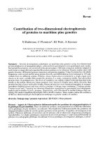
depending on the tissue, cell-type, and species of origin (Fig. 4). Acidic GSLs
include the ganglio series, which contain sialic acids and the sulfatides, which
often contain sulfate esters attached to galactosylceramides. Neutral GSLs
range from those containing only one monosaccharide, such as globosides, to
those containing variable length repeating structures such as the lactoside and
globoside series. The structural variability of the glycan portions of GSLs is
very large and rivals that seen for the glycosylation of proteins. In fact, glyco-
proteins and GSLs have many of the same terminal saccharide structures (27).
Unlike glycoproteins which display enormous numbers of glycan structures at
a single glycosylation site, even when made by clonal cell populations
(23,65,66), each glycan structure on a GSL is classified as a different species.
Given that the amphipathic character of GSLs greatly facilitates their separa-
tion and study, recent methods for the study of the glycans on glycoproteins
have resorted to first releasing the glycans from the protein and chemically
converting them to so-called “neoglycolipids” prior to study. Formation of
neoglycolipids from released glycans not only improves the chromatographic
or electrophoretic behavior of the glycans, but also allows for the introduction
of fluorescent or charged residues which greatly facilitate physical separations
and detection.
Saccharide Diversity 11
3. Glycoproteins
As mentioned previously, glycoproteins are classified by how the major
saccharide side chain is attached to the polypeptide core (Table 2). While
most is known about the biosynthesis, structures and functions of the aspar-
agine-linked (N-linked) glycoproteins (67), it is clear that the “mucin-type”
O-linked glycoproteins, which contain saccharides linked via
N-acetylgalactosamine to serine or threonine (GalNAc-Ser[Thr]) residues,
are likely as abundant, and just as important to many biological processes,
including the trafficking of blood cells, and defenses against microorgan-
isms. Collagens are among the most abundant glycoproteins and represent
the only common example of glycosylated hydroxylysine residues in higher
organisms. Of the collagen types, those species found enriched in basement
membranes are the most heavily glycosylated (68).
Fig. 4. Classification of glycosphingolipids according to their glycan structures.
Table 2
Major Types of Glyconjugates
Glycoproteins: Asn-linked; GalNAc-Ser(Thr); GlcNAc-Ser(Thr); collagens; glycogen
Proteoglycans: Many diverse types; contain one or more glycosaminoglycans
Glycosphingolipids: Glycosylated ceramides: gangliosides; neutral GSLs, sulfatides
Phosphatidylinositol Glycans: GPI anchors; free GPIs
12 Hart
3.1. Mucin-Type
O
-Glycans
The complex glycans derived from mucin-type glycoproteins can readily be
released from the protein by alkali-induced β-elimination (69–72). However,
due to “peeling” reactions that destroy the saccharides from the newly exposed
reducing end, these eliminations must generally be performed in the presence
of a reducing agent such as borohydride, complicating the easy modification of
the released saccharides with chromophores. Fortunately, as described in later
chapters, methods such as hydrazinolysis (73–75) have circumvented these
problems. Analysis of mucin-type saccharides has also been slowed by the
lack of a nondelective enzyme that will release intact O-glycans from proteins,
as exists for N-linked glycans (e.g., peptide:N-glycosidase F) (76,77) and GSLs
(glycoceramidase) (78). O-glycanase, which is commercially available, is
unfortunately specific only for Galβ1–3-GalNAc-Ser(Thr) structures and will
not release glycans from more complex O-linked glycoproteins (79).
GalNAc-Ser(Thr)-linked saccharides have been most well studied on
mucins, which contain a very heterogeneous population of clustered regions of
short saccharides that often terminate in sialic acids. The protein core and sac-
charide modifications on mucins are different for each cell type in which they
are made, and molecular biological studies have now described several distinct
types of core proteins (18,80–82). Most of these core proteins have regions
rich in proline, serine, threonine, glycine, and other amino acids that give rise
to distinctive mucin-like motifs. These motifs are often very extensively
glycosylated. Such mucin regions form rigid rod structures in solution owing
to the close spacing of bulky hydrophilic groups and negative charges along
their backbone. Mucins not only serve to lubricate epithelial surfaces and pro-
tect them from desiccation, but also, owing to their almost infinite structural
diversity, they serve as “decoy” binding sites for pathogenic microorganisms,
protecting host cells from invasion. Heavily glycosylated mucin domains also
serve a structural role in many receptors by creating a rigid rod domain that
allows the business end of the receptor to be displayed above the glycocalyx of
the cell (83). Given their comparatively small size, enormous diversity, and the
ability to be derivatized at their reducing termini, capillary electrophoresis
should prove to be a valuable tool in the study of these important but largely
neglected class of glycoproteins.
3.2.
O
-Linked
N
-Acetylglucosamine
The dynamic modification of nuclear and cytosolic proteins by
N-acetylglucosamine at specific serine and threonine residues (termed
O-GlcNAc, Fig. 3) is now known to be ubiquitous and abundant in virtually all
eukaryotic cells (30,84), with the possible exception of baker’s yeast.
O-GlcNAc has not yet been described in prokaryotes and does not appear to
Saccharide Diversity 13
occur in lumenal or extracellular compartments, locations where other forms
of glycosylation predominate. O-GlcNAc is found on myriad proteins in the
nucleus. As summarized in Table 3, many important regulatory proteins
are dynamically modified by O-GlcNAc. O-GlcNAc is added to proteins by
the O-GlcNAc transferase (85,86), which has been recently shown to be essen-
tial for single cell viability. O-GlcNAc is removed by O-GlcNAcase, one of
which has been characterized (87). These two enzymes are analogous to kinases
and phosphatases, respectively, for phosphorylation. In several cases,
O-GlcNAcylation and phosphorylation are reciprocal events, suggesting a
“yin–yang” relationship in terms of biological functions (88). Current evidence
suggests that O-GlcNAcylation may play an important role in the regulation of
transcription, translation, nuclear transport, cytoskeletal assembly, the cell
cycle, diabetes, and in the regulation of protein turnover.
Biochemical analyses of O-GlcNAcylation is complicated by the low abun-
dance and rapid turnover of most regulatory proteins, the low stoichiometry of
O-GlcNAc at individual sites, and the lack of sensitive detection methods. As
mentioned earlier, most currently used separation methods do not detect the
addition and removal of O-GlcNAc on most proteins. In addition, O-GlcNAc
is very labile, both due to the abundance of N-acetylglucosaminidases in cells,
Table 3
Identified
O
-GlcNAc Proteins
Nucleus
Nucleoporins
RNA polymerase II
Transcription factors: TBP, SP1, SRF, IPF-1
Kinases and splicing proteins: CK2 and SRs
Nuclear oncoproteins: c-Myc, v-Erb, SV40
Estrogen receptors: α and β
Tumor suppressors: Rb, p53
Many chromatin proteins: polytene
Fungal DNA binding, tyrosine phosphatase
Cytoplasm
Intermediate filaments: cytokeratins, neurofilaments
Bridging proteins: talin, vinculin, ankyrin, synapsins, 4.1
Microtubule-associated proteins: (MAPS): tau
Clathrin assembly protein
Many synapse and neuron proteins: APP
Small heat shock proteins
Signaling proteins: Raf
Many viral and parasite proteins
14 Hart
and the chemical/physical stability of the linkage itself. For example, it is dif-
ficult to detect O-GlcNAcylation even by mass spectrometry (MS). In
electrospray techniques, the saccharide is readily cleaved at commonly used
orifice voltages and is almost always lost prior to peptide fragmentation, mak-
ing MS/MS site mapping problematic. However, prior β-elimination of the
saccharide followed by electrospray mass spectrometry has allowed for direct
site mapping (89,90). In matrix-assisted laser desorption (MALDI) mass spec-
trometric methods the presence of the GlcNAc moiety typically lowers the
sensitivity of detection by at least five fold compared to that for the unmodified
peptide. In mixtures, suppression of the glycopeptide signals by unmodified
peptides makes analyses even more difficult. Generally, reverse-phase HPLC
does not readily resolve O-GlcNAc modified and unmodified peptides, but this
depends a great deal on the relative hydrophobicity of the peptide to which the
sugar is attached. In contrast, under the right conditions, capillary electrophore-
sis has the resolving power to readily separate O-GlcNAc, O-phosphate, and
unmodified peptides from each other (32). We anticipate that the combined use
of capillary electrophoresis and nanospray MS will play an important role in
elucidating the functions of O-GlcNAc on many key regulatory proteins.
3.3.
N
-Glycans
Asparagine-linked (N-linked) glycans are the most extensively studied form
of protein glycosylation (67). N-glycans are attached to nascent polypeptides
as they enter the lumen of the rough endoplasmic reticulum (RER) at specific
asparagine residues in the sequon Asn-X-Ser(Thr), where X can be almost any
amino acid, but generally is not proline or aspartate. In the RER, a large
oligosaccharide, Glc
3
Man
9
GlcNAc
2
-, is transferred directly to the protein en bloc
from a C
95
isoprenoid lipid donor, dolichol phosphate. The oligosaccharyl
dolichol phosphate donor substrate is preassembled in the RER. The enzyme
complex that accomplishes the transfer of the oligosaccharide to the protein is
the oligosaccharyl transferase (91). Immediately after transfer to the nascent
chain, an unusual processing of the N-glycan begins in which the outer glucose
residues and mannose residues are enzymatically removed as the protein is
transported through the secretory pathway (20). We now realize that the glu-
cose residues are part of an exquisite quality control mechanism involving the
glucose binding lectins calreticulin and calnexin (92,93) and reglucosylation
by an “unfolded protein” specific glucosyltransferase (94,95) that together pre-
vent misfolded proteins from leaving the RER. On entering the Golgi, typi-
cally, trimming of the N-glycans reaches a branch point at the oligosaccharide
Man
5
GlcNAc
2
, where if the saccharide is acted on by N-acetylglucos-
aminyltransferase I, it will be processed further to become a complex N-gly-
can, containing outer sugars such as galactose and sialic acids. If the N-glycan
Saccharide Diversity 15
is not acted upon by the N-acetylglucosaminyltransferase, it will remain a
“high-mannose” type saccharide.
A characteristic feature of N-glycans is their extensive branching which is
controlled by a number of specific N-acetylglucosaminyltransferases (96,97).
Perhaps the most important aspect of N-linked glycosylation in terms of biol-
ogy is site-specific oligosaccharide microheterogeneity. On most populations
of a glycoprotein, there can be many different glycan structures at a single site,
even though the amino acid sequences are identical in the population. The
amount and distribution of these glycoforms are highly reproducible depend-
ing on the growth conditions of the cell and the glycoforms are usually
cell-type specific (glycotypes). It appears that the purpose of the elaborate
biosynthetic/processing pathway for N-linked glycoproteins is not only to regu-
late trafficking and folding, but also to allow the cell to structurally remodel the
proteins it is synthesizing in response to its environment and developmental state.
3.4. Proteoglycans
By definition, a proteoglycan is any polypeptide that contains one or more
glycosaminoglycan (GAG) side chains (98–100). Clearly, many proteoglycans
also contain other types of sugar modifications. GAGs are long linear poly-
mers composed of repeating disaccharide sequences typically containing an
amino sugar and a uronic acid (except for keratan sulfates, which contain an
amino sugar and galactose residues). Except for hyaluronic acids, GAGs are
also extensively modified by sulfate esters. Table 4 summarizes the major
types of GAGs and their linkage to protein. Virtually every imaginable type of
proteoglycan has been found in various cell types. Some proteoglycans are mem-
brane proteins with only one or a few GAG chains, whereas others are secreted
molecules with more than 100 different GAG and other saccharide modifica-
tions. Many of the proteoglycan core proteins have been cloned and character-
ized, yet we still know little about the detailed structures of the intact molecules
of even the simplest proteoglycans. Proteoglycans are important structural
components, they serve to regulate development and fibrillogenesis of collagen,
and they regulate growth hormone functions. Capillary electrophoresis is play-
ing an important role in the structural elucidation of GAG chains, particularly
with respect to the separation of GAG fragments produced by controlled chemi-
cal or enzymatic degradations (101,102).
3.5. GPI Anchors
Until the mid-1980s it was widely believed that most integral membrane
proteins were anchored to the lipid bilayer by stretches of hydrophobic amino
acids. Initially studies with phospholipases (103) suggested that some proteins
were anchored by covalently attached lipid components. Structural studies in
16 Hart
parasites documented that certain proteins are anchored to the membrane by
GPI anchor structures (104) at their C-termini. Figure 5 summarizes the struc-
ture of a GPI anchor and illustrates the growing structural heterogeneity that
has been found in various organisms and cell types. The GPI anchor is
assembled in the RER by first attaching GlcNAc to phosphatidylinositol (105).
The GlcNAc is deacetylated and the mannosyl core is added. Ethanolamine
phosphate is attached using phosphatidylethanolamine as the donor. Proteins
to receive a GPI anchor have a hydrophobic signal sequence at their C-termi-
nus, which serves to temporarily anchor them to the RER membrane. A
transpeptidase cleaves the signal sequence and concomitantly transfers the pep-
tide to the lipid anchor. Outer sugars, such as galactose, are added to the anchor
in the Golgi (for review, see ref. 106). GPI anchors are another remarkable
example of how important posttranslational modifications can be completely
overlooked. In fact, it is now clear that most membrane proteins in protozoans
are anchored by GPI anchors (107), whereas the majority of membrane
proteins in eukaryotes are anchored by hydrophobic peptides. There are also
several examples of proteins that are bound to the membrane by both GPI
anchors and by peptide sequences, depending on RNA splicing. While there
has been much speculation about the purpose of GPI anchors in terms of mem-
brane mobility, role or lack thereof in signaling, and in the controlled release of
proteins, the functions of this mode of attachment remain unclear (108).
Table 4
Classification of Glycosaminoglycans
Repeating disaccharide (A–B)
n
Sulfate per
GAG Mol Wt. Monosaccharide A Monosaccharide B disaccharide
Hyaluronic 4000–
D-Glucuronic acid N-acetylglucosamine 0
acids 8 × 10
6
Chondroitin 5000– D-Glucuronic acid N-Acetylgalactosamine 0.2–2.3
sulfates 50,000
Dermatan 15,000– D-Glucuronic acid or N-Acetylgalactosamine 1.0–2.0
sulfates 40,000 L-Iduronic acid
Heparan 5000– D-Glucuronic acid or N-Acetylglucosamine 0.2–2.0
sulfates 12,000 L-Iduronic acid
Heparin 6000– D-Glucuronic acid or N-Acetylglucosamine 2.0–3.0
25,000 L-Iduronic acid
(mostly)
Keratan 4000– D-Galactose N-Acetylglucosamine 0.9–1.8
sulfates 19,000
Saccharide Diversity 17
4. Conclusions and Generalizations
In recent years, we have come to appreciate that most eukaryotic proteins
are covalently modified by the attachment of sugars. Glycobiology, which is
now the name of the field attempting to elucidate the structural/functional
importance of protein glycosylation, has become one of the most rapidly grow-
ing areas of biochemistry and cell biology. The enormous structural diversity of
complex glycans potentially allows the cell to express vast amounts of biological
information. Indeed, glycoconjugates are critical molecules in virtually every
biological process in eukaryotic organisms, including almost every infectious
and noninfectious disease afflicting mankind. Protein-bound saccharides are
thought to modify or fine-tune a protein’s functions at the structural level.
However, unlike proteins or nucleic acids, which are genetically encoded, the
structures of glycans are highly responsive to, and dependent on, both the meta-
bolic and developmental state of a cell. The study of glycoproteins has, until
recently, been hindered by the inherent complexities and structural diversity of
the molecules themselves, by the lack of tools for their study at the structural
level, and by a lack of knowledge about multicellular systems in which many
of the functions of protein-bound glycans reveal themselves. For conve-
nience, the reader is referred to the Appendix of this book for a description of
the structures of typical mono-, oligo-, and polysaccharides found in prokary-
otic and eukaryotic cells. Many of the methods described herein provide
much needed approaches toward our better understanding of these enigmatic
molecules.
Fig. 5. Illustration of the structural diversity of GPI anchors.
18 Hart
References
1. Varki, A., Cummings, R., Esko, J., Freeze, H., Hart, G., and Marth, J. (1999)
Essentials of Glycobiology, Cold Spring Harbor Laboratory Press, Cold Spring
Harbor, NY.
2. Hart, G. W. (1992) Glycosylation. Curr. Opin. Cell Biol. 4, 1017–1023.
3. Hart, G. W., Haltiwanger, R. S., Holt, G. D., and Kelly, W. G. (1989)
Glycosylation in the nucleus and cytoplasm, Annu. Rev. Biochem. 58, 841–874.
4. Frey, A., Giannasca, K. T., Weltzin, R., Giannasca, P. J., Reggio, H., Lencer, W. I.,
and Neutra, M. R. (1996) Role of the glycocalyx in regulating access of microparticles
to apical plasma membranes of intestinal epithelial cells: implications for microbial
attachment and oral vaccine targeting. J. Exp. Med. 184, 1045–1059.
5. Debbage, P. L. (1996) A systematic histochemical investigation in mammals of
the dense glycocalyx glycosylations common to all cells bordering the interstitial
fluid compartment of the brain. Acta. Histochem. 98, 9–28.
6. Viitala, J. and Jarnefelt, J. (1985) The red cell surface revisited. TIBS 14, 392–395.
7. Varki, A. (1993) Biological roles of oligosaccharides: all of the theories are cor-
rect. Glycobiology 3, 97–130.
8. Scott, J. E. (1991) Proteoglycan: collagen interactions and corneal ultrastructure.
Biochem. Soc. Trans. 19, 877–881.
9. Jester, J. V., Moller-Pedersen, T., Huang, J. Y., Sax, C. M., Kays, W. T., Cavangh,
H. D., et al. (1999) The cellular basis of corneal transparency: evidence for ‘cor-
neal crystallins.’ J. Cell Sci. 112, 613–622.
10. Nakazawa, K., Suzuki, S., and Wada, K. (1995) Proteoglycan synthesis by corneal
explants from developing embryonic chicken. J. Biochem. (Tokyo) 117, 707–718.
11. Kim, Y. J., Grodzinsky, A. J., and Plaas, A. H. K. (1996) Compression of carti-
lage results in differential effects on biosynthetic pathways for aggrecan, link
protein, and hyaluronan. Arch. Biochem. Biophys. 328, 331–340.
12. Wallis, G. A. (1995) Cartilage disorders: the importance of being sulphated. Curr.
Biol. 5, 225–227.
13. Caplan, A. I., Fiszman, M. Y., and Eppenberger, H. M. (1983) Molecular and cell
isoforms during development. Science 221, 921–927.
14. Dietrich, C. P., Tersariol, I. L. S., Toma, L., Moraes, C. T., Porcionatto, M. A.,
Oliveira, F. W., and Nader, H. B. (1998) Structure of heparan sulfate: identifica-
tion of variable and constant oligosaccharide domains in eight heparan sulfates
of different origins. Cell Mol. Biol. 44, 417–429.
15. Salmivirta, M., Lidholt, K., and Lindahl, U. (1996) Heparan sulfate: a piece of
information. FASEB J. 10, 1270–1279.
16. Faham, S., Hileman, R. E., Fromm, J. R., Linhardt, R. J., and Rees, D. C. (1996)
Heparin structure and interactions with basic fibroblast growth factor. Science
271, 1116–1120.
17. Keller, J. M. (1994) Specificity in heparin/heparan sulphate-protein interactions.
Glycobiology 4, 1–2.
18. Devine, P. L. and McKenzie, I. F. C. (1992) Mucins: structure, function, and
associations with malignancy. BioEssays 14, 619–625.
Saccharide Diversity 19
19. Strous, G. J. and Dekker, J. (1992) Mucin-type glycoproteins. CRC Crit. Rev.
Biochem. Mol. Biol. 27, 57–92.
20. Kornfeld, R. and Kornfeld, S. (1985) Assembly of asparagine-linked oligosac-
charides. Annu. Rev. Biochem. 54, 631–664.
21. Muir, H. (1990) Structure, biology and pathology of proteoglycans. The coming
of age of proteoglycans. Biochem. Soc. Trans. 18, 787–789.
22. Oldberg, A., Antonsson, P., Hedbom, E., and Heinegård, D. (1990) Structure and
function of extracellular matrix proteoglycans. Biochem. Soc. Trans. 18, 789–792.
23. Swiedler, S. J., Freed, J. H., Tarentino, A. L., Plummer, T. H., Jr., and Hart, G.
W. (1985) Oligosaccharide microheterogeneity of the murine major histocom-
patibility antigens. Reproducible site-specific patterns of sialylation and branch-
ing in asparagine-linked oligosaccharides. J. Biol. Chem. 260, 4046–4054.
24. Rademacher, T. W., Parekh, R. B., and Dwek, R. A. (1988) Glycobiology. Annu.
Rev. Biochem. 57, 785–838.
25. Parekh, R. B., Dwek, R. A., Thomas, J. R., Opdenakker, G., and Rademacher, T.
W. (1989) Cell-type-specific and site-specific N-glycosylation of type I and type
II human tissue plasminogen activator. Biochemistry 28, 7644–7662.
26. Rudd, P. M., Joao, H. C., Coghill, E., Fiten, P., Saunders, M. R., Opdenakker, G.,
and Dwek, R. A. (1994) Glycoforms modify the dynamic stability and functional
activity of an enzyme. Biochemistry 33, 17–22.
27. Rauvala, H. and Finne, J. (1979) Structural similarity of the terminal carbohy-
drate sequences of glycoproteins and glycolipids. FEBS Lett. 97, 1–8.
28. Hakomori, S. and Igarashi, Y. (1995) Functional role of glycosphingolipids in
cell recognition and signaling. J. Biochem. (Tokyo) 118, 1091–1103.
29. Hakomori, S. and Igarashi, Y. (1993) Gangliosides and glycosphingolipids as
modulators of cell growth, adhesion, and transmembrane signaling. Adv. Lipid
Res. 25, 147–162.
30. Hart, G. W. (1997) Dynamic O-Linked glycosylation of nuclear and cytoskeletal
proteins. Ann. Rev. Biochem. 66, 315–335.
31. Roquemore, E. P., Chou, T Y., and Hart, G. W. (1994) Detection of O-linked
N-acetylglucosamine (O-GlcNAc) on cytoplasmic and nuclear proteins. Methods
Enzymol. 230, 443–460.
32. Snow, D. M., Shaper, J. H., Shaper, N. L., and Hart, G. W. (1999) Determination
of ß1,4-galactosyltransferase enzymatic activity by capillary electrophoresis and
laser-induced fluorescence detection. Analyt. Biochem. 271, 36–42.
33. Alonso, M. D., Lomako, J., Lomako, W. M., and Whelan, W. J. (1995) A new
look at the biogenesis of glycogen. FASEB J. 9, 1126–1137.
34. Lomako, J., Lomako, W. M., and Whelan, W. J. (1990) The nature of the primer
for glycogen synthesis in muscle. FEBS Lett. 268, 8–12.
35. Marchase, R. B., Bounelis, P., Brumley, L. M., Dey, N., Browne, B., Auger, D., et al.
(1993) Phosphoglucomutase in Saccharomyces cerevisiae is a cytoplasmic glycopro-
tein and the acceptor for a Glc-phosphotransferase. J. Biol. Chem. 268, 8341–8349.
36. Veyna, N. A., Jay, J. C., Srisomsap, C., Bounelis, P., and Marchase, R. B. (1994)
The addition of glucose-1-phosphate to the cytoplasmic glycoprotein phospho-
20 Hart
glucomutase is modulated by intracellular calcium in PC12 cells and rat cortical
synaptosomes. J. Neurochem. 62, 456–64.
37. Fu, L., Bounelis, P., Dey, N., Browne, B. L., Marchase, R. B., and Bedwell, D.
M. (1995) The posttranslational modification of phosphoglucomutase is regu-
lated by galactose induction and glucose repression in Saccharomyces cerevisiae.
J. Bacteriol. 177, 3087–94.
38. Teng-umnua, P., Morris, H. R., Dell, A., Panico, M., Paxton, T., West, C. M.
(1998) The cytoplasmic F-box binding protein SKP1 contains a novel
pentasaccharide linked to hydroxyproline in dictyostelium. J. Biol. Chem. 273,
18,242–18,249.
39. Heese-Peck, A., Cole, R. N., Borkhsenious, O. N., Hart, G. W., and Raikhel, N.
V. (1995) Plant nuclear pore complex proteins are modified by novel oligosac-
charides with terminal N-acetylglucosamine. Plant Cell 7, 1459–1471.
40. Heese-Peck, A. and Raikhel, N. V. (1998) A glycoprotein modified with terminal
N-acetylglucosamine and localized at the nuclear rim shows sequence similarity
to aldose-1-epimerases. Plant Cell 10, 599–612.
41. Kinoshita, S. and Saiga, H. (1979) The role of proteoglycan in the development
of sea urchins. I Abnormal development of sea urchin embryos caused by the
disturbance of proteoglycan synthesis. Exp.Cell Res. 123, 229–236.
42. Kinoshita, S. (1974) Some observations on a protein-mucopolysaccharide com-
plex found in sea urchin embryos. Exp. Cell Res. 85, 31–40.
43. Hiscock, D. R. R., Yanagishita, M., and Hascall, V. C. (1994) Nuclear localization of
glycosaminoglycans in rat ovarian granulosa cells. J. Biol. Chem. 269, 4539–4546.
44. Fukuda, M., Bierhuizen, M. F. A., and Nakayama, J. (1996) Expression cloning
of glycosyltransferases. Glycobiology 6, 683–689.
45. Baenziger, J. U. (1994) Protein-specific glycosyltransferases: how and why they
do it! FASEB J. 8, 1019–1025.
46. Joziasse, D. H. (1992) Mammalian glycosyltransferases: genomic organization
and protein structure. Glycobiology 2, 271–277.
47. Hirschberg, C. B., Robbins, P. W., and Abeijon, C. (1998) Transporters of nucle-
otide sugars, ATP, and nucleotide sulfate in the endoplasmic reticulum and Golgi
apparatus. Annu. Rev. Biochem. 67, 49–69.
48. Abeijon, C., Mandon, E. C., and Hirschberg, C. B. (1997) Transporters of nucleotide
sugars, nucleotide sulfate and ATP in the Golgi apparagus. TIBS 22, 203–207.
49. Hennebicq, S., Tetaert, D., Soudan, B., Briand, G., Richet, C., Demeyer, D., et al.
(1998) Polypeptide:N-acetylgalactosaminyltransferase activities towards the
mucin MUC5AC peptide motif using microsomal preparations of normal and
tumoral digestive mucosa. Biochimie 80, 69–73.
50. Suzuki, S. and Honda, S. (1998) A tabulated review of capillary electrophoresis
of carbohydrates. Electrophoresis 19, 2539–2560.
51. Soga, T. and Heiger, D. N. (1998) Simultaneous determination of monosaccha-
rides in glycoproteins by capillary electrophoresis. Analyt. Biochem. 261, 73–78.
52. Chen, F. T. A. and Evangelista, R. A. (1998) Profiling glycoprotein N-linked
oligosaccharide by capillary electrophoresis. Electrophoresis 19, 2639–2644.
Saccharide Diversity 21
53. Denuzière, A., Taverna, M., Ferrier, D., and Domard, A. (1997) Capillary elec-
trophoresis of glycosaminoglycan-derived disaccharides: application to stability
studies of glycosaminoglycan chitosan complexes. Electrophoresis 18, 745–750.
54. Sato, K., Okubo, A., and Yamazaki, S. (1997) Determination of monosaccha-
rides derivatized with 2-aminobenzoic acid by capillary electrophoresis.
Analyt.Biochem. 251, 119–121.
55. El Rassi, Z. (1997) Recent developments in capillary electrophoresis of carbohy-
drate species. Electrophoresis 18, 2400–2407.
56. Scapol, L., Marchi, E., and Viscomi, G. C. (1996) Capillary electrophoresis of
heparin and dermatan sulfate unsaturated disaccharides with triethylamine and
acetonitrile as electrolyte additives. J. Chromatogr. A. 735, 367–74.
57. Hoffstetter-Kuhn, S., Alt, G., and Kuhn, R. (1996) Profiling of oligosaccharide-
mediated microheterogeneity of a monoclonal antibody by capillary electrophore-
sis. Electrophoresis 17, 418–422.
58. Kakehi, K. and Honda, S. (1996) Analysis of glycoproteins, glycopeptides and
glycoprotein-derived oligosaccharides by high-performance capillary electro-
phoresis. [Review] [48 refs]. J. Chromatogr. A. 720, 377–393.
59. Zhou, W. and Baldwin, R. P. (1996) Capillary electrophoresis and electrochemi-
cal detection of underivatized oligo- and polysaccharides with surfactant-con-
trolled electroosmotic flow. Electrophoresis 17, 319–324.
60. Hakomori, S. I., Yamamura, S., and Handa, K. (1998) Signal transduction through
glyco(sphingo)lipids - Introduction and recent studies on glyco(sphingo)lipid-
enriched microdomains. Ann. NY Acad. Sci. 845, 1–10.
61. McConville, M. J. and Blackwell, J. M. (1991) Developmental changes in the
glycosylated phosphatidylinositols of Leishmania donovani. Characterization of
the promastigote and amastigote glycolipids. J. Biol. Chem. 266, 15,170–15,179.
62. McConville, M. J., Homans, S. W., Thomas-Oates, J. E., Dell, A., and Bacic, A.
(1990) Structures of the glycoinositolphospholipids from Leishmania major. A fam-
ily of novel galactofuranose-containing glycolipids. J. Biol. Chem. 265, 7385–7394.
63. Frick, W., Bauer, A., Bauer, J., Wied, S., and Müller, G. (1998) Structure-activ-
ity relationship of synthetic phosphoinositolglycans mimicking metabolic insu-
lin action. Biochemistry 37, 13,421–13,436.
64. Gaulton, G., Kelly, K. L., Pawlowski, J., Mato, J. M., and Jarett, L. (1988) Regu-
lation and function of an insulin-sensitive glycosyl-phosphatidylinositol during
T lymphocyte activation. Cell 53, 963–970.
65. Ashford, D. A., Alafi, C. D., Gamble, V. M., Mackay, D. J. G., Rademacher, T.
W., Williams, P. J., et al. (1993) Site-specific glycosylation of recombinant rat
and human soluble CD4 variants expressed in Chinese hamster ovary cells.
J. Biol. Chem. 268, 3260–3267.
66. Rudd, P. M., Leatherbarrow, R. J., Rademacher, T. W., and Dwek, R. A. (1991)
Diversification of the IgG molecule by oligosaccharides. Mol. Immunol. 28,
1369–1378.
67. Lennarz, W. J. (1980) The Biochemistry of Glycoproteins and Proteoglycans, 1st
ed., Plenum Press, New York, NY.
22 Hart
68. Yurchenco, P. D. and Schittny, J. C. (1990) Molecular architecture of basement
membranes. FASEB J. 4, 1578–1590.
69. Cancilla, M. T., Penn, S. G., and Lebrilla, C. B. (1998) Alkaline degradation of
oligosaccharides coupled with matrix-assisted laser desorption/ionization Fou-
rier transform mass spectrometry: a method for sequencing oligosaccharides.
Analyt. Chem. 70, 663–672.
70. Duk, M., Ugorski, M., and Lisowska, E. (1997) Beta-elimination of O-glycans
from glycoproteins transferred to Immobilon P membranes: method and some
applications. Anal. Biochem. 253, 98–102.
71. Greis, K. D., Hayes, B. K., Comer, F. I., Kirk, M., Barnes, S., Lowary, T. L., and
Hart, G. W. (1996) Selective detection and site-analysis of O-GlcNAc-modified
glycopeptides by beta-elimination and tandem electrospray mass spectrometry.
Analyt. Biochem. 234, 38–49.
72. Vercellotti, J. R., Nienaber, N., and Chang, C. J. (1970) Monosaccharides as
O-glycosyl leaving groups from 3-hydroxy amino acids during base-catalyzed
elimination. Carbohydr. Res. 13, 63–74.
73. Mellors, A. and Sutherland, D. R. (1994) Tools to cleave glycoproteins. Trends
Biotechnol. 12, 15–18.
74. Patel, T., Bruce, J., Merry, A., Bigge, C., Wormald, M., Jaques, A., and Parekh,
R. (1993) Use of hydrazine to release in intact and unreduced form both N- and
O-linked oligosaccharides from glycoproteins. Biochemistry 32, 679–693.
75. Patel, T. P. and Parekh, R. B. (1994) Release of oligosaccharides from glycopro-
teins by hydrazinolysis. Methods Enzymol. 230, 57–66.
76. Nuck, R., Zimmermann, M., Sauvageot, D., Josic, D., and Reutter, W. (1990)
Optimized deglycosylation of glycoproteins by peptide-N4-(N-acetyl-beta-
glucosaminyl)-asparagine amidase from Flavobacterium meningosepticum.
Glycoconjugate J. 7, 279–286.
77. Tarentino, A. L. and Plummer, T. H., Jr. (1994) Enzymatic deglycosylation of
asparagine-linked glycans: purification, properties, and specificity of oligosac-
charide-cleaving enzymes from Flavobacterium meningosepticum. Methods
Enzymol. 230, 44–57.
78. Yamagata, T. and Ito, M. (1992) In CRC Handbook of Endoglycosidases and
Glycoamidases (Takahashi, N. and Muramatsu, T., eds.), CRC Press, Boca Raton,
FL, pp. 133–182.
79. Fan, J Q., Yamamoto, K., Matsumoto, Y., Hirabayashi, Y., Kumagai, H., and
Tochikura, T. (1990) Action of endo-alpha-N-acetylgalactosaminidase from
Alcaligenes sp. on amino acid-O-glycans: comparison with the enzyme from
Diplococcus pneumoniae. Biochem. Biophys. Res. Commun. 169, 751–757.
80. Wu, A. M., Csako, G., and Herp, A. (1994) Structure, biosynthesis, and function
of salivary mucins. Mol. Cell. Biochem. 137, 39–55.
81. Shimizu, Y. and Shaw, S. (1993) Cell adhesion: mucins in the mainstream. Nature
366, 630,631.
82. Thornton, D. J., Carlstedt, I., Howard, M., Devine, P. L., Price, M. R., and
Sheehan, J. K. (1996) Respiratory mucins: identification of core proteins and
glycoforms. Biochem. J. 316, 967–975.
Saccharide Diversity 23
83. Jentoft, N. (1990) Why are proteins O-glycosylated? TIBS 15, 291–294.
84. Snow, D. M. and Hart, G. W. (1998) Nuclear and cytoplasmic glycosylation. Int.
Rev. Cytol. 181, 43–74.
85. Kreppel, L. K. and Hart,G. W. (1999) Regulation of a cytosolic and nuclear O-
GlcNAc transferase: role of the tetratricopeptide repeats. J. Biol. Chem. 274,
32,015–32,022.
86. Kreppel, L. K., Blomberg, M. A., and Hart, G. W. (1997) Dynamic glycosylation of
nuclear and cytosolic proteins. Cloning and characterization of a unique O-GlcNAc
transferase with multiple tetratricopeptide repeats. J. Biol. Chem. 272, 9308–9315.
87. Dong, D. L Y. and Hart, G. W. (1994) Purification and characterization of an
O-GlcNAc selective N-acetyl-beta-D-glucosaminidase from rat spleen cytosol.
J. Biol. Chem. 269, 19,321–19,330.
88. Hart, G. W., Greis, K. D., Dong, L. Y. D., Blomberg, M. A., Chou, T. Y., Jiang,
M. S., et al. (1995) O-linked N-acetylglucosamine: the “yin-yang” of ser/thr phos-
phorylation? Nuclear and cytoplasmic glycosylation. Adv. Exp. Med. Biol. 376,
115–123.
89. Greis, K. D. and Hart, G. W. (1997) In Methods in Molecular Biology, Vol. 76:
Glycoanalysis Protocols (Hounsell, E. F., ed.), Humana Press, Totowa, NJ.
90. Greis, K. D., Hayes, B. K., Comer, F. I., Kirk, M., Barnes, S., Lowary, T. L., and
Hart, G. W. (1996) Selective detection and site-analysis of O-GlcNAc-modified
glycopeptides by beta-elimination and tandem electrospray mass spectrometry.
Anal. Biochem. 234, 38–49.
91. Silberstein, S. and Gilmore, R. (1996) Biochemistry, molecular biology, and
genetics of the oligosaccharyltransferase. FASEB J. 10, 849–858.
92. Vassilakos, A., Michalak, M., Lehrman, M. A., and Williams, D. B. (1998) Oli-
gosaccharide binding characteristics of the molecular chaperones calnexin and
calreticulin. Biochemistry 37, 3480–3490.
93. Williams, D. B. (1995) Calnexin leads glycoproteins into the fold.
Glycoconjugate J. 12, 3,4.
94. Fernández, F., D’Alessio, C., Fanchiotti, S., and Parodi, A. J. (1998) A misfolded
protein conformation is not a sufficient condition for in vivo glucosylation by the
UDP-Glc:glycoprotein glucosyltransferase. EMBO J. 17, 5877–5886.
95. Ganan, S., Cazzulo, J. J., and Parodi, A. J. (1991) A major proportion of
N-glycoptoteins are transiently glucosylated in the endoplasmic reticulum.
Biochemistry 30, 3098–3104.
96. Brockhausen, I., Möller, G., Yang, J M., Khan, S. H., Matta, K. L., Paulsen, H., et
al. (1992) Control of glycoprotein synthesis. Characterization of (1→4)-N-acetyl-
beta-D-glucosaminyltransferases acting on the alpha-D-(1→3)- and alpha-D-
(1→6)-linked arms of N-linked oligosaccharides. Carbohydr. Res. 236, 281–299.
97. Schachter, H. (1991) The ‘yellow brick road’ to branched complex N-glycans.
Glycobiology 1, 453–461.
98. Iozzo, R. V. and Murdoch, A. D. (1996) Proteoglycans of the extracellular envi-
ronment: clues from the gene and protein side offer novel perspectives in
molecular diversity and function. FASEB J. 10, 598–614.
24 Hart
99. Salmivirta, M. and Jalkanen, M. (1995) Syndecan family of cell surface
proteoglycans: developmentally regulated receptors for extracellular effector
molecules. Experientia 51, 863–872.
100. Hascall, V. C., Calabro, A., Midura, R. J., and Yanagishita, M. (1994) Isolation
and characterization of proteoglycans. Methods Enzymol. 230, 390–417.
101. Grimshaw, J. (1997) Analysis of glycosaminoglycans and their oligosaccharide
fragments by capillary electrophoresis. Electrophoresis 18, 2408–2414.
102. Al-Hakim, A. and Linhardt, R. J. (1991) Capillary electrophoresis for the analy-
sis of chondroitin sulfate- and dermatan sulfate-derived disaccharides. Analyt.
Biochem. 195, 68–73.
103. Low, M. G. and Zilversmit, D. B. (1980) Role of phosphatidylinositol in attach-
ment of alkaline phosphate to membranes. Biochemistry 19, 3913–3918.
104. Ferguson, M. A., Homans, S. W., Dwek, R. A., and Rademacher, T. W. (1988)
Glycosyl-phosphatidylinositol moiety that anchors Trypanosoma brucei variant
surface glycoprotein to the membrane. Science 239, 753–759.
105. Doering, T. L., Masterson, W. J., Englund, P. T., and Hart, G. W. (1989) Biosyn-
thesis of the glycosyl phosphatidylinositol membrane anchor of the trypanosome
variant surface glycoprotein. Origin of the non-acetylated glucosamine. J. Biol.
Chem. 264, 11,168–11,173.
106. Doering, T. L., Masterson, W. J., Hart, G. W., and Englund, P. T. (1990) Biosyn-
thesis of glycosyl phosphatidylinositol membrane anchors. J. Biol. Chem. 265,
611–614.
107. Ferguson, M. A. (1991) Evolutionary aspects of GPI metabolism in kinetoplastid
parasites. Cell Biol. Int. Rep. 11, 991–1005.
108. Ferguson, M. A. J. (1994) What can GPI do for you? Parasitol. Today 10, 48–52.
Release of Glycans from Glycoproteins 27
2
Chemical and Enzymatic Release
of Glycans from Glycoproteins
Tony Merry and Sviatlana Astrautsova
1. Introduction
The majority of proteins are posttranslationally modified, and the most sig-
nificant modification to many secreted and membrane-associated proteins of
eukaryotic cells is glycosylation, that is, the attachment of one or more oli-
gosaccharide (glycan) chains. Glycans may be attached to the peptide back-
bone through different types of linkage but they usually are subdivided into
those attached to glycoproteins primarily through an amide linkage to aspar-
agine residues (N-linked glycans), and those attached through an O-glycosidic
linkage to serine or threonine residues (O-linked glycans) or where the carbo-
hydrates form part of a glycosylphosphatidyl inositol moiety (GPI) attached to
the C-terminus of the peptide. Other types of linkage occur in certain other
glycoconjugates such as the linkage to hydroxylysine residues in collagen and
β-xylose of glycosaminoglycan chains in proteoglycans to serine residues in
the peptide core.
The structural diversity of glycans attached to proteins (1), as well as the
fact that each glycosylated polypeptide is generally associated with a popula-
tion of different glycan structures (2) leads to the considerable glycosylation
heterogeneity observed in many glycoproteins. With current techniques the
analysis is generally not possible on the intact glycoprotein. For this reason
oligosaccharide analysis is performed mainly following release of the oligosac-
charides from the polypeptide. A number of important considerations need to
be taken into account regarding the release procedure, and the following crite-
ria may be set:
27
From:
Methods in Molecular Biology, Vol. 213: Capillary Electrophoresis of Carbohydrates
Edited by: P. Thibault and S. Honda © Humana Press Inc., Totowa, NJ
28 Merry and Astrautsova
1. Release should be nonselective with regard to the types of glycan; otherwise a
representative profile will not be obtained.
2. The release should cause no modification of the glycan.
3. It should be suitably efficient to allow recovery of sufficient material for study of
the chosen sample.
4. The peptide material should be separated from the released glycans.
An additional consideration is that a free reducing terminal will make subsequent
derivatization for analysis of the glycans much simpler and is very desirable.
Techniques for glycan release have been devised based on either an enzy-
matic or a chemical procedure. Each type of technique has its own merits, and
the choice of technique will depend on such factors as the type of glycosylation
present and the nature and amount of the sample. In this chapter we concen-
trate on the release of the O- and N-linked and GPI-linked glycans attached to
glycoproteins.
Historically, chemical methods have been used to release O- and N-linked
oligosaccharides. A number of chemical techniques for release have been
described and used for several years but principally those most commonly used
are hydrazinolysis and alkali/reducing conditions (β-elimination) (3,4). The
use of anhydrous hydrazine for release of N-linked glycans was developed
mainly by the group of Kobata (4) and has now been applied to the analysis of
a large number of glycoproteins by many groups. It is thus a well established
and validated technique. More recently it has been shown (5,6) that the tech-
nique may be modified for the release of O-glycan structures.
In the last two decades, a growing repertoire of enzymes, including
endoglycosidases and glycosamidases, able to release glycoprotein oligosac-
charides under mild conditions have been available. The use of these enzymes
enables convenient and nonselective release of N-linked oligosaccharides from
glycoproteins. Some of these have a high degree of specificity with respect to
the type of N-linked oligosaccharides released. These have been well charac-
terized and some of them have been cloned (3). The specificity may cause
problems; for example, endoglycosidases able to release O-linked sugars
exhibit very restricted substrate specificity that limits their use.
In the cases when the protein is difficult to purify or when there are limited
amounts of sample, the N-glycan may be released directly from a band on a
sodium dodecyl sulfate-polyacrylamide gel electrophoresis (SDS-PAGE) gel
or a spot on two-dimensional electrophoresis using peptide N-glycosidase F
(PNGase F) (7). Following release, sequential exoglycosidase digestion using
highly specific enzymes can be used for simultaneously sequencing the glycan
in a standard panel of enzyme arrays, with analysis of the product using high-
performance liquid chromatography (HPLC). The new approaches involve the
digestion of aliquots of a total pool of oligosaccharides (flourescently labeled)