detection and analysis of genetic alterations
Bạn đang xem bản rút gọn của tài liệu. Xem và tải ngay bản đầy đủ của tài liệu tại đây (731.83 KB, 81 trang )
Detection and Analysis of
Genetic Alterations in Normal
Skin and Skin Tumours
Åsa Sivertsson
Med.Lic.
Royal Institute of Technology
Department of Biotechnology
Stockholm 2002
Åsa Sivertsson
Department of Biotechnology
Royal Institute of Technology
Stockholm Center for Physics, Astronomy and Biotechnology
SE-106 91 Stockholm
Sweden
Printed at Universitetsservice US AB
Box 700 14
100 44 Stockholm
Sweden
ISBN 91-7283-379-3
Åsa Sivertsson (2002): Detection and analysis of genetic alterations in normal skin
and skin tumours
Department of Biotechnology, Royal Institute of Technology
Stockholm, Sweden
ISBN 91-7283-379-3
ABSTRACT
The investigation of genetic alterations in cancer-related genes is useful for research,
prognostic and therapeutic purposes. However, the genetic heterogeneity that often
occurs during tumour progression can make correct analysis challenging. The
objective of this work has been to develop, evaluate and apply techniques that are
sufficiently sensitive and specific to detect and analyse genetic alterations in skin
tumours as well as in normal skin.
Initially, a method based on laser-assisted microdissection in combination with
conventional dideoxy sequencing was developed and evaluated for the analysis of the
p53 tumour suppressor gene in small tissue samples. This method was shown to
facilitate the analysis of single somatic cells from histologic tissue sections. In two
subsequent studies the method was used to analyse single cells to investigate the
effects of ultraviolet (UV) light on normal skin. Single p53 immunoreactive and non-
immunoreactive cells from different layers of sunexposed skin, as well as skin
protected from exposure, were analysed for mutations in the p53 gene. The results
revealed the structure of a clandestine p53 clone and provided new insight into the
possible events involved in normal differentiation by suggesting a role for allele
dropout. The mutational effect of physiological doses of ultraviolet light A (UVA) on
normal skin was then investigated by analysing the p53 gene status in single
immunoreactive cells at different time-points. Strong indications were found that
UVA (even at low doses) is indeed a mutagen and that its role should not be
disregarded in skin carcinogenesis.
After slight modifications, the p53 mutation analysis strategy was then used to
complement an x-chromosome inactivation assay for investigation of basal cell cancer
(BCC) clonality. The conclusion was that although the majority of BCC’s are of
monoclonal origin, an occasional tumour with apparently polyclonal origin exists.
Finally, a pyrosequencing-based mutation detection method was developed and
evaluated for detection of hot-spot mutations in the N-ras gene of malignant
melanoma. More than 80 melanoma metastasis samples were analysed by the
standard approach of single strand conformation polymorphism analysis
(SSCP)/DNA sequencing and by this pyrosequencing strategy. Pyrosequencing was
found to be a good alternative to SSCP/DNA sequencing and showed equivalent
reproducibility and sensitivity in addition to being a simple and rapid technique.
Keywords: single cell, DNA sequencing, p53, mutation, UV, BCC, pyrosequencing,
malignant melanoma, N-ras
Åsa Sivertsson, 2002
There is much pleasure to be gained from useless knowledge
BB
BB
ee
ee
rr
rr
tt
tt
rr
rr
aa
aa
nn
nn
dd
dd
RR
RR
uu
uu
ss
ss
ss
ss
ee
ee
ll
ll
ll
ll
ISBN 91-7283-379-3
This thesis is based on the following publications, which in the text will be referred to
by their Roman numerals:
LIST OF PUBLICATIONS
I. Persson Å, Ling G, Williams C, Bäckvall H, Ponten J, Ponten F, Lundeberg J.
(2000) Analysis of p53 mutations in single cells obtained from histological
tissue sections. Anal Biochem 287(1): 25-31.
II. Persson Å/Ling G, Berne B, Uhlén M, Lundeberg J, Ponten F. (2001)
Persistent p53 mutations in single cells from normal human skin. Am J Pathol
159:1247-1253.
III. Persson Å, Wiegleb Edström D, Bäckvall H, Lundeberg J, Pontén F, Ros A-
M, Williams C. (2002) The Mutagenic Effect of Ultraviolet A1 in Human
Skin-demonstrated by sequencing the p53 gene in single keratinocytes.
Photodermatology, Photoimmunology and Photomedicine. In press.
IV. Asplund A, Sivertsson Å, Lundeberg J, Pontén F (2002) Analysis of x-
chromosome inactivation patterns in human basal cell carcinoma reveals
diverse clonal organization: evidence of multicellular origin. Manuscript.
V. Sivertsson Å, Platz A, Hansson J, Lundeberg J. (2002) Pyrosequencing as an
alternative to SSCP for detection of N-ras mutations in human melanoma
metastases. Clin Chem. In press.
INTRODUCTION___________________________________________________ 1
Mutation detection_______________________________________________________ 1
Scanning methods _______________________________________________________ 2
DNA sequencing ______________________________________________________________ 2
Sanger sequencing_____________________________________________________________ 2
SSCP _______________________________________________________________________ 4
Other conformation-based methods________________________________________________ 6
Cleavage –based techniques _____________________________________________________ 8
Specific methods ________________________________________________________11
Hybridisation-based techniques__________________________________________________ 11
Methods based on allele-specific amplification ______________________________________ 12
Oligonucleotide ligation assays __________________________________________________ 12
Techniques based on polymerase extension _________________________________________ 13
Pyrosequencing ______________________________________________________________ 16
CARCINOGENESIS________________________________________________ 19
Cancer of the skin _______________________________________________________21
Ultraviolet radiation __________________________________________________________ 21
Normal skin _________________________________________________________________ 24
The tumour suppressor p53 _____________________________________________________ 26
The p53 patch _______________________________________________________________ 27
Non-melanoma skin cancer _____________________________________________________ 29
Malignant melanoma__________________________________________________________ 30
PRESENT INVESTIGATION________________________________________ 32
Genetic analysis of skin and skin tumour samples. _____________________________32
Sample handling and preparation for analysis_______________________________________ 32
Sample preparation _________________________________________________________ 32
Microdissection____________________________________________________________ 33
Laser microdissection _______________________________________________________ 33
p53 gene analysis ________________________________________________________35
Development of method for single cell genetic analysis (I). _____________________________ 35
Applications ____________________________________________________________38
Effects of UV radiation on the p53 gene in normal human epidermis (II, III) _______________ 38
Sun-exposure and persistent p53 mutations (II) _____________________________________ 38
p53 mutations induced by UVA1 (III) _____________________________________________ 41
The clonality of BCC (IV) ______________________________________________________ 43
Detection of N-ras hot-spot mutations in melanoma using pyrosequencing (V)______________ 45
Abbreviations______________________________________________________ 47
Acknowledgements _________________________________________________ 48
References ________________________________________________________ 50
Å Sivertsson
1
INTRODUCTION
The human body consists of approximately 2x10
12
cells, each containing the
hereditary information of the genome in the form of 3x10
9
basepairs of
deoxyribonucleic acid (DNA). This DNA is arranged into 46 chromosomes and the
composition of the DNA bases within these macromolecules determines the genotype
and contributes to the phenotype of an individual. Consequently, alterations in
chromosomes may lead to genetically related diseases. Cells are constantly exposed to
agents and events that can be harmful to the DNA. Depurination, deamination,
oxidation and methylation of bases are endogenous events that can change the base
composition of DNA, while exogenous agents such as UV light, ionising radiation
and genotoxic substances may damage DNA by creating pyrimidine dimers, double-
strand breaks and adducts. Several different gatekeeper and DNA repair systems exist
to maintain DNA integrity, but occasionally these backup systems fail and mutations
arise. Mutations can be divided into two classes; gross alterations and subtle
alterations. Gross alterations involve changes in chromosome number, partial
deletions of chromosomes, chromosomal translocations and gene duplications, while
subtle alterations comprise of single base substitutions, insertions and deletions.
Mutations are common events in cancer and the ability to monitor them is therefore of
great interest for diagnostic and prognostic purposes as well as in understanding gene
function.
Mutation detection
Gross chromosomal aberrations can usually be detected by cytogenetical methods or
by DNA fragment analysis, while methods that are more sensitive and usually more
expensive must be used for detection of subtle alterations. A wide range of methods
already exists for detection of single (or a few) basepair changes. Specificity,
sensitivity, technical complexity and cost vary between the different methods and the
multitude of different techniques present indicates that the perfect technique for
detection of subtle alterations has not yet been described. The mutation detection
strategies can be divided into two categories depending on the alteration they identify.
Scanning methods detect uncharacterised sequence variations, while specific methods
Detection and analysis of genetic alterations in normal skin and skin tumours
2
identify previously characterised sequence variations such as polymorphisms and
mutation hotspots. Some of the more widely used methods as well as some promising
upcoming techniques will be described briefly below.
Scanning methods
In order to perform a comprehensive mutation analysis of a stretch of DNA the use of
scanning methods or DNA sequencing is required. The principle of some scanning
methods described below is shown in Figure 1. DNA sequencing is considered the
golden standard for identification of mutations while any mutation found by a
scanning method is also usually confirmed by DNA sequencing.
DNA sequencing
In 1977, two different DNA sequencing techniques based on the generation of single-
strand DNA ladders were described. Maxam-Gilbert sequencing takes advantage of
chemicals that cleave DNA at specific bases to create fragments of different lengths
subsequently separated by electrophoresis (Maxam, 1977). The toxicity of the
chemicals used for cleavage has precluded widespread use of this method, but it
provides an approach for sequencing templates unavailable for enzymatic sequencing
such as adduct modified DNA (Wilkins, 1985; Mao, 1995; Mao, 1992). The Sanger
sequencing technique, which is more widely used, relies on enzymatic chain
termination to generate a sequencing ladder for size separation (Sanger, 1977).
Sanger sequencing
Although Sanger sequencing was introduced 25 years ago, to date it is still the only
method able to scrutinise each position in an unknown sample of up to 800 basepairs
in length. This feature has lead to the continuous development of the technique via
automation, nucleotide labelling and enzyme engineering, despite it being a laborious
and expensive method.
The method is based on primer extension in the presence of a dideoxynucleotide,
which causes chain termination. Four parallel reactions are performed in which a
primer is hybridised to the single-stranded DNA template and extended by a DNA
polymerase in the presence of a low concentration of a chain terminating
dideoxynucleotide (ddNTP) (one in each reaction) and all four deoxynucleotides
(dNTPs). The synthesis is terminated whenever a ddNTP is incorporated instead of
Å Sivertsson
3
the corresponding dNTP resulting in a ladder of fragments terminated at stepwise
intervals. The fragments are separated according to size by electrophoresis and the
sequence of the target DNA is determined by reading the band pattern.
Conventionally, radioactively labelled nucleotides or primers have been used to
facilitate detection of the band patterns, but these labels have now generally been
replaced by fluorescent-based labelling strategies. Fluorescent dyes can be introduced
by using primers with a 5´ end-label (dye-primers) (Smith, 1986; Ansorge, 1986) or
alternatively by incorporation of labelled ddNTPs (dye-terminators) (Prober, 1987) or
labelled dNTPs (internal labelling) (Ansorge, 1992) during extension. The dye-primer
and the dye-terminator set-ups are compatible with the two different formats available
for slab-gel electrophoresis i. e. the four lane-one dye and the one lane-four dye
strategies. The four lane-one dye format generates easily interpreted data, since a
single dye is used and the mobility is equal in all lanes. The throughput, however, is
slow but an automated set-up is available in the automated laser fluorescence
sequencer (ALF) from Amersham Pharmacia. In the one lane-four dye format, four
different fluorophores are used and all four sequencing ladders are separated
simultaneously in a single lane using the set-up described by Smith (Smith, 1986).
This approach has become more common due to its higher throughput and is used in
the automated sequencers commercially available from manufacturers such as
Applied Biosystems. The requirement for processing raw data using base calling
algorithms may however slightly hamper the sensitivity of mutation detection and
quantification.
The ever-increasing need for higher throughput and cost reduction in DNA
sequencing has lead to the gradual replacement of the slab gels by capillary gel
electrophoresis (CGE) (Smith, 1991). CGE requires smaller sample volumes allowing
for a reduction in the amount of template DNA and reagents, thereby resulting in a
reduction in overall costs. In addition, faster sequencing runs are facilitated, since the
capillaries can more effectively dissipate heat allowing the use of higher voltages
during electrophoresis. Also in terms of sample analysis capillaries have an advantage
over slab gels in that tracking of lanes no longer needs to be considered.
In addition to the development of fluorescent labelling and automation, a significant
advance in the technology has been the modification and engineering of the enzymes
used in the DNA sequencing reaction. The first enzyme used was the Klenow
Detection and analysis of genetic alterations in normal skin and skin tumours
4
fragment, which due to its sequence dependent discrimination between ddNTPs
generated variations in the raw data and thus uneven peaks (Klenow, 1970). Higher
quality and more easily interpreted data was generated using the T7 DNA polymerase,
which showed even incorporation of all four ddNTPs (Tabor, 1987; Tabor, 1989;
Kristensen, 1988). This enzyme still offers the most uniform sequence data but has
the disadvantage of being temperature sensitive and easily degraded. However, after
the introduction of the thermostable polymerase derived from Thermus aquaticus
(Taq DNA polymerase) (Chien, 1976) the use of a linear PCR amplification mode for
the dideoxy reactions has become very popular. In cycle sequencing, one primer is
used to generate the Sanger fragments in an amplification reaction, which gives the
advantages of lower template requirement, generation of single stranded DNA
without time-consuming denaturation of double-strands and ease of automation
(Innis, 1988; Murray, 1989). Manipulations of the Taq DNA polymerase solved
initial problems with discrimination of the enzyme for dNTPs over ddNTPs and a
uniform peak pattern comparable to that of T7 DNA polymerase can now be
generated (Reeve, 1995; Tabor, 1995).
SSCP
Scanning methods usually provide a simple and low-cost assay to simultaneously
compare larger regions of DNA between normal and unknown samples to rapidly
eliminate non-mutant samples. Since its introduction in 1989, single strand
conformation polymorphism (SSCP) analysis has become one of the most popular
scanning strategies, which is probably due to its technical simplicity in combination
with its low cost and relatively high sensitivity (Orita, 1989). The method is based on
the sequence dependent mobility of single stranded DNA during electrophoresis.
Amplified DNA fragments are first denatured using agents such as formamide,
sodium hydroxide, urea or methylmercuric hydroxide (Humphries, 1997; Xie, 1997).
Despite the superior performance of methylmercuric hydroxide in some applications
its toxicity precludes its widespread use, with the result that formamide is the most
commonly used reagent today (Weghorst, 1993; Hongyo, 1993). The single stranded
DNA is then subjected to electrophoresis through a non-denaturing gel with the
mobility of the DNA fragment dependent on its adopted conformation, which is
influenced by its sequence and size. Thus, a single base alteration can be detected by
SSCP if a conformer displaying different mobility through the gel is generated.
Å Sivertsson
5
However, due to the absence of theoretical models to predict either the three-
dimensional structure of single-stranded DNA under a given set of conditions or the
resulting mobility of any given conformer, the optimal conditions for discriminating
between any two fragments differing by a single base must be empirically determined.
In addition to the sequence context and size of the DNA fragment, several parameters
such as the gel matrix composition, temperature during electrophoresis and
concentration of DNA have been found to affect the sensitivity of SSCP analysis. The
pH, ionic strength and composition of the electrophoresis buffer can also affect the
mobility of DNA during SSCP. However, it has been postulated that virtually all
mutations can be detected by using a combination of the three following conditions;
electrophoresis at room temperature with or without 5%-10% glycerol and at 4°C
without glycerol (Hayashi, 1993; Hayashi, 1991; Sheffield, 1993). The composition
of the gel matrix is important and optimal sensitivity have been achieved using high
concentrations of acrylamide with low crosslinking or alternatively by using the
commercially available Mutation Detection Enhancement (MED) gel (Ravnik-Glavac,
1994; Glavac, 1993). The difference in migration between fragments longer than 300
basepairs can be further enhanced by addition of 10-15 % glycerol or sucrose, or 5 %
of a denaturing agent such as urea. Addition of glycerol decreases the pH and thereby
weakens the electrostatic repulsion between the negative charges in the nucleic acid
backbone, which in turn may allow for greater stabilisation of the tertiary structure
(Kukita, 1997). The same effect can also be achieved by lowering the temperature
during electrophoresis or by using buffers with a low pH or higher salt concentration
(Kukita, 1997). Addition of primers to the SSCP template has also been shown to
stabilise the single stranded conformation resulting in better separation of the DNA
fragments (Almeida, 1998). In relation to the DNA fragment length, Sheffield and co-
workers has reported an effective size limit of 150-200 basepairs in order to achieve a
maximum sensitivity of 97 % (Sheffield, 1993), while other studies have shown
sensitivity greater than 95 % for fragments ranging from 200 to 900 basepairs (Fan,
1993; Ravnik-Glavac, 1994; Ravnik-Glavac, 1994; Highsmith, 1999). These varying
results may in part depend on the sequence composition of the samples. The detection
sensitivity is greater in GC rich templates which is due to the higher proportion of
hydrogen bonds formed between G and C residues resulting in a more intricate, and
thus more easily influenced folding of the strands (Highsmith, 1999).
Detection and analysis of genetic alterations in normal skin and skin tumours
6
The detection of fragments after electrophoresis can be achieved by autoradiography,
silver staining or through the use of fluorescent dyes (Hoshino, 1992; Hiort, 1994;
Iacopetta, 1998; Law, 1996). Autoradiography is time-consuming and involves the
use of radioactively labelled primers and has therefore gradually been replaced by
other methods. Analysis on automated sequencers using fluorescent-labelled primers
is another commonly used alternative which offers high-throughput in addition to
high sensitivity. The use of fluorescent dyes further allows for the loading of low
concentrations of DNA which prevents disturbing reannealing (Makino, 1992;
Iwahana, 1996; Ellison, 1996; Ellison, 1993; Iwahana, 1994).
Depending on the sequence analysed and the conditions used it has been reported that
between 60 % to 100 % of mutations present can be detected by SSCP (Martincic,
1996; Vidal-Puig, 1994; Ellis, 2000; Moore, 2000; Ellison, 1993; Ravnik-Glavac,
1994). Usually a sensitivity of 90% or less is reported using a single condition for
electrophoresis while a sensitivity of greater than 95 % is reached when several
different conditions are applied.
Recently, high-throughput methods combining SSCP and heteroduplex analysis (HA)
have been described (Kozlowski, 2001; Kourkine, 2002). By thermally denaturing and
rapidly cooling a mixture of wild type and mutant double-stranded DNA, single
stranded conformers as well as homo -and heteroduplexes will be formed. The sample
is then subjected to simultaneous SSCP and HA using capillary electrophoresis or
capillary array electrophoresis and fluorescent detection. This tandem approach
results in 100 % sensitivity for mutation detection, compared to a sensitivity of 90-
93% for SSCP and 75-81 % for HA.
Other conformation-based methods
Denaturing gradient gel electrophoresis (DGGE), HA and denaturing high
performance liquid chromatography (DHPLC) belong to a group of scanning methods
which are based on mobility shifts created during electrophoresis between DNA
fragments of different sequences but of equal lengths. Denaturing gradient gel
electrophoresis (Myers, 1985)[(Fischer, 1980; Fisher, 1983) uses a linearly
increasing gradient of a denaturing agent to exploit differences in the melting
properties of DNA duplexes with different sequences. During migration in the gel the
duplexes will progressively dissociate at discrete domains that have lower melting
temperatures (dependent on the sequence composition) which will cause a marked
Å Sivertsson
7
retardation of the fragments. A single base change may result in a different migration
pattern, thus allowing mutations to be detected. If homo- and heteroduplexes of wild-
type and mutant sequences are formed via denaturation and reannealing of the
amplified fragments and a GC-clamp is introduced (Myers, 1985; Sheffield, 1989;
Myers, 1985) (to prevent complete denaturation of high melting domains), the
sensitivity can be further increased. Under these conditions, detection of almost 100
% of single nucleotide changes in fragments ≤ 500 basepairs has been shown (Myers,
1985). Several variants of DGGE have been developed such as genomic DGGE
(Borresen, 1988), temperature gradient gel electrophoresis (TGGE) (Wartell, 1990)
and constant denaturant gel electrophoresis (CDGE) (Hovig, 1991). Denaturing
gradient gel electrophoresis is a sensitive method and has an advantage over other
similar methods in that it is possible to optimise the method through computer
simulation. A limitation of DGGE is that establishing the method is labour intensive
and it can only analyse fragments of less than 600 basepairs. Heteroduplex analysis
(Nagamine, 1989; Keen, 1991) is based on the difference in mobility through a native
gel due to conformational differences between homoduplex and heteroduplex DNA.
The sensitivity is high for detection of insertions and deletions but lower for detection
of single base substitutions (Bhattacharyya, 1989; Bhattacharyya, 1989). By using a
special gene matrix, the sensitivity for single base changes can be improved reaching
an estimated mutation detection rate of 80% (Perry, 1992). Conformation-sensitive
gel electrophoresis (CSGE) is a variant of this method where conditions are created to
increase the local denaturation around a mismatch to enhance the retardation
(Ganguly, 1993; Ganguly, 2002). Denaturing high-performance liquid
chromatography is based on the same principle, but the separation is performed in a
special HPLC column and thus gel pouring is avoided (Liu, 1998). Under partly
denaturing conditions, the method detects single-base substitutions as well as small
deletions and insertions in fragments of 100 to 1,500 basepairs with high efficiency
and sensitivity (Wagner, 1999; Jones, 1999). Detection of mutations in fragments
smaller than 100 basepairs is also possible but completely denaturing conditions must
be used (Oefner, 2000). The method has recently been adapted so that it can be
performed in capillaries, which gives the same degree of separation but increases the
throughput and decreases the sample and solution volumes (Xiao, 2001; Huber,
2001). Dideoxy fingerprinting (ddF) combines a modified DNA sequencing reaction
with non-denaturing gel electrophoresis (Sarkar, 1992). In ddF, PCR products are
Detection and analysis of genetic alterations in normal skin and skin tumours
8
subjected to a sequencing reaction containing only one dideoxy terminator and the
analysis is based on migration differences or the loss or gain of fragments. Although a
detection sensitivity of up to 100 % for detection of p53 mutations has been
demonstrated, the establishment of the method is difficult and time-consuming
(Sarkar, 1992; Martincic, 1996; Blaszyk, 1995).
Cleavage –based techniques
Another group of scanning methods relies on chemical or enzymatic cleavage of
mismatches. Chemical cleavage of mismatch (CCM) was developed by Cotton in
1988 as a modification of the Maxam-Gilbert DNA-sequencing method (Cotton,
1988). Amplified DNA fragments are first denatured and reannealed (forming homo-
and heteroduplexes), followed by chemical modification of mismatched cytosines and
thymines by hydroxylamine and osmium tetroxide, respectively. The modified bases
can then be cleaved by hot piperidine treatment, which facilitates the localisation of
mismatches through detection of the cleavage products. The throughput of CCM has
been increased through the use of single tube reactions and by adapting the method to
a solid phase assay format (Hansen, 1996; Ramus, 1996). Fluorescence assisted
mutation assay (FAMA) is a modification of CCM that uses fluorescently labelled
primers in the PCR to achieve a higher sensitivity in visualisation of the cleaved
product (Verpy, 1994). Chemical cleavage of mismatch is the most sensitive and
specific cleavage method (Cotton, 1989) and these modifications has allowed CCM to
be performed in a semi-automated fashion. However, the biohazardous chemicals
involved make the method less attractive for routine use, although improvements have
been made by for example substituting osmium tetroxide for potassium permanganate
(Lambrinakos, 1999).
Methods based on enzymatic cleavage have an advantage over CCM in that they
avoid the use of hazardous chemicals. However, they display varying sensitivities and
most enzymes do not cleave all types of mismatches. The types of enzymes
commonly used in various cleavage assays include endonucleases, ribonucleases and
exonucleases. Several different methods take advantage of enzymatic cleavage by
endonucleases. Cleavage Fragment Length Polymorphism (CFLP) (Brow, 1996)
relies on the enzyme Cleavase which induces endonucleolytic cleavage at the base of
single stranded stem-loop structures formed after cooling of the DNA without
Å Sivertsson
9
reannealing. These stem-loop structures are dependent on the primary sequence and
thus changes in the sequence may result in alterations of the cleavage profile, which
subsequently can be detected by capillary or denaturing gel electrophoresis. In
contrast to CFLP, other endonuclease assays rely on cleavage at or near the site of
mismatches in heteroduplexes. The bacteriophage proteins T4 endonuclease VII
(T4E7) and T7E1, sometimes referred to as resolvases, can localise and cleave
mismatches in large DNA duplexes with a sensitivity approaching 100 % (Mashal,
1995; Youil, 1995; Youil, 1996). Ribonuclease A cleavage is another screening
method for large fragments (1Kb), which was first described by Myers using
DNA:RNA hybrids (Myers, 1985). Mismatches formed after hybridisation of labelled
RNA probes with amplified DNA are cleaved by RnaseA and visualised by
electrophoresis. The sensitivity of the original method is approximately 60 % but a
sensitivity of 88-90 % can be achieved using a modified method described by
Goldrick. In the Non-Isotopic RNase Cleavage Assay (NIRCA
TM
) (Goldrick, 1996;
Goldrick, 2001), RNA:RNA heteroduplexes formed by in vitro transcription of PCR
products are cleaved by RnaseTI and Rnase1 which cleave a wider range of
mismatches than RnaseA. The cleaved products are then separated by non-denaturing
agarose gel electrophoresis and visualised by ethidium bromide.
Exonuclease protection assays take advantage of mismatch-binding proteins for
mutation detection. The E. coli mismatch recognition protein MutS detects and binds
preferentially to single base mismatches and the resulting protein /DNA complex can
be visualised by a gel mobility shift assay (Lishanski, 1994). However, MutS is
unable to detect C:C mismatches and the low stability of MutS/DNA complexes may
also result in a loss of signal (Jiricny, 1988). MutS can also be used in the MutEx
exonuclease protection assay to localise mutations (Ellis, 1994) and in an in vitro
constructed MutHLS system to detect mismatches in heteroduplexes after PCR
amplification (Smith, 1996). Another E. coli protein Mut Y, has also been used by
itself or in combination with thymine glycosylase for mismatch detection (Hsu, 1994;
Lu, 1992). The enzyme is highly sensitive but recognises only G:A mismatches and
therefore detection of G:G or C:C mismatches is not possible even when glycosylase
is used. In Base Excision Sequence Scanning (BESS) (Hawkins, 1997; Hawkins,
1999) a limiting amount of dUTP is present during a single PCR amplification. The
amplified product is then cleaved at sites of dUTP incorporation and at dGTP sites
Detection and analysis of genetic alterations in normal skin and skin tumours
10
using two different excision reactions involving uracil-N-glycosylase/E. coli
endonuclease IV and dGTP modifications respectively. The resulting sets of
fragments correspond to the positions of deoxyguanine and deoxythymidine in the
sequence and can be analysed using standard sequencing gels or on an automated
sequencer. All mutation types can be detected and in most cases, exact identification
of the mutation and determination of its position is possible.
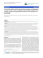
Figure 1. Principles of scanning methods used for detection of mutations at non-defined positions.
S
Scanning methods
SSCP
DGGE
HA
CCM
CFLP
MutS
MutHLS
RNase
MutY
Normal Mutant
S
S
H
L
Y
R
Gelshift
Gelshift
Gelshift
Gelshift
Cleavage
Cleavage
Cleavage
Cleavage
Cleavage
MutEx
Cleavage protection
Exo
Exo
Exo
Exo
BESS
Cleavage
U
G
U
Å Sivertsson
1
1
Specific methods
In contrast to scanning technologies, specific methods identify alterations at pre-
defined sites. Two types of alterations are of particular interest; mutations and single
nucleotide polymorphisms. Mutations can be distributed across the genome, but at
some sites a clustering tendency is observed. These so-called “hotspot mutations”
have been identified in for example cancer-related genes such as ras and p53. Single
nucleotide polymorphisms (SNP) have been estimated to occur once in every
thousand bases in the human genome. They represent nucleotide variations at certain
positions in coding and non-coding regions of the genome. SNPs in coding regions
may account for differences in drug response between individuals and also may be a
contributing factor in disease susceptibility (Evans, 1999), (Davignon, 1988; Bertina,
1994). The possible effect of SNPs in non-coding regions is not yet determined but
they are extremely useful as markers in population and genetics studies (Jorde, 2000;
Hacia, 1999). Some methods that have been developed to analyse both mutations and
SNPs and are tailor-made for detection of variations at specific sites are described
below and shown in Figure 2.
Hybridisation-based techniques
The possibility of detecting a single-base mismatch using hybridisation with allele
specific oligonucleotides (ASO) was demonstrated as early as 1979 (Wallace, 1979).
Since the invention of PCR, the principle of ASO has become widely used in various
assays. The method relies on the differences in hybridisation efficiency to the target
DNA between a fully complementary ASO probe and a probe containing a single
mismatch. The early ASO assays were performed in a dot-blot or reverse dot-blot
format immobilising either the amplified target DNA or the oligonucleotide probe set
respectively on a membrane (Saiki, 1988; Saiki, 1989). The hybridisation product was
detected using autoradiography or enzyme conjugates. The assay was later adapted to
a high-density microarray format, but neither format allows for perfect allele
discrimination (Wang, 1998; Cho, 1999). Increased stringency can be achieved using
DASH (dynamic allele-specific hybridisation) where the hybridisation is monitored
over a temperature gradient (Ririe, 1997),(Prince, 2001) or by using higher affinity
LNA (locked nucleic acid) probes instead of DNA (Orum, 1999). In some recently
developed real-time PCR-based ASO assays, fluorescence is emitted as a result of a
change in the physical distance between a fluorophore and a quencher molecule. In
Detection and analysis of genetic alterations in normal skin and skin tumours
12
the Molecular Beacon assay (Giesendorf, 1998; Vet, 1999; Tyagi, 1996; Smit, 2001)
fluorescence is emitted when the stem-loop structure of the probe is opened upon
perfect hybridisation to the DNA target sequence during the primer-annealing phase.
In the TaqMan
TM
assay release of the quencher after exonuclease degradation of
perfectly annealed probes by the polymerase allows the fluorophore to emit a
fluorescent signal (Livak, 1995; Livak, 1999). Both assays are compatible with 96-
well or 384-well microtiter plate formats and facilitate the use of multiple
fluorophores. However, the need for fluorescent and quencher moieties on the probes
make these assays rather expensive.
The concept of ASO was the first step towards today’s oligonucleotide arrays, which
are used in various applications ranging from mutation detection to gene expression
studies. The theoretical principle of sequencing by hybridisation (SBH) was
independently described by two groups in the late 1980’s and is based on arrays of all
possible combinations of short immobilised oligonucleotides (Drmanac, 1989) (Lysov
Iu, 1988). Labelled target DNA is then hybridised to the array and the hybridisation
pattern is used to in silico reconstruct the target sequence. This method proved less
suitable for de novo sequencing but has been successfully used for mutation detection
in the p53 gene (Drmanac, 1998).
Methods based on allele-specific amplification
Several allele-specific PCR-based methods have also been used for sequencing of
defined alterations. Allele specific PCR primers with the 3´ terminus annealing at the
variant position are used in the PCR reaction, which results in amplification with only
the perfectly matched primer. Assays based on this principle are for example ASA
(allele specific amplification) (Okayama, 1989), ASPCR (allele specific PCR) (Wu,
1989) and PASA (PCR amplification of specific alleles) (Sommer, 1992).
Oligonucleotide ligation assays
To increase the specificity of ASO hybridisation a number of assays based on ASO
ligation have been developed. In the oligonucleotide ligation assay (OLA)
(Landegren, 1988) a ligation probe and probes specific to wild type and mutant alleles
are hybridised adjacent to each other on the target sequence. Since ligases can
effectively discriminate against mismatches, only perfectly matched probes will be
Å Sivertsson
1
3
ligated. Depending on the label used, the detection of the ligated products can be
carried out in an ELISA format or by electrophoretic separation in a DNA sequencer
instrument (Nickerson, 1990; Samiotaki, 1994), (Grossman, 1994). The OLA assays
have also been used in different microarray formats (Broude, 2001; Gerry, 1999) but
the need for several differently modified probes increases the cost significantly. In
ligase chain reaction (LCR) two pairs of probes are used together with a thermostable
ligase in a cyclic ligation reaction resulting in amplification of the target sequence in
cases where the probes are perfectly matched (Barany, 1991). Padlock probes
(Nilsson, 1994) (Nilsson, 1997) are linear oligonucleotides that have complementary
target sequences at both ends separated by a random DNA sequence. Upon
hybridisation to a target, the probe ends are ligated to form a circularised probe if
there is complete homology to the target. Signal amplification can then be achieved
by using the circularised product of successful padlock probe ligation as a template
for rolling circle amplification (RCA) which creates a long single stranded DNA
composed of tandem-repeats complementary to the padlock probe (Fire, 1995; Baner,
1998; Lizardi, 1998). A single tube assay combining ligation and rolling circle
amplification has also been described which increases the throughput significantly
(Qi, 2001).
Techniques based on polymerase extension
Minisequencing, also denoted PEX for single nucleotide primer extension (Syvänen,
1990), is based on discrimination of variants by single nucleotide extension at the site
of a mutation. A primer is hybridised to the target sequence immediately adjacent to
the variable position and a DNA polymerase is then used to extend the 3´ end of the
primer with a labelled dideoxynucleotide complementary to the nucleotide at the
variable site. The method has been adapted to various formats and detection
strategies, resulting in ELISA, electrophoresis and microarray based formats
(Nikiforov, 1994; Pastinen, 1996; Tully, 1996; Pastinen, 1997; Lindroos, 2001). The
minisequencing concept is also used in the commercially available SNaPshot
TM
assay
(Applied Biosystems) and in the improved variant MAPA (multiplex automated
primer extension) described by Makridakis and Reichardt (Makridakis, 2001).
MALDI-TOF–MS (matrix-assisted laser desorption ionisation - time-of-flight - mass
spectrometry) emerged as a method for DNA sequencing in the late 1980’s. An
innovation in the field of ionisation of macromolecules made it possible to perform
Detection and analysis of genetic alterations in normal skin and skin tumours
14
analysis of DNA in a system which was earlier limited to peptide analysis (Karas,
1988). DNA samples are desorbed, ionised and subjected to an electric field where the
molecules are accelerated proportional to their mass/charge ratio. Detection is based
on the time required for the molecules to reach the detector. MALDI-TOF-MS is a
rapid sequence analysis technique that permits simultaneous analysis of many DNA
strands in a heterogenous mixture. However, the limited read-length of 100
nucleotides resulting from sequencing of dideoxy generated DNA ladders make it less
attractive for de novo sequencing (Wu, 1994; Taranenko, 1998; Monforte, 1997). The
method has been successfully used to sequence exon 5 to 8 of the p53 gene (Fu,
1998), but the true potential probably lies in typing single point mutations and SNPs
(Griffin, 2000; Griffin, 2000; Li, 1999; Tang, 1999; Griffin, 1999). Several different
approaches for MALDI-TOF-MS have been described. In the PINPOINT assay (Haff,
1997; Haff, 1997; Ross, 1998) the target is subjected to primer extension by a single
base in the presence of all four ddNTPs and the mass added onto the primer then
defines the variable base in the subsequent MALDI-TOF-MS analysis. In
MassEXTEND (Little, 1997; Little, 1997), (Braun, 1997; Braun, 1997) and VSET
(very short extension)(Sun, 2000) which are similar assays, dNTP mixes containing a
single ddNTP or ddNTP mixes containing a single dNTP respectively, are used in the
primer extension reaction. Apyrase-Mediated Allele-Specific Extension (AMASE) is
another single-step extension approach recently described (Ahmadian, 2001). This
assay relies on extension of paired allele-specific primers in the presence of the
nucleotide degrading enzyme apyrase. Only a perfectly matched primer will give rise
to extension, since the slower reaction kinetics of a mismatched primer-template
configuration will allow for the apyrase to degrade the nucleotides before extension
can occur. Thus the acknowledged difficulties that polymerases have in
discriminating between certain mismatches can be circumvented. The assay has
recently been adapted to a microarray format and successfully used for SNP typing
(O'Meara, 2002).
Å Sivertsson
1
5
Figure 2. Principles of methods used for detection of mutations/variations at defined positions.
ASO
ASA
OLA
PEX
LCR
No amplification
No duplex
No ligation
No ligation / amplification
No extension
Normal
Mutant
Specific methods
Beacons
Padlocks
AMASE
A
C
A
T
Fluorescence
No ligation / amplification
No extension