in vitro transcription and translation protocols
Bạn đang xem bản rút gọn của tài liệu. Xem và tải ngay bản đầy đủ của tài liệu tại đây (28.82 MB, 422 trang )
CHAPTER
1
Transcription In Vitro Using
Bacteriophage RNA Polymerases
EZaine l! Schenborn
1. Introduction
Synthesis of specific RNA sequences in vitro is simplified because
of the availability of bacteriophage RNA polymerases and specially
designed DNA vectors. RNA polymerases encoded by SP6, T’7, or T3
bacteriophage genomes recognize particular phage promoter sequences
of their respective viral genes with a high degree of specificity (I-3).
These RNA polymerases also transcribe DNA templates containing their
cognate promoters under defined conditions in vitro (4,5). Standard reac-
tion conditions for transcription in vitro can be adjusted for synthesis of
large amounts of RNA or for smaller amounts of labeled RNA probes.
Larger-scale in vitro synthesis produces RNA that mimics biologically
active RNA in many applications. The following examples represent
some of the different uses for RNA synthesized in vitro. RNA transcripts
are particularly well suited for the study of RNA virus gene regulation,
For example, the in vitro transcribed RNA genomes of poliovirus (6) and
cowpea mosaic virus (7) produce infectious particles in transfected cells.
For other types of studies, messenger RNA-like transcripts are used as
substrates to study RNA processing activities, such as splicing (8) and
3’-end maturation
(9,lO).
RNA transcripts synthesized in vitro also are
widely used as templates for protein synthesis in cell-free extracts
designed for in vitro translation (II). Transfer RNA-like transcripts have
been used as substrates to study RNase P cleavage specificities (12), and
other mechanisms of RNA cleavage have been investigated using RNA
From: Methods in Molecular Bology, Vol. 37: In Vitro Transcript/on and Translation Protocols
Edlted by: M J Tymms Copynght Q 1995 Humana Press Inc , Totowa, NJ
2 Schenborn
substrates and ribozymes synthesized in vitro (13). Gene regulation stud-
ies using antisense RNA also have taken advantage of the ease of in vitro
RNA synthesis. In vitro translation of a targeted message has been shown
to be inhibited in the presence of antisense RNA in vitro (14), and in vivo
translation has been blocked in
Xenopus
oocytes by antisense RNA (15).
The ability to synthesize discrete RNA templates in vitro also facilitates
studies of RNA and protein interactions (16,17).
The generation of radioactively labeled RNA hybridization probes is a
widely used application for RNA synthesized in vitro. RNA probes are
synthesized predominantly by incorporation of a radiolabeled ribonucle-
otide, 32P-, 3H-, or 35S-rNTP, into the transcript. Nonisotopic probes can
be synthesized by incorporation of biotinylated (18) or digoxigenin (19)
modified bases. For Northern blots, single-stranded RNA probes are gen-
erally more sensitive than the corresponding DNA probe because of the
higher thermal stability of RNA:RNA hybrids compared to RNA:DNA
hybrids and the absence of self-complementary sequences in the probe
preparation (4).
RNA probes also are more sensitive than DNA probes for the detec-
tion of DNA sequences transferred to membranes from Southern blots,
plaque lifts, and colony lifts (20). The lower background and increased
signal sensitivity of RNA probes are possible because of higher stability
of RNA:DNA hybrids compared to DNA:DNA hybrids. This increased
stability allows more stringent conditions to be used for the hybridiza-
tion and washing procedures (21). Another advantage of RNA probes is
that RNase A can be added after the hybridization reaction to eliminate
nonspecific binding of the probe to the membrane. High sensitivity also
has been achieved with RNA probes used for
in situ
hybridization (22)
and localization of genes in chromosome spreads (23). RNase mapping
is another application that takes advantage of the superior properties of
RNA probes for hybridization to complementary sequences. In this appli-
cation, a radiolabeled RNA probe is hybridized in solution to cellular
RNA, then the nonhybridized, single-stranded regions of the probe are
later digested with RNase A and RNase Tl, and the protected, hybrid-
ized regions are identified by gel analysis. This type of mapping is used
to quantitate low-abundance species of RNA, and to map exons, tran-
scription start sites, and point mutations ($24).
The DNA templates used for in vitro transcription contain the cloned
sequence of interest immediately “downstream” of an SP6, T7, or T3
Transcription In Vitro 3
I
Llneanze DNA
with an appropriate
restnctlon enzyme
Add RNA synthesis reaction
components and Incubate
yF!5$
I
run-off transcripts
;;tN;;$ template with
AA-
E z punfled RNA
transcripts
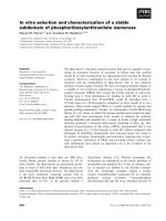
Fig. 1. Synthesis of RNA by transcription in vitro from a linear DNA template.
promoter sequence. Plasmid vectors are commercially available with the
phage promoter sequence adjacent to a cloning region. One example is
the pGEM@ series of vectors (Promega, Madison, WI) designed with
multiple cloning sites flanked by opposed SP6 and T7 promoters, allow-
ing the synthesis of either sense or antisense RNA from a single recom-
binant plasmid. Discrete RNAs, corresponding to the cloned sequence of
interest, are synthesized as “run-off” transcripts from a linear DNA tem-
plate. To prepare the linear template, the recombinant plasmid DNA is
cut with a restriction enzyme cleaving within, or shortly downstream of,
the cloned insert. The linear DNA is then added to the reaction mixture
for in vitro synthesis of RNA (see Fig. 1).
2. Materials
1. Transcription buffer (5X): 200 mM Tris-HCl, pH 7.5, 30 mM MgC&
10 mM spermidine, and 50 mM NaCl. Store at -2OOC.
4
Schenborn
2. ATP, GT’P, CTP, UTP: 10 mM stocks prepared in sterile, nuclease-free
water and adjusted to pH 7.0. Store at -20°C.
3.
100 mM DlT: Store at -20°C.
4. RNasin@ Ribonuclease Inhibitor: (Promega) Store at -20°C.
5. Nuclease-free water: Prepare by adding 0.1% diethyl pyrocarbonate
(DEPC) to the water. Autoclave to remove the DEPC. Caution: DEPC is a
suspected carcinogen.
6. TE buffer: 10 mM Tris-HCl, pH 8.0, and 1 mM EDTA. Prepare with stock
solutions that are nuclease-free.
7. TE-saturated phenol/chloroform: Mix equal parts of TE buffer and phenol,
and allow phases to separate. Mix 1 part of the lower, phenol phase with 1
part of chloroform:isoamyl alcohol (24: 1).
8. Chlorofornuisoamyl alcohol (24:l): Mix 24 parts of chloroform with 1
part isoamyl alcohol.
9. Ammonium acetate: 7.5 and 2SM.
10. 3M sodium acetate, pH 5.2.
11. Ethanol: Absolute (100%) and 70%.
12. Enzymes: SP6, T3, or T7 RNA polymerase at 15-20 U&L.
13. RNase-free DNase: RQl (Promega).
14. Restriction enzyme and appropriate buffer to linearize plasmid DNA
template.
15. DE-81 filters: 2.4 cm diameter (Whatman).
16. 0.5M Na2HP04, pH 7.0.
17. m’G(S’)ppp(S’)G: 5 r&f (New England BioLabs).
Microcentrifuge tubes, pipet tips, glassware: To provide a nuclease-
free environment,
use sterile, disposable microcentrifuge tubes and pipet
tips whenever possible for the preparation and storage of reagents.
Larger
volumes of reagents can be stored in bottles that have been baked at
250°C for four or more hours to inactivate RNases.
3. Methods
Throughout these procedures, precautions should be taken to protect
against ribonuclease contamination. These precautions include the use
of sterile, nuclease-free reagents and materials, and the use of disposable
gloves to prevent accidental contamination of samples with ribonucleases
present on the skin.
Three
steps are required for synthesis of
RNA
in vitro:
1. Preparation of the DNA template.
2. Transcription reaction.
3. Enrichment of the RNA product.
Transcription In Vitro 5
3.1.
Preparation of the DNA Template
The sequence of interest is cloned by established methods into an
appropriate vector, downstream of a promoter sequence for SP6, T7, or
T3 RNA polymerase. The recombinant plasmid DNA is purified, and either
added directly to the in vitro transcription reaction or linearized prior to
the run-off transcription reaction. Transcription of supercoiled plasmid
DNA results in the synthesis of high-mol-wt RNA, which contains vector
sequences. Discrete RNA sequences of interest, without vector sequence,
are generated by run-off transcription from linear templates prepared in
the following manner:
1. Determine the restriction site downstream of, or within, the cloned insert,
which will generate the desired run-off transcrtpt. Whenever possible,
select a restriction enzyme that produces 5’ overhanging or blunt ends. If
an enzyme that generates a 3’ overhang is selected, see Note 1. Set up the
restriction digest according to the enzyme supplier’s directions.
2. Check for completeness of digestion by agarose gel electrophorests. Dur-
ing this analysis, keep the DNA sample on ice. If digestion is complete,
proceed with step 3. Otherwise, add additional restriction enzyme to the
DNA, incubate an additional 30 min, and repeat the agarose gel analysis.
3. Extract the DNA by adding an equal volume of TE-saturated phenol/chlo-
roform, vortex for 1 min, and centrifuge at 12,000g for 2 min. Transfer the
upper phase to a fresh tube, and add 1 vol of chloroform:isoamyl alcohol
(24:l). Vortex for 1 min, and centrifuge at 12,000g for 2 min.
4. Precipitate the DNA by transferring the upper, aqueous phase to a fresh
tube, and adding 0.1 vol of 3M sodium acetate, pH 5.2, and 2 vol of abso-
lute ethanol. Cool 30 min at -7O”C, and centrifuge at 12,000g for 5 min.
5. Carefully pour off the supernatant, wash the pellet briefly with 1 mL of
70% ethanol, spin at 12,000g for 2 min, and remove the supernatant. Dry
briefly in a vacuum desiccator. Resuspend the pellet in nuclease-free water
or TE buffer to a final DNA concentration of approx 1 mg/mL.
3.2. Synthesis of Radiolabeled RNA Probes
(See Notes 2-5)
RNA probes at a specific activity of 6-9 x lo8 cprn&g can be gener-
ated by transcribing DNA in the presence of a limiting concentration
(12-24 ClM) of one radiolabeled ribonucleotide and saturating concen-
trations (0.5 n&f) of the other three rNTPs (see Notes 2 and 3). The
following example uses 50 l.tCi of a-[32P]CTP at a specific activity of
400 Ci/mrnol/20 PL reaction, providing a final concentration of 6 l,tM of
6
Schenborn
w[~~P]CTP. An
additional 12 w of unlabeled CTP is added to bring the
total concentration to 18
pM CTP. Expect approx 1 mol of RNA/m01 of
DNA
template to be synthesized under these conditions.
1. To a sterile microcentrifuge tube, add the following components at room
temperature in the order listed. This order of addition prevents precipita-
tion of the DNA by spermidine: 4 pL of 5X transcription buffer, 2 p.L of
100 mit4 D’IT, 20 U RNasin* Ribonuclease Inhibitor, 4 pL of ATP, GTP,
and UTP (2.5 mM each; prepare by mixing 1 vol of each individual 10 mM
stock of ATP, GTP, and UTP, and 1 vol of water), 2.4 p.L of 100 p.M
CTP (dilute 10 mM stock 1:lOO with water), 1 uL of DNA template
(up to 2 pg; l-2 mg/mL in nuclease-free water or TE), 5 l4.L of a-[32P]CTP
(400 Ci/mmol; 10 mCi/mL). Bring to a final vol of 19 uL with nuclease-
free water.
2. Initiate the reaction by adding 1 p.L of SP6, T7, or T3 RNA polymerase (at
15-20 U/p.L) .
3. Incubate for 60 min at 37-4O”C.
4. Remove 1 p.L from the reaction at this point to determine the percent incor-
poration and specific activity of the probe. The remainder of the sample
can be digested by RQl RNase-free DNase (Section 3.6.).
3.3. Determination
of
Percent Incorporation
and Probe Specific Activity
1. Remove 1 uL of the labeled probe, and dilute into 19 uL of nuclease-free
water. Spot 3 pL of this 1:20 dilution onto 4 DE8 1 filters. Dry the filters at
room temperature or under a heat lamp.
2. Place two filters directly into separate scintillation vials, add scintillation
fluid, and count. Calculate the average cpm per filter, and determine the
total cpm per microliter of original reaction as follows:
Total cpm@L of original reaction = average cpm per filter
x (20-fold dilution/3 uL)
(1)
3. Wash the unincorporated nucleotides from the remaining two filters by
placing the filters in a small beaker containing 50-100 mL of 0.5M
Na2HP04 (pH 7.0). Swirl the filters occasionally for 5 min, then decant,
and replace with fresh buffer. Repeat the wash procedure two more times.
Dip the filters briefly into 70% ethanol, and dry at room temperature or
under a heat lamp.
4. Place each filter into a scintillation vial, add scintillation fluid, and count.
Calculate the amount of labeled nucleotide incorporated into RNA (incor-
porated cpm) per microliter of original reaction as follows:
Transcription In Vitro 7
Incorporated cpm/pL of original reaction = average cpm per filter
x (20-fold dilution/3 uL)
(2)
This value will also be used in estimating the probe specific activity in
step 6.
5. Calculate the percent incorporation from the values determined above in
steps 2 and 4.
% Incorporation = (incorporated cpmkotal cpm) x 100
(3)
The percentage of incorporation under the conditions described generally
ranges from 70 to nearly 100%. A low incorporation of radiolabeled nucle-
otide (for example, below 50%) reflects a low yield of RNA product (see
Note 5).
6. Calculate the specific activity of the probe as cpm/ug RNA synthesized.
To do this, first calculate the total incorporated cpm in the reaction:
Total incorporated cpm = (incorporated cpmQ.tL of reaction)
x 20 uL reaction vol
(4)
Next we need to calculate the total nmoles of nucleotide in the reac-
tion to determine how many micrograms of RNA were synthesized;
50 FCi of ~F[~~P]CTP at 400 @/nmol corresponds to 0.12 nmol of
32P-CTP/reaction. Adding in the 12 @4 of unlabeled CTP (0.24 nmol)
gives a total of 0.36 nmol of CTP. If a maximum 100% incorporation
was achieved and CTP represents one-fourth of all the nucleotides in the
probe, then the total amount of nucleotides incorporated into the probe
would be (0.36 nmol x 4) or 1.44 nmol. Assuming an average PW/nucle-
otide of 330, the amount of RNA synthesized in this example would be
1.44 nmol x (330 ng/nmol) = 475 ng of RNA synthesized. If the percent-
age of incorporation calculated from step 5 was 80%, for example, then
the actual amount of RNA synthesized in the reaction would be 475 ng x
0.80 = 380 ng RNA.
SA = total incorporated cprn/ug RNA
(5)
In this example, the total incorporated CPM would be divided by 0.380
Pg RNA.
3.4. Synthesis of Large Quantities of RNA
(See Notes 2-6)
Using the following reaction conditions in which all four rNTPs are at
a saturating concentration, yields of 5-10 pg of RNA&g of DNA tem-
8
Schenborn
plate can be obtained (see Note 6). This represents up to 20 mol of RNA/
mol of DNA template. Incubation with additional polymerase after the
initial 60-min reaction can increase the yield of RNA up to twofold. The
following reaction can be scaled up or down as desired.
1. To a sterile microcentnfuge tube, add the following components at room
temperature in the order listed. This order of addition prevents precipita-
tion of the DNA by spermidine: 20 pL of 5X transcription buffer, 10 l.rL of
100 mM DTT, 100 U RNasin Ribonuclease Inhibitor, 20 pL of ATP, GTP,
UTP, and CTP (2.5 rnM each; prepare by mixing 1 vol of each individual
10 mM stock of ATP, GTP, UTP, and CTP), 2-5 pL of DNA template (5-
10 pg total; l-2 mg/rnL in nuclease-free water or TEZ). Add nuclease-free
water to a final vol of 98 pL.
2. Initiate the reaction by adding 2 lrL of SP6, T7, or T3 RNA polymerase (at
15-20 U&L).
3. Incubate for 60 min at 37aO°C.
4. Add an additional 2 pL of SP6, T7, or T3 RNA polymerase. Incubate for
60 min at 37-4O”C.
The DNA
template can now be digested by RQl RNase-free DNase
(Section 3.6.).
3.6. Synthesis
of
5’ Capped Transcripts
Some RNA transcripts require a m7G(5’)ppp(5’)G cap at the 5’ end for
higher translation efficiency, either in cell-free extracts or in
Xenopus
oocytes (25). Methylated capped transcripts also have been reported to
function more efficiently for in vitro splicing reactions (8) and are more
resistant to ribonucleases in nuclear extracts. The following reaction can
be scaled up or down as desired.
1. To a sterile microcentrifuge tube, add the following components at room
temperature m the order listed. This order of addition prevents precipita-
tion of the DNA by spermidine: 4 pL of 5X transcription buffer, 2 PL of
100 rnM DTT’, 20 U RNasin Rtbonuclease Inhibitor, 4 pL of ATP, UTP,
and CTP (2.5 rnM each; prepare by mixing 1 vol of each individual
10 mM stock of ATP, UTP, and CTP, and 1 vol of water), 2 PL of GTP
(0.5 mM, dilute 10 rnM stock 1:20 with water), 2 p.L of the cap analog
m7G(5’)ppp(5’)G (5 mM), and 1 pL of DNA template: l-2 pg (l-2 mg/mL
in nuclease-free water or TE). Add nuclease-free water, if necessary, to a
final vol of 19 j.rL.
2. Initiate the reaction by adding 1 pL of SP6, T7, or T3 RNA polymerase (at
15-20 U&L).
3. Incubate for 60 min at 37AOOC.
Transcription In Vitro 9
The DNA template can now be digested by RQl RNase-free DNase
(Section 3.6).
3.6. Digestion
of
the DNA Template Posttranscription
To achieve maximal sensitivities with RNA probes, the DNA tem-
plate must be eliminated after the transcription reaction. Elimination of
the DNA template also may be required for the preparation of biolog-
ically active RNAs. DNase can be used to digest the DNA template, but
during this enzymatic step, it is critical to maintain the integrity of the
RNA. RQ 1 DNase (Promega) is certified to be RNase-free and is recom-
mended for the following protocol.
1. After the in vitro transcription reaction, add RQl RNase-free DNase to a
concentration of 1 U&g of template DNA.
2. Incubate for 15 min at 37OC.
3. Extract with 1 vol of TE-saturated phenol/chloroform. Vortex for 1 min,
and centrifuge at 12,000g for 2 min.
4. Transfer the upper, aqueous phase to a fresh tube. Add 1 vol of chloro-
fornuisoamyl alcohol (24:l). Vortex for 1 min and centrifuge as in step 3.
5. Transfer the upper, aqueous phase to
a fresh tube. At
this point, a small
aliquot can be taken for electrophoretic analysis on a denaturing gel, and
the remainder of the sample can be precipitated (Section 3.7.).
3.7. Precipitation
of
RNA
1. Add 0.5 vol of 7.94 ammonium acetate to the aqueous RNA sample pre-
pared in Section 3.6. If the RNA sample was not digested with RQl DNase,
extract the RNA after the transcription reaction with TE-saturated phenol/
chloroform followed by a chloroform extraction, as described in Section
3.6., steps 3-5.
2. Add 2.5 vol of ethanol, mix, and place at -70°C for 30 min.
3. Centrifuge at 12,000g for 5 min. Carefully remove the supernatant.
4. Resuspend the RNA pellet in 100 pL of 2.5M ammonium acetate and mix.
5. Repeat the ethanol precipitation as described in steps 2 and 3 above.
6. Dry the pellet briefly under vacuum, and resuspend in 20 pL or other suit-
able volume of sterile TE or nuclease-free water. Store the RNA at -70°C.
4. Notes
1. Extraneous transcripts complementary to the opposite strand and vector
sequences are generated from DNA templates with 3’ overhanging ends
(26). The ends of these templates can be made blunt in the following man-
ner using the 3’-5’ exonuclease activity of the Klenow fragment of DNA
polymerase I. Set up the transcription reaction, but without nucleotides
10 Schenborn
Table 1
SA and Concentration of rNTPs Used for Transcription In Vitro
Nucleotide Specific activity @Meaction Final cont.
~G[~~P]
rNTP 400 Wmmol 50 pCi
6W
CX-[~%] rNTP 1300
Ci/mmoi 300 PCi 12cLM
5,6[3H] rNTP 40
Wmmol 25 pCi 31 w
and RNA polymerase. Add 5 U of Klenow fragment&g DNA, and incu-
bate for 15 min at 22OC. Then initiate the transcription reaction by adding
nucleotides and RNA polymerase, and incubate for 60 min at 3740°C.
2. Incomplete transcripts are more likely to be generated under the condi-
tions used for probe synthesis, in which the concentration of a radiolabeled
nucleotide becomes limiting. Of the four nucleotides, rGTP yields the high-
est percentage of full-length transcripts when present in limiting concen-
trations (4). However, for best results, radiolabeled rGTP should be used
within 1 wk of the reference
date. rATP yields the lowest percentage of
full-length transcripts and lowest incorporation when present at a limiting
concentration (5). In some cases, the amount of full-length transcripts
increases when the incubation temperature is lowered to 30°C. Another
possible cause for incomplete transcripts can be the presence of a sequence
within the DNA template that acts as a terminator for that particular poly-
merase. In this case, one can subclone the sequence of interest behind a
different RNA polymerase promoter.
3. The specific activity of a probe can be increased by using more than one
radiolabeled nucleotide per reaction at a limiting concentration. Also,
more than 5 p,L of the radionucleotide can be used per 20 p.L reaction if the
nucleotide is first aliquoted into the reaction tube and dried down under
vacuum. Table 1 lists the final concentration (final cont.) of radionucleotides
commonly used in RNA probe synthesis, in a 20-pL reaction volume.
Thiol-substituted rNTPs are incorporated less efficiently by the RNA poly-
merases than the corresponding 32P or 3H rNTPs (5).
4. Biotinylated rNTP can be added during the transcription reaction, but the
yield of RNA may be lowered. Alternatively, RNA can be modified after
transcription using photoactivatable biotin (27).
5. A low yield of RNA product can be caused by several conditions, includ-
ing precipitation of DNA by spermidine in the transcription buffer, RNase
contamination, carryover of residual contaminants or salts in the DNA
preparation, or inactive RNA polymerase.
6. High yields of RNA synthesized by SP6 or ‘IT RNA polymerase recently
have been reported using a transcription buffer containing 80 mM HEPES-
Transcription In Vitro
11
KOH, pH 7.5,2 mM spermidine, 1040 mM DlT, 3 mM each rNTP, 12-
16 mA4 MgCl,, and 1200-1800 U/mL RNA polymerase. Under these con-
ditions, yields up to 80 p.g of RNA/pg DNA were reported (28).
References
1. Butler, E. T. and Chamberlin, M. J. (1982) Bacteriophage SP6-specific RNA poly-
merase. J. Biol. Chem. 257,5772-5778.
2. Davanloo, P., Rosenberg, A. H., Dunn, J. J., and Studier, F. W. (1984) Cloning and
expression of the gene for bacteriophage T7 RNA polymerase. Proc. Natl. Acad.
Sci. USA
81,2035-2039.
3. Jorgensen, E. D., Joho, K., Risman, S., Moorefield, M. B., and McAllister, W. T.
(1989) Promoter recognition by bacterophage T3 and T7 RNA polymerases, in DNA-
Protein Interaction in Transcription (Gralla, J. D., ed.), Liss, New York, pp. 79-88.
4. Melton, D. A., Krieg, P. A., Rebagliati, M. R., Maniatis, T., Zinn, K., and Green,
M. R. (1984) Efficient in vitro synthesis of biologically active RNA and RNA
hybridization probes from plasmids containing a bacteriophage SP6 promoter.
Nucleic Acids Res.
12,7035-7056.
5. Krieg, P. A. and Melton, D. A. (1987) In vitro RNA synthesis with SP6 RNA
polymerase. Methods Enzymal.
155,397-4
15.
6. Kaplan, G., Lubinski, J., Dasgupta, A., and Racaniello, V. R. (1985) In vitro syn-
thesis of infectious poliovirus RNA. Proc. Natl. Acad, Sci. USA 82,8424-8248.
7. Eggen, R., Verver, J., Wellink, J., DeJong, A., Goldbach, R., and van Kammen, A.
(1989) Improvements of the infectivity of in vitro transcripts from cloned cowpea
mosaic virus cDNA: impact of terminal nucleotide sequences. Virology
173,
447-455.
8.
Kramer, A. R., Maniatis, T., Ruskin, B., and Green, M. R. (1984) Normal and
mutant human /3-globin pre-mRNAs are faithfully and efficiently spliced in vitro.
Cell
36,993-1005.
9. Krieg, P. A. and Melton, D. A. (1984) Formation of the 3’ end of histone mRNA by
post-transcriptional processing. Nature 308,203-206.
10. Georgiev, O., MOUS, J., and Birnstiel, M. (1984) Processing and nucleo-cytoplas-
mic transport of histone gene transcripts. Nucleic Acids Res.
12,8539-8551.
11. Krieg, P. A. and Melton, D. A. (1984) Functional messenger RNAs are produced
by SP6 in vitro transcription of cloned cDNAs. Nucleic Acids Res.
12,7057-7070.
12. Burgin, A. B. and Pace, N. R. (1990) Mapping the active site of ribonuclease P
RNA using a substrate containing a photoaffinity agent. EMBO J. 9,4111-4118.
13. Heus, H. A., Uhlenbeck, 0. C., and Pardi, A. (1990) Sequence-dependent struc-
tural variations of hammerhead RNA enzymes. Nucleic Acids Res.
18,1103-l
108.
14. Nicole, L. M. and Tanguay, R. M. (1987) On the specificity of antisense RNA to
arrest in vitro translation of mRNA coding for Drosophila hsp 23. Biosci. Rep. 7,
239-246.
15. Melton, D. A. (1985) Injected antisense RNAs specifically block messenger RNA
translation in vivo. Proc. Natl. Acad. Sci. USA 82, 144-148.
16. Witherell, G. W., Wu, H N., and Uhlenbeck, 0. C. (1990) Cooperative binding of
R17 coat protein to RNA. Biochemistry 29, 11,05 l-l 1,057.
12 Schenborn
17. Turek, C. and Gold, L. (1990) Systematic evolution of ligands by exponential
enrichment: RNA ligands to bacteriophage T4 DNA polymerase. Science 249,
505-5 10.
18. Langer, P. R., Waldrop, A. A., and Ward, D. C. (1982) Enzymatic synthesis of
biotin-labeled polynucleotides: novel nucleic acid affinity probes. Proc. Natl. Acad.
Sci. USA 70,6633-6637.
19 Aigner, S. and Pette, D. (1990) In situ hybridization of slow myosm heavy chain
mRNA in normal and transforming rabbit muscles with the use of a nonradioac-
tively labeled cRNA. Histochemistry 95,1 l-l 8.
20. Sambrook, J., Fritsch, E. F., and Maniatis, T. (1989) Molecular Cloning, A Lube-
ratory Manual, 2nd ed., Cold Spring Harbor Laboratory, Cold Spring Harbor, NY.
21. Casey, J. and Davidson, N. (1977) Rates of formation and thermal stabilities of
RNA:DNA and DNA:DNA duplexes at high concentrations of formamide. Nuclezc
Acids Res. 4, 1539-1552.
22. Uhlig, H., Saeger, W., Fehr, S., and Ludecke, D. K. (1991) Detection of growth
hormone, prolactin and human beta-chorionic gonadotropm messenger RNA in
growth-hormone-secreting pituitary adenomas by in situ hybridization. Virchows
Arch. Pathol. Anat. Histopathol. 418,539-546.
23. Matthaei, K. I. and Reed, K. C. (1986) Chromosome assignment m somatic hybrids
by in situ hybridization with tritium labeled Riboprobe@ RNA probes. Promega
Notes 5,5-6.
24. Zinn, K., DiMaio, D., and Maniatis, T. (1983) Identification of two distinct regula-
tory regions adjacent to the human p-interferon gene. Cell 34,865-879.
25. Contreras, R., Cheroutre, H., Degrave, W., and Fiers, W. (1982) Simple, efficient
in vitro synthesis of capped RNA useful for direct expression of cloned eukaryotic
genes. Nucleic Acids Res. 10,6353-6362.
26. Schenborn, E. T. and Mierendorf, R. C. (1985) A novel transcription property of
SP6 and T7 RNA polymerases: dependence on template structure. Nucleic Acids
Res. 13,6223-6236.
27. Forster, A. C., Mclnnes, J. L., Skingle, D. C., and Symons, R. H. (1985) Non-
radioactive hybridization probes prepared by the chemical labelling of DNA and
RNA with a novel reagent, photobiotin. Nucleic Acids Res. 13,745-761.
28. Gurevich, V. V., Pokrovskaya, I. D., Obukhova, T. A., and Zozulya, S. A. (1991)
Preparative in vitro mRNA synthesis using SP6 and T7 RNA polymerases. Analyt.
Biochem. 195,207-2 13.
CHAPTER 2
Subtraction Hybridization
cDNA Libraries
Clifford W. Schweinfest, Peter S. Nelson,
Michael W. Graber, Rita I. Demopoulos,
and Takis S. Papas
1. Introduction
Subtraction-hybridization cDNA libraries (14) are libraries enriched
for sequences representing mRNAs whose expression in one biological
source (e.g., tissues, cell lines) is different than in a second source.
Single-stranded cDNAs from both sources are allowed to hybridize so
that sequences common to the two sources will anneal. The annealed,
double-stranded DNAs are “subtracted” from the hybridization solution,
leaving a population of cDNA molecules enriched for sequences pref-
erentially expressed (or repressed) in the biological source of interest.
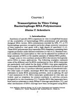
Figure 1 diagrammatically represents the scheme for subtraction hybrid-
ization currently employed in our laboratory.
The subtraction technique is particularly helpful for isolating differen-
tially expressed genes for which there is no
apriuri
knowledge (e.g., loss
of heterozygosity). Therefore, the subtraction technique may ask the
question, “What gene expression is different between two selected cell
types, such as tumor vs normal?” As such, it is important that such
matched sets of tumor and normal tissue be as similar as possible. For
example, a colon tumor is typically a benign or cancerous outgrowth of
epithelial cells of the mucosal layer. Its matched normal should be nor-
mal mucosa from the same patient. Further, where possible, the tumor
sample should be as homogenous as possible (7040% is usually suffi-
From:
Methods m Molecular Biology, Vol. 37: In V&o Transcnption and Translation Protocols
Edited by: M J Tymms CopyrIght 0 1995 Humana Press Inc., Totowa, NJ
13
14 Schweinfest et al.
cDNA Subtraction Hybridization
Normal
Tumor
4 4
mRNAu
mRNAr
4 4
ds cDNAu with
ds cDNAT with
5’ EcoRl end 5’ XhoI end
3’ Xhol end
3’ EcoRl end
\r
k
Clone into EcoRlIXhoI
digested hzAPI1
Selection with
In v/v0 excision to
amp, Kan
I
rescue ss phagemlds
using VCS Ml3 helper
S.S. cDNAu
4 Photoblotinylatlon
b-sscDNAu
S.S. cDNAT
Hybridize L 2nd round of
(1O:l *: b-sscDNAu : SSCDNAT)
hybridizahon for
J-
further enrichment
Subtract biotlnylated sscDNA
and hybrids with avidin agarose
or streptavldin
+
Subtract sscDNA enriched
/
for tumor sequences
+
Ethanol precipitate - PCR amplify
+
Convert to ds cDNA with
Klenow DNA polymerase
I
and phagemid primer
4
Use as enrmhed probe
to screen libraries
Transform competent 15 co/i
(XL%Blue, NM522)
) X-gal/lPTG screening
Pick white colonies into
96.well mmrotiter plate
J-
Differential Colony
Hybridization
+
Differential Southern
Hybridization
Fig. 1. Flow chart representing the strategy used to perform subtraction
hybridization. Although only one direction of subtraction is shown, we typi-
cally perform the subtractions in both directions.
Subtraction Hybridization
15
cient). Nonetheless, in spite of these precautions, tissues from organisms
will undoubtedly contain other cell types not necessarily desired (undif-
ferentiated fibroblasts, blood cells, and so on). A more controlled sub-
traction can be achieved when working with cell lines. Here, a subtraction
is typically performed on identical cell types, except that one may be
cultured under different growth conditions (e.g., high serum, growth fac-
tor addition) or in the presence of an inducing agent for differentiation or
after transfection with a cloned gene.
Prior to the subtraction-hybridization technique, differential hybridization
was used to identify differentially expressed cDNAs (5~5). The limit of sen-
sitivity of this method was that of cDNA (mRNA) species of approx 0.1%
abundance. This limit is imposed partly by the absolute amount of a specific
differentially expressed sequence in a total cDNA probe population and
partly by the kinetics of the pseudo-first-order hybridization with these
cDNA probes to total cDNA libraries. In our hands, subtraction hybridization
provides a sensitivity sufficient to isolate mRNAs with a 0.01% abundance.
For most subtractions, it is advantageous to start with two cDNA libraries
whose inserts are unidirectional and in opposite orientation to each other
(see Fig. 1). In this way, the induced single-strand phage DNA will contain
vectors of the same polarity (hence, nonhybridizing) and inserts of opposite
polarity. Therefore, only interlibrary hybridization events will occur. Also,
two libraries make it possible to perform subtractions in both directions,
which, in turn, allows both induced and repressed cDNAs to be enriched
and isolated. Nondirectional libraries will also undergo intralibrary hybrid-
ization events that are not helpful for enrichment of differentially expres-
sed clones. On the other hand, an advantage of nondirectional libraries is
that they can be randomly primed (as opposed to oligo dT primed) so as
to maximize sequence representation within the library. This can be help-
ful in representing 5’ ends that may not otherwise be reverse transcribed
because of mRNA length or secondary structure. As a general rule, how-
ever, we prefer to use directional cDNA libraries since hybridization and
subtraction of such libraries maximize enrichment. The protocol that fol-
lows is for subtraction with unidirectional libraries.
2. Materials
2.1. RNA Preparation
1. Guanidine isothiocyanate (GTC, Gibco-BRL, Gaithersburg, MD).
2. CsCl (Gibco-BRL).
16 Schweinfest et al.
3. Lysis solution: 4M GTC, 100 n&f Tris-HCl, pH 7.5, and 0.5% sodium
sarkosyl. This GTC solution is made up in RNase-free Hz0 (HZ0 treated
for 30 min with 0.1% diethylpyrocarbonate and then autoclaved) then fil-
tered through a 0.4Qtm filter, and stored at 4OC. Just before use, p-mercap-
toethanol may be added to a concentration of O.lM in the aliquot to be used.
4. CsCl solution: 5.7M CsCl and 0. 1M EDTA, pH 7.0, are prepared in RNase-
free H,O, and then autoclaved.
5. Mortar and pestle (baked to be RNase-free).
6. Dounce homogenizer (baked).
7. 3M sodium acetate, pH 5.5 (made RNase-free).
8. TE: 10 mM Tris-HCl, pH 7.5, and 1 mM EDTA (RNase-free).
9. mRNA purification kit (Pharmacia, Piscataway, NJ).
10. Methyl mercury hydroxide (Alfa, Danvers, MA).
Caution:
extremely
toxic.
2.2. cDNA Synthesis and Subtraction
1. Reverse transcriptase (Gibco-BRL).
2. RNaseH (Gibco-BRL).
3.
E. coli
DNA polymerase I (Boehringer Mannheim, Indianapolis, IN).
4. T4 DNA ligase (Boehringer Mannheim).
5. Polynucleotide kinase (Boehringer Mannheim).
6. Klenow fragment (Boehringer Mannheim).
7. T4 DNA polymerase (New England Biolabs, Beverly, MA).
8.
E. cob
DNA ligase (New England Biolabs).
9. RNasin @omega, Madison, WI).
10. dNTPs: All four deoxynucleotide triphosphates, as well as 5-methyl deoxy-
cytidine triphosphate (m5dCTP) and adenosine triphosphate (Pharmacia),
are in solution where possible. The m5dCTP is made up as a lOO-mM solu-
tion in RNase-free 10 mM Tris, pH 7.5.
11. 20X first-strand nucleotides: 10 n&f dATP, 10 mM dGTP, 10 mM dTTP,
and 5 mM mSdCTP
12. 50X second-strand RX nucleotides: 7.5 mM dATP, 7.5 mM dGTP, 7.5
mA4 dTTP, and 35 mM dCTP.
13. 50X second-strand XR nucleotides: 7.5 n&f dATP, 7.5 mM dGTP, 7.5
mM dTTP, and 10 mM m5dCTP.
14. Linker-primers: synthesized on an Applied Biosystems (Foster City, CA)
381A DNA synthesizer and purified on an oligonucleotide purification
cartridge.
XhoI:
5’ GAGAGAGAGAGAACTAGTCTCGAG~ 3’
EcoRk
5’ GAGAGAGAGAGAACI’ACTGAA
3’
For each, 5 A,&mL = 140 ug = 11 nmol.
Subtraction Hybridization 17
15. X/roe&t adapter oligonucleotides:
5’ TCGAGGCGGCCGC 3’ 5 A&nL = 38.2 nmol = 155 l,tg (“long” oligo)
3’ CCGCCGGCG 5’ 5 A&nL = 60 nmol = 167 pg (“short” oligo)
16. EcoRI.Not adapter (Pharmacia) is 5’-d[AAlTCGCGGCCGCT]-3’.
(GCGCCGGCGA)p-5’
17. 5X Superscript buffer (supplied by Gibco-BRL with Superscript): 250 mit4
Tris-HCI, pH 8.3, 375 mM KCl, and 15 mM MgC&.
18. 10X second-strand buffer: 200 m&f Tris-HCl, pH 7.5, 50 mM MgC12, 1M
KCl, and 100 mM ammonium sulfate.
19. 10X ligation buffer: 500 m&f Tris-HCl, pH 7.5, 100 r&f MgC12, 100 mM
D’IT, 10 mM spermidine, and 500 pg/mL BSA.
20. 10X kinase buffer: 500 mM Tris-HCl, pH 7.5, 100 mM MgC&, 50 mM
D’IT, 1 mM spermidine, and 1 mM EDTA.
21. 10X annealing buffer: 200 mM Tris-HCI, pH 7.5, and 500 mM NaCl.
22. 10X STE: 100 mM Tris-HCl, pH 7.5,1.5M NaCl, and 10 mM EDTA.
23. Sephacryl-200 (Pharmacia).
24. Isolab (Akron, OH) QS-P columns are used for spin-column chromatography.
25. Phenol: chloroform (1: 1).
26. XLl-Blue, PLK-F’, SURE, Uni-ZAP XR, helper phage (VCSM13), and
Gigapack II Gold are all purchased from Stratagene (La Jolla, CA).
27. Kanamycin (Gibco-BRL) and ampicillin (Sigma, St. Louis, MO).
28. Phage precipitation solution: 3.5M ammonium acetate, pH 7.5, and 20%
polyethylene glycol (PEG8000).
29. Photoprobe Biotin and Avidin D agarose resin (Vector Laboratories,
Burlingame, CA): Resin is prepared by washing the slurry three to four
times in resin buffer (see step 26), removing the last wash, and working
with the packed resin. Photobiotin and Streptavidin from Gibco-BRL can
also be used.
30. GE Sunlamp Model RSK with 275-W bulb.
3 1. HE buffer: 10 mM HEPES, pH 7.5, and 1 mM EDTA.
32. 2-Butanol (Baker, Phillipsburg, NJ).
33. 2X hybridization mix: 1,5MNaC1,50 mM HEPES, pH 7.5,lO mM EDTA,
and 0.2% SDS.
34. Resin buffer: 1M NaCl and 20 mA4 HEPES, pH 7.5.
35. 2X YT media: 10 g NaCl, 10 g yeast extract, and 16 g bacto-tryptone/L.
36. Superbroth media: 35 g bactotryptone, 20 g yeast extract, and 5 g NaCl,
pH 7.5/L.
37. pBluescript primers: Reverse primer, T3 primer, Ml3 primer, T7 primer,
and SK primer (Stratagene).
38. 10X Klenow buffer: 100 mMTris-HCl, pH 7.5,70 mi14MgC12, and 10 mA.4
DTT.
18
Schweinfest et al.
39. Sterile 50% glycerol.
40. Gene Amp PCR Kit (Perkin-Elmer Cetus, Norwalk, CT): This includes a
10X buffer, nucleotides, and Taq DNA polymerase.
41. Random Primers Labeling Kit (Gibco-BRL).
42. Quik Hyb hybridization solution (Stratagene).
43. 20X SSPE stock: 3.6A4 NaCl, 0.2M NaH2P04, and 20 miI4 EDTA, pH 7.4.
44. TE: 10 rnMTris-HCl, pH7.5, and 1 mMEDTA.
45. O.lM DTI’.
46. Superscript (BRL).
47. 15 mA4 PNAD.
48. a-[32P]dATP.
49. 10 mg/mL BSA.
3. Methods
To obtain successful cDNA subtraction libraries, it is imperative that
high-quality cDNA libraries be constructed. This, in turn, requires high-
quality mRNA template. Therefore, some effort will be made describing
mRNA preparation and cDNA synthesis.
3.1. mRNA Isolation
Tissues to be used for mRNA isolation should be quickly dissected of
heterogenous tissue and snap frozen in liquid nitrogen until used. Frozen
tissue should be ground to a powder with a mortar and pestle, occasion-
ally adding liquid nitrogen to maintain a frozen “crunchy” state. The
powder is then lysed in the GTC reagent. Cell-culture sources should be
healthy and well fed before harvesting. Avoid using confluent cultures,
if possible. Cells should be harvested quickly, washed one to two times in
sterile saline, and lysed immediately in the GTC reagent (see Note 1). RNA
from the GTC-lysed material is purified by centrifugation through a CsCl
cushion, and the RNA recovered according to published protocols (7).
1. Grind the frozen tissue to a powder with a mortar and pestle, and then
transfer the frozen powder to a Dounce homogenizer.
2. Add the GTC reagent (“8 r&/g of starting tissue). As the frozen tissue/
powder thaws in the GTC reagent, dounce homogenize until the sample IS
uniformly lysed.
3. Layer the lysate on top of a 4-4.5 mL solution of 5.7M CsCl and O.lM
EDTA, pH 7.0, in a quick-seal tube for a 50Ti Beckman rotor. Fill the tube
to the top with the GTC solution, and seal the tube. Centrifuge at 34,000 rpm
for 15-18 h at 15OC. Alternatively, a swinging bucket rotor, such as an
SW41, may be used, but the centrifugation time should be increased to 20 h.
Subtraction Hybridization 19
4. After centrifugation, recover the clear, glassy pellet by carefully aspirating
away all the liquid in the tube. Resuspend the pellet in 0.5 mL TE.
5. Extract once or twice with an equal volume of phenol: CHCl, (l:l), and
then precipitate with 2 vol of ethanol in the presence of 0.3M sodium
acetate for 15 min at -80°C (on dry ice).
6. Collect the precipitate at maximum speed for 15 min at 4°C in a microfuge,
and then resuspend the ethanol precipitate in RNase-free H,O.
7. mRNA should be purified by at least two rounds of binding and elution
from oligo dT cellulose. (We find it convenient to use the spin-column kit
and method from Pharmacia, especially when multiple samples are being
processed.)
8. If the source of tissue or cells is abundant, we typically process 1 mg of
total RNA and expect yields of about 20 pg of mRNA. When the source
is nonabundant (e.g., human tissues), a yield of 2% of the input total RNA
is assumed (not measured by absorbance) for the purpose of cDNA syn-
thesis (see Note 2).
3.2. cDNA Library Construction
The
synthesis is performed essentially by the method
of Gubler and
Hoffman (8) with some modifications from Stratagene’s Uni-Zap Kit
and some of our own.
1. Heat denature l-2 pg of mRNA in 2 l.tL RNase-free HZ0 at 65°C for 5
min, and then chill on ice (see Note 3).
2. Add 2 p,L of 10 mM CHsHgOH (caution: toxic), and incubate for 10 min
at room temperature.
3. Add 1 pL of 75 mM P-mercaptoethanol (to sequester the mercury), and
incubate for 5 min at room temperature. The denatured mRNA is now
in 5 pL and is ready for cDNA synthesis.
4. Prepare a Master Mix #l, which contains the following components per each
first-strand cDNA synthesis to be performed: 4 l.tL of 5X Superscript buffer,
2 pL of O.lM DTT, 1 pL of RNasin, 0.4 pL of 10 mCi/mL a-[32P]dATP,
3.6 j.tL of H20, and 1 p,L of 200 U/pL Superscript.
5. Combine the following reagents to perform the first-strand cDNA synthe-
sis: 5 pL denatured mRNA, 1 yL of 20X first-strand nucleotides, 2 p,L of
1.4 pg/mL appropriate linker primer (XhoI or EcoRI linker primer), and 12
pL of Master Mix #l .
6. Incubate 1 h at 37OC. You may save 1 pL after first-strand synthesis for
later analysis (see Notes 4 and 5). Dilute it to 10 pL with 10 mMTris-HCl,
pH 7.5, and 1 mM EDTA. Analyze by TCA precipitation and alkaline aga-
rose gel electrophoresis, if desired.
20
Schweinfest et al.
7. Toward the end of first-strand synthesis, prepare a Master Mix #2 contain-
ing the following components per each second-strand reaction to be per-
formed: 10 p.L of 10X second-strand buffer, 3.75 p,L of O.lM DTT, 0.6 p,L
of 10 mCi/mL a-[32P]dATP, 1 PL of 15 mM PNAD, 0.5 pL of 10 mg/mL
BSA, 5 PL of 5 U/pL E. cull DNA polymerase I, 0.5 PL of 2 U/p,L RNaseH,
0.25 l.tL of 4 U@L
E. coli
DNA ligase, and 56.4 l,tL of H20.
8. Immediately after first-strand synthesis, dilute the 20 pL reaction into the
Master Mix #2 along with appropriate nucleotide mixtures: 20 p,L of first-
strand reaction, 2 PL of 50X appropriate nucleotides (RX nucleotides
for XhoI-primed, XR nucleotides for EcoRI-primed) and 78 p,L of Master
Mix #2.
9. Incubate the second-strand reaction 1.5 h at 14”C, and then 30 min at room
temperature.
10. Add 10 U of T4 DNA polymerase, and then incubate 30 min at 37°C. Heat
kill the reaction at 65°C for 10 min.
11. Extract once in phenol: CHC13 (1: 1).
12. Purify the samples through a Sephacryl-200 spin column: A 2-mL (bed
volume) Sephacryl-200 column is prepared in an IsoLab QS-P column
tube. It is equilibrated in 1X STE, allowed to run dry by gravity, and then
prespun for 2 min at 4OOg in a swinging bucket configuration. The -100
l.tL sample is carefully applied to the top of the column resin (now a cylin-
der that has somewhat shrunken back from the sides of the column) and
spun for 2 min at 400g. Approximately 100 p.L are recovered. One to five
microliters may be saved for later analysis (see Notes 4 and 5).
13. Precipitate the purified cDNA by adding l/20 vol of 3M sodium acetate,
pH 5.5, and 2.5 vol of ethanol. Wash the pellet once in 80% ethanol. Lyo-
philize to dryness.
14. Kinase 10 nmol of the “short” oligo of the XhoaNot adapter in the follow-
ing 20 l.tL reaction mixture: 10 l.tL of 1 nmol&L “short” oligo, 2 l.tL of 1 OX
kinase buffer, 1 pL of 100 mA4 rATP, 6 pL of H20, and 1 p.L of 10 U&L
polynucleotide kinase.
15. Incubate kinase reaction for 30 min at 37OC.
16. Heat inactivate the polynucleotide kinase by incubating the reaction at
70°C
for 30 min.
17. Combine the kinased “short” ohgo with the “long” oligo in the following
annealing mixture: 20 pL of kinased “short” oligo, 10 ILL of 1 nmol/p,L
“long” oligo, 10 PL of 10X annealing buffer, and 60 p,L of H20.
18. Boil the annealing mixture 5 min and then allow to slowly cool to < 3O’C.
The XhoG’Vot adapter is now ready to ligate to the cDNA. (The EcoRIJVut
adapter is purchased from Pharmacia ready to use.) The annealed adapter
is now 100 pm0Vp.L.
Subtraction Hybridization 21
19. Ligate the appropriate adapter to each cDNA (the XhoI-primed cDNA
receives the EcoRIJVot adapter; the EcoRI-primed cDNA receives the
Xho.Not adapter) by resuspending the lyophilized cDNA (step 13) in the
following 10 I.~L reaction: 5 l.tL of 100 pmol&L appropriate adapter, 2 i.tL
of HzO, 1 ,FLL of 10X ligase buffer, 1 PL of 10 rnM rATP, and 1 l.tL of 2-5
U/l & T4 DNA ligase.
20. Incubate the ligation reaction overnight at 4”C, and then inactivate the
ligase at 68°C for 30 min.
21. Kinase the adapter cDNA in the following 20 PL reaction: 10 p.L of
adapter-cDNA, 1 pL of 10X kinase buffer, 2 l.tL of 10 rnM rATP, 6 p,L of
HzO, and 1 p,L of 10 U/pL polynucleotide kinase.
22. Incubate the reaction at 37OC for 30 min, and then inactivate the enzyme at
70°C for 30 min.
23. Digest each cDNA at its 3’ end (XhoI or EC&I) with the appropriate enzyme
for 1 h at 37°C in a total volume of 50-60 pL. For the X/z01 digestion, use
100 U of XhoI&tg of cDNA to be digested (see Note 4 for cDNA quantitation).
For the EC&I digestion, divide the cDNA into three equal ahquots, and digest
in a volume of 20 p,L using 40,80, and 160 U&g cDNA. The digestions are
always performed with the manufacturer’s supplied buffers (see Note 6).
24. Following EcoRI digestion, pool the three aliquots, and proceed immedi-
ately to the next step.
25. Adjust the digested cDNA to 100 l.rL vol and 1X STE.
26. Extract once with 100 l.tL phenol:CHCls (1: l), and purify through a
Sephacryl-200 spin column as in step 12. Recovery is approx 100 pL (see
Note 7).
27. Count l-2 pL of the cDNA by liquid scintillation in order to determine its
concentration using the specific activity determined earlier (see Note 4).
28. Coprecipitate equimolar amounts of the vector (EcoRI.XhoI digested
XZAPII) and cDNA with ethanol (see Note 8). The precipitation mixture is
1 pL of 1 l.tg/pL vector DNA, an equimolar amount of cDNA (typically
c20 I.~L, see Note 8), 1X STE up to a volume of 20 pL, 1 l.tL of 3M sodium
acetate, and then 50 l,t.L of 100% ethanol (see Note 9).
29. Precipitate at -80°C (dry-ice powder) for 15 min, and then collect precipi-
tate by centrifugation at maximum speed in a microfuge for 15 min at 4°C.
30. Wash the pellet once in 80% ethanol, and air-dry briefly (do not lyophihze).
31. Resuspend the pellet in 5 l.tL of ligation mixture (0.5 l,tL of 10X ligation
buffer, 0.5 l.tL of 10 mM rATP, 2 U of T4 DNA ligase, and Hz0 up to 5 JJL
final ~01).
32. Ligate overnight at 12OC, and then allow 2 h at room temperature.
33. Package l-2 j.tL of the ligation reaction with Stratagene’s Gigapack II Gold
exactly according to the manufacturer’s protocol. Titer the cDNA librar-
22
Schweinfest et al.
ies, expecting at least l@-lo6 PFU/mL for the cDNAs (the EcoRI linker-
primed library is usually on the low end of this range) and at least lo6 PFU/
mL for the test insert. This lrbrary (primary recombinants) must be titered
on Stratagene strains PLK-F’, XLl-Blue MRF’, or SURE, which allow the
growth of phage that contain methylated DNA (see Note 10).
3.3. Mass Rescue
of
the cDNA Libraries
Rescue is the conversion of the h library to the single-stranded phag-
emid library by the process of in vivo excision. During in vivo excision,
a helper phage recognizes the initiation site of the origin of replication
for the pBluescript phagemid embedded, along with the cloned cDNA,
within the h vector. Replication proceeds, copying the pBluescript
phagemid and your cloned cDNA, until the termination site of the origin
of replication is reached, where the newly synthesized single strand is
circularized, packaged as a phagemid, and secreted from the E.
coli
host.
It is important to rescue the once-amplified library in a manner that
minimizes possible differential growth of the individual cDNAs, while
maximizing the yield of recombinant single-stranded phage. It is also
helpful, though not imperative, to minimize the amount of helper phage
input (and subsequent output) during the rescue in order to generate as
pure a yield as possible. The following procedure is our current “state-
of-the-art” method for achieving those goals:
1. Combine 3 x log XLl-Blue cells in 2X YT medium (10 mL of cells grown
to OD6a0 of 0.4), 3 x log recombinant hZAP phage particles from a once-
amplified library, and lOi VCS Ml3 helper phage.
2. Allow 15 min absorption at 37OC.
3. Grow, shaking, at 37°C for 2-3 h (do not exceed this time).
4. Heat the sample at 70°C for 20 min.
5. Pellet cells and debris by centrifugation at 6000g for 5-10 min.
6. Decant and save the supernatant containing rescued phage and helper.
7. Combine 1 mL of supernatant and 20 mL of exponentially growing XLl-
Blue cells (ODsoo = 0.4) grown m superbroth.
8. Grow for 02 h (until OD = l.O), and then dilute 50-fold into prewarmed
superbroth. After 30-60 mm growth at 37OC, add kanamycin and
ampicillin to 50 pg/mL each, and grow at 37°C for 8-16 h.
9. Pellet cells and debris. Save supernatant.
10. Clarify supernatant with a second centrifugation (at a higher speed) to pel-
let any remaining material.
11. Precipitate the phage from the supernatant by adding 114 vol of 3.5M ammon-
ium acetate, pH 7.5, and 20% polyethylene glycol (PEG 8000).
Subtraction Hybridization
23
12. Allow at least 1 h at 4°C for precipitation. Collect the phage by centrifuga-
tion for 30 min at 11,OOOg.
13. For direct isolation of phage DNA, resuspend the phage in 10 mM Tris-
HCl, pH 7.5, and 10 mM EDTA.
14. Heat 20 min at 70°C.
15. Extract one time each with an equal volume phenol, phenol:CHCl, (l:l),
and then CHCls.
16. Ethanol precipitate the DNA (see Notes 11-15).
3.4. Biotinylation
1. Aliquot 100 l.tg ss DNA, and adjust volume up to 0.5 mL in HE.
2. Sonicate twice for 60 s.
3. Ethanol precipitate and resuspend in 100 p,L HE.
4. Under a safe light, add 100 ILL of 1 mg/mL photoprobe biotin to the DNA.
5. Mix and place the open tube, open, in ice bath at a distance of 10 cm from
a GE sunlamp (Model RSK-6) equipped with a 275-W bulb. Irradiate for
15 min.
6. Adjust the solution to O.lM Tris-HCl, pH 9.0.
7. Extract twice with an equal volume of 2-butanol.
8. Ethanol precipitate. The pellet should have a reddish brown or purple color.
If not, repeat the photobiotinylation.
9. Resuspend the biotinylated ss DNA (b-ss DNA) in 100 l.tL HE.
3.5. Subtraction Hybridization
This method is essentially that of Duguid et al. (2).
1. In a total volume of 400 l.tL or less, combine a lo-fold excess of
biotinylated ss DNA with nonbiotinylated ss DNA in the following mix-
ture: 50-100 pg b-ss DNA, 5-10 l.tg ss DNA (this is the DNA to be
enriched), 5 pg poly (A), and 5 pg poly (C).
2. Ethanol precipitate the mixture by adjusting it to 0.3M sodium acetate and
adding 2-2.5 vol of ethanol. Incubate at -8OOC for 15 min, then collect the
precipitate at 4°C for 15 mm at maximum speed in a microfuge, and resus-
pend in 10 pL HzO.
3. Add 10 p,L of 2X hybridization mix.
4. Seal the mixture into a sihconized 100 p.L capillary tube.
5. Boil l-2 min at 100°C.
6. Allow to hybridize at 68°C for 20 h.
7. After hybridization, carefully shake the contents down to one end of the
capillary, break it open, and recover the DNA with a drawn-out capillary
or other narrow pipeting device.
8. Dilute the reaction up to 200 FL with HE buffer.
24
Schweinfest et al.
9. Adjust to 1M NaCl and 20 mM HEPES, pH 7.5 (Resin Buffer = RB).
10. Add 200 pL of packed Avidin D agarose resin.
11. Incubate 30 min at room temperature while gently rocking or rotating the
mixture.
12. Microfuge 30 s at 3000g. Save supernatant.
13. Wash the resin three times in 200 pL RB. Save each supernatant.
14. Pool the supematants and combine with 100 uL fresh-packed resin. Incu-
bate 30 min on a rotator, as above.
15. Pellet resin 30 s at 3000g. Save the supematant.
16. Wash the pellet three times in 100 pL RB. Save the supematants.
17. Dilute the mixture to 0.5M NaCl and ethanol precipitate overnight at
-2OOC.
18. Since the yield of subtracted ss DNA is small, recover the precipitated
DNA by centrifugation for 30 min at 20,000 rpm in a SW41
rotor at 4°C.
19. Resuspend the pellet m 20 PL 5 rniV Tris-HCl, pH 7.5, and 0.1 mM EDTA.
(See Notes 16-l 8).
3.6. Conversion of Subtracted ss cDNA
into a Plasmid Library
In order to make permanent subtractive libraries, the ss cDNA is con-
verted to double-stranded and transfected into E. coli.
3.6.1. Conversion
1. Anneal the subtracted ss cDNA to a primer (reverse primer or T3) in the
following lo-pL mixture: 5 pL of subtracted ss cDNA, 1 pL of 10X anneal-
ing buffer, 0.5 l.tL of 10 w primer, and 3.5 pL of H20.
2. Heat to 68OC for 3-5 min, and allow to cool slowly to ~30°C.
3. Proceed with synthesis of the second strand: 10 p.L of annealed DNA, 5 p.L
of 10X Klenow buffer, 0.5 pL of 5 mM 4 dNTPs, 10 U of Klenow frag-
ment, and Hz0 up to a final volume of 50 pL.
4. Incubate at 37OC for 2 h.
3.6.2. E. coli Transforming and Library Formation
1. Transform up to 5 uL into competent
E. coli
(e.g., XLl-Blue, NM522)
exactly according to the supplier’s protocol. It is important to include the
X-gal/FIG color selection as well.
2. Pick the white colonies into 96-well microtiter plates containing 100 pL of
LB + 50 pg/mL ampicillin Grow overnight on an orbital shaker at 37°C.
3. Add 100 pL sterile 50% glycerol, shake another 15 min, and then freeze at
-7OOC. Subtraction libraries organized in this way can be replica plated
onto a 150-mm Petri dish without thawing the library.
Subtraction Hybridization
25
3.7. Screening for Differentially Expressed cDNAs
Depending on the extent to which subtraction removed
common
sequences and depending on the abundance of a given differentially
expressed cDNA, anywhere from a few to a few hundred subtracted
library clones may have to be screened. The approach we favor is listed
below. For other approaches, see Notes 19 and 20.
3.7.1. PCR Amplification
of
Subtracted ss cDNA
1. Utilizing a GeneAmp PCR Kit and the subtracted ss cDNA (Section 3.5.,
step 19), assemble the following PCR reaction: 5 PL of subtracted ss
cDNA, 5 pL 10X PCR buffer, 2.5 p.L of 20 l.tJ4 T3 primer, 2.5 PL of 20
@4 Ml3 primer, 1 PL 10 mJ4 dN’I’Ps (all four), 2.5 U of Taq polymerase,
and Hz0 up to 50 pL.
2. Overlay with 50 pL mineral oil.
3. Amplify using the following regime: 94°C for 7 min, followed by 25 cycles
of 94°C for 1 min, 41°C for 1 min, and 72°C for 1 min with a 5-s autoexten-
sioukycle.
4. Recover PCR products by removing as much of the aqueous reaction as
possible from underneath the mineral oil.
5. Purify PCR products by spin-column chromatography with Sephacryl-200
(see cDNA synthesis, Section 3.2., step 12).
6. If necessary, do a second round of PCR on the products from the first
round (see Note 21).
7. Fifty nanograms of the PCR amplified subtracted cDNA are labeled with
32P-nucleotides exactly according to the instructions provided with the
Random Primers Labeling Kit.
3.7.2. Differential Screening
of
;1 cDNA Libraries
with Subtracted PCR-Amplified Probes (See Notes 24 and 25)
Since the subtractions are performed in two directions, the two sub-
tracted cDNAs are separately enriched for sequences preferentially
gained or preferentially lost in one library relative to the other. These
subtracted DNAs are amplified by PCR as described above. Once ampli-
fied and labeled, they make highly sensitive differential probes to be
used on the original libraries.
1. Plate out 50,000-250,000 plaques from the original library at a density of
50,000 PFU/lSO mm plate (or 250,000/23 x 23 cm plate), and grow approx
6 h at 37°C.
2. Make duplicate lifts from each plate, and fix the DNA by any preferred
method (see ref. 9).