in vitro mutagenesis protocols
Bạn đang xem bản rút gọn của tài liệu. Xem và tải ngay bản đầy đủ của tài liệu tại đây (24.72 MB, 386 trang )
CHAPTER 1
Site-Directed Mutagenesis
Using Positive Antibiotic Selection
Richard N. Bohnsack
1. Introduction
A number protocols have been established for site-directed mutagen-
esis based on the work of Smith (1) and Hutchinson et al. (2) that use
hybridization of a mismatched oligonucleotide to a DNA template fol-
lowed by second-strand synthesis by a DNA polymerase. These tech-
niques provide efficient means for incorporating and selecting for the
desired mutation (3-5). Oligonucleotide hybridization techniques use
single-stranded DNA, usually derived from Ml3 phagemid vectors,
which is hybridized to a mutagenic oligonucleotide. Second-strand syn-
thesis is primed by the mutagenic oligonucleotide to provide a heterodu-
plex containing the desired mutation. If no selection method for mutants
is employed, the theoretical yield of mutants using this procedure is 50%
(owing to the semiconservative mode of DNA replication). In practice,
however, the yield of mutants may be much lower. This is assumed to be
owing to such factors as incomplete in vitro DNA polymerization, primer
displacement by the DNA polymerase used to synthesis the second
strand, and in vivo host-directed mismatch repair mechanisms that favor
the repair of the nonmethylated newly synthesized mutant strand (6).
Several improvements have been developed that increase the efficiency
of mutagenesis to the point where greater than 90% of recovered clones
incorporate the desired point mutation. The Altered Sites II Mutagenesis
Systems use antibiotic resistance to select for the mutant strand to pro-
vide a reliable procedure for highly efficient site-directed mutagenesis.
From Methods m Molecular Biology, Vol 57 In V&o MUtag8n8SlS Protocols
Edtted by M K Trower Humana Press Inc. Totowa, NJ
1
2
Bohnsack
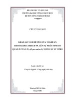
Figure 1 is a schematic outline of the Altered Sites II protocol. The
mutagenic oligonucleotide and an oligonucleotide that restores anti-
biotic resistance to the phagemid, the antibiotic repair oligonucle-
otide, are simultaneously annealed to the template DNA, either
ssDNA (5) or alkaline-denatured dsDNA. Synthesis and ligation of
the mutant strand by T4 DNA polymerase and T4 DNA ligase links
the two oligonucleotides. The mutant plasmids are replicated in a
mismatch repair deficient Escherichia coli m&S strain, either ES 130 1
(7,s) or BMH 7 1 - 18 (6), following clonal segregation in a second host
such as JM109. In addition to the repair oligonucleotide and the muta-
genic oligonucleotide, a third oligonucleotide can be incorporated in
the annealing and synthesis reactions that inactivates the alternate
antibiotic resistance. The alternate repair and inactivation of the anti-
biotic resistance genes in the Altered Sites II vectors allows multiple
rounds of mutagenesis to be performed without the need for additional
subcloning steps.
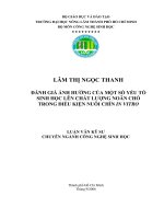
Figure 2 is a plasmid map for the pALEER- vector that is included
with the Altered-Sites II system (5). The pALTER-1 vector contains
a multiple cloning site flanked by opposmg SP6 and T7 RNA poly-
merase promoters, inserted into the DNA encoding the 1acZ a-pep-
tide. Cloning of a DNA insert into the multiple cloning site results in
inactivation of the a-peptide. The vector contains the gene sequences
for ampicillin and tetracycline resistance. The plasmid provided has a
frameshift in the ampillicin gene that is repaired in the first round of
mutagenesis. Propagation of the plasmid and recombmants is per-
formed under tetracycline resistance. The pALTER- 1 vector also con-
tains the fl origin of replication, which allows for the production of
ssDNA on infection with the helper phage R408 or Ml 3K07 (9-I 1).
Two other vectors are available, pALTER-Exl and pALTER-Ex2.
The pALTER-Exl is identical to pALTER-1 but contains a novel
multiple cloning site with an expression cassette (12). The pALTER-
Ex2 vector has the same multiple cloning site, expression cassette, and
fl origin as pALTER-Exl, but has a ColEl-compatible P15a origin of
replication and gene sequences for tetracycline and chloramphenicol
resistance (12).
Protocols for the preparation of template DNA and competent cells
are given in the Materials section. Design of the mutagenic oligonucle-
otide is discussed in Note 1, ref. 13, and Chapters 11 and 15 of ref. 14.
Positive Antibiotic Selection
3
multiple
cloning site
insert
+-
1’
1
1. Clone insert into
pALTER-1 Vector.
I
2. Isolate dsDNA.
insert
t’
3. Alkaline denature and
anneal mutagenic oligo,
Ampicillin Repair Oligo
t
4. Synthesize mutant strand
with T4 DNA Polymerase
and T4 DNA Ligase.
Amp’
ts
t
5. Transform ES 1301
8. Perform
mutS. Grow in media
additional
with selective antibiotic.
rounds of
mutagenesis
t
6. Prepare mini-prep. DNA.
using selection
7. Transform JM109.
for Tet repair
t
Select mutants on plates
alternatina
with appropriate
antibiotic.
Tet*
.
Fig. 1. Schematic diagram of the Altered Sites II in vitro mutagenesis proce-
dure using the pALTER-1 vector as an example.
4 Bohnsack
start
Fig. 2. pALTER-1 vector cncle map.
2. Materials
2.1. Reagents for Preparation of ssDNA
and Plasmid Miniprep DNA Templates
1. Helper phage (Either R408 or M13K07).
2. 3.75M Ammonium acetate in 20% polyethylene glycol (mol wt = 8000).
3. Chloroformxoamyl alcohol (24: 1).
4. TE-Saturated phenol:chloroform:tsoamyl alcohol (25:24: 1).
5. 5MNaCl stock.
6. Resuspension buffer. 25 mM Tris-HCl, pH 8.0, 10 mMEDTA, and 50 mM
glucose.
7. Lysis buffer: 0.2MNaOH, 1% SDS. Prepare fresh.
8. Neutralization solutton: 3.5Mpotassium acetate, pH 4.8.
9. DNase-free RNase A (100 mg/mL)
2.2. Reagents for Denaturation of dsDNA Template
1, 2M NaOH, 2 miI4 EDTA
2. 2M Ammonium acetate, pH 4.6.
3. 70 and 100% Ethanol.
4. TE buffer: 10 mMTris-HCl, pH 8.0, 1 WEDTA.
2.3. Regents for the Annealing Reaction
and Mutant Strand Synthesis
1. Oligonucleotides (see Table 1 and Note 1).
2. 10X Annealing buffer: 200 mM Trts-HCl, pH 7.5, 100 mA4 MgCl,,
500
mA4 NaCl
Positive Antibiotic Selection 5
Table 1
Repair and Knockout Oligonucleottde to be Used m Annealing Reactton@
Plasmid
pALTER- 1 and
pALTER& 1
pALTER- 1 and
pALTER-Ex 1
pALTER-Ex2
pALTER-Ex2
Selection
AmpSTet’ to Amp’TeV
First round
AmprTetS to AmpSTetr
Second round
CmSTetr to CmrTetS
First round
CmrTetS to CmSTetr
Second round
Repair oltgo
Amp repair
Tet repair
Cm repair
Tet repair
Knockout oligo
Tet knockout
Amp knockout
Tet knockout
Cm knockout
aAbbrevlatlons. Amp’, ampldhn reslstant, amps, ampidlm sensitive, Cm’, chloramphemcol
resistant, Cms, chloramphemcol sensitive; Tet’, tetracychne resistant, TeP, tetracycline sensitive
3. 10X Synthesis buffer: 100 mM Trts-HCl, pH 7.5, 5 mM dNTPs, 10 mM
ATP, 20 mA4 DTT.
4. T4 DNA polymerase (10 U/uL).
5. T4 DNA ligase (20 U/l.tL).
2.4. Reagents
for
Preparation
of Competent Cells and Transformation
1. Solution A: 30 mM potassium acetate, 100 mM RbCl, 10 mM CaCl,, 50
mM MnClz, and 15% (w/v) glycerol; adjust to pH 5.8 with acetic acid.
Filter-sterilize prior to use.
2. Solution B: 10 mM MOPS, 75 mA4 CaCl,, 10 mA4 RbCl, and 15% (w/v)
glycerol; adjust to a final pH of 6.8 with KOH. Filter-sterilize prior to use.
3.
E. coli
strains ES1301 mutSand JM109 (Promega, Madison, WI).
3. Methods
3.1. Preparation of Template
Templates may be either single-stranded phagmid DNA or double-
stranded plasmid DNA (see Note 2)
3.1.1. Preparation
of Single-Stranded DNA Template
1. Prepare an overnight culture of cells containing recombinant phagemtd
DNA by picking a single antibiotic resistant colony from a fresh plate.
Inoculate 3 mL of LB broth containing the appropriate antibiotic and shake
at 37OC.
2. The next morning, inoculate 50 mL of LB broth with 1 mL of the overnight
culture. Shake vigorously at 37°C for 30 min m a 250-mL flask.
6 Bohnsack
3. Infect the culture with helper phage at a multiplicity of infection (MOI) of
10. Continue shaking for 6 h. The volume of phage to be added to arrive at
an MO1 of approx 10-20 can be calculated by assuming that the cell con-
centratton of the starting culture ranges from 5 x lo7 to 1 x lo8 cells/ml.
An MO1 of 10 requires 5 x 1 O8 to 1 x lo9 phage/mL.
4. Harvest the supernatant by pelleting the cells at 12,000g for 15 mm. Trans-
fer the supernatant into a fresh tube and centrifuge at 12,000g for 15 mm to
remove any remaining cells.
5. Prectpitate the phage by adding 0.25 volumes of 3.75Mammonmm acetate
in 20% polyethylene glycol (mol wt 8000) to the supernatant. Allow solu-
tion to stand on ice for 30 mm then centrifuge at 12,000g for 15 mm. Thor-
oughly drain the supernatant.
6. Resuspend the pellet m 1 mL of TE buffer, pH 8.0, and transfer 500 pL of
the sample to each of two microcentrifuge tubes.
7. To each tube, add 500 nL of chlorofornnisoamyl alcohol (24: 1) to lyse the
phage, vortex for 1 mm. Separate phases by centrifuging for 2 mm m a
mtcrocentrifuge. Transfer the upper aqueous phases to fresh microcentrt-
fuge tubes.
8. Add an equal volume of TE-saturated phenol:chloroform:isoamyl alcohol
(25:24: 1) to each tube, vortex 1 mm, and centrifuge as in step 7
9. Transfer the aqueous phases to fresh tubes and repeat the phenol extraction
as m step 8. Repeat the extractton until there is no material visible at the
interface of the two phases. Transfer the aqueous phases to fresh micro-
centrifuge tubes and add NaCl to a final concentration of 0.25M (0.05 vol
of a 5MNaCI stock). Add 2 vol of 100% ethanol and mcubate on ice for 30
mm. Precipitate ssDNA by centrifuging at top speed in a microcentrifuge
for 15 min. Carefully rinse the pellet with 1 mL of 70% ethanol and dry
the pellet under vacuum. Resuspend the pellet in a small volume of Hz0
and estimate the concentration of DNA (see Note 3). The ssDNA is ready
for use in the annealing reaction (see Section 3.3.).
3.1.2. Plasmid Miniprep Procedure
1. Place 1.5 mL of an overnight culture into a mtcrocentrifuge tube and cen-
trifuge at 12,OOOg for 2 mm. The remaining overnight culture can be stored
at 4OC.
2. Remove the medium by aspiration, leaving the bacterial pellet as dry
as possible.
3. Resuspend the pellet by vortexing m 100 pL of ice-cold resuspension
buffer.
4. Add 200 pL of lysis buffer. Mix by inversion. Do not vortex. Incubate on
ice for 5 min.
Positive Antibiotic Selection
7
5. Add 150 pL of ice-cold neutralization solution. Mix by inversion and
incubate on ice for 5 min.
6. Centrifuge at 12,000g for 5 min.
7. Transfer the supernatant to a fresh tube, avoiding the white precipitate.
8. Add 1 vol of TE-saturated phenol:chloroform:isoamyl alcohol (25:24: 1).
Vortex for 1 min and centrifuge at 12,000g for 2 min.
9. Transfer the upper aqueous phase to an fresh tube and add 1 volume of chloro-
fotmisoamyl alcohol (24: 1). Vortex for 1 mm and centrifuge as in step 8.
10. Transfer the upper aqueous phase to a fresh tube and add 2.5 vol of 100%
ethanol. Mix and incubate on dry ice for 30 mm.
11. Centrifuge at 12,000g for 15 min. Rinse the pellet with cold 70% ethanol
and dry the pellet under vacuum.
12. Dissolve the pellet in 50 pL of sterile deionized H20. Add 0.5 pL of
DNase-free RNase A.
13. The concentratton of plasmid DNA can be estimated by electrophoresls on
an agarose gel.
3.2. Denaturation of Double-Stranded DNA Template
Double-stranded DNA must be alkaline denatured prior to use in the
mutagenesis protocol.
1. Set up the following alkaline denaturation reaction. This generates enough
DNA for one mutagenesis reaction: dsDNA template, 0.05 pmol (approx
0.2 pg); 2MNaOH, 2 mM EDTA, 2 pL; sterile deionized HZ0 to 20 pL
final volume.
2. Incubate for 5 min at room temperature.
3. Add 2 pL of 2M ammonium acetate, pH 4.6, and 75 pL of 100% ethanol.
4. Incubate for 30 min at -7OOC.
5. Precipitate the DNA by centrifugation at top speed in a microcentrifuge
for 15 min.
6. Dram and wash the pellet with 200 pL of 70% ethanol. Centrifuge again as
in step 5. Dry pellet under vacuum.
7. Dissolve pellet in 10 pL of TE buffer and proceed immediately to the
annealing reaction (see Section 3.3.).
3.3. Annealing Reaction
and Mutant Strand Synthesis
In the following example, both the antibiotic repair and knockout oli-
gonucleotides are included in the reaction mixture. It is not necessary to
include the antibiotic knockout oligonucleotide in the mutagenesis if a
second round of mutagenesis is not desired.
Bohnsack
1. Prepare the mutagenesis annealing reaction as described in the following
using the appropriate antibtotic repair and knockout oligonucleotides
(see Table I and Notes 1 and 4): 0.05 pmol dsDNA or ssDNA mutagenesis
template (200 ng dsDNA, 100 ng ssDNA), 1 pL (0.25 pmol) antibiotic
repair oligonucleotide (2.2 ng/pL), 1 pL (0.25 pmol) antibrotic knock-
out oligonucleotide (2.2 ng/nL), 1.25 pmol mutagenic ohgonucleotide
(phosphorylated), 2 PL annealing 10X buffer, stertle deionized H,O to a
final volume of 20 pL.
2. Heat the annealing reactions to 75°C for 5 min and allow them to cool
slowly to room temperature. Slow cooling mimmizes nonspecific annealing
of the oligonucleotides. Cooling at a rate of approx l”C/min to 45°C fol-
lowed by more rapid cooling to room temperature (22°C) is recommended.
3. Place the annealmg reactions on ice and add the following: 3 PL synthesis
10X buffer, 1 PL T4 DNA polymerase, 1 FL T4 DNA hgase, 5 pL (final
~0130 pL) sterile deionized H20.
4. Incubate the reaction at 37’C for 90 min.
The
mutagenesis reaction
is
then transformed into competent cells of
the
E. coli strain
ES1301
mutS
(see Section 3.5. and Note 5).
3.4. Preparation of Competent Cells
The following is the rubidium chloride method of Hanahan (15)
and may be used to prepare compentent cells of both ES 130 1
mu6
and
JM109.
1, Inoculate 5 mL of LB medium with 10 I ~L of a glycerol stock of either
ES1301
mutSor
JM109 cells. Incubate at 37°C overnight.
2. Inoculate 50 mL of LB medium with 0.5 mL of the overnight bacterial culture.
3. Grow cells until the OD600 reaches 0.4-0.6 (approx 2-3 h at 37°C).
4. Centrifuge cells for 5 mm at 5OOOg, 4°C m a sterile disposable tube.
5. Decant the supernatant and resuspend the cells in 1 mL of solution A. Bring
the volume up to 20 mL with solution A.
6. Incubate cells on ice for 5 min then pellet the cells as described in step 4.
7. Decant the supernatant and resuspend the cells in 2 mL of ice-cold solution
B. Incubate on ice for 15-60 min.
8. Freeze the cells on crushed dry ice in 0.2-mL ahquots. Competent cells
prepared by this method can be stored at -70°C for 5-6 wk.
3.5. Transformation into
ES1301
mutS Strain
1. Thaw competent ES 1301
m&S
cells (see Section 3.4.) on ice. Add 15 I.~L
of the mutagenesis reaction to 100 pL of competent cells and mix gently.
2. Incubate cells on ice for 30 min.
Positive Antibiotic Selection 9
3. Heat shock the cells at 42°C for 90 s after the incubation on ice to improve
the transformation efficiency.
4. Add 4 mL of LB medium without antibiotic and mcubate for 1 h at 37OC
with shaking.
5. After 1 h, add selective antibiotic to the culture. Final concentrations should
be 125 pg/mL ampicillin, 10 pg/mL tetracycline, or 20 pg/mL chloram-
phemcol depending on the vector and antibiotic repair oligonucleottde used
in the mutagenesis reaction.
6. Incubate culture overnight at 37°C with shaking.
7. Isolate plasmid DNA by alkaline lysis procedure as outlined in Section 3.1.2.
3.6. Transformation into JiMlO Strain
and Clonal Segregation
1. Thaw JM109 competent cells (see Section 3.4.) on ice. Add 0.05-0.1 pg of
plasmid DNA prepared from the overnight culture of ES1301 mutS cells
and mix briefly.
2. Let the cells stand on ice for 30 min.
3. Heat shock for 90 s at 42OC.
4. Add 2 mL LB medium and incubate at 37°C for 1 h to allow the cells to
recover.
5. Aliquot the culture into two microcentrifuge tubes and centrifuge for 1
min in a microcentrifuge.
6. Decant the supernatant and resuspend the cell pellets in 50 PL of LB medium.
7. Plate the cells in each tube on an LB plate contaimng the appropriate selec-
tive antibiotic.
The Altered Sites II protocol generally produces 60-90% mutants, so
colonies may be screened by direct sequencing. Assuming greater than
60% mutants are obtained, screening five colonies will give a greater
than 95% chance of finding the mutation. The SP6 and T7 sequencing
primers can be used for sequencing if the mutation is within 200-300 bp
from the end of the DNA insert. Often it is convenient to incorporate a
unique restriction site into the mutagenic oligonucleotide without alter-
ing the amino acid sequence. These sites can be used to screen for plas-
mids that have incorporated the mutagenic oligonucleotide.
When using this technique for doing multiple rounds of mutagenesis,
it is convenient to screen simultaneously for antibiotic sensitive isolates.
Simply inoculate each isolate into two tubes of media, one containing
each antibiotic; antibiotic clones will be identified easily. Antibiotic sen-
sitive isolates can also be identified by replicate plating in a grid format.
10 Bohnsack
Single colonies can be picked and used to inoculate two plates contain-
ing selective antibiotic in sequence.
4. Notes
1. The mutagenic oligonucleotide must be comphmentary to the ssDNA
strand produced by the mutagenesis vectors in the presence of helper
phage. This is true for double-stranded mutagenesis as well, since the
mutagenic oligonucleotide must hybridize to the same strand as the antibi-
otic repatr oligonucleotide for the coupling to be effective.
The stability of the complex between the oligonucleotide and the tem-
plate is determined by the base compositton of the oligonucleotide and the
conditions under which it IS annealed. In general, a 17-20 base oligonucle-
ottde with the mismatch located in the center will be sufficient for single base
mutations. This gives 8-10 perfectly matched nucleotides on either side of the
mismatch. For mutations involving two or more mismatches, ohgonucleotides
25 bases or longer are needed to allow for 12-l 5 perfectly matched bases on
either side of the mtsmatch. Larger deletions may require an oligonucle-
otide having 2&30 matches on either side of the mismatched region.
2. Mutagenesis can be performed usmg either dsDNA or ssDNA templates.
The double-strand procedure 1s faster and does not require the prior prepa-
ration of ssDNA. The single-strand procedure maybe useful, however,
when trying to maximize the total number of transformants, such as for
generating mutant libraries. Double-stranded DNA must be alkaline dena-
tured before use in the mutagenesis reaction. Poor quality dsDNA inhibits
second-strand synthesis during mutagenesis, therefore, tt is recommended
that sequencing quality DNA be used for the mutagenesis reaction.
3. Differences in yields of ssDNA have been observed to be dependent on the
particular combination of host, vector, and helper phage. Generally, higher
yields have been observed using the Altered Sites II vectors in combina-
tion with R408 helper phage and the JM109 train.
4. The annealing condittons required may vary with the composition of the
oligonucleotide. AT-rich complexes tend to be less stable than GC-rtch
complexes and may require a lower annealing temperature to be stabthzed.
Routinely, oligonucleotides can be annealed to a DNA template by heating
to 75°C for 5 min followed by slow cooling to room temperature. For more
detailed discussions of ohgonucleottde design and annealing condttions,
see refs. 13 and 14. The amount of ohgonucleottdes used m the annealing
reaction may vary, depending on the size and amount of DNA template. A
25: 1 oligonucleotide:template molar ratto for the mutagenic oligonucle-
otide and a 5: 1 oltgonucleotide:template molar ratio for the antibiotic repair
and knockout ohgonucleottdes is recommended for a typical annealing
Positive Antibiotic Selection 11
reaction. For efficient ligation, the mutagenic oligonucleotide should
be phosphorylated.
5. Mutant plasmid may be rapidly transferred from the
mutS
host into a more
suitable host for long-term maintenance and clonal segregation. The
mutagenesis reaction products are cotransformed into the ES1301 mutS
strain along with R408 rfDNA. The cotransformed rfDNA causes the
mutant phagemid to be replicated and packaged as an infectious particle
which is secreted into the media. These particles are used to infect a suit-
able F+ host such as JM 109, and the tranfectants are selected by their anti-
biotic resistance encoded by the phagemid. The procedure requires only a
single transformation step into ES1301 mutS and reduces the total time
required for the mutagenesis protocol by elimmating the plasmid miniprep
and transformation into the final host strain. The number of colonies
obtained after the cotransformation procedure is very dependent on the
competency of the ES1301 mutS cells; at least 106-lo7 cfu/pg DNA is
required for efficient cotransfotmation.
a. Thaw competent ES1 301 m&S cells on ice. To 100 l.tL of cells add 15
pL of the mutagenesis reaction from Section 3.3. and 100 ng of R408
rfDNA, mix briefly.
b. Incubate cells on ice for 30 min.
c. Heat shock the cells at 42°C for 90 s to increase transformation efficiency.
d. Add 4 mL of LB medium without antibiotic and incubate at 37OC
for 3 h with shaking to allow the cells to recover and produce infec-
tious phagemid.
e. After the 3-hr mcubation period:
i. Transfer 3 mL of the described culture to two tubes and pellet cells
by centrifugation at top speed in a microcentrifuge for 5 min.
Remove the supernatants, combine, and add to 100 PL of an over-
night culture of JMlO9 cells.
ii. To the remaining 1 mL of unpelleted transformed ES 1301 mutS cells,
add 4 mL of LB medium containing the appropriate selective antibi-
otic and incubate at 37OC overnight with shaking. This culture will
serve as a backup, to be used if the cotransformation procedure yields
too few transformants (see Section 3.5.).
f. Incubate the 3 mL JM109 culture from step 5a for 30 mm at 37°C with
shaking and plate 100 PL on each of four to five plates containing
the appropriate selective media. A typical cotransformation should
yield approx 50 colonies per plate. To obtain more colonies, plate the
entire 3-n& culture. Pellet the cells by centrifuging 1 min in a micro-
centrifuge. Resuspend the cells in 500 PL of LB and plate 100 yL on
each of five plates.
12 Bohnsack
Acknowledgments
I thank Scott Lesley for his assistance in developing the Altered Sites II
vectors and protocols and Ken Lewis and Dave Thompson for their work
on the original Altered Sites protocols. I also thank Jerry Hildebrand for
his assistance in preparing the figures.
References
1. Smith, M (1985) In vitro mutagenesis. Ann. Rev Genet.
19,423-462.
2. Hutchmson, C. A., Phillips, S , Edgell, M. H., Gillam, S., Jahnke, P., and Smith,
M. (1978) Mutagenesis at a specific position in a DNA sequence. J. Biol. Chem
253,655 l-6559
3, Wu, R. and Grossman, L. (1987) Site-specific mutagenesis and protein engmeer-
ing, Section IV, Chapters 17-20. Methods Enzymol.
154,329-403.
4. Kunkel, T. A. (1985) Rapid and efficient site-specific mutagenesis without pheno-
type selection. Proc Natl. Acad Scl USA 82,488-492.
5. Lewis, K. and Thompson, D V. (1990) Efficient site directed m vitro mutagenesis
using ampicillm selection, Nucleic Aczds Res 18, 3439-3443.
6. Kramer, B., Kramer, W., and Fritz, H. J (1990) Different base/base mismatched
are corrected with different efficiencies by the methyl-directed DNA mismatch-
repair system of E. Colt. Cell 38, 879-887.
7. Siegel, E. C., Wain, S. L., Meltzer, S F., Bmion, M. L , and Steinberg, J L. (1982)
Mutator mutations m Escherlchia colr induced by the insertion of phage mu and
the transposable resistance elements Tn5 and Tn 10. Mutat Res 93,25-33
8. Zell, R. and Frttz, H. J (1987) DNA mismatch-repair m Escherichza toll coun-
teracting the hydrolytic deamination of 5-methyl-cytosine restdues. EMBO J
6, 1809-1815.
9 Dotto, G. P., Enea, V., and Zinder, N D. (1981) Functional analysis of bacterioph-
age fl intergenic region. Vzrology
114,463-473
10 Dotto, G. P. and Zinder, N. D (1983) The morphogenetic signal of bacteriophage
fl. Vzrology
130,252-256.
Il. Dotto, G. P., Hormchi, K., and Zmder, N. D. (1984) The functional origm of bacte-
riophage fl DNA replication. Its signals and domains. J. Mof Biol
172,507-52 I
12. Altered Sites ZZ in vitro Mutagenesis Systems Manual, #TM001 (1994) Promega
Corp., Madison, WI.
13. Ptechocki, M. P. and Hmes, R N. (1994) Oligonucleotide design and optimized
protocol for site-directed mutagenesis. BioTechniques
16,702-707.
14. Sambrook, J., Fritsch, E. F., and Mamatts, T. (1989) Molecular Clonzng, A Labo-
ratory Manual Cold Spring Harbor Laboratory Press, Cold Spring Harbor, NY.
15. Hanahan, D. (1985) Techmques for transformation ofE. ~011, in DNA Clonrng, vol
1 (Glover, G. M., ed), IRL Press, Oxford, UK
CHAPTER 2
In Vitro Site-Directed Mutagenesis
Using the Unique Restriction
Site Elimination (USE) Method
Li Zhu
1. Introduction
In vitro site-directed mutagenesis has been widely used in vector modi-
fication, and in gene and protein structure/function studies (1,2). This
procedure typically employs one or more oligonucleotrdes to introduce
defined mutations into a DNA target of known sequence (2-9). A variation
of this procedure, termed the USE (Unique Restriction Site Elimination)
mutagenesis method (I), offers two important-and unique-advan-
tages: specific base changes can be introduced into virtually any double-
stranded plasmid; and plasmids carrying the desired mutation can be
highly enriched by selecting against the parental (wild-type) plasmid.
The USE strategy employs two oligonucleotide primers: one primer (the
mutagenic primer) produces the desired mutation, whereas the second
primer (the selection primer) mutates a restriction site unique to the plas-
mid for the purpose of selection.
Unlike most other methods of in vitro mutagenesis (4,7), the USE
method does not require single-stranded vectors or specialized double-
stranded plasmids. Cloned genes may be mutated in whatever vector they
reside, thus eliminating days or even weeks of subcloning steps. The
only requirement for the USE method is that the vector contain a unique
restriction enzyme recognition site and an antibiotic-resistance gene that
can be used in transformation as a selectable marker conditions easily met
From Methods m Molecular Biology, Vol 57’ In Wtro Mutagenesrs Protocols
Edlted by* M K. Trower Humana Press Inc , Totowa, NJ
13
by most plasmids. Generally, any unique restriction site present in the
plasmid can be used as the selection site in the mutagenesis experiment.
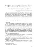
To carry out site-directed mutagenesis with the USE method, the
mutagenic and selection primers are simultaneously annealed to one
strand of the denatured target (parental) plasmid (Fig. 1). The annealing
conditions favor the formation of hybrids between the primers and the
DNA template, although some parental plasmids will simply reanneal.
After new DNA strands are synthesized and ligated to the primer-
annealed plasmids, the mixture of parental and hybrid plasmids is
digested with a restriction enzyme whose recognition site is altered by
annealing of the selection primer. This preliminary digestion with the
selection enzyme linearizes parental plasmids, rendering them at least
100 times less efficient than closed circular forms in transformation of
bacterial cells (10, II). However, hybrid plasmids containing a mismatch
in the enzyme recognition site are resistant to digestion and will remain
in circular form. Because of the very high probability that both the selec-
tion primer and the mutagenic primer will simultaneously anneal to the
same template, a plasmid that has an altered unique restriction site will
have a high probability (>90%) of containing the targeted mutation (12).
Thus, this preliminary digestion step enriches for hybrid (mutant) plas-
mids while selecting against parental duplex plasmids.
The hybrid (mutant) plasmids are transformed into an Escherichia coli
strain (mutS) defective in mismatch repair (first transformation), which
generates both mutant and parental duplex plasmids. Transformants are
pooled, and plasmid DNA is prepared from the resulting mixed plasmid
population. The isolated DNA is then subjected to a second selective
restriction enzyme digestion to eliminate the parental-type plasmids.
Mutant plasmids lacking the restriction enzyme recognition site are
resistant to digestion. A final transformation using the thoroughly
digested DNA will result in highly efficient recovery of the desired
mutated plasmids.
The combined use of two oligonucleotide primers in the USE method
results in mutation efficiencies of 70-90%. The actual mutation effi-
ciency achievable in any given experiment depends on a number of fac-
tors, including:
1. The ability of the restriction enzyme chosen for the selection steps to effi-
ciently digest parental (unmutated) plasmids (see Note 1);
2. The complete denaturation of the target plasmid before annealing the primers;
1. Denature dsDNA
2. Anneal Primers
3. Synthesize second strand
with T4 DNA Polymerase and
seal gaps with T4 DNA Ligase,
primary digestion with selection
restricbon enzyme
4 Transform muf.9 E co/r
FIRST TRANSFOfiMATfON
5 Isolate DNA from transformant pool
6 Secondary digestion with
selection enzyme
1
SElKllON
PRIMER
+
MUTAGENIC
PRIMER
1
0
1
+
7 Transform E.
coli
SECOND (FINAL) TRANSFORMATION
1
8. Isolate DNA from
indlvldual transformants
to confltm presence of
desired mutation
a
Plasmid
Fig. 1. Site-directed mutagenesis using the USE method. Note that the mutagenrc
primer contains the desired mutation and the selection primer contains a mutation
to either eliminate a unique restriction site or to change it to a different unique site.
Zhu
3. Simultaneous and saturated annealing of selection and mutagenic primers
to the denatured target plasmid (see Note 2); and
4. The stable incorporation of the base changes brought about by the anneal-
ing of the primers (see Note 3).
Mutations that can be introduced using the USE system are: single or
multiple specific base changes (I, 12-14); deletion of one or a few nucle-
otides (1,12); precise, large deletions (13) (see Note 4); and addition
(insertion) of a short stretch of DNA (15).
Another useful feature of this method is that multiple successive rounds
of mutagenesis may be performed on the gene of interest without recloning
if the selection step is designed so that it changes the original unique restric-
tion site into another unique restriction site-with no net loss of unique sites.
A list of ready-made selection primers available from Clontech (Palo
Alto, CA) is shown in Table 1. Trans Oligos are designed to be suitable
for use with many commonly used vectors and will maintain the reading
frame as well as the amino acid sequence encoded by the target gene.
Switch Oligos (also shown in Table 1) may be used to convert the
mutated site back to the original restriction site when multiple rounds of
mutagenesis are required. All Trans Oligos and Switch Oligos are phos-
phorylated at the 5’ end during their synthesis, and therefore are ready for
immediate use in the mutagenesis procedure.
2. Materials
All materials are stored at -20°C unless stated otherwise.
2.1. Reagents
for
USE Mutagenesis
1. 10X Annealing buffer:
200 m1I4 TrwHCl, pH 7.5, 100 mM MgCI,, 500
mA4 NaCl (store at 4°C).
2. 10X Synthesis buffer: 100
mA4 Tris-HCl, pH 7.5, 5 nwI4 each of dATP,
dCTP, dGTP, and dTTP, 10 mM ATP, 20 mM DTT.
3. E coli strains (store at -70°C in 50% glycerol):
a. BMH 71-18 mutS, a mismatch repair-deficient strain:
thi,
supE, A(lac-
proAB), (mutS::TnlO)(F’proAB, lad ZAM15) (16) (see Note 5).
b. Wild-type mutS+ strains, such as DH5cx.
4. T4 DNA polymerase (2-4 U/pL).
5. T4 DNA ligase (4-6 U/pL).
Example of materials that can be used for a control mutagenesis (see
Note 6 for discussion of the control materials provided in the Transformer
Site-Directed Mutagenesis System from Clontech):
Name of primer Catalog no
Table I
Premade Selectton Pruners”
Prtmer sequence Applicable vectors
Trans Oligo AatWEcoRV
Swatch Oligo EcoRVMafII
Trans Ohgo &ZIII/BgflI
Swttch Ohgo BglrI/AfnII
Trans Oligo AZwNU’peI
Switch Oligo S’eUAZwNI
Trans Ohgo EcoO 109I/StuI
Switch Oligo StuI/EcoO 1091
Trans Ohgo EcoRuEcoRV
Swnch Ohgo EcoRVIEcoRl
Trans Oligo HindIIIMuI
G
Swatch Oligo MluUHrndIII
Trans Ohgo MfeVAkoI
Switch Oligo Ncoh’NdeI
Trans Oligo ScaIMuI
Switch Ohgo StuIKcaI
Trans Oligo SspI/EcoRV
Switch Oligo
EcoRVISspI
Trans Ohgo XmnI!EcoRV
Switch Oligo EcoRVIXmnI
(#6487- 1)
(#6378- 1)
(#6494- 1)
(#6372-l)
(#6488-l)
(#6373-l)
(#6490- 1)
(#6379-l)
(#6496- 1)
(#6374- 1)
(#6497- 1)
(#6376- 1)
(#6493- 1)
(#6377- 1)
(#6495-l)
(#6380-l)
(#6498-l)
(#6381-l)
(#6499- 1)
(#6375-l)
GTGCCACCTGATATCTAAGAAACC 1,2,4-7, 10, 11
GTGCCACCTGACGTCTAAGAAACC
1
CAGGAAAGAAGATCTGAGCAAAAG l-3,8, 11
CAGGAAAGAACATGTGAGCAAAAG
1
GCAGCCACTAGTAACAGGATT 1-3,5,6,8-l 1
GwCCAmGTAACAGGATT
1
GTATCACGAGG’CCTTTCGTCTC 1,6, 11
GTATCACGAGGCCCTTTCGTCTC
1
CGGCCAGTGATATCGAGCTCGG
176
CGGCCAGTGAATTCGAGCTCGG 1
CAGGCATGCACGCGTGGCGTAATC
46
CAGGCATGCAAGCTTGGCGTAATC 1
GAGTGCACCATGGGCGGTGTGAAAT
1,496
GAGTGCACCATATGCGGTGTGAAAT
1
GTGACTGGTGAGGCCTCAACCAAGTC
l-l 1
GTGACTGGTGAGTACTCAACCAAGTC
1
CTTCCTTTTTCGATATCATTGAAGCATTT
1,2,44
CTTCCTTTTTCAATATTATTGAAGCATTT 1
GCTCATCATTGGATATCGTTCTTCGGG
1,3,4,6,8,9
GCTCATCATTGmAACGnTTCGGG
1
‘The Tram Ohgo or Switch Ohgo name denotes that a umque parental restnctlon site IS replaced by a new unique restnctlon site The underlmed
portions of the sequences represent the second restnctlon enzyme sites (after site conversion) Basepairs shown m bold are changed or deleted during
mutagenesis, and A represents a basepalr that has been deleted to create the new site The vectors listed are examples of vectors that contam the m&cated
Trans Ohgo sequences only once and thus are smtable for the USE method Each Switch Ohgo will anneal after mutagenesis to the same region that its
correspondmg Trans Ohgo anneals Some of the Tram Ohgos may be used with addltlonal vectors, for example, Trans Ohgo NdeIINotI IS unique in 116
vectors found m GenBank Note that all Tram Ohgo and Switch Ohgo sequences are umque m pUC19 However, Switch Ohgo sequences may not be
umque m other vectors after they have been mutated with the correspondmg Trans Ohgo
Before usmg a Trans Ohgo or Swatch Ohgo wltb another vector
not on the list, be sure to verify that the chosen restncbon site 1s present m the target plasmld only once Also verify that the base pan sequences flankmg
both sides of the restncfion site (1 e , the primer arms) match with the plasmld sequence Vector 1 pUCl9,2 pBR322,3 pBluescnpt SKII+, 4 pGem3Z,
5 pET1 lc, 6 pNEBl93,7 pGemex-I,8 pSPORTl,9 pIBI25, lo- pGAD424, 11 pGBT9 All Trans Ohgos and Switch Ohgos are 5’-phosphorylated.
18
Zhu
6. Control plasmid: pUCl9M, 0.1 p.g/uL (see Note 7).
7. Control mutagenic primer: 5’-P1 GAGGGTTTTCCCAGTCACGACG 3’,
0.05
ug/pL (see Note 8).
8. Control selection primer: 5’ P1 GAGTGCACCATGGGCGGTGTGAAAT
3’, 0.05
ug/pL (see Note 9).
9. NdeI restrIction enzyme (20 U/FL, for the control experiment).
Additional materials required for the experimental mutagenesis (see
Notes 1 O-1 6 for tips on primer design).
10. 0.1 l.tg/uL Target plasmid (see Notes 17 and 18).
11. 0.05 pg/pL Mutagenic primer.
12. 0.05 pg/pL SelectIon primer.
13. 5-20 U/uL Selection restriction enzyme (see Note 19).
2.2. Primer Phosphorylation, Preparation
of
Competent E. coli Cells, and Transformations
1, T4 polynucleotlde kinase (10 U/uL).
2. 10X T4 Kinase buffer: 500 mMTr1s-HCl, pH 7.5, 100 mMMgCl,, 50 mM
DTT, 10 mA4 ATP
3 Amplclllin: 100 mg/mL (1000X) stock solution in water. Filter sterilize
and store at 4°C for no more than 1 mo.
4. Competent cells: Either electrocompetent cells or chemically competent
cells (prepared ahead of time) may be used in the transformations.
Electrocompetent BMH 71-18
mutS
cells (#C2020-1) or DH5a cells
(#2022-l), and chemically competent BMH 7 l- 18
mutS
cells (#C20 lo- 1)
may be purchased from Clontech.
5. IPTG (isopropyl P-o-thiogalactopyranoside): 20-d stock solution in ster-
ile, distilled water. Store at 4OC. Use 10 pL/lO-cm plate.
6. LB agar plates containing 50-l 00 ug/mL amp1c1111n (LB + amp agar): LB
+ amp agar plates are used when performing the control mutagenesis wrth
pUC19M and the control primers. LB agar plates containing a different
antibiotic may be required for other target plasmlds.
7. LB medmm: 10 g/L bacto-tryptone, 5 g/L bacto-yeast extract, 10 g/L NaCl.
Adjust pH to 7.0 with 5NNaOH Autoclave to sterilize. For detailed infor-
mat1on on the preparation of media for bacteriological work, please refer
to the laboratory manual by Sambrook et al. (2).
8. TE buffer: 10 mA4Tns-HCl, pH 7.5, 1 mMEDTA.
9. Tetracycline: 5 mg/mL (100X) stock solution 1n ethanol. Wrap tube with
aluminum foil and store at -20°C.
IO. X-Gal (5-bromo, 4-chloro, 3-indolyl P-n-galactoside): 20 mg/mL stock solu-
tion in dimethylformamide (DMF). Store at -20°C. Use 40 pL/lO-cm plate.
Mutagenesis Using the USE Method
19
3. Methods
3.1. Primer Phosphorylation
Both the mutagenic and selection primers must be phosphorylated at
their 5’ end before being used in a USE mutagenesis experiment. Highly
efficient 5’ phosphorylation is commonly achieved by an enzymatic
reaction using T4 polynucleotide kinase. (See Note 20 for an alternative
phosphorylation procedure.) The control primers provided in the Trans-
former Kit have been phosphorylated and purified.
1. To a 0.5-mL microcentrifuge tube, add 2.0 pL of 10X kinase buffer, 1.0
pL of T4 polynucleotide kinase (10 U/uL), and 1 ~18 of primer (20-30
nucleotides long). Adjust the volume to 20 uL with water. Mix and centri-
fuge briefly.
2. Incubate at 37OC for 60 min.
3. Stop the reaction by heating at 65°C for 10 mm.
4. Use 2.0 pL of the phosphorylated primer solution in each mutagenesis
reaction.
5. Unused phosphorylated primers can be stored at -20°C for several weeks.
3.2. Denaturation and Annealing of Plasmid DNA
The following conditions are recommended for the annealing of phos-
phorylated primers to most plasmids (I, 12,13). Slow cooling is not nec-
essary and, in many cases, may be detrimental. The alternative annealing
protocol given in Note 2 1 is recommended for plasmids larger than 10 kb.
1. Prewarm a water bath to boiling (1 OOOC) (see Note 22).
2. Set up the primer/plasmid annealing reaction m a 0.5-mL microcentrifuge
tube as follows: 2.0 ltL of 10X annealing buffer, 2.0 JJL of plasmrd DNA
(0.05 pg/pL), 2.0 uL of selection primer (0.05 pg/pL), and 2.0 pL of muta-
genic primer (0.05 ug/pL) (see Note 23).
3. Adjust with water to a total volume of 20 PL. Mix well. Briefly centrifuge
the tube.
4. Incubate at 100°C for 3 mm.
5. Chill immediately m an ice water bath (OOC) for 5 min. Briefly centrifuge
to collect the sample.
3.3. Synthesis of the Mutant DNA Strand
1. Add to the annealed primer/plasmid mixture: 3.0 PL of 10X synthesis
buffer, 1.0 pL of T4 DNA polymerase (24 U/pL), 1.0 uL of T4 DNA
ligase (4-6 U&L), and 5.0 PL of water.
2. Mix well and centrifuge briefly. Incubate at 37OC for 2 h.
3. Stop the reaction by heating at 70°C for 10 min to inactivate the enzymes
4. Let the tube cool to room temperature for a few minutes.
3.4. Primary Selection
by Restriction Enzyme Digestion
After the mutant DNA strand synthesis and Iigation, a majority (>95%)
of the plasmids present in the total plasmid pool will be parental-type
plasmids. The purpose of the first restriction enzyme digestion is to
selectively linearize the parental DNA and thereby greatly enrich for mutant
plasmids within the total DNA pool before the first transformation. This
primary selection step facilitates the second (final) restriction digestion
by reducing the percentage of plasmids that are susceptible to digestion.
1, For the control mutagenesrs, simply add 1 VL of NdeI to the synthesis/
ligation mrxture and incubate at 37°C for l-2 h.
2. For the experimental mutagenesis, use the buffer condmons that resulted
in the most efficient digestion of the target plasmid, as determmed by the
relative reduction m the number of transformants in the preliminary test
(see Note 19). If drgestton was satisfactory (i.e., >99.9% of target plasmrds
were cut) using the annealing buffer, then simply add 20 U of the chosen
restriction enzyme to the synthesrs/ligatton mixture and incubate at 37°C
for l-2 h. If the digestion was significantly better using the enzyme
manufacturer’s recommended buffer, then change or adjust the buffer
accordmgly (see Note 24).
3. After the primary restrictron digestion, heat the tube containing the DNA
at 70°C for 5 min to inactivate possible endo- or exonuclease contaminants
that could damage the mutated DNA.
3.5. First Transformation
The purpose of the first transformation is to amplify the mutated strand
(as well as the parental strand) in the BMH 7 l-l 8 repair-deficient &W.&S)
strain of E.
coli.
Either electroporation or chemical transformation may
be used in this as well as all subsequent transformations. A detailed pro-
tocol for preparation of both types of competent cells can be found in
refs. I7 and 18. For best mutagenesis results, your transformation proce-
dure should yield at least
1
x 1 O7 transformants per microgram of DNA using
chemical transformation, or at least 1 x 1 OS transformants per microgram
of DNA using electrotransformation. The amount of plasmid/primer
DNA solution and competent cell suspension used per tube depends on
whether you are using chemical transformation or electrotransformation.
Mutagenesis Using the USE Method
21
3.5.1. Chemical Transformation
1. Preheat a heating block or water bath to 42OC.
2. Add 5-10 pL of the primary restriction-digested plasmid/primer DNA
solution to a 15mL Falcon tube containing 100 pL of competent BMH
71-18 mutS cells and incubate on ice for 20 min.
3. Transfer to 42OC for 1 min. Proceed to step 2.
3.5.2 Electroporation
1. Dilute the primary restriction-digested plasmid/primer DNA solution five-
fold with water.
2. Add 2 pL of the diluted DNA to a separate tube containing 40 ltL of
electrocompetent BMH 7 l- 18 mutS cells on ice.
3. Perform the electroporation according to the manufacturer’s instructions.
(For example, if you are using the Bio-Rad [Hercules, CA] E. cob Pulser
Electroporator, use a 0. l-cm cuvet and set the mstrument at 1.8 kV, with
constant capacitance at 10 pF and internal resistance at 600 W.)
3.5.3
Recovery
This applies to both chemical transformation and electroporation:
1. Immediately add 1 mL of LB medium (with no antibiotic) to each tube.
2. Incubate at 37OC for 60 mm with shaking at 220 rpm.
3.5.4. Amplification
1. Add 4 mL of LB medium containing the appropriate selection antibi-
otic. (In the case of the control experiment with pUC19M, use LB
medium containing 50 pg/mL ampicillin.)
2. Incubate the culture at 37OC overmght with shaking at 220 rpm.
3.6. Isolation of the Plasmid Pool
and Second Restriction Enzyme Digestion
After overnight growth of transformed BMH 71-18
mutS
cells, both
parental and mutated plasmid strands are segregated and amplified. Now
the plasmid pool can be purified from the cells and subjected to a second
restriction digestion to further enrich for the desired mutants by selecting
against the parental (nonmutated) plasmids. A quick boiling-lysis method
for plasmid preparation (19) is recommended, since it consistently results
in clean “miniprep” DNA. However, other standard miniprep procedures
such as the alkaline lysis method (2) may be used.
1, Dissolve the DNA pellets m 100 yL of TE buffer (each). The normal yield
of plasmid DNA using the quick boiling-lysis method is approx 2-5 pg.
22
2. (Optional) Purify DNA on a spin column (e.g., Chroma SPIN + TE-400
Column, Clonetech, cat. no. K1323-1).
3. Set up the followmg restriction enzyme digestion: 5.0 uL ofpurtfied mtxed
plasmid DNA (approx 100 ng), 2.0 uL of 10X restrtctton enzyme buffer,
and 1 uL of restriction enzyme (1 O-20 U). For the control experiment, use
NdeI and 1 OX annealing buffer (whtch IS functtonally equtvalent to 1 OX
NdeI buffer). Adjust the final volume to 20 uL with sterile water.
4. Mix well. Incubate at 37’C for 1 h.
5. Add an additional 10 U of the appropriate restriction enzyme, and continue
incubation at 37°C for another 1 h.
3.7. Final Transformation
The purpose of the final transformation is to amplify and stably clone
the mutated plasmid in a mismatch repair-competent (MutS) RecA-
strain of E.
coli
to avoid accumulation of random mutations in the plas-
mids. For this reason, the mismatch repair-deficient
E.
coli
strain used for
the first transformation (BMH 7 l-l 8 mutS) should not be used in the second
transformation. For blue/white colony color conversion of transformants on
X-gal/IPTG plates, an E.
coli
strain that is capable of lacZa complemen-
tation should be used; examples are DHSa, MV 1190, or JM109. (Other
E.
co/i
strains can also be used if no color conversion is required.)
1. Use 5.0 uL of the digested plasmtd (approx 25 ng) for transformatton of
chemtcally competent cells, or 1 .O pL of fivefold diluted (with water) plas-
mid (approx 1 ng) for transformation of electrocompetent cells. Follow the
same procedure as for the first transformatton.
2.
Recovery:
a. Immediately add 1 .O mL of LB medium (wtth no anttbiottc) to each tube.
b. Incubate at 37°C for 60 mm with shaking at 220 t-pm.
3. Prepare lo-, loo-, and lOOO-fold dilutions of the cell/DNA mixture (100
PL each).
4. For transformation experiments m which you expect to see a blue/white
colony color conversion, add 40 uL of a 20 mg/mL X-gal solutton and 10 pL
of a 20-W IPTG solutton to each tube containing cells (includmg controls).
5. Mix well and spread each suspension evenly onto LB agar plates contain-
ing the appropriate antibiotic for selectron of transformants (50 ug/mL
ampicillin for the control experiment).
6. Incubate the plates at 37°C overnight.
For pUC 19M control transformations, the mutatron efficiency IS estimated
by the number of blue (mutated) colonies divided by the total number of
Mutagenesis Using the USE Method 23
blue and white (wnmutated) colonies on X-gal/IPTG plates. An efficiency
rate of 70-90% is expected if the mutagenesis is performed successfully.
For mutagenesis experiments that do not involve a visible phenotype,
such as colony color, nutrition requirement, resistance to another antibi-
otic, or hybridization to a particular DNA probe, it may be necessary to
isolate plasmid DNA to characterize the mutation. Depending on the type
of mutation generated (such as a large deletion), the putative mutant plas-
mids may be screened by digestion with appropriate restriction enzymes.
In any case, the mutations should be verified by directly sequencing the
mutagenized region(s).
4. Notes
1. The success of this mutagenesis procedure is directly correlated with the
success of the restriction enzyme digestions used for mutant enrichment
Thus, it is important to obtain complete digestion of the parental plasmid
before each transformation. The chances of obtaining complete digestion
will be maximized if the plasmid DNA IS purified before the digestion
steps as explamed in Note 18 and the Methods section, and if you make
sure that the target plasmid at low concentrations can be efficiently digested
with the selection enzyme before beginning the mutagenesis experiment
(Note 19). The extent of digestion of the plasmid DNA after the first and
second digestion steps can be checked by electrophoresing two 5.0~pL
samples on a 0.8% agarose mmigel. Digested (Imearized) plasmid DNA
will run as a discrete band; undigested (circular) DNA will run as two
bands, correspondmg to the relaxed circular form and the supercoiled form,
with relative mobilitres less than and greater than the linearized form,
respectively. Since the parental plasmid makes up a greater part (>95%) of
the total plasmid pool at the first selection step, the bands corresponding to
the uncut (mutant) plasmids may not be visible or, if visible, will be quite
faint compared to the cut (parental) plasmid band. However, after the second
selection digestion, the resistant bands will become significantly more intense.
2. If only one of the two mutations is incorporated into the newly synthesized
strands, then the background of unmutated molecules following the sec-
ond E. coli transformation will be high. Tips to ensure high coupling of the
two types of mutations are given in Notes 13 and 23.
3. T4 DNA polymerase 1s used to extend the primers and to synthesize the
mutant strand. Unlike the Klenow fragment
of DNA polymerase I (an
enzyme commonly used in m vitro mutagenesis), T4 DNA polymerase
does not have strand displacement activity (20,21). Thus, with T4 DNA
polymerase, the primers (along with the desired base changes) will be
mcorporated into the newly synthesized strand. This property of T4 DNA
Zhu
polymerase makes it possible to perform multiple site-directed mutagen-
eses simultaneously using more than one mutagenic primer in the reaction
mixture (22). T4 DNA hgase is used to ligate the newly synthesized DNA
strand to the 5’ (phosphorylated) end of the oligonucleotide primer, a step
that is necessary to obtain covalently closed circular DNA with high trans-
forming ability. A specialized E. colz strain, BMH 71-18 mutS, defective
m mismatch repair (26), IS used in the first transformation to prevent unde-
sirable repair of the mutant DNA strand. The first transformation step also
serves to amplify the entire mutagenesis process.
4. The USE method has been applied to generate precise deletions as large as
several hundred basepans. However, the efficiency is generally much
lower than that of site-directed mutations (13). The deletion mutagenesis
efficiency will be improved if a umque restriction site within the targeted
deletion region can be used for the mutant enrichment (our unpublished
observations). This techmque, which allows for a more direct selection for
mutant plasmids, IS the basis of the Quantum Leap Nested Deletion method
described in Chapter 11.
5. E coli strain BMH 71-18 mutScarrles a TnlO msertron m mutS; this TnlO
insertion causes a repair-deficiency phenotype as well as tetracylme resis-
tance. For the repair-deficiency phenotype and TnlO insertion to be mam-
tained, this strain must be grown on mednnn containing 50 pg/mL tetracycline.
6. It is recommended to run a control mutagenesis in advance of or concur-
rently with-the experimental mutagenesis. The control experiment should
be designed to result in a specific, well-characterized mutation that can be
detected easily, for example, by a change m colony phenotype. The control
plasmid (pUC19M) and primers included m the Transformer Stte-
Directed Mutagenesis Kit (Clontech #Kl600-1), result in a blue/white
colony color conversion.
7. pUC 19M has a stop codon in its lad gene and thereby makes white colo-
nies on X-gal/IPTG plates when transformed into an appropriate host
strain. pUC 19M was derived from the wild-type pUC 19 by a USE site-
directed mutagenesis. pUC 19M contams a mutation at nucleotide positton
366, which interrupts the codmg sequence of the 1ac.Z gene on pUC 19 by
convertmg the UGG tryptophan codon to the amber stop codon UAG. (The
amber mutation m the pUC I9M lad gene is not sufficiently suppressed
by the suppressor tRNA gene [supE] present in BMH 7 I- 18 mu6 strain.)
Since this plasmid does not make functional P-galactosidase, rt will form
white colonies on LB agar plates containing X-gal and IPTG, when trans-
formed into a 1acZ bacterial host (such as BMH 71-18 mutS or DHSa).
The control plasmrd, pUC 19M, carries the gene for ampicillin resistance,
which is used as the bacterial selectable marker. E. coli cells trans-
Mutagenesis Using the USE Method
formed by pUC19M are selected by plating on medium containing 50
pg/mL ampicillin.
8. The control mutagenic primer reverts the stop codon in the 1uc.Z gene of
pUC19M back into a functional tryptophan codon. The reverted plasmid
will produce blue colonies on X-gal/IPTG plates, thus allowing visual color
discrimmation of the base change when transformed into an appropriate E.
coli
host strain. Since there is no selective pressure on the mutagenic
primer, it gives an objective indication of the mutagenesis process. The
efficiency of mutagenesis can be determined by the number of blue
(mutant) vs white (parental) colonies on X-gal/IPTG plates. If the mutagen-
esis has been successful, 70-90% of the ampicillin-resistant transformants
will form blue colonies. For mutation experiments in which colony color
conversion is not applicable, restriction analysis or sequencing is neces-
sary to verify and characterize the mutations.
9. The control selection primer alters a unique iWe recognition site on
pUC19M, thus allowing for selection against parental (unmutated) plas-
mids by digestion with NdeI. Available premade selection primers are
listed in Table 1.
10. The mutagenic and selection primers must anneal to the same strand of the
plasmid. The distance between the selection primer and the mutagenic
primer is not critical. These primers have been placed within 50 bp of each
other or as far apart as 5 kb (1,12). If possible, design the selection and
mutagenic primers so that they will be relatively evenly spaced after
annealing to the template; this will allow the DNA polymerase to extend
both primers an equivalent distance. In rare cases, where the unique restric-
tion site and the targeted mutagenic site are very close to each other, one
single primer can be designed to introduce both mutations simultaneously.
11. The function of the selection primer is to eliminate the origmal unique
restriction enzyme site. The selection primer can be designed by incorpo-
rating one or more basepair changes within the targeted unique restriction
site. Since restriction enzymes recognize an exact DNA sequence, any base
changes within the recognition sequence will abolish the restriction digestion.
12. If possible, use a restriction site located in an intergemc region as the
selection site. If the selection restriction site must be located within a gene,
avoid using a selection primer that will introduce changes that could inter-
fere with the expression of that gene (e.g., by causing a reading-frame shift
or a premature termination codon).
13. Length of primers: In most cases, 10 nucleotides of uninterrupted matched
sequences on both ends of the primer (flanking the mismatch site) should
give sufficient annealing stability, provided that the GC content of the
primer is greater than 50%. If the GC content is less than 50%, the lengths