physics of bio-molecules and cells
Bạn đang xem bản rút gọn của tài liệu. Xem và tải ngay bản đầy đủ của tài liệu tại đây (11.04 MB, 585 trang )
CONTENTS
Lecturers xi
Participants xiii
Pr´eface xvii
Preface xxi
Contents xxv
Course 1. Physics of Protein-DNA Interaction
by R.F. Bruinsma 1
1 Introduction 3
1.1 Thecentraldogmaandbacterialgeneexpression 3
1.1.1 Two families . . . 3
1.1.2 Prokaryotegeneexpression 5
1.2 Molecularstructure 8
1.2.1 ChemicalstructureofDNA 8
1.2.2 PhysicalstructureofDNA 10
1.2.3 Chemicalstructureofproteins 12
1.2.4 Physicalstructureofproteins 14
2 Thermodynamics and kinetics of repressor-DNA interaction 16
2.1 Thermodynamicsandthelacrepressor 16
2.1.1 Thelawofmassaction 16
2.1.2 Statisticalmechanicsandoperatoroccupancy 19
2.1.3 Entropy,enthalpy,anddirectread-out 20
2.1.4 Thelacrepressorcomplex:Amolecularmachine 23
2.2 Kineticsofrepressor-DNAinteraction 26
2.2.1 Reactionkinetics 26
2.2.2 Debye–Smoluchowskitheory 28
2.2.3 BWHtheory 30
2.2.4 Indirect read-out and induced fit . . . 32
xxvi
3 DNA deformability and protein-DNA interaction 34
3.1 Introduction 34
3.1.1 Eukaryotic gene expression and Chromatin condensation . . 34
3.1.2 A mathematical experiment and White’s theorem . . . . . . 37
3.2 Theworm-likechain 40
3.2.1 CircularDNAandthepersistencelength 42
3.2.2 Nucleosomes and the Marky–Manning transition . . . . . . 42
3.2.3 Protein-DNA interaction under tension 45
3.2.4 Force-ExtensionCurves 47
3.3 TheRSTmodel 50
3.3.1 Structuralsequencesensitivity 50
3.3.2 Thermalfluctuations 52
4 Electrostatics in water and protein-DNA interaction 53
4.1 Macro-ionsandaqueouselectrostatics 54
4.2 Theprimitivemodel 56
4.2.1 Theprimitivemodel:Ion-free 57
4.2.2 Theprimitivemodel:DHregime 57
4.3 Manningcondensation 58
4.3.1 Chargerenormalization 58
4.3.2 Primitivemodel:Oosawatheory 59
4.3.3 Primitivemodel:Freeenergy 61
4.4 Counter-ion release and non-specific protein-DNA interaction . . . 63
4.4.1 Counter-ionrelease 63
4.4.2 Nucleosome formation and the isoelectric instability . . . . 64
Course 2. Mechanics of Motor Proteins
by J. Howard 69
1 Introduction 71
2 Cell motility and motor proteins 72
3 Motility assays 73
4 Single-molecules assays 75
5 Atomic structures 77
6 Proteins as machines 78
7 Chemical forces 80
8 Effect of force on chemical equilibria 81
9 Effect of force on the rates of chemical reactions 82
xxvii
10 Absolute rate theories 85
11 Role of thermal fluctuations in motor reactions 87
12 A mechanochemical model for kinesin 89
13 Conclusions and outlook 92
Course 3. Modelling Motor Protein Systems
by T. Duke 95
1 Making a move: Principles of energy transduction 98
1.1 MotorproteinsandCarnotengines 98
1.2 SimpleBrownianratchet 99
1.3 Polymerizationratchet 100
1.4 Isothermalratchets 103
1.5 Motorproteinsasisothermalratchets 104
1.6 Designprinciplesforeffectivemotors 105
2 Pulling together: Mechano-chemical model of actomyosin 108
2.1 Swinginglever-armmodel 108
2.2 Mechano-chemicalcoupling 110
2.3 Equivalentisothermalratchet 111
2.4 Manymotorsworkingtogether 112
2.5 Designedtowork 115
2.6 Force-velocityrelation 116
2.7 Dynamical instability and biochemical synchronization . . 118
2.8 Transientresponseofmuscle 119
3 Motors at work: Collective properties of motor proteins 119
3.1 Dynamical instabilities . . 119
3.2 Bidirectionalmovement 120
3.3 Criticalbehaviour 121
3.4 Oscillations . . . . 124
3.5 Dynamic buckling instability . . . . . 125
3.6 Undulation of flagella . . 127
4 Sense and sensitivity: Mechano-sensation in hearing 129
4.1 Systemperformance 129
4.2 Mechano-sensors: Hair bundles . . . . 130
4.3 Activeamplification 131
4.4 Self-tunedcriticality 133
4.5 Motor-driven oscillations . 134
4.6 Channel compliance and relaxation oscillations . . . . . . 136
xxviii
4.7 Channel-driven oscillations 138
4.8 Hearingatthenoiselimit 139
Course 4. Dynamic Force Spectroscopy
by E. Evans and P. Williams 145
Part 1: E. Evans and P. Williams 147
1 Dynamic force spectroscopy. I. Single bonds 147
1.1 Introduction 147
1.1.1 Intrinsic dependence of bond strength on time frame
forbreakage 148
1.1.2 Biomolecular complexity and role for dynamic force
spectroscopy 148
1.1.3 Biochemical and mechanical perspectives of bond strength . 150
1.1.4 Relevant scales for length, force, energy, and time . . . . . . 153
1.2 Brownian kinetics in condensed liquids: Old-time physics . . . . . 154
1.2.1 Two-statetransitionsinaliquid 155
1.2.2 Kineticsoffirst-orderreactionsinsolution 156
1.3 Linkbetweenforce–time–andbondchemistry 158
1.3.1 Dissociation of a simple bond under force . . . 158
1.3.2 Dissociation of a complex bond under force:
Stationaryrateapproximation 159
1.3.3 Evolutionofstatesincomplexbonds 163
1.4 Testing bond strength and the method of dynamic force
spectroscopy 164
1.4.1 Probemechanicsandbondloadingdynamics 165
1.4.2 Stochastic process of bond failure under rising force . . . . 168
1.4.3 Distributionsofbondlifetimeandruptureforce 169
1.4.4 Crossover from near equilibrium to far from equilibrium
unbonding 172
1.4.5 Effect of soft-polymer linkages on dynamic strengths
ofbonds 175
1.4.6 Failure of a complex bond and unexpected transitions
instrength 177
1.5 Summary 185
Part 2: P. Williams and E. Evans 186
2 Dynamic force spectroscopy. II. Multiple bonds 187
2.1 Hiddenmechanicsindetachmentofmultiplebonds 187
2.2 Impactofcooperativity 188
2.3 Uncorrelatedfailureofbondsloadedinseries 191
2.3.1 Markovsequenceofrandomfailures 191
2.3.2 Multiple-complexbonds 193
xxix
2.3.3 Multiple-idealbonds 194
2.3.4 Equivalentsingle-bondapproximation 195
2.4 Uncorrelatedfailureofbondsloadedinparallel 198
2.4.1 Markovsequenceofrandomfailures 198
2.4.2 Equivalentsingle-bondapproximation 198
2.5 Poissonstatisticsandbondformation 199
2.6 Summary 203
Seminar 1. Polymerization Forces
by M. Dogterom 205
Course 5. The Physics of Listeria Propulsion
by J. Prost 215
1 Introduction 217
2 A genuine gel 218
2.1 Alittlechemistry 218
2.2 Elasticbehaviour 220
3 Hydrodynamics and mechanics 220
3.1 Motioninthelaboratoryframe 220
3.2 Propulsionandsteadyvelocityregimes 221
3.3 Gel/bacteriumfrictionandsaltatorybehaviour 223
4 Biomimetic approach 225
4.1 A spherical Listeria 225
4.2 Spherical symmetry . . . 226
4.3 Steadystate 227
4.4 Growthwithsphericalsymmetry 229
4.5 Symmetrybreaking 229
4.6 Limitationsoftheapproachandpossibleimprovements 231
5 Conclusion 234
xxx
Course 6. Physics of Composite Cell Membrane
andActinBasedCytoskeleton
by E. Sackmann, A.R. Bausch and L. Vonna 237
1 Architecture of composite cell membranes 239
1.1 The lipid/protein bilayer is a multicomponent smectic phase
withmosaiclikearchitecture 239
1.2 The spectrin/actin cytoskeleton as hyperelastic cell stabilizer . . . 242
1.3 Theactincortex:Architectureandfunction 245
2 Physics of the actin based cytoskeleton 249
2.1 Actinisalivingsemiflexiblepolymer 249
2.2 Actinnetworkasviscoelasticbody 253
2.3 Correlation between macroscopic viscoelasticity and molecular
motionalprocesses 258
3 Heterogeneous actin gels in cells and biological function 260
3.1 Manipulationofactingels 260
3.2 Control of organization and function of actin cortex
by cell signalling . . 265
4 Micromechanics and microrheometry of cells 267
5 Activation of endothelial cells: On the possibility
of formation of stress fibers as phase transition of actin-network
triggered by cell signalling pathways 271
6 On cells as adaptive viscoplastic bodies 274
7 Controll of cellular protrusions controlled by actin/myosin
cortex 278
Course 7. Cell Adhesion as Wetting Transition?
by E. Sackmann and R. Bruinsma 285
1 Introduction 287
2 Mimicking cell adhesion 292
3 Microinterferometry: A versatile tool to evaluate adhesion
strength and forces 294
4 Soft shell adhesion is controlled by a double well interfacial
potential 294
xxxi
5 How is adhesion controlled by membrane elasticity? 297
6 Measurement of adhesion strength by interferometric contour
analysis 299
7 Switching on specific forces: Adhesion as localized dewetting
process 300
8 Measurement of unbinding forces, receptor-ligand leverage
and a new role for stress fibers 300
9 An application: Modification of cellular adhesion strength
by cytoskeletal mutations 303
10 Conclusions 303
A Appendix: Generic interfacial forces 304
Course 8. Biological Physics in Silico
by R.H. Austin 311
1 Why micro/nanofabrication? 315
Lecture 1a: Hydrodynamic Transport 319
1 Introduction: The need to control flows in 2 1/2 D 319
2 Somewhat simple hydrodynamics in 2 1/2 D 321
3 The N-port injector idea 328
4 Conclusion 333
Lecture 1b: Dielectrophoresis and Microfabrication 335
1 Introduction 335
2 Methods 337
2.1 Fabrication 337
2.2 Viscosity 338
2.3 Electronicsandimaging 338
2.4 DNAsamples 338
3 Results 339
3.1 Basicresultsanddielectrophoreticforceextraction 339
4 Data and analysis 343
xxxii
5 Origin of the low frequency dielectrophoretic force in DNA 347
6 Conclusion 353
Lecture 2a: Hex Arrays 356
1 Introduction 356
2 Experimental approach 360
3 Conclusions 364
Lecture 2b: The DNA Prism 366
1 Introduction 366
2 Design 366
3 Results 367
4 Conclusions 372
Lecture 2c: Bigger is Better in Rachets 374
1 The problems with insulators in rachets 374
2 An experimental test 375
3 Conclusions 381
Lecture 3: Going After Epigenetics 382
1 Introduction 382
2 The nearfield scanner 383
3 The chip 384
4 Experiments with molecules 387
5 Conclusions 391
Lecture 4: Fractionating Cells 392
1 Introduction 392
2 Blood specifics 392
3 Magnetic separation 397
xxxiii
4 Microfabrication 398
5 Magnetic field gradients 399
6 Device interface 401
7 A preliminary blood cell run 406
8 Conclusions 409
Lecture 5: Protein Folding on a Chip 411
1 Introduction 411
2 Technology 412
3 Experiments 415
4 Conclusions 418
Course 9. Some Physical Problems in Bioinformatics
by E.D. Siggia 421
1 Introduction 423
2 New technologies 425
3 Sequence comparison 427
4 Clustering 430
5 Gene regulation 432
Course 10. Three Lectures on Biological Networks
by M.O. Magnasco 435
1 Enzymatic networks. Proofreading knots:
How DNA topoisomerases disentangle DNA 438
1.1 Lengthscalesandenergyscales 439
1.2 DNAtopology 440
1.3 Topoisomerases 441
1.4 Knotsandsupercoils 444
1.5 Topological equilibrium . 446
1.6 Cantopoisomerasesrecognizetopology? 447
1.7 Proposal:Kineticproofreading 448
xxxiv
1.8 Howtodoittwice 449
1.9 Thecareandproofreadingofknots 451
1.10 Suppression of supercoils . . 453
1.11Problemsandoutlook 455
1.12Disquisition 457
2 Gene expression networks. Methods for analysis
of DNA chip experiments 457
2.1 Theregulationofgeneexpression 457
2.2 Geneexpressionarrays 460
2.3 Analysisofarraydata 463
2.4 Somesimplifyingassumptions 464
2.5 Probesetanalysis 466
2.6 Discussion 470
3 Neural and gene expression networks: Song-induced gene
expression in the canary brain 471
3.1 Thestudyofsongbirds 472
3.2 Canarysong 473
3.3 ZENK 474
3.4 Theblush 476
3.5 Histologicalanalysis 476
3.6 Natural vs. artificial 479
3.7 TheBlushII:gAP 480
3.8 Meditation 481
Course 11. Thinking About the Brain
by W. Bialek 485
1 Introduction 487
2 Photon counting 491
3 Optimal performance at more complex tasks 501
4 Toward a general principle? 518
5 Learning and complexity 538
6 A little bit about molecules 552
7 Speculative thoughts about the hard problems 564
Seminars by participants 579
Preface
Matter has many states, including soft condensed, inert or alive. The latter is far
from thermodynamic equilibrium, and apparently has an agenda of its own. Yet
the same physical laws apply to all matter. The difference is in the complexity to
which living systems have evolved, to states that gather and process information,
replicate themselves, etc.
Molecular and cell biology have dramatically expanded our knowledge
about this complexity in the last decades. This knowledge is the foundation of
biological physics, which is currently expanding rapidly and is itself adding to
this knowledge. Its role in biology is a wonderful challenge: to draw the line
between necessity and possibility, between results of immutable physical laws
and results of evolution that may be specific to the one natural history we have
access to. The study of life is, after all, similar to reverse engineering
1
. What
fascinating engineering it describes, however! The deeper one gets into the
details, the more captivating the study becomes: these systems were “designed”
bottom-up, so answers to some of the biggest questions about Life are hidden in
their smallest parts.
The 75th Les Houches summer school addressed the physics of
biomolecules and cells. In biological systems ranging from single biomolecules
to entire cells and larger biological systems, it focused on aspects that require
concepts and methods from physics for their analysis and understanding. The
school opened with two parallel lecture series by Robijn Bruinsma and Jonathon
Howard. Physics of Protein-DNA Interaction by Robijn Bruinsma started from
the structure of DNA and associated proteins, and lead to discussions of
electrostatic interactions between proteins and DNA, and the diffusive search for
specific binding sites. Joe Howard’s lectures on Mechanics of Motor Proteins
discussed mechanical properties of individual proteins and motors, and of
complex cytoskeletal structures. Simultaneously, Evan Evans’ shorter series
Using Force to Probe Chemistry of Biomolecular Bonds and Structural
Transitions explored the rich dynamic behaviors of rupturing individual
biomolecular bonds. These lectures were followed by Erich Sackmann’s
discussion of Micro-rheometry of Actin Networks and Cellular Scaffolds. He
gave an introduction to membranes and the cytoskeleton and discussed the
mechanical properties of cells and the physics of cell adhesion. Robijn Bruinsma
1
Reverse engineering: “the process of analysing a subject system to identify the
system’s components and their interrelationships and create representations of
the system in another form or at a higher level of abstraction”. (E.J. Chikofsky
and J.H. Cross, II. IEEE Software 7 (1990) 13-17.)
xxii
complemented Erich Sackmann’s lectures with theoretical lectures on Statistical
Mechanics and Bioadhesion. The second half of the school contained two long,
parallel lecture series by Thomas Duke and Bill Bialek. Tom Duke
complemented Joe Howard’s course with Modelling Motor Protein Systems,
which focused more on theoretical approaches. Starting with physical models for
motor proteins, he discussed physical aspects of cilia and flagella and showed
that active physical phenomena on the cellular scale are important in hearing.
Another example of motion generation was discussed in Jacques Prost’s lectures
Physics of Listeria Propulsion, which provided a general description of how the
controlled polymerization of an actin gel can be used for propulsion. Bill
Bialek’s long series of lectures Thinking About the Brain gave an introduction to
the principles governing sensory and nervous systems. Starting from simple
examples of information processing in the visual system of the fly, he moved to
fundamental questions on how nervous systems process information. In
Bioinformatics and Statistical Mechanics Eric Siggia reviewed decoding of
genetic information obtained from genome projects. It was followed by Bob
Austin’s lectures on Micro- and Nanotechnology-Physics in Biotechnology, new
technologies which make it possible to study and manipulate biomolecules in
artificial arrays and structures. Marcelo Magnasco wrapped up the school
excellently with his Three Lectures on Biological Networks, which covered the
unknotting of DNA, the analysis of gene chip data, and studies of gene
expression in learning canaries, all in a style that kept the attention of the
audience to the last minute of four weeks.
The lectures were complemented by invited seminars given by Albert
Libchaber (RecA Polymerization on Single-Stranded DNA and Directed
Evolution: A Molecular Study) and Marileen Dogterom (Polymerization Forces).
Two public lectures were given in the town of Les Houches, by Albert Libchaber
(Qu'est-ce que la vie ?) and Thomas Duke (Les moteurs de la vie). Furthermore,
Phil Williams gave an invited lecture within Evan Evan's series, and Tom
McLeish and Chris Wiggins contributed with seminars: The Mysterious Case of
Too Many
β
-Sheets and Into Physical Models of Biopolymers, respectively.
During a study period, Tom McLeish gave a well-attended tutorial on thermally
activated barrier crossing, on the school's lawn, with Mt. Blanc as a backdrop
and most illustrative barrier. We also organized sixteen short student
presentations over four evenings, and two poster sessions with a total of
seventeen posters; see titles and presenters at the end of this volume. The
students had great energy and enthusiasm, and, amazingly in view of their
schedule, kept it up till the very end.
This school had three times as many applicants as there are seats in the
lecture hall, and we had to turn down many strong applicants. We hope this book
to some extent makes up for this unfortunate restriction on admission.
xxiii
The relative isolation of the Les Houches Physics School on the mountain
side vis-à-vis Mt. Blanc is perfect for learning and interacting. As are long hikes
in the mountains on weekends. Life-long friendships are formed, we know: Two
of this school’s organizers first met as students in a Les Houches summer school.
If the present school has taught and inspired its participants as much as that
school did years ago, we have done well.
Acknowledgements
The four weeks spent in Les Houches went smoothly, thanks to the staff of the
school: Ghislaine D'Henry, Isabel Lelièvre and Brigitte Rousset. The school was
sponsored by NATO as an Advanced Study Institute, by the EU as a
Eurosummerschool, by CNRS as an École Théematique, and by the Danish
Research Agency through its Graduate School of Biophysics. NSF covered travel
costs for some US residents. We thank them all for making the school possible.
We are convinced that this book presents outstanding examples of biological
physics, and thank the contributors again for their great efforts of lecturing and
writing.
Henrik Flyvbjerg
Frank Jülicher
Pál Ormos
François David
“bruinsma”
2002/8/8
page 1
✐
✐
✐
✐
✐
✐
✐
✐
COURSE 1
PHYSICS OF PROTEIN-DNA INTERACTION
R.F. BRUINSMA
Department of Physics and
Astronomy, University of California,
Los Angeles, CA 90024, USA,
and
Instituut-Lorentz for Theoretical
Physics, Universiteit Leiden,
Postbus 9506, 2300 Leiden,
The Netherlands
“bruinsma”
2002/8/8
page 2
✐
✐
✐
✐
✐
✐
✐
✐
Contents
1 Introduction 3
1.1 Thecentraldogmaandbacterialgeneexpression 3
1.2 Molecularstructure 8
2 Thermodynamics and kinetics of repressor-DNA interaction 16
2.1 Thermodynamicsandthelacrepressor 16
2.2 Kineticsofrepressor-DNAinteraction 26
3 DNA deformability and protein-DNA interaction 34
3.1 Introduction 34
3.2 Theworm-likechain 40
3.3 TheRSTmodel 50
4 Electrostatics in water and protein-DNA interaction 53
4.1 Macro-ionsandaqueouselectrostatics 54
4.2 Theprimitivemodel 56
4.3 Manningcondensation 58
4.4 Counter-ion release and non-specific protein-DNA interaction . . . 63
“bruinsma”
2002/8/8
page 3
✐
✐
✐
✐
✐
✐
✐
✐
PHYSICS OF PROTEIN-DNA INTERACTION
R.F. Bruinsma
1 Introduction
1.1 The central dogma and bacterial gene expression
1.1.1 Two families
Life is based on a symbiotic relationship between two families of biopoly-
mers: DNA and RNA, constituted of nucleic-acids, and proteins, consti-
tuted of amino-acids [1]. Proteins are the active agents of the cell. As
enzymes, they control the rates
of biochemical reactions taking place inside
the cell. They are responsible for the transcription of the genetic code, i.e.,
the production of copies of short segments of the genetic code that are used
as blue-prints for the production of new proteins, and for the duplication
of the genetic code, i.e., the production of a full copy of the genetic code
during cell division. Synthesis of other macromolecules, such as lipids and
sugars, is carried out by proteins, the mechanical force of our muscles is gen-
erated by specialized proteins adept at “mechano-chemistry”, they detect
light, sound, and smell, and maintain the structural integrity of cells.
If we view the cell as a miniature chemical factory that simultaneously
runs many chemical processes, then the proteins form the control system of
the factory, turning reactions on and off. The control system obeys orders
from the central office: the cell nucleus. The DNA inside the nucleus can
be considered as the memory of the computer system of the central office:
it is the information storage system of the cell. Blueprints for the synthesis
of proteins are stored in the form of DNA base-pair sequences, much like
strings of zero’s and one’s store information in digital computers. A gene is
the data string required for the production of one protein (actually, multiple
variants of a protein can be produced from the same gene). The beginning
and end points of a gene are marked by special “start” and a “stop” signals.
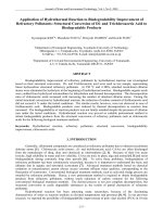
When a protein has to be synthesized, a specialized copying protein, RNA
polymerase, transcribes a copy of a gene beginning at the start signal and
ending at the stop signal (see Fig. 1).
c
EDP S ciences, Springer-Verlag 2002
“bruinsma”
2002/8/8
page 4
✐
✐
✐
✐
✐
✐
✐
✐
4 Physics of Bio-Molecules and Cells
Fig. 1. Gene transcription.
This copy is in the form of an RNA strand known as mRNA (or “mes-
senger” RNA). A huge molecular machine, the Ribosome, synthesizes the
protein from the mRNA blueprint. Interestingly, these Ribosomes are com-
pound constructs of RNA strands (known as rRNA) and proteins, with the
active biochemistry carried out not by the protein part, as you might have
expected, but by the RNA part. Indeed, unlike DNA, RNA strands are in
fact capable to act as enzymes.
The information stream is strictly one way: DNA contains the informa-
tion required for the synthesis of proteins. The genetic code is not altered
by the transcription, and RNA strands do not insert their code into DNA.
We call this basic principle of biochemical information flow the “central
dogma”. We know next to nothing about how this elaborate relationship
between the nucleic and amino acids developed. The basic chemical struc-
ture of the two families is quite different. The molecular biology of living
organisms is all highly similar and based on the central dogma and we do
not know of the existence of more primitive molecular information and con-
trol systems from which we could somehow infer a developmental history
(though we suspect that once upon a time both information storage and en-
zymatic activity was based purely on RNA since RNA is able to carry out
enzymatic activity as we saw). The central dogma applies to living organ-
isms. Retroviruses are able to insert their RNA code into host DNA, using a
special enzyme called “reverse transcriptase”. This looks like an exception
to the central dogma but viruses are not considered living organisms since
they are not able to reproduce themselves independently nor do they carry
out metabolic activity, the two defining requirements of a living organism.
“bruinsma”
2002/8/8
page 5
✐
✐
✐
✐
✐
✐
✐
✐
R.F. Bruinsma: Physics of Protein-DNA Interaction 5
It is reasonable to ask why biopolymers should be of special interest to
physicists. The physics of polymers – particular synthetic polymers – has
been studied for decades and an elegant, general theoretical framework is
available. The motivation behind a study of the interaction between DNA
and proteins is quite different from that of a study of synthetic polymers.
In polymer physics, we want to compute the free energy and correlation
functions of a typical polymer in a solution or melt, with results that are
as much as possible independent of the detailed molecular structure of the
polymers. That philosophy does not apply to biopolymers where we are
dealing with highly a-typical molecules that carry out certain functions.
Their structure presumably evolved under the adaptive pressures exerted
on micro-organisms that relied for their survival on efficient performance of
the functions these molecules are involved with. A molecular biophysicist
tries to shed light on how functional molecular devices work and how their
design constraints are met. These are of course very complex systems, so it
is a good strategy to focus as much as possible on basic principles of physics
of general validity and relying as little as possible on assumptions concerning
the detailed molecular structure. The hope is that this will provide us with
constraints on the design and operation of functional biopolymers in the way
that the Second Law of Thermodynamics constrains the maximum efficiency
of steam engines.
In order to illustrate this approach, we will focus on two special cases
that have been particularly important in the development of our under-
standing of protein-DNA interaction, the lac repressor and the Nucleosome
Complex. These two systems have been studied in such detail that we may
hope to understand how they “work” as molecular devices. In these lectures,
we will see what insights thermodynamics, statistical mechanics, elasticity
theory, and electrostatics can provide us in this respect.
1.1.2 Prokaryote gene expression
How does an organism “know” when to turn gene transcription on and
when to turn it off? We divide cells in two groups: eukaryotes and prokary-
otes. The cells of animals and plants – the eukaryotes – have their DNA
sequestered inside a nucleus and the cell has a complex set of internal “or-
gans” called organelles. Gene expression of eukaryotic cells, the focus of
much current research, is a complex affair, which we will discuss in a later
section. Bacteria, prokaryotes, lack a nucleus and organelles and their gene
expression is much better understood [3]. We will discuss a simple example:
the expression of the “lac” gene of the bacterium Escirichia Coli (E.Coli for
short) [4].
Large numbers of the E.Coli parasitic bacteria live inside your intestines
(“colon”). When you drink a glass of milk, part of it will be metabolized
“bruinsma”
2002/8/8
page 6
✐
✐
✐
✐
✐
✐
✐
✐
6 Physics of Bio-Molecules and Cells
not by you but by your E.Coli bacteria. The first step is the breakdown
of lactose, sugar molecules consisting of two linked molecular rings. Lac-
tose is broken down into two single-ring glucose molecules. This chemical
reaction requires an enzyme, called “β Galactosidase”, to proceed because
lactose does not dissociate spontaneously (an enzyme speeds up a reac-
tion by lowering the activation energy barrier). First though, the lactose
molecules must be transferred from the exterior of the bacterium to the cell
interior (or “cytoplasm”) across the membrane that surrounds E.Coli. This
is done by another protein, called “Permease”. Finally, a third protein,
called “Transacetylase”, is required for chemical modification of the sugar
molecules.
The DNA of E.Coli carries three separate genes for the production of
these three enzymes: lacY, lacZ, and lacA. Expression of the three genes
starts when the environmental lactose concentration rises, and it stops when
the lactose concentration drops (to avoid wasteful use of precious macro-
molecular material). The three genes are located right behind each other on
the DNA, and – sensibly – they are transcribed collectively. Such a cluster
of functionally connected genes is called an “operon”. The lac operon also
contains three regulatory sequences:
a) Promoter Sequence
This sequence is “recognized” by RNA Polymerase. By that we mean
that RNA Polymerase molecules in solution bind to Promoter
Sequences on the DNA but not to other sequences. From this start
site, RNA polymerase can transcribe RNA in either direction.In
one direction, “downstream”, it produces the RNA code of our three
enzymes. In the other direction, “upstream”, it transcribes the neigh-
boring “Regulator” sequence.
b) Regulator Sequence
The Regulator sequence is the code of a fourth protein: lac repressor.
The lac repressor, which is not involved in the metabolic of lactose,
plays a key regulatory role in turning the gene “on” or “off”.
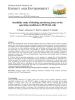
c) Operator Sequence
The operator sequence is a DNA sequence that is recognized by lac
repressor. If lac repressor is bound to the operator sequence, then
downstream gene expression is blocked. The Figure 2 shows how this
“genetic switch” works.
First, assume that the concentration of lactose in the environment is high.
Lactose molecules bind reversibly to the repressor protein. For high lactose
concentrations, the lactose-bound form is favored under conditions of chem-
ical equilibrium. In the lactose-bound (or “induced”) form, the repressor
“bruinsma”
2002/8/8
page 7
✐
✐
✐
✐
✐
✐
✐
✐
R.F. Bruinsma: Physics of Protein-DNA Interaction 7
Fig. 2. The lac operon and gene regulation.
has a different structure in which it does not bind to the operator sequence.
RNA polymerase proteins binding to the promoter sequence are free to tran-
scribe in the down-stream (and up-stream) direction. Along the downstream
direction, it will produce an RNA copy of the genes of the three enzymes
required for lactose breakdown. Transcription along the up-stream direction
produces an RNA copy of the lac repressor gene. Production of repressor
proteins at a low level is necessary to maintain their concentration since
proteins have a finite lifetime (after a certain period, a protein receives a
molecular “tag” targeting it for future breakdown as part of the scheduled
maintenance program of the cell).
Next, assume that the lactose concentration has dropped. The chemical
equilibrium now favors the lactose-free conformation of the repressor. Lac
“bruinsma”
2002/8/8
page 8
✐
✐
✐
✐
✐
✐
✐
✐
8 Physics of Bio-Molecules and Cells
repressor binds to the operator sequence and downstream gene transcription
is blocked. Genetic switches of this type are used by E.Coli (and other
bacteria) to respond to changes in temperature, salinity, acidity, and the
oxygen level. Efficiency of these switches clearly is a matter of life and
death for the bacterium so we should expect that the structure of proteins
like the lac repressor has been “sharpened” by natural selection for optimal
performance. If you would put yourself the task of designing a lac repressor
protein some obvious minimum engineering requirements would be:
Specificity
: the lac repressor must be able to recognize the operator se-
quence. Repressor proteins must be able to efficiently “read” the DNA
code.
Reversibility
: the lac Repressor must bind reversibly to lactose or else gene
expression could not be turned off. Similarly, it must bind reversibly to
DNAorelsegeneexpressioncould not be turned on.
Reactivity
: The lac Repressor must locate the operator sequence within
minutes after the lactose concentration drops. If it takes too long to turn a
genetic switch then the bacterium could be dead before it had the change
to respond to the changing environment.
In the next sections, we will see what thermodynamics, statistical mechan-
ics, and elasticity theory have to say about these requirements. First, we
have to learn more about the molecular structure of the two biopolymer
families [2].
1.2 Molecular structure
1.2.1 Chemical structure of DNA
The basic monomer unit – the polymer repeat unit – of double-stranded
DNA is shown in Figure 3.
The parts marked B and B
∗
are large, planar organic groups consisting of
one or two 5-atom aromatic rings. They resemble benzene and, like benzene,
these groups do not dissolve very easily in water. The symbols B and B
∗
stand either for the smaller single- ring Cytosine and Thymine (the “pyrim-
idines”), or the larger two- ring Guanine and Adenine (the “purines”). We
will use the notation G, T, C, and A for short. The four groups all have the
chemical character of a base (i.e., they are proton acceptors).
Not every combination of bases is permitted: in particular only B-B
∗
pairs of purines and pyrimidines are possible. The Watson–Crick base-
pairing consists of combining A with T and G with C. An A-T pair is
connected by two hydrogen bonds and a G-C pair by three hydrogen bonds,
so they have a higher binding energy. Other purine-pyrimidine pairings
“bruinsma”
2002/8/8
page 9
✐
✐
✐
✐
✐
✐
✐
✐
R.F. Bruinsma: Physics of Protein-DNA Interaction 9
Fig. 3. Double-stranded D NA repeat unit.
(like G with T) are possible, but they do have a lower binding energy. The
genetic code of an organism, or “genome”, is simply a listing of the different
base-pairings along the DNA sequence of that organism. Note that if you
know the sequence of bases of one strand, you always can reconstruct the
other “complementary” strand, assuming that Watson–Crick base pairing
is valid.
The bases are connected to sugar groups (indicated by S in the figure).
Sugars have the general formula (CH
2
O)
n
and usually are water soluble.
The particular sugar of DNA belong to the group of pentoses, 5-atom sugar
rings, and is known as deoxyribose. The deoxyriboses of two adjacent bases
are connected together by tetrahedral phosphate groups (PO
−
4
) to form to-
gether the sugar-phosphate “backbone”. Adjacent sugar groups are sepa-
rated by 6
˚
A. The backbone strands have a directionality: they start with
a deoxyribose at the 3
end and end with a phosphate at the 5
end. The
backbone has two important physical characteristics for our purposes: it is
highly flexible and, in water at room temperature, it is highly charged.The
negative charge of the backbone is due to the fact that the phosphate groups
in water at physiological acidity levels are fully dissociated. Charged molec-
ular groups are usually soluble in water and the sugar-phosphate backbone
is indeed highly soluble in water. The flexibility is due to the fact that the
covalent P-O bonds can freely rotate around so adjacent PO
−
4
tetrahedra
and ribose rings along the backbone can rotate around their joining axis.
We can describe the backbone as a charged, freely jointed chain.
“bruinsma”
2002/8/8
page 10
✐
✐
✐
✐
✐
✐
✐
✐
10 Physics of Bio-Molecules and Cells
RNA molecules are similar to single stranded DNA molecules with two
differences. First, the base Thymine is replaced by another base, Uracil,
and second, the sugar group has an extra OH group and is called a ribose.
Intermezzo: Hydrogen bonding and the hydrophobic force
Hydrogen bonding provides the binding mechanism between complementary
bases. Hydrogen bonding plays in general a central role whenever macro-
molecules are dissolved in water. The hydrogen bond is an electrostatic
bond with a positively charged proton from one molecular group associat-
ing with a negatively charged atom of another molecular group, usually an
oxygen (O
−
), Carbon (C
−
) or nitrogen (N
−
) atom. The cohesion of wa-
ter is due to hydrogen bonding between water molecules, with the proton
of one water molecule binding to the oxygen of another water molecule.
The characteristic energy scale of the hydrogen bond is of the order of the
thermal energy k
B
T , so it is a relatively weak bond. At room tempera-
ture, a thermally fluctuating network of hydrogen bonds connects the water
molecules.
Molecules such as alcohol that are easy to dissolve in water are called
“hydrophilic” while molecules, such as hydrocarbons, that are not soluble
in water are called “hydrophobic” [5]. Hydrophobic molecules cannot be
incorporated in the thermally fluctuating network of the hydrogen bonds.
They are surrounded by a shell of water molecules that have a reduced
entropy, since they have fewer potential partners for the formation of a
hydrogen bonding network. As far as the water molecules are concerned,
the surface of a large hydrophobic molecule resembles the air-water surface,
which has a surface energy γ of about 70 dynes/cm. We thus can estimate
the solvation free energy – the free energy cost of inserting a molecule in
a solvent – as the surface area of the hydrophobic molecule times γ.If
we wanted to dissolve a certain number of hydrophobic molecules we could
reduce the total exposed surface area in order to minimize the free energy
cost by collecting the hydrophobic molecules in dense clusters. This effect
is known as the “hydrophobic interaction”, though it obviously is not a
pair-wise interaction between molecules. Ultimately, the clustering leads to
phase-separation, which you can observe when you try to mix oil with water.
An important thermodynamic characteristic of the hydrophobic interaction
is that it is predominantly entropic in nature.
1.2.2 Physical structure of DNA
The physical structure of double-stranded DNA is determined by the fact
that it is neither hydrophobic nor hydrophilic. It belongs to a special
intermediate group the “amphiphiles” that share properties from both
“bruinsma”
2002/8/8
page 11
✐
✐
✐
✐
✐
✐
✐
✐
R.F. Bruinsma: Physics of Protein-DNA Interaction 11
classes: one part of DNA – the backbone – is hydrophilic and another
part of DNA – the bases – is hydrophobic. This frustrated, amphiphlic
character of DNA, plus the flexibility of the backbone, produces the famous
double-helical structure of DNA. To see how, imagine DNA stretched out
like a (straight) ladder (Fig. 4).
Fig. 4. Geometry of stretched DNA.
It turns out that the gaps between the rungs of the ladder, 2.7
˚
A, are
wide enough to allow water molecules to slip in between the bases. Under
the action of the hydrophobic force, the bases attract each other. The fixed
6
˚
A spacing between the sugar groups prevents a contraction of the ladder,
but there is another way to bring the bases in contact. Imagine that we
gradually twist the ladder, thereby forming a double spiral. This is possible
because of the flexibility of the backbone. As we increase the twisting, the
bases are brought into closer contact and the water molecules are squeezed
out. For a twist angle T of about 32 degrees between adjacent bases, the
gap is completely closed. This produces the classical double helix shown
below. The repeat length is 360/T bases, or about 11 bases. The repeat
distance along the helix, or pitch, is about 35
˚
A (using Fig. 4, compute T
yourself).
The DNA double-helix is thus held together by the hydrophobic at-
traction between bases, sometimes called the stacking interaction, and the
hydrogen bonding between complementary bases. The double-helix is not
very stable. If you heat DNA, the two strands start to fall apart for temper-
atures in the range of 70–80
◦
C. In addition, a number of different variants
of the double helix can be realized by modest changes in the environmen-
tal conditions. Under conditions relevant for the life of cells, the dominant
structure is the “B form”, a right-handed helix with a 24
˚
Adiameter. In-
creasing the salt concentration somewhat weakens the electrostatic repulsion
between the two backbones. A new structure, known as “A DNA”, appears,
“bruinsma”
2002/8/8
page 12
✐
✐
✐
✐
✐
✐
✐
✐
12 Physics of Bio-Molecules and Cells
Fig. 5. B DNA.
with a smaller 18
˚
A diameter for the double helix and larger pitch of about
45
˚
A. This structural flexibility of DNA is actually essential for its function:
in order for the genetic code to be “read” by RNA Polymerase and other
proteins, you must be able to “open-up” the double-helix. Storing the ge-
netic code in an overly rigid and stable storage device would be like having
a library with no doors.
1.2.3 Chemical structure of proteins
There are 20 different monomer units, or “residues”, that can be used to
construct protein biopolymers. These are the naturally occurring amino-
acids. Amino-acids have the form of a tetrahedron with a Carbon atom at
the center, denoted by C
α
. Recall that carbon has 4 electrons available to
make chemical bonds. For the central C
α
atom, these four electrons occupy
four electronic orbitals (the “sp
4
” orbitals) directed towards the vertices of a
tetrahedron (as in diamond). At the four vertices are placed, respectively, a
hydrogen atom, an NH
2
“amino” group, an acidic COOH “carboxyl” group,