small gtpases and their regulators, part a
Bạn đang xem bản rút gọn của tài liệu. Xem và tải ngay bản đầy đủ của tài liệu tại đây (10.16 MB, 543 trang )
Preface
The frequent association of mutated Ras proteins with human cancers
has stimulated considerable interest in the role of these small GTPases. A
continuing expansion of interest in Ras family proteins has prompted the
compilation of the chapters in this volume which cover four broad experi-
mental approaches for studying Ras biochemistry and biology. The first
section describes methods for purifying recombinant Ras proteins and the
analysis of their posttranslational modifications. In particular, two chapters
describe the use of farnesyltransferase inhibitors to study Ras function in
vivo. The second section describes
in vitro
and
in vivo
approaches to evalu-
ate the guanine nucleotide binding properties of Ras proteins. The third
section emphasizes approaches to measure protein-protein interactions
between components of the Ras signal transduction pathway. The final
section describes diverse protocols for evaluating the biological properties
of Ras proteins.
It is now evident that Ras proteins are members of a large superfamily
of small GTPases. These Ras-related proteins function in diverse cellular
processes such as growth control (Ras family proteins), actin cytoskeletal
organization (Rho family proteins), and intracellular transport (Rab, ARF,
Sarl, and Ran family proteins). Because of the rapid expansion of interest
in these new areas of study, Rho and transport GTPases are covered in
depth in two companion volumes of
Methods in Enzymology,
256 and 257.
Techniques applicable to one family are frequently useful for studying other
families. This three-volume series provides a comprehensive collection of
techniques that will greatly benefit research in the field of small GTPase
function, providing both an experimental reference for the many scientists
who are now working in the field and a starting point for newcomers who
are likely to be enticed into it in the years to come.
We are very grateful to all the authors for their time and expertise in
compiling this collection of experimental protocols. These volumes should
provide a resource for addressing the role of members of the Ras superfam-
ily in the biology of normal and transformed cells.
CHANNING J. DER
W.E. BALCH
ALANHALL
Contributors to Volume 255
Article numbers
are. in parentheses following the names
Affiliations listed are current.
of contributors.
NILS B. ADEY
(50) Department of Biology, tory for Physiological Chemistry, University
University of North Carolina at Chapel Hill,
of Utrecht, Utrecht, The Netherlands
Chapel Hill, North Carolina 27599
HONG
CAI (23) Dana-Farber Cancer Institute
DARIO R. ALESSI
(29), MRC Protein Phos-
and Department of Pathology, Harvard
phorylation Unit, Department of Biochem-
Medical School, Boston, Massachusetts
istry, University of Dundee, Dundee DDI
02115
4HN, Scotland
SHARON L. CAMPBELL-BURK
(l), Department
ALAN ASHWORTH
(29) Chester Beatty Labo-
of Biochemistry and Biophysics, University
ratories, Institute of Cancer Research, Lon-
of North Carolina at Chapel Hill, Chapel
don SW3 6JB, United Kingdom
Hill, North Carolina 27599
JOSEPH AVRUCH
(33), Diabetes Unit and Med-
JOHN W. CARPENTER
(l), Department of Bio-
ical Services, Department of Medicine,
chemistry and Biophysics, University of
Harvard Medical School, Massachusetts
North Carolina at Chapel Hill, Chapel Hill,
General Hospital East, Cambridge, Massa-
North Carolina 27599
chusens 02129
DAVID CASTLE
(27), Department of Cell Biol-
DAFNA BAR-SAGI
(13,43), Cold Spring Har-
ogy and Anatomy, University of Virginia
bor Laboratory, Cold Spring Harbor, New
Health Sciences Center, Charlottesville, Vir-
York II 724
ginia 22908
RHONDA L. BOCK
(38) Department of Cancer
ANDREW
D.
CATLING
(25), Department of Mi-
Research, Merck Research Laboratories,
crobiology and Cancer Center, School of
West Point, Pennsylvania 19486
Medicine, University of Virginia, Char-
GIDEON
E.
BOLLAG
(2,3,18), Onyx Pharma-
lottesville, Virginia 22908
ceuticals, Richmond, California 94806
RITA
S.
CHA
(44), Center for Environmental
JOHANNES
L. Bos (17, 22) Laboratory for
Health Sciences, Massachusetts Institute of
Physiological Chemistry, University of
Technology, Cambridge, Massachusetts
02139
Utrecht, Utrecht, The Netherlands
PIERRE
CHARDIN (13), Institute de Pharma-
DAVID
A. BRENNER (35), Departments of
cologie Moleculaire et Cellulaire, 06560 Val-
Medicine, Biochemistry and Biophysics,
bonne, France
University of North Carolina at Chapel Hill,
Chapel Hill, North Carolina 27599
LI CHEN (46), Onyx Pharmaceuticals, Rich-
mond, California 94806
DANIEL BROEK
(15), Department of Biochem-
ROBIN CLARK
(2), Onyx Pharmaceuticals,
istry and Molecular Biology, Norris Com-
Richmond, California 94806
prehensive Cancer Center, University of
Southern California School of Medicine,
GEOFFREY J. CLARK
(40), Department of
Los Angeles, California 90033
Pharmacology, School of Medicine, Univer-
sity of North Carolina at Chapel Hill,
MICHAEL
S.
BROWN
(5), Department of Mo-
Chapel Hill, North Carolina 27599
lecular Genetics, University of Texas South-
western Medical Center, Dallas, Texas
PHILIP COHEN
(29) MRC Protein Phosphory-
75235
lation Unit, Department of Biochemistry,
University of Dundee, Dundee DDI 4HN
BOUDEWUN
M. T.
BURGERING
(22) Labora-
Scotland
ix
X
CONTRIBUTORS
ROBBERT
H. COOL (lo), Max-Planck-Institut
fiir Molekulare Physiologie, 44139 Dort-
mund, Germany
GEOFFREY
M.
COOPER
(23), Dana-Farber
Cancer Institute and Department of Pathol-
ogy, Harvard Medical School, Boston, Mas-
sachusetts 02115
SALLY COWLEY
(29), Chester Beatty Labora-
tories, Institute of Cancer Research, London
SW3 6JB, United Kingdom
ADRIENNE
D. Cox (21, 40), Departments of
Radiation Oncology and Pharmacology,
School of Medicine, University of North
Carolina at Chapel Hill, Chapel Hill, North
Carolina 27599
DIDIER CUSSAC (13), Institutede Pharmacolo-
gie Moleculaire et Cellulaire, 06560 Val-
bonne, France
ALIDA M. M. DE VRIES-SMITS (17,22), Labo-
ratory for Physiological Chemistry, Univer-
sity of Utrecht, Utrecht, The Netherlands
PAUL DENT
(27) Howard Hughes Medical
Institute, and Markey Center for Signal
Transduction, University of Virginia Health
Sciences Center, Charlottesville, Virginia
22908
CHANNING
J. DER (6,21,40), Department of
Pharmacology, The University of North
Carolina at Chapel Hill, Chapel Hill, North
Carolina 27599
JULIAN DOWNWARD
(11,17), Imperial Cancer
Research Fund, London, WC2A 3PX,
United Kingdom
CHRISTINE ELLIS
(20), Institute of Cancer Re-
search, Chester Beatty Laboratories, Lon-
don SW3 6JB, United Kingdom
TONY EVANS
(2) Onyx Pharmaceuticals,
Richmond, California 94806
STEPHAN M. FELLER
(37), Laboratory of Mo-
lecular Oncology, Rockefeller University,
New York, New York 10021
JEFFREY FIELD
(47) Department of Pharma-
cology, University of Pennsylvania School
of Medicine, Philadelphia, Pennsylvania
I9104
CATHY FINLAY
(39), Department of Cell Biol-
ogy, Glaxo Inc., Research Triangle Park,
North Carolina 27709
TO VOLUME 255
ROBERT FINNEY
(32), Molecular Cancer Biol-
ogy, Cell Therapeutics, Seattle, Washing-
ton 98119
MA~HIAS FRECH
(13) Institute de Pharma-
cologie Moleculaire et Cellulaire, 06560 Val-
bonne, France
JACKSON
B.
GIBBS
(12, 19, 38), Department
of Cancer Research, Merck Research Labo-
ratories, West Point, Pennsylvania 19486
JOSEPH
L.
GOLDSTEIN
(5) Department of Mo-
lecular Genetics, University of Texas South-
western Medical Center, Dallas, Texas
75235
SUZANNE
M.
GRAHAM
(40), Department of
Pharmacology, School of Medicine, Univer-
sity of North Carolina at Chapel Hil, Chapel
Hill, North Carolina 27599
HIDESABURO HANAFUSA
(37) Laboratory of
Molecular Oncology, Rockefeller Univer-
sity, New York, New York 10021
JOHN
F.
HANCOCK
(2,7,24), Onyx Pharma-
ceuticals, Richmond, California 94806
MATT
J.
HART
(14), Onyx Pharmaceuticals,
Richmond, California 94806
CRAIG
A.
HAUSER
(41), Cancer Research
Center, La Jolla Cancer Research Founda-
tion, La Jolla, California 92037
DESIREE HERRERA
(32), Molecular Cancer
Biology, Cell Therapeutics, Seattle, Wash-
ington 98119
STANLEY
M.
HOLLENBERG
(34) Vellum Insti-
tute, Portland, Oregon 97201
GUY L.
JAMES
(5) Department of Molecular
Genetics, University of Texas Southwestern
Medical Center, Dallas, Texas 75235
MICHEL JANICOT
(42), Rhone-Poulenc Rorer,
Centre de Recherche de Vitry/Alfortville,
94403 Vitry sur Seine, France
ALGIRDAS J. JESAITIS
(48) Department of Mi-
crobiology, Montana State University,
Bozeman, Montana 59717
WEI JIANG
(45) Molecular Biology and Virol-
ogy Laboratory, The Salk Institute, La
Jolla, California 92037
GARY L. JOHNSON
(30) Division of Basic Sci-
ences, National Jewish Center for Immunol-
ogy and Respiratory Medicine, Denver,
Colorado 80206, and Department of Phar-
CONTRIBUTORS TO VOLUME
255
xi
macology, University of Colorado Medical
School, Denver, Colorado 80262
J. DEDRICK JORDAN (21), Department of
Chemistry, School of Medicine, University
of North Carolina at Chapel Hill, Chapel
Hill, North Carolina 27599
Scold M. KAHN (45), Center for Radiological
Research, Columbia University, New York,
New York 10032
BRIAN K. KAY (50) Curriculum in Genetics
and Department of Biology, University of
North Carolina at Chapel Hill, Chapel Hill,
North Carolina 27599
YOSHITO KAZIRO (16) Faculty of Bioscience
and Biotechnology, Tokyo Institute of
Technology, Yokohama 226, Japan
MIREI~LE KENIGSBERG (42), Rhone-Poulenc
Rorer, Centre de Recherche de Vitry/Alfort-
ville, 94403 Vitry sur Seine, France
ROYA KHOSRAVI-FAR (6) Department of
Pharmacology, School of Medicine, Univer-
sity of North Carolina at Chapel Hill,
Chapel Hill, North Carolina 27599
BEATRICE KNUDSEN (37), Laboratory of Mo-
lecular Oncology, Rockefeller University,
New York, New York 10021
NANCY E. KOHL (38) Department of Cancer
Research, Merck Research Laboratories,
West Point, Pennsylvania 19486
SHINYA KURODA (26) Department of Molec-
ular Biology and Biochemistry, Osaka Uni-
versity Medical School, Okazaki 444, Ja-
pan, and Department of Cell Physiology,
National Institute for Physiological Sci-
ences, Okazaki 444, Japan
CAROL A. LANGE-CARTER (30) Division of
Basic Sciences, National Jewish Center for
Immunology and Respiratory Medicine,
Denver, Colorado 80206, and Department
of Pharmacology, University of Colorado
Medical School, Denver, Colorado 80262
SALLY
J.
LEEVERS (28, 29), Chester Beatty
Laboratories, Institute of Cancer Research,
London SW3 6JB, United Kingdom
CHRISTIAN LENZEN (lo), Max-Planck-Insti-
tute fur Molekulare Physiologie, 44139
Dortmund, Germany
BEN MARGOLIS (36), Department of Pharma-
cology, and Kaplan Cancer Center, New
York University Medical Center, New York,
New York 10016
CHRISTOPHER J. MARSHALL (28, 29), Chester
Beatty Laboratories, Institute of Cancer Re-
search, London SW3 6JB, United Kingdom
MARK S. MARSHALL (33) Department of
Medicine, Division of Hematology and On-
cology, and Walther Oncology Center, Indi-
ana University, Indianapolis, Indiana 46202
FRANK MCCORMICK (3, 18), Onyx Pharma-
ceuticals, Richmond, California 94806
VIVIEN MEASDAY (20), Banting and Best De-
partment of Medical Research, University
of Toronto, Toronto, Canada M5G IL6
ANDREI MIKHEEV (44), Center for Environ-
mental Health Sciences, Massachusetts Insti-
tute of Technology, Cambridge, Massachu-
setts 02139
KEITH A. MINTZER (47), Department of Phar-
macology, University of Pennsylvania
School of Medicine, Philadelphia, Pennsyl-
vania 19104
HIROSHI MITSUZAWA (9), Department of Mi-
crobiology and Molecular Genetics, Univer-
sity of California at Los Angeles, Los
Angeles, California 90024
MICHAEL F. MORAN (20), Banting and Best
Department of Medical Research, Univer-
sity of Toronto, Toronto, Canada MSG I L6
DEBORAH K. MORRISON (31), Cellular
Growth Mechanisms Group, ABL-Basic
Research Program, NCI-FCRDC, Freder-
ick, Maryland 21702
SCOTT D. MOSSER (38) Department of Cancer
Research, Merck Research Laboratories,
West Point, Pennsylvania 19486
RAYMOND D. MOSTELLER (15), Department
of Biochemistry and Molecular Biology,
Norris Comprehensive Cancer Center, Uni-
versity of Southern California School of
Medicine, Los Angeles, California 90033
ALLEN OLIFF (38) Department of Cancer Re-
search, Merck Research Laboratories, West
Point, Pennsylvania, 19486
WEONMEE PARK (15), Department of Biologi-
cal Sciences, Molecular Biology Program,
xii
CONTRIBUTORS TO VOLUME
255
University of Southern California, Los
Angeles, California 90089
CHARLES
A.
PARKOS (48), Department of Pa-
thology, Brigham and Women’s Hospital,
Boston, Massachusetts 02115
MANUEL PEIwCHO (45), California Institute
of Biological Research, La Jolla, Califor-
nia 92037
PAUL POLAKIS (A), GnyX Pharmaceuticals,
Richmond, California 94806
EMILIO PORFIRI (2), Onyx Pharmaceuticals,
Richmond, California 94806
PATRICK POULLET (49), Department of Micro-
biology and Molecular Genetics, University
of California at Los Angeles, Los Angeles,
California 90024
Scan POWERS (14, 46) Onyx Pharmaceuti-
cals, Richmond, California 94806
LAWRENCE
A.
QUILLIAM (41,50), Department
of Pharmacology, University of North Car-
olina at Chapel Hill, Chapel Hill, North
Carolina 27599
MARK
T.
QUINN (48) Veterinary Molecular
Biology, Montana State University, Boze-
man, Montana 59717
CHRISTOPH
W. M.
REUTER (25), Department
of Microbiology and Cancer Center, School
of Medicine, University of Virginia, Char-
lottesville, Virginia 22908
GUILLERMO ROMERO (27) Department of
Pharmacology, University of Pittsburgh,
Pittsburgh, Pennsylvania 15261
BONNEE RUBINFELD (4), Onyx Pharmaceuti-
cals, Richmond, California 94806
TAKAYA SATOH (16), Faculty of Bioscience
and Biotechnology, Tokyo Institute of
Technology, Yokohama 226, Japan
MICHAEL D. SCHABER (19) Department of
Cancer Research, Merck Research Labora-
tories, West Point, Pennsylvania 19486
JOSEPH SCHLESSINGER (36) Department of
Pharmacology, New York University, Med-
ical Center, New York, New York 10016
KAZUVA SHIMIZU (26) Department of Molec-
ular Biology and Biochemistry, Osaka Uni-
versity Medical School, Okazaki 444, Ja-
pan, and Department of Cell Physiology,
National Institute for Physiological Sci-
ences, Okazaki 444, Japan
EDWARD Y. SKOLNIK (36) Departments of
Pharmacology and Internal Medicine, Skir-
ball Institute for Biomolecular Medicine,
New York University Medical Center, New
York, New York 10016
PATRICIA
A.
SOLSKI
(21),
Department of
Pharmacology, School of Medicine, Univer-
sity of North Carolina at Chapel Hill,
Chapel Hill, North Carolina 27599
ANDREW
B.
SPARKS (50) Curriculum in Ge-
netics and Molecular Biology, University of
North Carolina at Chapel Hill, Chapel Hill,
North Carolina 27599
JEFFRY
B.
STOCK (8), Departments of Molecu-
lar Biology and Chemistry, Lewis Thomas
Laboratory,
Princeton University,
Princeton, New Jersey 08544
THOMAS
W.
STIJRGILL (27) Howard Hughes
Medical Institute, and Markey Center for
Signal Transduction, University of Virginia
Health Sciences Center, Charlottesville, Vir-
ginia 22908
YOSHIMI TAKAI (26) Department of Molecu-
lar Biology and Biochemistry, Medical
School, Osaka University, Osaka 565,
Japan
FUYUHIKO TAMANOI (9, 49) Department of
Microbiology and Molecular Genetics, Uni-
versity of California at Los Angeles, Los
Angeles, California 90024
TRAC(
J.
THOMAS (38) Department of Cancer
Research, Merck Research Laboratories,
West Point, Pennsylvania 19486
JUDITH
M.
THORN (50) Department of Biol-
ogy, University of North Carolina at Chapel
Hill, Chapel Hill, North Carolina 27599
BRUNO TOCQUE (42), Rhone-Poulenc Rorer,
Centre de Recherche de Vitry/Alfortville,
94403 Vitry sur Seine, France
LOESJE VANDERVOORN (17),Laboratory for
Physiological Chemistry, University of
Utrecht, Utrecht, The Netherlands
ANNE B. VOITEK (34) Fred Hutchinson Can-
cer Research Center, Seattle, Washington
98104
CRAIG VOLKER (8), Departments of Molecu-
lar Biology and Chemistry, Lewis Thomas
CONTRIBUTORS TO VOLUME
255
. . .
x111
Laboratory, Princeton University,
TAI W~I WONG
(37) Department
of
Bio-
Princeton, New Jersey 08544 chemistry, University
of
Medicine and Den-
MICHAEL
J.
WEBER
(25), Department
of
Mi-
tistry
of
New Jersey (UMDNJ), Piscataway,
crobiology and Cancer Center, School of
New Jersey 08854
Medicine, University of Virginia, Char-
BUNPEI YAMAMORI
(26) Department
of
Mo-
lottesville, Virginia 22908
lecular Biology and Biochemistry, Osaka
I.
BERNARD WEINSTEIN
(45), Columbia Pres-
University Medical School, Okazaki 444,
byterian Cancer Center, New York, New
Japan, and Department
of
Cell Physiology,
York 10032
National Institute
for
Physiological Sci-
JOHN K. WESTWICK
(35, 41), Department
of
ences, Okazaki 444, Japan
Pharmacology, University
of
North Caro-
HELMUT ZARBL
(44) Fred Hutchinson Can-
lina at Chapel Hill, Chapel Hill, North Car-
cer Research Center, Seattle, Washington
olina 27599
98104, and Massachusetts Institute
of
Tech-
FRANCINE R. WILSON
(38) Department
of
nology,
Cambridge, Massachusetts 02139
Cancer Research, Merck Research Labora-
XIAN-FENG ZHANG
(33), Diabetes Unit and
tories, West Point, Pennsylvania 19486
Medical Services, Department
of
Medicine,
ALFRED WITTINGHOFER
(lo), Max-Planck- Harvard Medical School, Massachusetts
Institut ftir Molekulare Physiologie, 44139 General Hospital, Charlestown, Massachu-
Dortmund, Germany setts 02129
[i] REFOLDING AND PURIFICATION OF Ras PROTEINS 3
[1] Refolding and Purification of Ras Proteins
By
SHARON L. CAMI'BELL-BURK and JOHN W. CARPENTER
Introduction
Ras proteins are essential components of cellular processes, providing
a link between growth factor receptors at the cell surface and gene expres-
sion in the nucleus to regulate normal cell growth and differentiation. ~'~-
They are often referred to as "molecular switches" because they regulate
intracellular signaling by a cyclic process involving interconversion between
GTP (on) and GDP (off) states. The
ras
gene product, p21, has become
an essential reagent in many laboratories interested in Ras-mediated sig-
nal transduction.
Our laboratory has been investigating the structural basis for Ras func-
tion using nuclear magnetic resonance (NMR) spectroscopy. These studies
require tens of milligrams of isotopically 15N,13C-enriched material, and
therefore efforts have been made to increase the yield and reduce the
cost associated with isolation of isotopically enriched Ras by optimizing
purification methods. When H-Ras is produced using the expression system
of Feig
et al., 3
95-99% is localized in the inclusion bodies as insoluble
protein, whereas 1-5% is expressed in the soluble fraction. Consequently,
we have worked out a procedure for refolding Ras proteins from inclu-
sion bodies, to optimize the overall yield of Ras protein isolated from
Escherichia coll.
Here we describe purification methods for isolating
Ras proteins in high yield from both soluble and particulate fractions of
E. coll.
Ras protein refolded from inclusion bodies possesses biochemical
activities comparable to Ras protein purified from the soluble fraction.
Furthermore, NMR data indicate that the refolded Ras protein is structur-
ally similar to Ras isolated from the soluble fraction. The purification
procedures should be applicable to a number of low molecular weight
Ras-related proteins that share sequence and mechanistic homology with
Ras proteins.
1 M. Barbacid,
Annu. Rev. Biochem.
56, 779 (1987).
~J. L. Bos,
Cancer Res.
49, 4682 (1989).
3 L. A. Feig, B. T. Pan, T. M. Roberts, and G. M. Cooper,
Proc. Natl. Acad. Sci. USA
83,
4607 (1986).
Copyright (c? 1995 by Academic Press. Inc.
METHODS IN ENZYMOLOGY. VOI. 255 All rights of reproduclion in any form reserved
4
EXPRESSION, PURIFICATION, AND MODIFICATION
[1]
Methods
Protein Expression and Cell Growth
The E. coli expression vectors pAT-RasH 4 and pTACC-RasC', 5 encod-
ing the first 166 residues of the human Ras p21 protein [Ras p21 (1-166)],
have been kindly provided by C. Der and A. Wittinghofer, respectively.
The plasmids are transformed into E. coli strain JM105. Conditions for cell
growth of selectively and uniformly ~SN]3C-enriched H-Ras have been
described previously. 67 Ras is expressed by growing bacteria at 33 ° in Luria
broth. At an optical density of -2.3 (600 nm), expression of the protein
is induced by the addition of 1 mM isopropyl-/3-D-thiogalactopyranoside
(IPTG). Samples are collected hourly and the fermentor chilled when the
glucose concentration falls to zero (-4 hr). Cells are harvested by centrifu-
gation at 3300 g, 4 °, for 30 rain and the cell paste is stored at -80 °. All
subsequent steps are performed at 4 °. The cell paste is resuspended to 0.1
g of cell paste/ml with sonication buffer [20 mM Tris-HC1 (pH 7.2), 100
mM NaC1, 5 mM MgCI2, 1 mM dithiothreitol (DTT), and 1 mM phenyl-
methylsulfonyl fluoride (PMSF)] and the cells are washed once by pelleting
at 16,000 g for 10 rain. The cells are resuspended again to 0.1 g of cell
paste/ml with sonication buffer, and then broken by sonication in a 250-
ml Rossett cup (VWR Scientific, Marietta, GA) at maximum output pulsed
50% duty cycle for 45 rain, using a Heat Systems (VWR Scientific, Marietta,
GA) W-375 sonicator equipped with a 0.5-in. button tip. We have also
employed the French press as an alternative method for cell lysis. Soluble
and insoluble fractions are fractionated by centrifugation at 17,000g for 30
rain. If the soluble fraction is not used immediately, ammonium sulfate is
added to 80% saturation, and the resultant mixture is stored at 4 ° . The
insoluble fraction is resuspended to 0.1 vol of sonicated material. All purifi-
cation procedures are performed at 4 ° .
Purification qf Soluble H-Ras Protein
DNA is precipitated from the soluble fraction by the slow addition of
10% polyethyleneimine dissolved in sonication buffer to a final concentra-
tion of 0.03%. It is important that the final concentration of polyethyleneim-
ine does not exceed 0.03%, as Ras protein will start to precipitate at higher
4 C. J. Der. T. Finkel, and G. M. Cooper,
(?ell (Cambridge, Mass.)
44, 167 (1986).
J. John, I. Schlichtin, E. Schiltz. P. Rosch. and A. Wininghofer,
J. Biol. Chem.
264,
13086 (1989).
~' P. J. Kraulis, P. J. Domaille, S. L. Campbell-Burk. 3'. Van Aken, and E. Laue,
Biochemistry
33, 3515 (1994).
v R. J. DeLoskey, D. E. Van Dyk, T. E. Van Aken. and S. Campbelt-Burk,
Arch. Biochem.
Biophys.
311, 72 (1994).
[1] REFOLDING AND PURIFICATION OF Ras PROTEINS 5
concentrations. The mixture is then stirred slowly for 20 min and the precipi-
tate pelleted at 27,000 g for 20 min. The resultant supernatant is dialyzed
for 22 hr against 2 × 10 vol of QFF buffer [20 mM Tris-HC1 (pH 8.0 at
4°), 50 mM NaC1, 30/xM GDP, 5 mM MgC12, 10% glycerol (v/v), and 1 mM
DTT] plus 1 mM PMSF. The dialyzed material is then loaded onto a Q-
Sepharose Fast Flow (Pharmacia, Piscataway, N J) anion-exchange column
(4.4 × 14.5 cm) equilibrated with QFF buffer at a flow rate of 4 ml/min.
H-Ras is eluted off the column with a 2-liter gradient of 50-1000 mM NaCI
in QFF buffer. Typically, H-Ras elutes off the column as a broad peak
at 250-450 mM NaC1. The fractions containing H-Ras are pooled and
concentrated to <10 ml using an Amicon (Danvers, MA) stirred cell with
a YM10 membrane.
Gel-filtration chromatography is performed using a Sepharose S-200
high-resolution column (2.5 × 100 cm; Pharmacia) equilibrated with S-200
buffer [20 mM Tris-HCl (pH 8.0, at 4°), 100 mM NaCI, 5 mM MgC12, 1
mM DTT, 10% (v/v) glycerol, and 30/xM GDP] at a flow rate of 2 ml/min.
The fractions containing H-Ras are pooled and concentrated using a YM10
membrane in an Amicon stirred cell and/or a Centricon 10 concentrator
to >20 mg/ml. Western blot analysis and GDP binding are performed on
aliquots from the various purification steps. Concentrated H-Ras protein
is stored at -20 ° after the addition of 1.6 vol of Ras freezing buffer [20
mM Tris-HC1 (pH 8.0), 10 mM NaC1, 5 mM MgC12, 1 mM DTT, 75%
(v/v) glycerol, and 30/xM GDP].
If the soluble fraction is stored as an ammonium sulfate precipitate,
the protein is resuspended with sonication buffer and dialyzed to remove
ammonium sulfate prior to use.
Purification of Guanidine Hydrochloride-Solubilized Ras Protein .f?om
Inclusion Bodies
The insoluble fraction is resuspended in sonication buffer and pelleted
at 17,000 g, The resultant pellet is resuspended to a protein concentration
of 10 mg/ml with solubilization buffer [5.0 M guanidine hydrochloride, 50
mM Tris-HC1 (pH 8.0), 50 mM NaCI, 5 mM MgC12, 1 mM EDTA, 5 mM
DTT, 1 mM PMSF, 30/,M GDP, and 5% (v/v) glycerol] and stirred for 1
hr. The insoluble material is then pelleted by centrifugation at 17,000 g for
30 min. The supernatant is diluted 100-fold with dilution buffer (same as
solubilization buffer, minus guanidine-HC1 and 1 mM DTT instead of 5
mM DTT) and incubated without stirring for 2 hr. The sample is then
dialyzed against 2 vol of dialysis buffer [20 mM Tris-HCl (pH 8.0), 5 mM
MgCI2, 1 mM DTT, 1 mM PMSF, 5% (v/v) glycerol, and 30/xM GDP] for
18 hr. Anion-exchange chromatography is performed using Q-Sepharose
Fast Flow (QFF) resin as described above for the soluble H-Ras protein.
6 EXPRESSION, PURIFICATION, AND MODIFICATION [ ]]
The QFF fractions are analyzed for GDP-binding activity and by sodium
dodecyl sulfate-polyacrylamide gel electrophoresis (SDS-PAGE) to deter-
mine which fractions contained H-Ras. The H-Ras fractions are pooled
and concentrated with a YM10 membrane in an Amicon stirred cell to >20
mg/ml and stored at -20 ° after dilution with 2 vol of Ras freezing buffer.
Western blot analysis is performed and GDP-binding activity is measured.
Purification of Urea-Solubilized Ras Protein from Inclusion Bodies
The insoluble fraction resuspended in sonication buffer is pelleted at
17,000 g. The resultant pellet is resuspended to a protein concentration of
10 mg/ml with solubilization buffer [6 M urea, 20 mM Tris-HC1 (pH 8.0),
50 mM NaC1, 5 mM MgC12, 1 mM EDTA, 1 mM 2-mercaptoethanol (2-ME),
1 mM PMSF, 30/xM GDP, and 5% (v/v) glycerol] and stirred for 2 hr. The
insoluble material is then pelleted by centrifugation at 17,000 g for 30 min.
The resultant pellet is resuspended to its previous volume with solubilization
buffer and stirred for an additional 2 hr. The insoluble material is then
pelleted by centrifugation at 17,000 g for 30 min. The supernatants from
both spins are combined and diluted 20-fold with dilution buffer [20 mM
Tris (pH 8.0), 50 mM NaC1, 5 mM MgCI2,30/xM GDP, 5% (v/v) glycerol, 1
mM 2-ME] and incubated with gentle stirring overnight at 4 °. Alternatively,
solubilized Ras may be dialyzed against the dilution buffer instead of dilut-
ing the sample 20-fold, to remove the urea and allow for refolding. This
alternative procedure reduces the total sample volume for ease of sample
manipulation in subsequent steps. The sample is then spun one more time
to remove insoluble material, and then loaded onto an anion-exchange
chromatography column using QFF resin. The column is washed with one
column volume of QFF buffer [20 mM Tris (pH 8.0), 50 mM NaC1, 5 mM
MgCI2, 30 txM GDP, 10% (v/v) glycerol, 1 mM DTT], then eluted with a
linear salt gradient from 50 to 1000 mM NaC1, over 10 column volumes.
A typical elution profile from the QFF column is shown in Fig. l. The
fractions eluted from the QFF column are analyzed for GDP-binding activ-
ity and by SDS-PAGE to determine which fractions contain H-Ras. The
H-Ras fractions are pooled and concentrated to about 10 ml, using a YM10
membrane in an Amicon stirred cell. The concentrated H-Ras pool is loaded
onto an S-200 gel-filtration column (2.5 × 100 cm) equilibrated with S-200
buffer and eluted at a flow rate of 2.0 ml/min. A representative elution
profile from the S-200 column is shown in Fig. 2. The fractions from the
S-200 column are analyzed by 15% SDS-PAGE gel electrophoresis to
determine where the H-Ras protein has eluted. The fractions containing
H-Ras are pooled and concentrated using a YM10 membrane in an Amicon
stirred cell to >20 mg/ml and stored at -20 ° after dilution with 2 vol of
[1]
REFOLDING AND PURIFICATION OF
Ras
PROTEINS
7
E
~D
¢
<
5.0
4.5
4.0
3.5
3.0
2.5
2.0
1.5
1.0
0.5
0.0
-0.5
5
Z
5 10 15 20 25 30 35 40 45 50 55 60 65 70-
Fraction No. (5 rnl each)
Fic;. 1. Ras elution prolile from a QFF column.
Ras freezing buffer [20 mM Tris (pH 8.0), 10 mM NaCI, 5 mM MgCI2, 30
/xM GDP, 75% (v/v) glycerol, 1 mM DTT]. The various stages of urea-
solubilized refolding and purification of Ras can be followed by SDS-
PAGE gel analysis, as shown in Fig. 3.
Alternate Batch Q-Sepharose Fast Flow Pur~l~cation Procedure
If the highest yield is not as important as speed, a batch binding proce-
dure may be used. Soluble Ras extracted from the soluble fraction or
refolded from inclusion bodies can be purified further by combining with
equilibrated QFF resin in a large container and nutated at 4 ° for I. hr. The
QFF mixture is then passed over a glass funnel with perforated plate (No.
36060-600C; Coming, Corning, NY) under vacuum. The gel is not allowed
to dry. The unbound material is then washed from the gel with QFF buffer.
At this stage, the gel can be packed into a column and eluted as normal.
Protein Determination
The Bio-Rad protein assay (Bio-Rad, Richmond, CA) is used to deter-
mine protein concentration using bovine serum albumin (BSA) (A-7906,
8 EXPRESSION, PURIFICATION, AND MODIFICATION [II
1,2
E
o~
t"q
<
1,0
0.8
0.6
0.4
0.2
pool
0.0 v
0 5 10 15 20 25 30 35 40 45 50
Fraction
No. (5
ml each)
FIG. 2. Elution profile of Ras from an S-200 column.
55 60
1 2 3 4 5 6 7 8
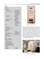
FIG. 3. A 15c/b SDS-PAGE gel of purification fractions. Lane 1, molecular weight markers:
lane 2, inclusion bodies; lane 3, solubilized inclusion bodies in 6M urea buffer: lane 4, refolded
Ras (QFF load): lane 5, insoluble material from the refold: lane 6, S-200 load; lane 7, purified
Ras from S-200 column: lane 8, molecular weight markers.
[1]
REFOLDING AND PURIFICATION OF
Ras
PROTEINS 9
Lot No. 11H0109; Sigma, St. Louis, MO) as the protein standard, s Standard-
ization is achieved using a known concentration of Ras determined by
amino acid composition analysis. The protein values for full-length Ras
calculated from the Bio-Rad assay and from amino acid analysis should be
the same. However, the protein value for truncated Ras calculated from
the Bio-Rad assay is 1.15-fold higher than the value obtained by amino
acid analysis.
SDS-PA GE and Gel Scanning. SDS-PAGE is performed using precast
Daiichi 10-20% polyacrylamide gels purchased from Integrated Separation
Systems or using standard 15% polyacrylamide gels and buffers reported
by Laemmli. ') Bio-Rad low-range molecular weight standards are used as
molecular weight markers. Gels are scanned using an LKB (Bromma, Swe-
den) Ultroscan XL laser densitometer or a Molecular Dynamics (Sunnyvale,
CA) computing densitometer and the data are processed using GelScan
XL version 1.2 software or ImageQuant version 3.15 software.
Guanine Nucleotide-Binding Assays
Ras proteins (200 nM) are labeled in 20 mM Tris (pH 8), l mM di-
thiothreitol, 1 mM EDTA, BSA (1 mg/ml) with 1/xM [c~-3eP]GTP or [8,5'-
3H]GDP (104 cpm/pmol) for 30 rain at 20 °. MgC12 is added to 5 mM and
proteins placed on ice. Ras-bound nucleotide is determined by vacuum
filtration on 0.1-tzm pore size cellulose nitrate filters (Schleicher and Schuell,
Inc., Keene, NH) and liquid scintillation counting. ~°
Results
Optimization of Protein Refolding
We have previously described procedures for isolation of both soluble
Ras and guanidine-solubilized Ras from inclusion bodies of E. coli. 7 The
refolding yield of Ras was further optimized using urea as the solubilization
agent. Hence, we focus our discussion on comparison of urea- and guanidine
hydrochloride-solubilized refolding of Ras, and describe the experimental
conditions optimized to yield refolded H-Ras with the highest recovery of
active protein. The following parameters were varied: solubilization agent,
protein concentration, temperature, and the presence of glycerol.
M. Bradford,
AnaL Biochem.
72, 248 (1976).
U. K. Laemmli,
Nature (London)
227, 680 (1970).
l0 L. A. Quilliam, C. J. Der, R. Clark, E. C. O'Rourkc, K, Zhang, F. McCormick, and G. M.
Bokoch,
Mol. CelL BioL
10, 2901 (1990).
10
EXPRESSION, PURIFICATION, AND MODIFICATION [1]
Protein concentration is an important parameter in refolding pro-
teins.11 14 The protein concentration during refolding must be low enough
that intramolecular interactions are favored over intermolecular interac-
tions, as intermolecular interactions can result in protein aggregation, thus
lowering the yield of correctly folded protein.
In guanidine hydrochloride-solubilized Ras, the refolding yield was as-
sessed at three different protein concentrations: 1.0, 0.1, or 0.01 mg/ml.
The diluted protein was then dialyzed to remove the denaturant. The yield
of soluble refolded H-Ras when solubilized inclusion body protein was
diluted from 10 to 1 mg/ml ranged from 27 to 40%. A precipitate formed
shortly after diluting the solubilized inclusion body protein to 1 mg/ml.
Protein dilution to either 0.01 or 0.1 mg/ml resulted in a 1.2- to 3.4-fold
increase in the yield of soluble refolded Ras protein compared to refolding
at l mg/ml. No precipitates were observed at protein concentrations of
0.01 and 0.1 mg/ml. Interestingly, the concentration of protein during refold-
ing does not significantly affect the GDP-binding stoichiometry of refolded
H-Ras, indicating that H-Ras tends to precipitate if it does not fold correctly.
However, with urea-solubilized inclusion body protein, we obtained a
higher refolding yield of 75% at 1 mg/ml. High refolding yields were also
demonstrated at protein concentrations as high as 10 mg/ml. The yield was
not improved further by refolding at lower protein concentrations, as was
observed for guanidine hydrochloride-solubilized Ras protein. A possible
explanation for the improved refolding yield, using urea as the solubilization
agent, is that urea is neutral and is less likely to salt out populated hydropho-
bic refolding intermediates compared to an ionic solubilization reagent such
as guanidine hydrochloride.
The effects of temperature and the presence of 10% (v/v) glycerol were
also examined. Ras was refolded at either 4 or 25°C. The yield of soluble
refolded H-Ras was slightly higher at 4°C compared to 25°C. Glycerol has
been used to stabilize the activity of enzymes and the native structure of
proteins for many years. 15 ~ The addition of glycerol to an aqueous protein
solution results in preferential binding of water to proteins. The hydrated
11 j. London, C. Skrzyna, and M. E. Goldberg,
Eur. J. Biochern.
47, 409 (1974).
~2 F. A. Marston, P. A. Lowe, M. T. Doel, J. M. Schoemaker, S. White, and S. Angul,
Bio/
Technology
2, 800 (1984).
~3 M. E. Winker, M. Blaber, G. Bennett. W. Hohnes, and G. A. Vehar,
Bio/Technology 3,
990 (1985).
14 F. A. Marston,
Biochem. J.
240(1), 1 (1986).
15 j. Jarakab. A. E. Seeds, Jr., and P. Tralalay,
Biochemistry
5, 1269 (1966).
1~ j. S. Myers and W. B. Jakoby,
Biochern. Biophys. Res. Comrnun.
51, 631 (1973).
17 H. Hoch,
.l. Biol. Chem.
248, 2992 (1973).
~8 K. Gekko and S. N. Timasheff,
Biochemistry
20, 4667 (1981).
[1]
REFOLD1NG AND PURIFICATION OF
Ras
PROTEINS
11
protein is less likely to unfold in the structured glycerol solvent than it
would in water alone] 9 The addition of glycerol to buffers used to refold
H-Ras did not result in any significant changes in the yield of refolded
H-Ras or the GDP-binding stoichiometry. The effects of glycerol on both
the stability and thermodynamics of denaturation/renaturation of refolded
H-Ras were not investigated.
The refold procedure was successfully scaled up from 2 mg of inclusion
body protein to approximately 1300 mg. The overall yield of guanidine
hydrochloride-refolded H-Ras protein from two wild-type H-Ras prepara-
tions using the same batch of inclusion bodies was 18 and 25%. In compari-
son, the overall yield of urea-solubilized Ras was 41%.
The GDP-binding stoichiometry values for all the refolded Ras samples
were essentially the same, ranging from 0.24 to 0.43, and were independent
of protein concentration, temperature, and the presence of glycerol during
refolding. The GDP stoichiometry values calculated from the filter-binding
assay were consistently lower than stoichiometries calculated from NMR
spectroscopy. In particular, both ,sip and tH,15N NMR data show one species
of H-Ras, predominantly bound to GDP, suggesting the GDP-binding stoi-
chiometry was >0.9. Moore et al. ~° have reported that they also observed
the same discrepancy. The GDP-binding stoichiometries that they calcu-
lated from the filter-binding assay were 35-45% of values obtained using
other methods to measure GDP binding to H-Ras.
The various Ras refolding and purification procedures described in this
manuscript are outlined in Figure 4. We have isolated and re, folded a
number of truncated wild-type and mutant H-Ras proteins using these
procedures. So far, all have comparable yields and no modifications were
necessary.
Comparison of Soluble H-Ras and Refolded H-Ras Proteins
Soluble H-Ras protein was purified as described in Methods from the
same cell paste used to isolate and refold wild-type and mutant Ras protein.
The yield of soluble and refolded wild-type H-Ras (purity >90% as deter-
mined by C4 reversed-phase high-performance liquid chromatography)
from 50.2 g of cell paste was 118 and 220 mg, respectively. The GDP-
binding stoichiometries and GTPase activities for soluble Ras and refolded
Ras isolated from the same cells were measured. Refolded H-Ras-bound
GDP and hydrolyzed GTP equally as well as the soluble form isolated from
the same E. coli cells.
I~ K. Gekko and S. N. Timasheff, Biochemistry 20, 4677 (1981).
2o K. J. M. Moore, M. R. Webb, and J. F. Eccleston, Biochemistry 32, 7451 (1993).
12 EXPRESSION, PURIFICATION, AND MODIFICATION [11
Refold and purify Ras Sonicate Cells, spin Refold and purify Ras
from inclusion bodies from inclusion bodies
Purify Soluble Ras
resuspend pellet to from supernatantj resuspend pellet to 10 mg/ml
10 mg/ml in 6 M Urea ~ in 5 M Guanidine-HCI
spin polyethyleneimine
Dialyze or dilute 20-fold ~ dilute 100-fold
T
/
Stir for 20 min, spin out DNA /
/
• J dialyze (2X) overnight
stir overflight at 4 ~ ~E: 'alyze (2Xl °ve rnig ht//z
next dayspin
; ,,//
Load on QFF
elute, 50-1000 mM NaCI
15% SDS-PAGE
pool fractions containing
Ras, concentrate
Gel filtration on S-200
15% SDS-PAGE
I ool fractions containing
Ras, concentrate
Freeze
FIG. 4. Flowchart of procedures for purification of Ras protein from both soluble and
particulate fractions of
E. coll.
Two-dimensional NMR spectra of soluble and refolded H-Ras were
also compared. Our NMR results indicate that the refolded protein has a
structure similar to that of protein purified from the soluble fraction and
that only one species predominantly exists in the refolded protein. 7
[2] PURIFICATION OF BACULOV1RUS-EXPRESSED Ras AND Rap 13
In summary, urea appears to be a better solubilization agent for refold-
ing Ras relative to guanidine hydrochloride. We can refold Ras at higher
protein concentrations and obtain superior recovery yields. Consequently,
purification procedures employing urea solubilization of Ras proteins can be
conducted at lower volumes, facilitating the time and reagent cost associated
with purification. Our refolding procedures have been successful in the
purification of full-length and truncated Ras proteins. The refolded protein
possesses similar GDP-binding stoichiometry and solution structure relative
to Ras isolated from the soluble fraction. Given the high degree of sequence
and mechanistic homology between Ras proteins and low molecular weight
guanine nucleotide-binding proteins, it is likely that these procedures will
be applicable to purification of various members of the Ras superfamily pro-
teins.
Acknowledgments
We are grateful to Bob Manhews and Jing Zhang at Pennsylvania State, who first demon-
strated the advantages of refotding Ras with urea. We also thank Richard DeLoskcy, who
initiated this project at Du Pont Merck, and worked out the refolding protocol in the presence
of guanidine hydrochloride. Last, we thank Channing Der and Alfred Wittinghofer for provid-
ing the plasmids that express full-length and truncated H-Ras, respectively.
[2] Purification of Baculovirus-Expressed Recombinant
Ras and Rap Proteins
By EMILIO PORF1R1, TONY EVANS, GIDEON BOLLAG, ROBIN CLARK,
and JOHN F. HANCOCK
Introduction
H-Ras, N-Ras, K-Ras(A), and K-Ras(B) are membrane-bound guanine
nucleotide-binding proteins that participate in the regulation of cell prolifer-
ation and differentiation. 1 Mutation of the
ras
genes, resulting in amino
acid changes at positions 12, 13, or 61, can trigger neoplastic transformation
and has been detected in about 20% of all human tumors. Rap proteins
(Rap 1 A, Rap l B, and Rap2) are Ras-related GTPases that share 53% amino
acid homology with Ras and are able to antagonize the effects of oncogenic
Ras
in vivo. 2
1 H. R. Bourne. D. A. Sanders, and F. McCormick,
Nature (London)
349, 117 (1991).
2 H. Kilayama, Y. Sugimoto. T. Matsuzaki. Y. Ikawa, and M. Noda,
Cell (Cambridge, Mass'.)
56, 77 (1989).
Copyright fL 1995 by Academic lhess, Inc.
METHODS IN ENZYMOLOGY, VOL 255 All rights o1 reproduclion in any torm reserved
14 EXPRESSION, PURIFICATION, AND MODIFICATION [9.]
Membrane localization of Ras and Rap is essential for their biological
activity and requires a series of posttranslational modifications occurring
at the carboxy-terminal
CAAX
motif (C, cysteine; A, aliphatic; X, any
amino acid)) These modifications comprise farnesylation (Ras) or geranyl-
geranylation (Rap) of the cysteine residue, removal of the
AAX
amino
acids, and carboxymethylationY In addition H-Ras, N-Ras, and K-Ras(A)
require palmitoylation, whereas K-Ras(B), which is not palmitoylated, re-
quires a polybasic domain within the hypervariable region for efficient
plasma membrane binding. 6
The critical role of Ras in the regulation of cell proliferation, and the
involvement of activated
ras
oncogenes in the development of many types of
cancer, have led to a great deal of research on the biological and biochemical
properties of Ras and Ras-related proteins. A variety of sources have been
used to produce recombinant Ras required for biochemical and crystallo-
graphic studies. Small amounts of naturally occurring Ras proteins have
been purified from mammalian tissue. 7 A high yield of recombinant H-Ras
or N-Ras proteins has been obtained by expressing them in
Escherichia
coli,
as described in [1] in this volume. The expression of K-Ras(4B) or
Rap is more problematic because polybasic domains are sensitive to E.
coli
proteases.
Bacterially produced Ras proteins do not undergo posttranslational
processing at the C-terminal
CAAX
motif. Given the importance of pro-
cessing for Ras function, alternative strategies have been used to generate
prenylated Ras proteins. Several observations have shown that Ras proteins
expressed in the baculovirus-insect cell system are processed in the same
way as in mammalian cells. H-Ras is farnesylated and palmitoylated, s K-Ras
is farnesylated, ~) and Rap is geranylgeranylated, u~ The series of posttransla-
tional modifications is completed by proteolysis of the
AAX
amino acids
3 B. M. Willumsen. K. Norris, A. G. Papageorge, N. L. Hubbert, and D. R. Lowy.
EMBO
J. 3, 2581 (1984).
4 j. F. Hancock, A. 1. Magee, J. E. Childs, and C. J. Marshall,
Cell (Cambridge, Mass.) 57,
1167 (1989).
5 S. Clarke,
Annu. Rev. Biochem.
61, 355 (1992).
J. F. Hancock, H. Patcrsom and C. J. Marshall,
Cell (Cambridge, Mass.)
63, 133 (1990).
7 T. Yamashita, K. Yamamoto, A. Kikuchi, M. Kawata, J. Kondo, T. Hishida, Y. Tcranishi,
H. Shiku, and Y. Takai,
J. Biol. Chem.
263, 17181 (1988).
s M. J. Page, A. Hall, S. Rhodes, R. H. Skinner, V. Murphy, M. Sydenham, and P. N. Lowe,
.1. Biol. Chem.
264, 19147 (1989).
~ P. N. Lowe. M. J. Page, S. Bradley, S. Rhodes, M. Sydenham. H. Paterson, and R. H.
Skinner L
Biol. Chem.
266, 1672 (1991).
l0 T. Mizuno, K. Kaibuchi, T. Yamamoto, M. Kawamura, T. Sakoda, H. Fujioka, Y. Matsuura,
and Y. Takai.
Proc. Natl. Acad. Sci. U.S.A. 88,
6442 (1991).
[2] PURIFICATION OF BACULOVIRUS-EXPRESSED
Ras
AND
Rap 15
and cysteine methylation. 11 It has been estimated that processed Ras can
constitute up to 20% of the total Ras protein expressed in insect cells, s
Ion-exchange chromatography on Mono Q, followed by gel-filtration
chromatography on Superose 12, has been used to purify processed and
unprocessed H-Ras, K-Ras, Rap, Rac, and RhoA from the membrane and
cytosolic fraction of insect cells, respectively. ~,m Fractionation on Mono S
has also been used to purify K-Ras(4B). ~) Purification by ion-exchange
chromatography yields 80-95% pure Ras proteins. However. this approach
is time consuming, because two column purification steps are often neces-
sary, and a considerable amount of work may be necessary to optimize
the system.
We use single-step immunoaffinity chromatography with peptide elution
to purify epitope-tagged H-Ras, K-Ras(4B), N-Ras, Rapl, Racl, and RhoA
expressed in the baculovirus-insect cell system. The tag we use, known as
"Glu-Glu" tag, includes six amino acid residues (EYMPME) and was de-
rived from the sequence of an internal region of the polyomavirus medium T
antigen (EEEEYMPME).12 An anti-Glu-Glu monoclonal antibody (MAb),
raised against an identical peptide (EEEEYMPME), specifically recognizes
the Glu-Glu tag and was originally used to purify medium T antigen from
polyomavirus-infected cells] 2 Other than the Glu-Glu tag, several epitope
tags are available and have been used to purify a wide range of peptides.
These include the KT3 tag (TPPPEPET) recognized by the anti-KT3
MAb, 13 the hemagglutinin tag (YPYDVPDYA) recognized by the 12CA5
MAb] 4 the Flag (Immunex, Seattle, WA) tag (DYKDDDDK) recognized
by the 4El 1 MAb,~5 and the tripeptide Glu-Glu-Phe tag recognized by the
YLI/2 MAb. ~(~ Compared to ion-exchange chromatography, purification
by immunoaffinity chromatography is a faster and gentler method, suitable
for small-scale preparation, which can be used under nondenaturing condi-
tions and which yields proteins purified to homogeneity. Elution with the
epitope tag is highly specific for the tagged protein. Furthermore, the small
El p. N. Lowe, M. Sydenham, and M. J. Page, Oncogene 5, 1045 (1990).
1~ T. Grussenmeyer, K. H. Scheidtmann, M. A. Hutchinson, W. Eckhart. and G. Walter, Proc.
Natl. Acad. Sci. U.S.A. 82, 7952 (1985).
13 G. A. Martin, D. Viskochil, G. Bollag, P. C. McCabe, W. J. Crosier, H. Haubruck, L.
Conroy, R. Clark, P. O'Connell, R. M. Cawlhon, M. A. Innis, and F. McCormick, Cell
(Cambridge. Mass. ) 63, 843 (199(I).
t4 H. L. Niman, R. A. Houghten, L. A. Walker, R. A. Reisfeld, I. A. Wilson. J. M. Hogle,
and R. A. Lerner, Proc. Natl. Acad, Sci. U.S.A. 80, 4949 (1983).
~5 T. P. Hopp, K. S. Prickett, V. L. Price, R. T. Libby~ C. J. March, D. P. Cerretti, 15). L. Urdal,
and P. J. Conlon, Bio/Technology 6, 1204 (1988).
i(, R. H. Skinner, S. Bradley, A. L. Brown, N. J. E. Johnson, S. Rhodes, D. K. Stammers. and
P, N. Lowc, J. Biol. Chem. 266, 14163 (1991).
16
EXPRESSION, PURIFICATION, AND MODIFICATION [2]
size of the epitope tag, usually 6-10 amino acids that are added at the N
or the C terminus, is unlikely to interfere in protein-protein interactions
or to affect the enzymatic activity of the protein of interest. However, tags
that can be placed only at the C terminus, like the KT3 tag or the tripeptide
tag, cannot be used with Ras proteins, because they would affect Ras pro-
cessing.
Expression of Epitope (Glu-Glu)-Tagged Ras and Rap Proteins in
Baculovirus-Insect Cell System
The DNA sequence (GAATACATGCCAATGGAA) encoding the
Glu-Glu epitope tag is placed into the 5' end of wild-type K-Ras(4B),
H-Ras, and Rap cDNA using the polymerase chain reaction (PCR). The
tagged cDNAs are cloned into the
Kpnl
and
XbaI
sites of the pAcC13 ~7
baculovirus transfer vector to place the
ras
genes under the control of the
polyhedrin promoter. To generate recombinant baculovirus 2/xg of Ras-
pAcC13 or Rap-pAcC13 DNA is cotransfected in Sf9
(Spodoptera frugi-
perda,
fall armyworm ovary) cells with 1 /xg of gapped and linearized
Autographa californica
nuclear polyhedrosis virus DNA (PharMingen, San
Diego). ~s Cotransfections are carried out by lipofection, using the synthetic
lipid N-L1
-(2,3-dioleyloxy)propyl]-N,N,N-triethylammonium
chloride
(DOTMA) mixed 1:1 with dioleoylphosphatidylethanolamine. Recombi-
nant virus is isolated from occlusion negative (occ) plaques following two
cycles of reinfection. 1~) To verify the expression of Ras or Rap, several
small-scale cultures are infected with independent isolates of recombinant
virus and analyzed by Western blot using the anti-Glu-Glu MAb, For large-
scale cultures (500 ml to 10 liters), Sf9 cells seeded at the density of 1 ×
106 cells/ml are infected with 5 to 10 × lff' plaque-forming units (PFU)/
ml of recombinant virus. Suspension cultures are grown in Grace's medium
containing 10% (v/v) fetal calf serum and 3.5% (v/v) yeast hydrolysate, for
48 hr at 37 ° prior to harvesting. Cells are collected by centrifugation at 500
g for 10 min at 4°: cell pellets, approximately 1 ml each (-100 × 10 (' cells),
are snap-frozen in liquid nitrogen and stored at -80 ° until use. We estimate
that Ras or Rap accounts for 1-1.5% of total Sf9 cell protein and some
20% of the total Ras or Rap protein recovered from the cell lysate is
prenylated.
17 S. Munenfitsu, M. A. Innis, R. Clark, F. McCormick, A. Ullrich, and P. Polakis,
Mol. (.'ell.
Biol.
10, 5977 (1990).
~s G. E. Smith. M. D. Summers. and M. J. Fraser,
Mol. Cell Biol. 3,
2156 (1983).
t~ D. R. O'Reilly, K. L. Miller. and V. A. Luckow, "Baculovirus Expression Vectors: A
Laboratory Manual." Freeman, New York, 1992.
[2] PURIFICATION OF BACULOVIRUS-EXPRESSED Ras AND Rap 17
Separation of Processed and Unprocessed Ras and Rap Proteins by
Triton X- 1 14 Partitioning
To separate prenylated (processed) from unprocessed Ras and Rap
proteins we use the Triton X-l14 partition method. 2° One milliliter of
previously snap-frozen Sf9 cells is thawed at room temperature, resus-
pended in 10 vol of ice-cold 50 mM Tris-HC1 (pH 7.5), 150 mM NaC1,
5 mM MgC12, 200 p,M GDP, 1 mM dithiothreitol (DTT), 1 mM Pefabloc
(Boehringer Mannheim Co., Indianapolis, IN), 10/,g/ml each of leupeptin,
aprotinin, and soybean trypsin inhibitor and homogenized with 20 strokes
in a Dounce (Wheaton, Millville, NJ) homogenizer. Following homogeniza-
tion, a 1/10 vol of 11% (w/v) Triton X-114 is added to the lysate to adjust
the final Triton X-114 concentration to 1% (v/v), and the lysate is mixed
for 10 min at 4 ° and centrifuged at 100,000 g at 4 ° for 30 min to get rid of
the insoluble material. The cleared supernatant is warmed at 37 ° for 1-2
min, until it becomes cloudy, then centrifuged at 400 g for 4 min at room
temperature to separate the aqueous (upper) phase from the detergent
(lower) phase. Both phases are adjusted to 1% (v/v) Triton X-114 on ice
and three sequential phase separations are performed to "wash" each of
the original aqueous and detergent-enriched phases.
Purification of Epitope (Glu-Glu)-Tagged Ras and Rap Proteins by
Immunoaffinity Chromatography
Ehttion of Prenylated Ras and Rap Proteins
Posttranslationally processed Ras proteins are purified by immunoaffin-
ity chromatography from the original detergent phase on a 1-ml protein
G-Sepharose column conjugated with anti-Glu-Glu MAb 12 (PG Glu-Glu
column). The detergent phase, adjusted to 1% (v/v) Triton X-114, is applied
to the PG Glu-Glu column for 1 hr at 4 °. The column is washed with 20
vol of buffer A [50 mM Tris-HCl (pH 7.5), 150 mM NaC1, 5 mM MgClx,
1 mM DTT, 1 mM Pefabloc, and 10 ~g/ml each of leupeptin, aprotinin,
and soybean trypsin inhibitor], containing 0.5% (w/v) sodium cholate. Pre-
nylated Ras proteins are eluted in six 1-ml fractions of buffer A, containing
0.5% (w/v) sodium cholate and a 50-p,g/ml concentration of N-terminally
acetylated ED peptide (EYMPTD), a peptide known to bind with high
affinity to the PG Glu-Glu column.
2, L. Gulierrez. A. I. Magee, C. J. Marshall. and J. F. Hancock. EMBO ,I. 8, 1093 (1989).
18 EXPRESSION, PURIFICATION, AND MODIFICATION [2]
Elution of Unprocessed Ras and Rap Proteins
Unprocessed Ras and Rap proteins are purified from the original aque-
ous phase, which is loaded onto a 1-ml PG Glu-Glu column for 1 hr at 4 °.
The column is washed with 40 vol of buffer A and Ras proteins are eluted in
six 1-ml fractions of buffer A, containing 50/xg of ED peptide per milliliter.
Unprocessed Ras is quantitatively recovered from the PG Glu-Glu
column, but solubilization of prenylated Ras or Rapl requires the addition
of 0.5% (w/v) sodium cholate to the elution buffer. The use of a higher
concentration of sodium cholate (e.g., 1%, w/v) did not result in a signifi-
cantly better recovery of prenylated Ras or Rapl. Moreover, such a high
concentration of detergent may affect the further use of the purified proteins
because it may interfere with the interaction of Ras with its effectors/
regulators
in vitro.
We have also tested n-octyl-/3-D-glucopyranoside at a
concentration of 1.2% (w/v). Using this detergent, the recovery of preny-
lated Ras was similar to that achieved using 0.5% (w/v) sodium cholate.
Large-Scale Purification of K-Ras(4B) Proteins
For large-scale preparation of K-Ras(4B), we separate unprocessed
from processed K-Ras(4B) using ion-exchange chromatography on
S-Sepharose, followed by purification by immunoaffinity chromatography
on a PG Glu-Glu column.
Sf9 cells (5-ml packed volume, 100 × 10 ~ cells/ml) expressing K-Ras(4B)
are resuspended in 20 ml of 20 mM NaPO4 (pH 7.5), 2 mM MgC12, 1 mM
DTT, 0.1 mM Pefabloc, 1 /xg/ml each of leupeptin, aprotinin, and soybean
trypsin inhibitor, 0.2/xg of E-64
[trans-epoxysuccinyl-L-leucylamido(4-gua-
nidino)butane (buffer B)] per milliliter, additionally containing 0.5%
(w/v) sodium cholate and 1 mM Pefabloc, 10 /xg/ml each of leupeptin,
aprotinin, and soybean trypsin inhibitor, and E-64 (2/~g/ml). Sf9 cells are
lysed by sonication (twice, 1 min each), and centrifuged at 30,000 g for 10
min at 4 °. The insoluble material is resuspended in 20 ml of buffer B
containing 0.5% (w/v) sodium cholate and protease inhibitors, then briefly
sonicated and recentrifuged. The supernatants from the first and second
centrifugation are combined and centrifuged at 100,000 g. The cleared
supernatant is loaded onto a 10-ml S-Sepharose column. The prenylated
protein does not bind the resin and is recovered from the column
flowthrough.
Purification of Prenylated K-Ras(4B) Proteins
The S-Sepharose column flowthrough is loaded onto a 0.5-ml PG Glu-
Glu column for 1 hr at 4 °. The column is washed with 30 vol of buffer B,
[2] PURIFICATION OF BACULOVIRUS-EXPRESSED Ras AND
Rap, 19
30 vol of buffer B containing 0.5% (w/v) sodium cholate, 30 vol of buffer
B containing 500 mM NaCI, and 30 vol of buffer B containing 0.5% (w/v)
sodium cholate. Processed K-Ras(4B) is eluted in ten 1-ml fractions of
buffer B containing 0.5% (w/v) sodium cholate and ED peptide (:25 ~g/ml).
Purification of Unprocessed K-Ras(4B) Proteins
The S-Sepharose column is washed with 10 vol of buffer B and eluted
with 45 ml of buffer B containing 200 mM NaC1. The eluate is loaded for
1 hr at 4 ° onto a 0.5-ml PG Glu-Glu column. The PG Glu-Glu column is
washed with 30 vol of buffer B, 30 vol of buffer B containing 0.5% Nonidet
P-40 (NP-40), 30 vol of buffer B containing 500 mM NaCI, and 30 vol of
buffer B. Unprocessed K-Ras(4B) is eluted in ten 1-ml fractions of buffer
B containing ED peptide (25/~g/ml).
Analysis of Purified Ras and Rap Proteins and Validation of
Separation Procedures
Following the elution, an aliquot (1/100) of each fraction is analyzed
by sodium dodecyl sulfate-polyacrylamide gel electrophoresis (SDS-
PAGE) on a 16% polyacrylamide gel, and protein concentration is deter-
mined using the Bradford reaction. The SDS-PAGE analysis shows that
Ras proteins are purified to near homogeneity and run between the 21-
and 30-kDa molecular mass markers (Fig. 1). Typically we purify 0.1-0.2
mg of prenylated Ras and 1.2 mg of unprocessed Ras from a 1-ml Sf9 cell
pellet (-100 x lff' cells). Ras proteins are snap-frozen in liquid nitrogen
(50/~l/aliquot) and stored at -80 ° until use.
Validation of" Triton X-114 Separation Procedure
As previously observed, the mobility on SDS-PAGE of farnesylated
Ras is greater than the mobility of unprocessed Ras (Fig. 2). To validate
the Triton X-114 separation procedure, purified Ras is incubated with
[~H]farnesyl pyrophosphate and farnesyltransferase in a cell-free system,
as described in [9] in this volume. At the end of a 60-rain incubation, only
the Ras proteins purified from the aqueous phase (unprocessed) incorporate
the radiolabel from [3H]farnesyl pyrophosphate whereas the Ras proteins
purified from the detergent phase do not.
Assessment of Guanine Nucleotide-Binding Capacity of Ras Proteins
To ascertain that the purified Ras proteins are biologically active we
determine their respective GTP-binding capacity. Ras proteins (4 pmol,
20
EXPRESSION, PURIFICATION, AND MODIFICATION [2]
Detergent phase Aqueous phase
D123456MA123456
FIG. 1. Purification of processed and unprocessed K-Ras(4B) protein by immunoaffinity
chromatography. Lysates from Sf9 cells expressing K-Ras(4B) were partitioned into detergent
and aqueous phases, using the Triton X-114 method. Processed and unprocessed K-Ras(4B)
proteins were purified by immunoaffinity chromatography on a PG Glu-Glu column. Samples
(10 ~1) of the original aqueous (A) and detergent (D) phases and of each fraction eluted
from the PG Glu-Glu column were analyzed by SDS-PAGE on a 16% polyacrylamide gel
stained with Coomassie blue. Lanes DI-6, samples of the fractions eluted from the column
loaded with the detergent phase; lanes A1-6, samples of the fractions eluted from the column
loaded with the aqueous phase; lane M, molecular weight markers.
100 nM) are incubated with I /xM [3H]GTP (31.5 Ci/mmol) in a 40-/,1
reaction mixture containing 20 mM Tris-HC1 (pH 7.5), 50 mM NaC1, 5 mM
MgC12, 10 mM EDTA, 1% (w/v) bovine serum albumin, 1 mM DTT, 0.05%
(w/v) sodium cholate. Following a 10-min incubation at room temperature,
the loading is stopped by adding 10 mM MgC12. After a further 5 min,
1 ml of ice-cold 20 mM Tris-HC1 (pH 7.5), 50 mM NaC1, 5 mM MgC12 is
added to each tube and the mixture is filtered through a nitrocellulose filter
30 kd -I~
M D
A D A D A
21 kd
FIG. 2. SDS-PAGE analysis of Ras proteins purified by Triton X-114 phase separation
followed by immunoaffinity chromatography. Five hundred nanograms of K-Ras(4B) and of
the K-Ras C-terminus mutants K6Q and (TAIL were analyzed by SDS PAGE on a 16%
polyacrylamide gel stained with Coomassie blue. Lanes A, unprocessed Ras purified from
the aqueous phase: lanes D, processed Ras purified from the detergent phase.
[3] PURIFICATION Of Ras GAPs 21
(pore size, 45/xm), using a 1225 sampling manifold apparatus (Millipore,
Bedford, MA). Filters are washed with 10 ml of ice-cold 20 mM Tris-HC1
(pH 7.5), 50 mM NaC1, 5 mM MgC12, dried, and counted in a scintillation
counter to determine the radioactivity bound to Ras. Following a 10-rain
incubation, 23,000 (_+4000) cpm/pmol is usually detected either in farnesy-
lated or unprocessed Ras proteins; no significant [3H]GTP binding to Ras
is detected when EDTA is omitted from the loading buffer.
Conclusions
Purification of epitope-tagged Ras by immunoaffinity chromatography
constitutes a fast and simple method yielding proteins purified to homogene-
ity. The six-amino acid Glu-Glu tag added at the N terminus of the protein
does not appear to interfere with Ras interactions with its regulators and
effectors. 21'22 The Triton X-114 partition represents a simple and reliable
method for separating processed and unprocessed Ras proteins that is
especially suitable for small-scale purifications.
Acknowledgments
We thank Jonathan Driller and David Lowe for excellent technical assistance with the
baculovirus-insect cell expression system.
el E. Porfiri, T. Evans, P. Chardin, and J. F. Hancock,
J. Biol.
Chem. 269, 2267:2 (1994).
_~2 M. Spaargaren. G. A. Martin, F. McCormick, M. J. Fernandez-Sarabia. and J. R. Bischoff.
13iochern l.
300, 303 (1994).
[3] Purification of Recombinant Ras
GTPase-Activating Proteins
By
GIDEON BOLLAG and FRANK McCORMICK
Introduction
Deactivation of Ras. GTP is achieved by catalyzed GTP hydrolysis. L In
human tissues, at least two proteins are capable of catalyzing this hydrolysis
on Ras, and they have been termed GAPs for GTPase-activating proteins.
The first GAP to be identified was a 120-kDa protein, which is denoted
t D. R. Lowy and B. M. Willumsen.
Anmc Rev. Biochem.
62, 851 (1993).
Copyrighl t2 1995 by Academic Press, lnc
METHODS IN ENZYMOLOGY, VOL. 255 All rights of repmduclion in any titan reserved.