small gtpases and their regulators, part c
Bạn đang xem bản rút gọn của tài liệu. Xem và tải ngay bản đầy đủ của tài liệu tại đây (6.02 MB, 369 trang )
Preface
GTPases are now recognized as essential components for protein traffic
between all compartments of the cell. This includes vesicular traffic through
the exocytic and endocytic pathways, where GTPases play key roles in the
assembly of vesicle coats (budding), in vesicle targeting and in fusion, as
well as in protein traffic in and out of the nucleus. GTPases involved in
transport include the Rab and ARF families, Sarl, Ran, dynamin, and
heterotrimeric G proteins. In addition to GTPase, a number of associated
accessory factors are critical for function. These include posttranslational
modifying enzymes (such as prenyl transferases and myristyl transferases),
factors that affect guanine nucleotide binding [guanine nucleotide dissocia-
tion inhibitors (GDIs) and guanine nucleotide exchange factors (GEFs)],
and factors that stimulate guanine nucleotide hydrolysis [GTPase-activating
proteins (GAPs)].
To understand the function of GTPases and their cognate factors, a
wealth of
in vitro biochemical and in vivo molecular genetic approaches are
currently being applied to individual proteins. Given the diverse spectrum of
compartments regulated by individual GTPases, techniques developed for
one particular member of a family are often applicable to other members.
In a broader sense, many of the techniques developed for a particular
gene family are also frequently applicable to other gene families given the
exceptional structural configuration of GTPases.
The purpose of this volume is to bring together the latest technologies
in the study of GTPase function involved in protein trafficking. It provides
concise descriptions of the recent methodological innovations that allow
both the novice and experienced investigator to explore the function of
these proteins in detail. We are extremely grateful to the many investigators
who have generously contributed their time and expertise to bring this
wealth of technical experience to one volume. It should provide a valuable
resource to address the many issues confronting our understanding of the
role of these GTPases in the biology of cell.
W. E. BALCH
CHANNING J. DER
ALAN HALL
xiii
Contributors to Volume 257
Article numbers are in parentheses following the names of contributors.
Affiliations listed are current.
KIRILL ALEXANDROV (27),
Cell Biology Pro-
gram, European Molecular Biology Labo-
ratory, 69012 Heidelberg, Germany
SCOTt A. ARMSTRONG (5),
Department of
Molecular Genetics, University of Texas
Southwestern Medical Center, Dallas,
Texas 75235
WILLIAM E. BALCH
(1,
7, 10, 20, 21),
Depart-
ments of Cell and Molecular Biology, The
Scripps Research Institute, La Jolla, Califor-
nia 92037
CHARLES BARLOWE (13),
Department of Bio-
chemistry, Dartmouth Medical School,
Hanover, New Hampshire 03755
F. RALF BISCHOFF (17),
Division for Molecu-
lar Biology of Mitosis, German Cancer Re-
search Center, D-69009 Heidelberg,
Germany
WILLIAM H. BRONDYK (14, 23),
Promega Cor-
poration, Madison, Wisconsin 53771
MICHAEL S. BROWN (5),
Department of Mo-
lecular Genetics, University of Texas South-
western Medical Center, Dallas, Texas
75235
H. ALEX BROWN (33),
Department of Phar-
macology, Southwestern Medical Center,
University of Texas, Dallas, Texas 75235
CECILIA BUCCI (2, 19),
Dipartimento di Bio-
logia e Patologia Cellulare e Molecolare
"L. Califano, '" 80131 Napoli, Italy
HERMAN BUJARD (24),
Zentrum far Moleku-
late Biologic der Universiti~t Heidelberg,
D-69120 Heidelberg, Germany
JANET L. BURTON (12),
Department of Cell
Biology, Howard Hughes Medical Institute,
Yale University School of Medicine, New
Haven, Connecticut 06510
ix
HANNA DAMKE (24),
Department of CeU Biol-
ogy, The Scripps Research Institute, La
Jolla, California 92037
CHRISTIANE DASCHER (20, 21),
Department
of Cell Biology, The Scripps Research Insti-
tute, La Jolla, California 92037
PIETRO DE CAMILLI (12),
Department of Cell
Biology, Howard Hughes Medical Institute,
Yale University School of Medicine, New
Haven, Connecticut 06510
A.
BARBARA DIRAC-SVEJSTRUP (3),
Depart-
ment of Biochemistry, Stanford University
School of Medicine, Stanford, California
943O5
CARLOS G. DOTTI (32),
Cell Biology Pro-
gram, European Molecular Biology Labo-
ratory, D-69117 Heidelberg, Germany
PAUL DUPREE (32),
Department of Plant Sci-
ences, Cambridge University, Cambridge
CB2 3HA, United Kingdom
MARILYN GIST FARQUHAR (29),
Division of
Cellular and Molecular Medicine, Univer-
sity of California, San Diego, La Jolla, Cali-
fornia 92093
SUSAN FERRO-NovICK (4),
Department of
Cell Biology, Howard Hughes Medical In-
stitute, Yale University School of Medicine,
New Haven, Connecticut 06510
SABINE FREUNDIAEB (24),
Zentrum far Mo-
lekulare Biologie der Universitiit Heidel-
berg, D-69120 Heidelberg, Germany
DIETER GALLWlTZ (15),
Department of Mo-
lecular Genetics, Max-Planck Institute for
Biophysical Chemistry, D-37018 GOt-
tingen, Germany
MICHELLE D. GARRE~rr (11, 26),
Onyx Phar-
maceuticals, Richmond, California 94806
X CONTRIBUTORS TO VOLUME
257
LARRY GERACE (30),
Department of Cell Bi-
ology, The Scripps Research Institute, La
Jolla, California 92037
JOSEPH L. GOLI~STEIN (5),
Department of Mo-
lecular Genetics, University of Texas South-
western Medical Center, Dallas, Texas
75235
MANFRED GOSSEN (24),
MCB Barker/Kosh-
land ASU, University of California, Berke-
ley, California 94720
RONALD W. HOLZ (25),
Department of Phar-
macology, University of Michigan Medical
School, Ann Arbor, Michigan 48109
HISANORI HORIUCHI (2, 27),
CelIBiology Pro-
gram, European Molecular Biology Labo-
ratory, 69012 Heidelberg, Germany
Lugs A. HUBER (32),
Department of Bio-
chemistry, University of Geneva, CH-1211
Geneva 4, Switzerland
Yu JIANG (4),
Department of Cell Biology,
Howard Hughes Medical Institute, Yale
University School of Medicine, New Haven,
Connecticut 06510
RICHARD A. KAHN (16),
Laboratory of Bio-
logical Chemistry, Division of Cancer
Treatment, National Cancer Institute, Na-
tional Institutes of Health, Bethesda, Mary-
land 20892
AKIRA KIKUCHI (8),
Department of Biochem-
istry, Hiroshima University School of Medi-
cine, Hiroshima 734, Japan
KEITaROU KIMU~ (6),
Genetics Engineering
Laboratory, National Food Research Insti-
tute, Tsukuba 305, Japan
IAN G. MaCARA (14, 23),
Department of Pa-
thology, University of Vermont, Burlington,
Vermont 05405
Luis MARTIN-PARRAS (22),
Cell Biology Pro-
gram, European Molecular Biology Labo-
ratory, D-69117 Heidelberg, Germany
J. MICHAEL MCCAFFERY (29),
Division of
Cellular and Molecular Medicine, Univer-
sity of California, San Diego, LaJolla, Cali-
fornia 92093
FRAUKE MELCHIOR (30),
Department of Cell
Biology, The Scripps Research Institute, La
Jolla, California 92037
CAROL MURPHY (34),
Cell Biology Program,
European Molecular Biology Laboratory,
D-69012 Heidelberg, Germany
HIROYUKI NAKANISHI (8),
Department of Mo-
lecular Biology and Biochemistry, Osaka
University Medical School, Suita 565, Japan
AKImRO NAga~YO (6),
Department of Biologi-
cal Sciences, Graduate School of Science,
University of Tokyo, Tokyo 113, Japan
PETER J. NOVICK (1l, 26),
Department of Cell
Biology, Yale University School of Medi-
cine, New Haven, Connecticut 06510
CLAUDE NUOFFER (1, 10),
Department of Cell
Biology, The Scripps Research Institute, La
Jolla, California 92037
TOSHmIKO OKA (6),
Department of Organic
Chemistry and Biochemistry, Institute of
Scientific and Industrial Research, Osaka
University, Osaka 567, Japan
FRANK PETER (1, 10),
Department of Cell Bi-
ology, The Scripps Research Institute, La
Jolla, California 92037
SUZANNE R. PFEFVER (3, 28),
Department of
Biochemistry, Stanford University School
of Medicine, Stanford, California 94305
HERWlG PONSa~NGL (17),
Division for Molec-
ular Biology of Mitosis, German Cancer
Research Center, D-69009 Heidelberg,
Germany
PAUL A. RANDAZZO (16),
Laboratory of Bio-
logical Chemistry, Division of Cancer
Treatment, National Cancer Institute, Na-
tional Institutes of Health, Bethesda, Mary-
land 20892
MARKUS A. RmDERER (3),
Department of
Biochemistry, Stanford University School
of Medicine, Stanford, California 94305
DENISE M. ROBERTS (11),
Department of Cell
Biology, Yale University School of Medi-
cine, New Haven, Connecticut 06510
GUENDALINA ROSSI (4),
Department of Cell
Biology, Howard Hughes Medical Institute,
Yale University School of Medicine, New
Haven, Connecticut 06510
TONY ROWE (7),
Department of Cell Biology,
The Scripps Research
Institute,
La Jolla,
California 92037
CONTRIBUTORS TO VOLUME 257 xi
TAKUYA SASAKI (9), Department of Molecu-
lar Biology and Biochemistry, Osaka Uni-
versity Medical School, Suita 565, Japan
ISABELLE SCHALK (10), Department of Cell
Biology, The Scripps Research Institute, La
Jolla, California 92037
RANDY SCHEK/vIAN (13, 18), Departments of
Molecular and Cell Biology, Howard
Hughes Medical Institute, University of
California, Berkeley, Berkeley, California
94720
SANDRA L. SCHMID (24), Department of Cell
Biology, The Scripps Research Institute, La
Jolla, California 92037
MIGUEL C. SEABRA (5), Department of Molec-
ular Genetics, University of Texas South-
western Medical Center, Dallas, Texas
75235
RUTH A. SENTER (25), Department of Phar-
macology, University of Michigan Medical
School, Ann Arbor, Michigan 48109
ALLAN D. SHAPIRO (28), Department of Bio-
chemistry, Stanford University School of
Medicine, Stanford, California 94305
HIROMICHI SHIRATAKI (31), Department of
Cell Biology, National Institute for Physio-
logical Sciences, Okazaki 444, Japan
THIERRY SOLDATI (3, 28), Department of Bio-
chemistry, Stanford University School of
Medicine, Stanford, California 94305
HARALD STENMARK (19), Cell Biology Pro-
gram, European Molecular Biology Labo-
ratory, D-69012 Heidelberg, Germany
PAUL C. STERNWEIS (33), Department of
Pharmacology, University of Texas South-
western Medical Center, Dallas, Texas
75235
DEBORAH J. SWEET (30), Department of Cell
Biology, The Scripps Research Institute, La
Jolla, California 92037
YOSHIMI TAKAI (8, 9, 31), Department of Mo-
lecular Biology and Biochemistry, Osaka
University Medical School and Department
of Cell Physiology, National Institute for
Physiological Sciences, Suita, Osaka 565,
Japan
LAUREL THOMAS (21), Vollum Institute, Ore-
gon Health Sciences University, Portland,
Oregon 97201
GARY THOMAS (21), VoUum Institute, Oregon
Health Sciences University, Portland, Ore-
gon 97201
ELLEN J. TISDALE (20), Department of Cell
Biology, The Scripps Research Institute, La
Jolla, California 92037
MICHAEL D. UHLER (25), Department of Bio-
logical Chemistry and The Mental Health
Research Institute, University of Michigan
Medical School, Ann Arbor, Michigan
48109
OLIVER ULLRICH
(2,
27), Cell Biology Pro-
gram, European Molecular Biology Labo-
ratory, 69012 Heidelberg, Germany
JUDY K. VANSLYKE (21), Vollum Institute,
Oregon Health Sciences University, Port-
land, Oregon 97201
PETRA VOLLMER (15), Department of Molecu-
lar Genetics, Max Planck Institute for Bio-
physical Chemistry, D-37018 Gottingen,
Germany
OFRA WEISS (16), Department of Endocrinol-
ogy and Metabolism, Hadassah University
Hospital, Jerusalem 91120, Israel
THOMAS YEUNG (18), Division of Biochemis-
try and Molecular Biology, Howard Hughes
Medical Institute, University of California,
Berkeley, Berkeley, California 94720
TOHRU YOSHIHISA (18), Department of Cell
Biology, Institute for Virus Research, Kyoto
University, Kyoto 606-01, Japan
MARINO ZERIAL (2, 19, 22, 27, 34), Cell Biol-
ogy Program, European Molecular Biology
Laboratory, D-69012 Heidelberg, Germany
[ 1] PURIFICATION OF His6-Rabl 3
[11 Purification of His6-Tagged Rabl Proteins Using
Bacterial and Insect Cell Expression Systems
By
CLAUDE NUOFFER, FRANK
PETER,
and WILLIAM E. BALCH
Introduction
The members of the Rab/YPT/SEC4 family of Ras-like GTPases are
likely to function as molecular switches' in regulating the assembly and/or
disassembly of protein complexes that mediate the vectorial movement
of transport vesicles between distinct subcellular compartments. We have
established that the Rabl proteins play an essential role in traffic through
the early secretory pathway in mammalian cells by showing that selected
RablA and RablB mutants with altered guanine nucleotide-binding prop-
erties act as potent trans dominant inhibitors of transport between the
endoplasmic reticulum (ER) and the Golgi complex both in vivo t and
in vitro. 2'3
This chapter describes the isolation of recombinant wild-type or
mutant forms of Rabl via expression in Escherichia coli and Spodoptera
frugiperda (Sf9) insect cells. Although the bacterial expression system
is more convenient from a technical point of view, the utility of Rabl
proteins prepared from E. coli is limited by the fact that these invariably
lack the COOH-terminal geranylgeranyl (GG) groups that are essential
for normal Rabl function. 2 In contrast, the eukaryotic expression system
allows the purification of membrane-associated, isoprenylated forms of
the proteins (RablGG). 4 Both expression systems require the purification
of relatively minor pools of functional protein. In the case of E. coli,
this is due to the strong tendency of Rabl proteins to form inclusion
bodies. To obtain active forms of the proteins we focus on the purification
of the soluble pool, which represents no more than 1-10% of the total
production. In Sf9 cells, the yields of isoprenylated Rabl proteins are
low as <5% of the expressed protein is posttranslationally processed
and incorporated into host cell membranes. To overcome these difficulties,
1 E. J. Tisdale, J. R. Bourne, R. Khosravi-Far, C. J. Der, and W. E. Balch, J.
Cell BioL
119,
749 (1992).
2 C. Nuoffer, H. W. Davidson, J. Matteson, J. Meinkoth, and W. E. Balch,
J. Cell Biol.
125,
225 (1994).
3 S. Pind, S. N. Pind, C. Nouffer, J. M. McCafffey, H. Plutner, H. W. Davidson, M. G. Farquhar,
and W. E. Balch,
J. Cell Biol.
125, 239 (1994).
4 F. Peter, C. Nuoffer, S. N. Pind, and W. E. Balch, J.
Cell Biol. 126,
1393 (1994).
Copyright © 1995 by Academic Press, Inc.
METHODS IN ENZYMOLOGY, VOL. 257 All rights of reproduction in any form reserved.
4 EXPRESSION, PURIFICATION, AND MODIFICATION [ 1]
we take advantage of N-terminal His6 tags that allow us to use metal
chelate chromatography as a rapid and efficient purification step. 5 These
N-terminal His6 modifications do not interfere with the function of
wild-type and mutant Rabl proteins in transport through the early
secretory pathway. 2
Methods
Purification of His6-Rab i from
Escherichia coli
His6-Rabl proteins are produced in
E. coli
using the T7 RNA polymer-
ase-dependent expression system developed by Studier
et al. 6
Briefly, the
cDNA is placed under control of a T7 promoter and the resulting expression
vector is introduced into
E. coli
strain BL21(DE3), which contains the T7
RNA polymerase gene under control of the
lacZ
promoter. Exposure of
the cells to isopropyl-/3-thiogalactopyranoside (IPTG) induces T7 RNA
polymerase production and triggers expression of the cDNA.
Procedures
Buffers
Lysis buffer: 50 mM Tris-HC1, pH 8,1 mM EDTA, 10 mM 2-mercapto-
ethanol
NTA buffer: 50 mM MES-NaOH, pH 6, 0.3 M NaCI, 1 mM MgCI2,
50/zM EGTA, 10 mM 2-mercaptoethanol
25/125:25 mM HEPES-KOH, pH 7.2, 125 mM potassium acetate
Construction of Expression Vectors
An expression vector for the production of Rab proteins with a
N-terminal His6 tag was first constructed using the Rab3A cDNA and
plasmid pETlld (Novagen) as follows: A
NcoI-NdeI
linker encoding an
initiator Met followed by six consecutive His residues was ligated along
with the Rab3a cDNA excised from pET3a-Rab3A as a
NdeI-BamHI
fragment into the
NcoI
and
BamHI
sites of pETlld. 2 Constructs directing
the expression of wild-type and mutant His6-Rabl proteins were obtained
through excision of the Rab3a sequence with
NdeI
and
BamHI
and inser-
tion of the corresponding Rabl fragments isolated from pET3a-Rabl
plasmids, m
5 E. Hochuli, W. Bannwarth, H. DObeli, R. Gentz, and D. StOber,
Bio Technology
6,1321 (1988).
6 F. W. Studier, A. H. Rosenberg, J. J. Dunn, and J. W. Dubendorff, this series, Vol. 185, p. 60.
[ 1] PURIFICATION OF His6-Rabl 5
Expression
The pETlld-His6-Rabl plasmids are introduced into competent
BL21(DE3) cells and transformants are selected on LB-agar plates con-
taining 100/xg/ml ampicillin overnight at 37 °. A single colony is transferred
into LB supplemented with 100 tzg/ml ampicillin, and the preculture is
grown to saturation overnight at 37 °. The culture is diluted 1 : 50 into fresh
medium and grown at 28! to
OD60o
of 0.6-1.0 with good aeration. Expres-
sion is induced by the addition of IPTG to a final concentration of 0.4 mM
and incubation is continued for 2-4 hr. The cultures are chilled on ice, the
cells are harvested by centrifugation, washed, and the cell pellets are frozen
in liquid N2 and stored at -80 °.
Note: The expression protocol just described results in levels of soluble
protein that vary considerably between different wild-type and mutant
forms of Rabl. In some cases, induction for 6-16 hr in the presence of
0.01-0.1 mM IPTG may result in higher yields of soluble protein. In general,
mutant forms of Rabl tend to be less soluble compared to the wild-type
proteins. This is most evident in the case of the RablA/B(N124/121I)
mutants, 3 which are extremely insoluble and remain prone to precipitation
throughout the purification process and cannot be kept in solution at con-
centrations >0.2 mg/ml.
Purification
Preparation of L ysis Supernatant
All subsequent manipulations are performed at 4 ° unless otherwise
stated. The pellets are thawed and resuspended in 10 vol lysis buffer supple-
mented with 1 mM phenylmethylsulfonyl fluoride (PMSF), 0.5/xg/ml leu-
peptin, and 1/xM pepstatin A. Lysozyme is added to a final concentration
of 0.4 mg/ml and the suspension is incubated for 30 rain at 4 ° with gentle
agitation. After lysis of the cells through two rounds of freezing in liquid
N2
followed by thawing at 32 ° with constant agitation, the lysate is adjusted
to 0.3 M NaCI, 10 mM MgCI2, and 0.2% deoxycholate. The viscosity is
reduced by incubation in the presence of 40 gg/ml DNase I for 30 min at
4 ° with gentle agitation, and the lysate is clarified by centrifugation at
22,000g (13,500 rpm in a Beckman JA-20 rotor) for 30 min. The resulting
supernatant serves as a source to purify the soluble His6-Rabl fraction by
metal chelate affinity chromatography and gel filtration chromatography
as described below.
Note: The inclusion of 10/xM GDP in the lysis buffer and throughout
the remainder of the purification process may slightly increase the stability
6
EXPRESSION, PURIFICATION. AND MODIFICATION
[1]
of Rabl mutants with low affinities for guanine nucleotides such as the
RablA/B(N124/121I) mutants. 3
Ni2+-NTA-Agarose Chromatography
The supernatant is applied to a column (0.5-5 ml bed volume) of Ni 2+-
saturated nitrilotriacetic acid (NTA)-agarose (Qiagen) equilibrated with
lysis buffer containing 0.3 M NaC1 and 10 mM MgCI2 (flow rate: 1 ml/
min). The column is washed with 10 vol of equilibration buffer, 10 vol of
NTA-buffer, and 10 vol of NTA-buffer supplemented with 25 mM imidaz-
ole. The column is eluted with NTA-buffer containing 250 mM imidazole,
and fractions containing His6-Rabl are identified by analyzing aliquots by
SDS-PAGE and Coomassie blue staining. Fractions containing His6-Rabl
are pooled, and the proteins are further purified by gel filtration chromatog-
raphy.
Note: To minimize the nonspecific adsorption of proteins to the NTA-
agarose resin, it is essential to adjust the bed volume of the NTA-agarose
column depending on the amount of His6-Rabl present in the lysate. For the
purification of wild-type proteins and mutants with comparable solubility
[RablA/B(S25/22N)2], we typically use -1-2 ml of resin for each liter of
culture. In the case of the RablA/B(N124/121I) mutants, better results are
obtained with -5-10x smaller columns.
S-I O0 Gel-Filtration Chromatography
The pooled fractions are applied to a 75 x 2.5-cm column (flow rate:
-0.5 ml/min) of Sephacryl S-100 (Pharmacia LKB) equilibrated with 25/125
supplemented with 1 mM MgCI2 and 1 mM sodium mercaptoethanesulfonic
acid. Fractions containing His6-Rabl are identified by analyzing aliquots
by SDS-PAGE and Coomassie blue staining. The proteins elute with an
apparent molecular mass of -24-26 kDa. Peak fractions are pooled and
concentrated by ultrafiltration using Centricon concentrators (Amicon
Danvers, MA). Aliquots are frozen in liquid N2 and stored at -80 °.
Note: In the case of the wild-type proteins and the RablA/B(S25/22N)
mutants, -1-2.5 mg of >95% pure His6-Rabl can be recovered per liter
of culture. The yields are typically -10-20x lower for the
RablA/B(N124/
1211) mutants.
Comment
Recombinant proteins isolated from
E. coli
have been used to determine
the guanine nucleotide-binding properties of various Rabl mutants. 2,3
Moreover, we have shown that the RablA/B(N124/121I) mutants do not
require posttranslational processing to perturb transport between the endo-
[ 1] PURIFICATION OF His6-Rabl 7
plasmic reticulum and the Golgi complex in vivo and in
12itro. 1'2
In contrast,
the COOH-terminal geranylgeranyl modifications are essential for wild-
type Rabl function and the inhibitory activity of the RablA/B(S25/22N)
mutants. 2 It is possible, however, to convert a fraction of these proteins
into the biologically active form in vitro by incubation in the presence of
exogenous geranylgeranyl pyrophosphate and rat liver cytosol as a source
of rab geranylgeranyltransferase, 2 even though the efficiency of this reaction
is relatively low.
Purification of His6-RablGG from Sf9 Membranes
His6-Rabl proteins are produced in Sf9 cells following infection of the
cells with high titer stocks of recombinant Autographa californica nuclear
polyhedrosis virus (AcMNPV) which direct the expression of the cloned
cDNAs under control of the viral polyhedrin promotor. 7
Procedures
Buffers
Lysis buffer: 50 mM HEPES-KOH, pH 7.2, 1 mM MgClz
Extraction buffer: Lysis buffer supplemented with 0.15 M NaC1 and
0.6% 3-[(3-cholamidopropyl)dimethylammonio]-l-propane sulfo-
nate (CHAPS)
Mono Q buffer: 25 mM Tris-HC1, pH 7.5, 1 mM MgCI2, 0.6% CHAPS
Generation of Recombinant Virus
Recombinant virus stocks were prepared using the MaxBac baculovirus
expression vector system (Invitrogen). cDNA fragments with flanking NheI
sites were amplified by polymerase chain reaction from the respective
pET-Rabl constructs (see above) using appropriate 5'- and 3'-oligonucleo-
tide primers according to standard procedures. The products were sub-
cloned, verified by DNA sequencing, and introduced into the NheI site of
the baculovirus transfer vector pBlueBac. Constructs containing a single
insert in the appropriate orientation were selected by restriction analysis,
and the pBlueBac-His6-Rabl plasmids were cotransfected along with linear
AcMNPV DNA into Sf9 cells. Viral recombinants were identified, purified,
amplified, and titered according to the instructions of the manufacturer.
High titer stocks ( 1-2 × 10⁸ plaque-forming units/ml) are stored in ali-
quots at 4 ° in the dark.
7 M. D. Summers and G. E. Smith, Tex., Agric. Exp. Stn. [Bull.] 1555 (1987).
8 EXPRESSION, PURIFICATION, AND MODIFICATION [1 ]
Expression
Sf9 cells are grown in Ex-Cel1400 (JRH Bioscience) supplemented with
5% fetal bovine serum to a density of -1.5-2.5 × 10⁶ cells/ml in spinner
flasks that are maintained at 26-27 ° . The cells are infected with recombinant
virus at a multiplicity of infection of 5-10 and incubation is continued for
72 hr. The cells are harvested and washed with phosphate-buffered saline,
and cell pellets are resuspended in 2 vol of lysis buffer, frozen in liquid N2,
and stored at -80 ° .
Preparation of Membrane Fraction and Membrane Extraction
All subsequent manipulations are performed at 4 ° unless otherwise
stated. The cell suspension is thawed and diluted with 1 vol of lysis buffer
supplemented with 0.3 M NaC1, 1 mM PMSF, 0.5/~g/ml leupeptin, and 1
/zM pepstatin. Lysis is accomplished by using a N2 cavitation bomb (25
min, 500 psi). The homogenate is centrifuged for 5 min at 900g to remove
cell debris and nuclei, and membranes are pelleted from the supernatant
by centrifugation at 100,000g for 1 hr (40,000 rpm in a Beckman Ti60 rotor).
The membranes are resuspended in 10 vol of lysis buffer supplemented
with 0.15 M NaCI and the protease inhibitor cocktail using a Dounce
homogenizer and centrifuged again as described earlier. The washed mem-
brane pellets are resuspended in 5 vol of extraction buffer supplemented
with the protease inhibitor cocktail, and the extracts are clarified by centrifu-
gation as described previously. The supernatant is used to purify His6-
RablGG by metal chelate chromatography followed by anion-exchange
chromatography on a Mono Q FPLC (fast protein liquid chromatography)
column as described below.
Note: Complete lysis of the cells prior to the high-speed centrifugation is
essential to minimize contamination of isoprenylated RablGG with soluble
cytosolic Rabl lacking the COOH-terminal geranylgeranyl groups. The
nonprocessed pool can be purified from the cytosolic fraction essentially
as described earlier for the purification of His6-Rabl from E. coli lysis super-
natants.
Purification
Ni2 +-NTA-Agarose Chromatography
Sf9 membrane extracts are processed on Ni2+-NTA-agarose columns
as described for E. coli lysates, except that all buffers are supplemented
with 0.6% CHAPS.
121 PURIFICATION OF Rab5 PROTEIN 9
Mono Q Chromatography
Eluates from the NiE÷-NTA-agarose columns are concentrated and
dialyzed against 50 vol of Mono Q buffer. The sample is diluted to 10 ml,
filtered through a 0.22-/zm Durapore membrane (Millipore), and loaded
onto an FPLC Mono Q HR5/5 column (Pharmacia) equilibrated with
Mono Q buffer (flow rate: 1 ml/min). After washing the column with
20 ml of Mono Q buffer, it is developed with a linear gradient of
0-0.25 M NaCI in Mono Q buffer over 20 min and 1-ml fractions
are collected. His6-RablGG, which elutes in the range of 50-100 mM
NaCI, is identified by analyzing aliquots of the fractions by SDS-PAGE
and Coomassie blue staining. The fractions are pooled, concentrated,
dialyzed against 25/125 containing 0.6% CHAPS, and stored in aliquots
at -80 ° .
Note: We routinely recover -0.2-0.5 mg of >95% pure His6-RablGG
from each liter of infected cells. The proportion of isoprenylated protein
is >90%, as estimated by phase separation in Triton X-114 solution. 8
Comments
His6-RablGG prepared from Sf9 membranes and recombinant GDP
dissociation inhibitor (GDI) isolated from
E. coli
can be used to reconstitute
a soluble GDI-Rabl complex
in vitro
(see [10] in this volume). This complex
has been shown to serve as a functional source of Rabl for vesicular
transport between the ER and the Golgi complex
in vitro. 4
8 C. Bordier, J.
Biol. Chem.
256, 1604 (1981).
[2] Purification of Posttranslationally Modified and
Unmodified Rab5 Protein Expressed in
Spodoptera frugiperda
Cells
By HISANORI HORIUCHI, OLIVER ULLRICH, CECILIA BUCCI,
and
MARINO ZERIAL
Introduction
Rab proteins are posttranslationally modified at their C termini by
addition of the 20-carbon isoprenoid, geranylgeranyl, mediated by Rab
Copyright © 1995 by Academic Press, Inc.
METHODS IN ENZYMOLOGY, VOL. 257 All rights of reproduction in any form reserved.
10 EXPRESSION, PURIFICATION, AND MODIFICATION [2]
geranylgeranyltransferase (Rab GGTase). 1 Although Rab proteins ex-
pressed in
Escherichia coli
do not undergo this modification, they are active
in guanine nucleotide binding and GTP hydrolysis. Factors that modulate
GDP/GTP exchange and GTP hydrolysis have been searched using these
proteins. 2 However, geranylgeranylation has been shown to be essential
for the function of Rab proteins
in vivo 3
and to interact with one regulatory
protein, Rab-GDP dissociation inhibitor (GDI),
in vitro.
Rab-GDI forms
a complex with, and inhibits GDP dissociation from, several Rab pro-
teins. 4-6 Furthermore, Rab-GDI modulates the membrane association of
Rab proteins and is required for their function. 7 Therefore, it is important
to obtain posttranslationally modified Rab proteins in order to study the
mechanism of their membrane association and function.
Rab5 is a 25-kDa GTP-binding protein localized to the plasma
membrane, clathrin-coated vesicles, and early endosomes, and functions
as a regulatory factor of endocytosis. 8-1° As for other Rab proteins,
Rab5 is geranylgeranylated at its C terminus 6 and this modification is
essential for its function, l° In order to obtain Rab5 in the isoprenylated
form, we have made use of a baculovirus expression system. This chapter
describes a method to purify both posttranslationally modified and
unmodified Rab5 from
Spodoptera frugiperda
(Sf9) insect cells overex-
pressing the protein. Purified posttranslationally modified and unmodified
Rab5 protein efficiently bind GTP and GDP. However, as expected,
Rab-GDI is active only on modified Rab5. When modified Rab5 com-
plexed with Rab-GDI is introduced into permeabilized cells, Rab5 is
localized to its correct site of function and induces the formation of
enlarged early endosomes as previously observed
in vivo, l°
indicating
that it is functionally active, ll
1M. C. Seabra, M. S. Brown, C. A. Slaughter, T. C. Stidhof, and J. L. Goldstein,
Cell
(Cambridge, Mass.)
70, 1049 (1992).
E. S. Burstein and I. G. Macara,
Proc. Natl. Acad. Sci. U.S.A.
89, 1154 (1992).
3 p. Chavrier, J P. Gorvel, E. Steltzer, K. Simons, J. Gruenberg, and M. Zerial,
Nature
(London)
353, 769 (1991).
4 T. Sasaki, A. Kikuchi, S. Araki, Y. Hata, M. Isomura, S. Kuroda, and Y. Takai, J.
Biol.
Chem. 265,
2333 (1990).
5 S. Araki, K. Kaibuchi, T. Sasaki, Y. Hata, and Y. Takai,
Mol. Cell, Biol.
11, 1438 (1991).
60. Ullrich, H. Stenmark, K. Alexandrov, L. A. Huber, K. Kaibuchi, T. Sasaki, Y. Takai,
and M. Zerial, J.
Biol. Chem.
268, 18143 (1993).
7 M. D. Garrett, J. E. Zahner, C. M. Cheney, and P. J. Novick,
EMBO
J. 13, 1718 (1994).
8 p. Chavrier, R. G. Parton, H. P. Hauri, K. Simons, and M. Zerial,
Cell (Cambridge, Mass.)
62, 317 (1990).
9 j_p. Gorvel, P. Chavrier, M. Zerial, and J. Gruenberg,
Cell (Cambridge, Mass.) 64,
915 (1991).
l0 C. Bucci, R. G. Parton, I. H. Mather, H. Stunnenberg, K. Simons, B. Hoflack, and M.
Zerial,
Cell (Cambridge, Mass.)
70, 715 (1992).
11 O. Ullrich, H. Horiuchi, C. Bucci, and M. Zerial,
Nature (London) 368,
157 (1994).
[2] PURIFICATION OF Rab5 PROTEIN 11
Purification of Posttranslationally Modified and Unmodified Rab5
from Sf9 Cells
Construction and Selection of Rab5-Containing Baculovirus
A full-length cDNA-encoding canine Rab512 is cloned in the
BamHI
site downstream of the polyhedrin promoter in the baculovirus transfer
vector pVL1393.13 A Rab5 recombinant
Autographa californica
multiple
nucleocapsid nuclear polyhedrosis virus (AcMNPV) is constructed by ho-
mologous recombinationJ 4 Briefly, 1 /xg of linear AcMNPV DNA 15 (In-
vitrogen) is mixed in a polypropylene tube with 5 tzg of the transfer vector
containing the cDNA encoding Rab5 in 120/xl of a buffer [20 mM N-2-
hydroxyethylpiperazine-N'-2-ethanesulfonic acid (HEPES), pH 7.4, and
150 mM NaC1]. In a separate tube, 60/zl of the transfection reagent DOTAP
(Boehringer-Mannheim) is added to 60 t~l of the same buffer. Both solutions
are mixed and incubated at room temperature for 15 min. Three milliliters
of serum-free Grace's medium (GIBCO, Grand Island, NY) is then added
to the transfection tube. Sf9 cells (2.0 × 10⁶ cells), seeded in a 25-cm 2 flask
1 hr before to be allowed to attach to the substratum, are washed twice
with the serum4ree medium and then the transfection solution is added.
After 7 hr, 3 ml of Grace's medium supplemented with 20% heat-inactivated
fetal calf serum (FCS) is added and the cells are further incubated at
27 °. After a week the medium is collected and used at different dilutions
(10-1-10 -6) to infect Sf9 cells plated 1 hr before at a density of 106/25-cm 2
flask. After 1 hr of infection, a plaque assay is performed as previously
described 14 and cells are left at 27 °. After 6-8 days, plaques containing
putative recombinant virus are selected. The virus is eluted in the medium
and is used for another plaque purification assay. Recombinant plaques
are identified for the absence of occlusions that are normally formed on
expression of the polyhedrin protein.
Expression of Rab5 in Sf9 Cells
Sf9 cells are grown in 165-cm 2 tissue culture flasks (Greiner) in Grace's
medium supplemented with 10% (v/v) heat-inactivated FCS, 100 U/ml
penicillin, and 100/zg/ml streptomycin at 27 °. A virus stock is prepared by
infecting Sf9 cells with the recombinant virus. On the 5th day after infection,
the medium is collected and centrifuged at 1000g for 10 rain at 4 ° to remove
lz p. Chavrier, M. Vingron, C. Sander, K. Simons, and M. Zerial,
MoL Cell Biol.
10, 6578 (1990).
13 V. A. Luckow,
in
"Recombinant DNA Technology and Applications" (A. Prokop, R. K.
Bajpai, and C. S. Ho, eds.), p. 97. McGraw-Hill, New York, 1991.
14 M. D. Summers and G. E. Smith,
Tex., Agric. Exp. Stn. [Bull.]
1555 (1987).
15 p, A. Kitts, M. D. Ayres, and R. D. Possee,
Nucleic Acids/Res.
18, 5667 (1991).
12
EXPRESSION, PURIFICATION, AND MODIFICATION
I21
floating cells. The supernatant containing the virus is stored at 4 ° as a virus
stock. For producing Rab5 protein, subconfluent Sf9 cells grown on three
24.5 × 24.5-cm tissue culture plates (Nunc) are infected with 7.5 ml of the
virus stock per plate in 75 ml of Grace's media supplemented with 10%
(v/v) heat-inactivated FCS, 100 U/ml penicillin, and 100/~g/ml streptomycin
and are incubated for 3 days at 27 °. The cells are harvested and pelleted
by centrifugation at 1000g for 10 min at 4 °. After one wash with 50 ml of
phosphate-buffered saline, cells are centrifuged again and the pellet (3 ml)
is stored at -80 ° until use. Subsequently, the cell pellet is fractionated
into a high-speed pellet (membrane fraction) and a supernatant (cytosol
fraction). The posttranslationally modified Rab5 is purified from the mem-
brane fraction, whereas the cytosol fraction contains large amount of un-
modified Rab5.
Preparation of Cytosol and Membrane Fractions from Sf9 Cells
The pellet of Rab5-expressing Sf9 cells is resuspended in 20 ml of ice-
cold buffer A [20 mM HEPES/KOH, pH 7.2, 2 mM ethylene glycol bis(/~-
aminoethyl
ether)-N,N,N',N'-tetraacetic
acid (EGTA), 5 mM MgC12, 10
mM 2-mercaptoethanol] containing 10/iM (p-amidinophenyl)methanesul-
fonyl fluoride and 100 mM KCI. This suspension is sonicated on ice 10
times each for 30 sec with 30-sec intervals to break the cells. Postnuclear
supernatant (PNS) is obtained by centrifugation of the homogenate at
1000g for 5 min at 4 °. The PNS is then centrifuged at 160,000g (Beckman
SW40 rotor, 30,000 rpm) for 30 min at 4 °. About 10% of Rab5 is recovered
in the pellet and 90% in the supernatant, as determined by sodium dodecyl
sulfate-polyacrylamide gel electrophoresis (SDS-PAGE) stained with
Coomassie blue (Fig. 1B, lanes 1-3) and by Western blot analysis using
anti-Rab5 monoclonal antibody (data not shown) (see [27] in this volume).
The main band of Rab5 in the pellet migrates slightly faster than that in
the supernatant on SDS-PAGE. This is an indication that Rab5 in the
pellet is posttranslationaUy modified while the protein in the supernatant
is not. a6 A further criterion to distinguish between the two forms is the
interaction with Rab-GDI (see below). The reason why most of the Rab5
is recovered in cytosol may be due to limitations of the Rab GGTase and/
or the substrate.
Purification of Posttranslationally Modified Rab5 from Membrane
of Sf9 Cells
For purification of modified Rab5, the pellet (membrane fraction) is
resuspended in 4 ml of ice-cold buffer A containing 0.6% (w/v) 3-[(3-cholam-
16 M. Peter, P. Chavrier, E. A. Nigg, and M. Zerial, J.
Cell Sci.
102, 857 (1992).
[2] PURIFICATION OF Rab5 PROTEIN 13
A
0
~ 2
0
-9,
.__ 0
o
0-
i i i i
Zl- 0
5 10 15 20 25
Fraction Number
E
t-
1.0 o
cO
Cq
0.5 ~
o
<
B
Mr (K)
106
80
49.5
32.5
27.5
18.5
MonoQ fractions
8 10 12 14 16 18 20 22 24
:1:: Unmodified Rab5
Modified Rab5
1 2 3 4 5 6 7 8 9 10 11 12 13 14
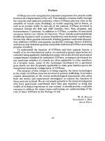
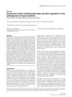
Fie. 1. Purification of posttranslationally modified Rab5 by Mono Q column chromatogra-
phy. (A) Chromatography profile and [35S]GTP3,S binding activity in the presence (O) and
in the absence (0) of Rab-GDI. , absorbance at 280 nm. (B) Analysis by SDS-PAGE
(12% acrylamide gel) stained with Coomassie blue showing the starting homogenate of Sf9
cells expressing Rab5 (lane 1), 2/zl out of 20 ml of the cytosol fraction of the cells (lane 2),
5/zl out of 4 ml of the sample loaded onto a Mono Q column (lane 3), and a 10-/zl aliquot
of fractions 4-24 (lanes 4-14). For details, see text.
idopropyl)dimethylammonio]-l-propanesulfonic acid (CHAPS) (Sigma)
with sonication for 10 sec on ice and is incubated for 1 hr at 4 ° on a rotating
wheel. The suspension is centrifuged at 160,000g for 30 min at 4 °, and the
supernatant (4 ml, 10 mg protein) is loaded onto a Mono Q HR5/5 column
(Pharmacia) equilibrated with degassed buffer A containing 0.6% (w/v)
CHAPS (Fig. 1A). After washing the column with 12 ml of the same buffer,
proteins are eluted with buffer A containing 0.6% CHAPS and 1 M NaC1.
Fractions (0.5 ml) are collected and analyzed by SDS-PAGE stained with
Coomassie blue (Fig. 1B) and immunoblotting using anti-Rab5 monoclonal
antibody (data not shown) (see [27] in this volume). Most of Rab5 is
detected in two peaks. The first consists of the flow-through fractions (frac-
14
EXPRESSION, PURIFICATION, AND MODIFICATION
[2]
tions 4-11; about 20% recovery) and the second consists of the washing
fractions (fractions 12-24, about 40% recovery), where Rab5 migrates
slightly faster compared to the protein contained in the first peak on SDS-
PAGE. These fractions are further characterized for the presence of post-
translationally modified Rab5. Since Rab-GDI has been shown to be active
only on posttranslationally modified but not on unmodified Rab proteins, 5
we tested each fraction for Rab-GDI to inhibit GDP/GTP exchange as
deduced by the binding of radiolabeled GTPTS 4 (Fig. 1A). An aliquot
(2/zl) of each fraction is incubated in the presence or in the absence of 5
/zM Rab-GDI, purified from overexpressing
E. coli
as a His6-tagged protein
(see [27] in this volume), in a buffer (20/zl) containing 20 mM HEPES/
KOH (pH 7.2), 10 mM EDTA, 5 mM MgC12, 1 mM dithiothreitol, and
1/zM [35S]GTPTS (20,000 cpm/pmol, DuPont-NEN) for 10 min at 30 °.
Protein-bound [35S]GTPTS is measured by passing the reaction mixture
through a nitrocellulose filter (0.45-/zm pore size, 2.5 cm diameter, BA85,
Schleicher & Schuell) immediately after adding 3 ml of filtration buffer [20
mM tris[hydroxymethyl]aminomethane hydrochloride (pH 7.5), 100 mM
NaCI, and 25 mM MgC12]. After three washes with 3 ml filtration buffer,
the filter is dried and the radioactivity is measured in 5 ml Ready Safe
scintillation liquid (Beckman) using a Beckman LS 6000SC type scintillation
counter. Proteins in these fractions effectively bind [35S]GTPyS. Although
Rab-GDI does not effect [35S]GTPyS binding to the proteins of fractions
4-11, it effectively inhibits [35S]GTPyS binding to the proteins of fractions
12-24, thus indicating that the second peak (fractions 12-24) Contains
posttranslationally modified Rab5. The Rab5 protein recovered in fractions
4-11 may come from the contaminating cytosol and/or aggregated cytosol
Rab5. The samples are analyzed by SDS-PAGE (12% acrylamide gel)
stained with Coomassie blue (Fig. 1B, lanes 4-14). Typically, about 200/zg
of highly purified posttranslationally modified Rab5 is obtained in frac-
tions 12-24.
Purification of Posttranslationally Unmodified Rab5 from Cytosol
of Sf9 Cells
The posttranslationally unmodified Rab5 is purified from the cytosol
of Rab5-expressing Sf9 cells by a one-step procedure using hydroxyapatite
column chromatography. Hydroxyapatite (Seikagakukogyo, Tokyo, Japan)
is swollen in distilled water and the fine particles are removed by changing
the water every 30 min until the supernatant is clear. Then, 1 ml of hydroxy-
apatite is transferred onto a Poly-Prep chromatography column (Bio-Rad),
followed by equilibration with buffer B (20 mM HEPES/KOH, pH 7.2, 5
mM MgCIz, 10 mM 2-mercaptoethanol). The cytosol (1 ml, 5 mg of protein)
[3] Rab9 PURIFICATION AND ISOPRENYLATION 15
is loaded onto the column. After washing the column with 5 ml of buffer
B, the column is eluted with buffer B containing 0.6% CHAPS. Fractions
(0.5 ml) are collected, and 150/zg of unmodified Rab5 is eluted in fractions
2-8. Because of the high level of expression and the particular property
of Rab5 to be eluted by CHAPS, the purity is over 90%. Purified unmodified
Rab5 efficiently binds GTP and GDP but, as expected, Rab-GDI does not
inhibit [35S]GTP3~S binding in the same assay mentioned earlier. In this
simple procedure, 3 mg of highly purified unmodified Rab5 can be expected
from one preparation of the cytosol (20 ml).
[31 Expression of Rab9 Protein in Escherichia coIi:
Purification and Isoprenylation/n Vitro
By
MARKUS
A.
RIEDERER, THIERRY SOLDATI,
A. BARBARA
DIRAC-SVEJSTRUP,
and SUZANNE R. PFEFFER
Introduction
This chapter describes the purification of canine Rab9 after expression
in Escherichia coli, and the small-scale and preparative-scale isoprenylation
of Rab9 in vitro. Escherichia coli-expressed Rab proteins are valuable
reagents in analyzing the biochemical properties, structural features, and
functional activities of individual rab proteins. In addition, characterization
of purified mutant forms of Rab proteins can provide valuable information
to complement functional studies of Rab proteins in in vitro systems or in
living cells.
The pET expression system developed by Studier et al. 1 is invaluable
for the production of milligram quantities of specific proteins in E. coli.
Rab9 cDNA was subcloned into the pET8c plasmid, which places the
cDNA under the control of a T7 RNA polymerase promoter. The resulting
expression vector, pET8c-Rab9, is transformed into the E. coli strain BL21
(DE3), which expresses the T7 RNA polymerase gene under the control
of the lacZ promoter. The addition of isopropyl-/3-D-thiogalactoside (IPTG)
induces the synthesis of T7 RNA polymerase, which, when present at high
levels, produces large amounts of Rab9 mRNA and thus large amounts of
Rab9 protein.
1 F. Studier, A. Rosenberg, J. Dunn, and J. Dubendorf, this series, Vol. 185, p. 60.
Copyright © 1995 by Academic Press, Inc.
METHODS IN ENZYMOLOGY, VOL. 257 All rights of reproduction in any form reserved.
16 EXPRESSION, PURIFICATION, AND MODIFICATION [31
Materials
IPTG, ampicillin, geranylgeranyl pyrophosphate, and [3H]geranylgera-
nyl pyrophosphate (GGPP and [3H]GGPP, American Radiolabeled Chem-
icals)
Plasmids
E. coli expression plasmid pET8c-Rab9wt, pET8c-Rab9N21
Cells
E. coli strain BL21(DE3) [F-, ompT, r-B, m-8] 1
Equipment~Columns
Pressure filtration cell (Amicon)
Q-Sepharose Fast Flow column (Pharmacia)
Sephacryl S-100 column (Pharmacia)
Buffers
Lysis buffer: 64 mM Tris-HCl (pH 8.0), 8 mM MgCI2,2 mM EDTA, 0.5
mM dithiothreitol (DTT), 10/zM GDP, 1 mM phenylmethylsulfonyl
fluoride (PMSF), 10 mM benzamidine, 10/zg/ml leupeptin, 1 tzM
pepstatin, 3/zg/ml aprotinin, and 1 mM NaNz
S-100 buffer: 64 mM Tris-HC1 (pH 8.0), 100 mM NaC1, 8 mM MgC12,
2 mM EDTA, 0.2 mM DTT, 10/.~M GDP, 1 mM PMSF, 10 mM
benzamidine, and 1 mM NaN3
Procedures
Expression and Purification of Rab9 Protein
The procedure was optimized for Rab9 purification based on a pre-
viously described method of Tucker et al. 2
1. The cDNA of rab9 was cloned into the E. coli expression vector,
pET8c. 1 The pET8c plasmid was linearized with BamHI, filled in using the
Klenow fragment of DNA polymerase I, and cut with NcoI. Both restriction
enzyme sites are located in the polylinker of pET8c. A pGEM1-Rab9
2 j. Tucker, G. Sczakiel, J. Feuerstein, J. John, R. Goody, and A. Wittinghofer, EMBO J. 5,
1351 (1986).
[3] Rab9 PURIFICATION AND ISOPRENYLATION 17
plasmid 3 was linearized with PstI and filled in with T4 DNA polymerase.
A second NcoI digestion liberated a fragment containing the rab9 gene.
The pET8c and Rab9 fragments were purified by agarose gel electrophoresis
prior to ligation by standard procedures. 4 The construct was confirmed by
restriction analysis and transformed into the E. coli strain BL21.
2. An overnight culture of BL21 + pET8c-Rab9wt is grown in LB +
ampicillin (100/zg/ml). Five hundred milliliters of LB + ampicillin (100
/zg/ml) is inoculated with 5 ml of overnight culture and grown to
an OD60o
of 0.4-0.6. Induction is started by the addition of IPTG to a final concentra-
tion of 0.4 mM. Induction at 37 ° is performed for 3.5 hr before the cells
are centrifuged for 5 min at 6000 rpm. The supernatant is discarded, and
the cell pellet is frozen in liquid N2 and stored at -20 °.
3. The bacterial pellet is resuspended in 15 ml ice-cold lysis buffer. The
cells are lysed by two passages through a French press at medium power
with 1400 units pressure. Subsequent steps are performed at 4 ° .
4. Protamine sulfate is added to a final concentration of 1 mg/ml and
the suspension is stirred for 2 min. The mixture is then centrifuged for 5
min at 16,000 rpm in a precooled Sorvall SS-34 rotor.
5. The supernatant is loaded onto a 15-ml Q-Sepharose Fast Flow col-
umn preequilibrated in lysis buffer and washed with 20 ml of lysis buffer.
Proteins are eluted with a 2 x 50-ml gradient of 0-200 mM NaC1 in lysis
buffer and 2-ml fractions are collected.
6. Alternate fractions (20/xl) are analyzed by polyacrylamide gel elec-
trophoresis (12%) and proteins are visualized by Coomassie blue staining.
Rab9 protein is determined by size comparison with control Rab9 protein
on the stained gel. The identity of the band is later confirmed by Western
blot and GTP overlay.
7. Fractions containing Rab9 are pooled, concentrated to a final volume
of 2 ml by pressure filtration in a stirred cell, and applied to a 240-ml
Sephacryl S-100 column. The column is run in S-100 buffer at a rate of
about 20 ml per hour; 80 fractions of 2.5 ml are collected.
8. Fractions containing Rab9 are pooled and concentrated by pressure
filtration to a final concentration of 0.4-1.0 mg/ml. Rab9 protein is either
rapidly frozen in liquid nitrogen and stored at -80 ° or stored in 40% glycerol
at -20 ° .
Notes: (1) Rab9 protein has the unique property of being very efficiently
proteolysed at the carboxy terminus. No commercially available protease
3 D. Lombardi, T. Soldati, M. A. Riederer, Y. Goda, M. Zerial, and S. R. Pfeffer,
EMBO J.
12, 677 (1993).
4 j. Sambrook, E. Fritsch, and T. Maniatis, "Molecular Cloning: A Laboratory Manual," 2nd
ed. Cold Spring Harbor Lab., Cold Spring Harbor, NY, 1989.
18
EXPRESSION, PURIFICATION, AND MODIFICATION [3]
inhibitor was found to inhibit this proteolytic step. The only significant way
to increase the yield of full-length Rab9 protein is to work at 4 ° and to
work as fast as possible. Rab9 and the unknown protease are resolved on
the Q-Sepharose column, where the protease activity elutes at a higher salt
concentration relative to Rab9 protein. (2) The Rab9S21N mutant is much
less soluble when expressed in E. coli. The modifications described below
increase the pool of soluble Rab9S21N and permit the purification of small
quantities of Rab9S21N protein. 5
Results
1. Induction: After induction for 3.5 hr, the 26-kDa Rab9 polypeptide
is clearly detectable in cell extracts subjected to SDS-PAGE and Coomassie
blue staining. The identity of the 26-kDA protein is confirmed by immu-
noblot analysis using a Rab9-specific antibody. In addition, the expressed
protein binds GTP as determined by the [o~-32p]GTP overlay of proteins
resolved by SDS-PAGE and transferred to nitrocellulose.
2. Ion exchange: Soluble fractions are subjected to ion-exchange chro-
matography on a column of Q-Sepharose. The [a-32p]GTP overlay of the
collected Q-Sepharose fractions reveals two peaks of GTP-binding activity.
Immunoblot analyses show that both peaks contain Rab9-immunoreactive
material: immunoreactive material in the first peak migrates as a 26-kDa
polypeptide and, in the second peak, as a 22-kDa species. The larger poly-
peptide comigrates with Rab9 protein present in the induced E. coli lysate
and is further purified. Amino-terminal sequencing and mass spectrometry
have confirmed that the 22-kDa polypeptide represents a truncated form
of Rab9 which lacks 22 amino acids at its carboxy terminus. 3 Typically,
30-50% of Rab9 is recovered in truncated form, which we refer to as
Rab9AC. This degradation product is completely resolved from intact Rab9
on Q-Sepharose chromatography and thus does not contaminate the final
preparation. In summary, Rab9 elutes from the Q-Sepharose at 120 mM
NaC1; the Q-Sepharose chromatography results in a sevenfold purification
relative to the initial cell lysate (Table I).
3. Gel filtration: The pooled Q-Sepharose fractions are concentrated
by pressure filtration and are subjected to gel filtration using Sephacryl
S-100. The Rab9 protein migrates with a retention coefficient of 0.6. The
S-100 column results in an additional -fourfold purification (Table I).
4. In summary, the two-step purification yields a 27-fold enrichment of
Rab9 protein and a final preparation that is ->95% pure. One gram of cell
paste yields 0.8 mg of Rab9. Rab9 preparations are typically 90% active
5 M. A. Riederer, T. Soldati, A. D. Shapiro, J. Lin, and S. R. Pfeffer, J.
Cell Biol.
125, 573 (1994).
[3] Rab9 PURIFICATION AND ISOPRENYLATION 19
TABLE I
PURIFICATION OF Rab9 PROTEIN
Total
nucleotide
Total Total binding Specific
protein volume activity activity Yield Purification
Fraction (mg) (ml) (nmol) (nmol/mg) (%) (-fold)
Lysate 77.7 18.5 120 1.5 100 1.0
Q-Sepharose 4.4 14.7 47 11 40 7.1
S-IO0 0.95 10.6 38 40 32 27
as judged by the extent of [ot-32p]GTP binding relative to applied protein. 6
No loss of binding activity has been detected after >2 months of storage
at -20 ° in S-100 column buffer containing 40% glycerol.
Notes: (1) We have also created a Rab9 protein that possesses a different
carboxy-terminal tetrapeptide which serves as a signal for isoprenylation
by prenyltransferase I. The resulting Rab9-CLLL is not degraded during
the purification process, suggesting that a carboxypeptidase initiates the
proteolytic processing and preferentially cleaves Rab9 protein terminating
in CC. (2) Other Rab9 mutant proteins: After induction for 6 hr at 30 °, a
small pool of Rab9S21N is soluble and can be purified using the same
procedure employed for Rab9. In contrast, Rab9140M and Rab9N127I are
insoluble under conditions that allow purification of Rab9S21N. Attempts
to solubilize these mutant proteins in 6 M guanidinium-chloride followed
by dilution have not been successful. (3) Rab7 can be expressed in E. coli
and purified using the identical procedure described earlier. Rab7 appears
to be resistant to proteases and is eluted from the Q-Sepharose column at
70 mM NaC1.
Isoprenylation of Rab9 in Vitro
Small-Scale in Vitro Prenylation
Small-scale prenylation reactions are extremely useful either to test in
vitro the prenylatability of a Rab protein or mutant thereof or, alternatively,
to optimize the conditions prior to preparative scale incubations. A standard
50-/zl reaction contains 5 ng Rab9, 50 ng GGPP, and 1.5 mg/ml crude ClIO
cytosol. The buffer conditions are similar to those used for in vitro endosome
6 A. D. Shapiro, M. A. Riederer, and S. R. Pfeffer, J.
Biol. Chem.
268, 6925 (1993).
20
EXPRESSION, PURIFICATION, AND MODIFICATION
[3]
to TGN transport 7 22 mM HEPES-KOH, pH 7.2, 20 mM Tris-HC1, 116
mM KC1, 4.3 mM magnesium acetate + MgC12), 2 mM DTT, and 0.2 mM
GDP, plus a protease inhibitor cocktail and an ATP-regenerating system.
After incubation at 37 ° for 1 to 2 hr, the prenylation reactions are clarified
by ultracentrifugation at 300,000g for 10 min in a TLA100.2 rotor (Beck-
man) and analyzed by 12.5% SDS-PAGE and anti-Rab9 immunoblotting.
As prenylated Rab9 migrates slightly faster than the unprenylated starting
material, the efficiency of the prenylation reaction can be easily monitored.
Alternatively, if no molecular size shift is expected, or for precise quantita-
tion analysis, small-scale reactions should include 1/xM GGPP and 0.1/xM
[3H]GGPP, and be followed by SDS-PAGE and fluorography.
Another small-scale prenylation assay used to assess prenylatability of
a construct is based on the cell-free translation of an in vitro-transcribed Rab
eDNA. Commercially available rabbit reticulocyte lysate (e.g., Promega) is
gel filtered and therefore does not contain enough endogenous GGPP to
ensure prenylation of newly translated proteins. Efficient prenylation can
be achieved by adding 10 tzM GGPP in the in vitro translation reaction
containing [35S]methionine (as judged then by a molecular size shift after
SDS-PAGE and autoradiography analysis) or 1/zM GGPP and 0.1/zM
[3H]GGPP in reactions carried out with unlabeled amino acids (as judged
by incorporation of radioactivity in the translation product analyzed by
SDS-PAGE and fluorography).
Preparative in Vitro Prenylation
In a standard 0.5-ml reaction, 1 tzg of purified Rab9 (100 nM) is preny-
lated in the presence of 5.6 mg/ml of crude Chinese hamster ovary (CHO)
cytosol (prepared as described in Goda and Pfeffer 7) and 10 tzM of geranyl-
geranyl pyrophosphate (GGPP, American Radiolabeled Chemicals, Inc)
by incubation for 1 hr at 37 °. Preparative prenylation of Rab9 protein is
usually about 50-80% efficient. 8 Separation of the prenylated Rab9 from
nonreacted or degraded material by Sephacryl S-100 gel filtration chroma-
tography (see below) is facilitated by the fact that prenylated Rab proteins
associate with GDP dissociation inhibitor (GDI) and hence fraetionate at
~80 kDa, whereas the other products will elute around 20-30 kDa.
Gel Filtration Chromatography and Fraction Analysis. Samples are ana-
lyzed on a 50-ml Sephacryl S-100 (Pharmacia) column equilibrated and
eluted in S-100 buffer (64 mM Tris/HC1, pH 8, 100 mM NaC1, 8 mM MgCI2,
2 mM EDTA, 0.2 mM DTI', 10 tzM GDP, and 1 mM PMSF). Forty 0.4-
ml fractions are collected; alternate fractions are subjected to 12.5% SDS-
7 y. Goda and S. R. Pfeffer,
Cell (Cambridge, Mass.) $5,
309 (1988).
s T. Soldati, M. A. Riederer, and S. R. Pfeffer,
Mol. Biol. Cell
4, 425 (1993).
[4] CHARACTERIZATION OF TYPE-II GGTase 21
PAGE and conventional immunoblotting. Rab9 protein is detected using
rabbit or mouse antibodies raised against native, recombinant Rab9 pro-
tein. 8 Detection of GDI is carried out using affinity-purified antibodies
raised against purified Rab3A-GDI. 8 Secondary antibodies are either goat
anti-rabbit or goat anti-mouse IgG conjugated to horseradish peroxidase
(Bio-Rad). All antibodies are used at 1:1000 dilution; antigen-antibody
complexes are detected by enhanced chemiluminescence (ECL, Amer-
sham). Quantitation of ECL signals on X-ray films (Kodak) is carried out
using a densitometric scanner (Model 300 A, Molecular Dynamics) or a
Phosphorlmager system (Molecular Dynamics).
[4] Characterization of Yeast Type-II
Geranylgeranyltransferase
By YU JIANG, GUENDALINA ROSSI, and SUSAN FERRO-NOVICK
Introduction
Members of the Rab GTP-binding protein family are involved in the
regulation of different exocytic and endocytic transport processes. 1 They
are localized to diverse intracellular compartments and participate in vari-
ous steps of vesicular traffic. 1 In yeast, two Rab GTPases, Sec4p and Yptlp,
have been shown to play a role on the exocytic pathway. 2'3 They are signifi-
cantly homologous to each other, but function at distinct stages of the
pathway. Although Yptlp is involved in mediating the transport of vesicles
from the endoplasmic reticulum (ER) to the Golgi complex, 3 Sec4p is
required for membrane traffic from the Golgi to the plasma membrane. 2
Like most small GTP-binding proteins, Yptlp and Sec4p are synthesized
in the cytosol, but become membrane bound after undergoing posttransla-
tional modification. Mutations that block the membrane attachment of these
proteins result in a block in secretion, a,5 Thus, the membrane association of
Yptlp and Sec4p is crucial for their function.
The ability of small GTP-binding proteins to bind to membranes is
conferred by the addition of geranylgeranyl, a 20-carbon isoprenoid deriva-
1 M. Zerial and H. Stenmark, Curr. Opin. Cell Biol. 5, 613 (1993).
2 A. Salminen and P. J. Novick, Cell (Cambridge, Mass.) 49, 527 (1987).
3 N. Segev, J. Mulholland, and D. Botstein, Cell (Cambridge, Mass.) 52, 915 (1988).
4 G. Rossi, Y. Jiang, A. P. Newman, and S. Ferro-Novick, Nature (London) 351, 158 (1991).
s R. Li, C. Havel, J. A. Watson, and A. W. Murray, Nature (London) 366, 82 (1993).
Copyright © 1995 by Academic Press, Inc.
METHODS IN ENZYMOLOGY, VOL. 257 All rights of reproduction in any form reserved.