The nicotinic acetylcholine receptor alpha 4 subunit contains a functionally relevant SNP Haplotype
Bạn đang xem bản rút gọn của tài liệu. Xem và tải ngay bản đầy đủ của tài liệu tại đây (527.92 KB, 7 trang )
Eggert et al. BMC Genetics (2015) 16:46
DOI 10.1186/s12863-015-0204-1
RESEARCH ARTICLE
Open Access
The nicotinic acetylcholine receptor alpha 4
subunit contains a functionally relevant SNP
Haplotype
Marlene Eggert1, Georg Winterer2, Mario Wanischeck3, Jean-Charles Hoda4, Daniel Bertrand5 and Ortrud Steinlein6*
Abstract
Background: Non-coding single nucleotide polymorphisms within the nicotinic acetylcholine receptor alpha 4 subunit
gene (CHRNA4) are robustly associated with various neurological and behavioral phenotypes including schizophrenia,
cognition and smoking. The most commonly associated polymorphisms are located in exon 5 and segregate as part of
a haplotype. So far it is unknown if this haplotype is indeed functional, or if the observed associations are an indirect
effect caused by linkage disequilibrium with not yet identified adjacent functional variants. We therefore analyzed the
functional relevance of the exon 5 haplotype alleles.
Results: Using voltage clamp experiments we were able to show that the CHRNA4 haplotype alleles differ with respect
to their functional effects on receptor sensitivity including reversal of receptor sensitivity between low and high
acetylcholine concentrations. The results indicate that underlying mechanisms might include differences in codon
usage bias and changes in mRNA stability.
Conclusions: Our data demonstrate that the complementary alleles of the CHRNA4 exon 5 haplotype are
functionally relevant, and might therefore be causative for the above mentioned associations.
Keywords: CHRNA4, Acetylcholine receptor, ACh sensitivity, Haplotype, mRNA stability
Background
Cholinergic effects on cortical information processing
and related cognitive performance are partly mediated
through stimulation of high-affinity heteromeric α4β2
nicotinic acetylcholine receptors (nAChRs) [1-4]. α4β2
receptors are abundantly expressed in human cortex and
hippocampus and possess high affinity to (partial) agonists including nicotine and varenicline [5-7]. Receptor
upregulation occurs with chronic exposure to agonists
and is thought to be regulated on the translational/posttranslational rather than transcriptional level [8-10].
In earlier work, we reported a causative relationship
between mutations in exon 5 of CHRNA4 (the nAChR
α4-subunit coding gene) and the autosomal dominant
nocturnal frontal lobe epilepsy (ADNFLE) - a rare seizure disorder that is frequently associated with neurocognitive deficits or psychiatric affections [11-14]. We then
* Correspondence:
6
Ortrud K Steinlein, Institute of Human Genetics,
Ludwig-Maximilians-University Hospital, 80336 Munich, Germany
Full list of author information is available at the end of the article
explored in a more recent study, whether association also
might exist between human information processing and
common CHRNA4 exon 5 single nucleotide polymorphisms (SNPs). Using functional magnetic resonance
imaging (fMRI), association, especially for SNP rs1044396,
was observed with prefrontal/parietal information processing during a selective attention-requiring task [15].
Complementary behavioral, electrophysiological and neuroimaging studies from other groups have later provided
converging evidence supporting the validity of this association. [16-25] Furthermore, we and others repeatedly observed that the common exon 5 SNPs are also associated
with endophenotypes of nicotine dependence [26-29].
The CHRNA4 SNPs that repeatedly showed association
with neurological and behavioral traits all have in common that they are silent mutations, i.e. are not changing
codons and therefore have no apparent effect on the protein sequence. However, during the last decade it has become obvious that not all silent SNPs are functionally
neutral. In the present study, we therefore addressed
the question if silent CHRNA4 exon 5 SNPs are able to
© 2015 Eggert et al.; licensee BioMed Central. This is an Open Access article distributed under the terms of the Creative
Commons Attribution License ( which permits unrestricted use, distribution, and
reproduction in any medium, provided the original work is properly credited. The Creative Commons Public Domain
Dedication waiver ( applies to the data made available in this article,
unless otherwise stated.
Eggert et al. BMC Genetics (2015) 16:46
modulate mRNA or receptor properties. For this purpose we conducted experiments on receptor sensitivity
and mRNA stability, and performed in silico analysis regarding possible codon usage differences introduced by
the haplotype alleles.
Results
Receptor sensitivity analysis
Exon 5 of the CHRNA4 gene contains a linkage group
(haplotype) of synonymous variants (haplotype 5’-rs1044393,
rs1044394, rs2229959, rs2229960, rs1044396, rs1044397-3’)
of hitherto unknown functional relevance. This haplotype rather than single SNPs was chosen for the here
reported functional studies because exon 5 SNPs are in
linkage disequilibrium with each other. It is therefore
not possible to decide if, for example, associations found
for a specific SNP are indeed caused by this SNP or by another one located on the same haplotype. Analysis of the
haplotype therefore allowed us to simultaneously include
all major exon 5 SNPs into our search for functional
effects.
Page 2 of 7
Haplotype allele hap1 (T-T-G-T-C-G) corresponds to
the NCBI reference sequence (NM000744.5) and has a
frequency of 9% in the general population, while the
complementary hap2 allele (C-C-T-C-T-A) accounts for
52% of all alleles (according to our reference student
population). Hap 1 and Hap 2 differ in each single SNP
position. Besides hap1 and hap2 at least six other alleles
of this haplotype are present in the normal population.
These haplotypes share one or more SNP allele with the
major haplotypes Hap1 and Hap2 and can therefore be
expected to produce intermediate results in functional
analysis. They were therefore not included in the present
study.
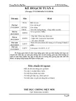
Heterologous expression experiments showed that α4β2receptors with both haplotypes yield functional receptors
with current amplitudes that increased in a dose-dependent
manner with the ACh concentrations (Figure 1). For
low ACh doses the currents from hap2 receptors (incl.
rs1044396 T-allele) were up to 130% larger than those
from hap1 receptors (incl. rs1044396 C-allele), resulting in a
shift of the hα4(hap2)β2 curve towards lower concentrations
with respect to the curve for hα4(hap1)β2. For higher doses
Figure 1 Graphical representation of electrophysiological experiments. a) Concentration activation curves for the α4(hap1)β2 and α4(hap2)β2
receptors. To minimize scatter, data were collected from large batches of cells. Data obtained from 70 cells expressing the α4(hap1)β2 are indicated by
stars whereas circles correspond to results collected for 65 cells expressing the α4(hap2)β2. Bars indicate the SEM. Continuous curve is the best fit
obtained with the sum of two Hill equations for the α4(hap2)β2 and dashed line for the α4(hap1)β2. Representative currents evoked by 100 nM and
30 μM ACh are illustrated in the lower panel. For comparison responses have been normalized to 100% for the largest evoked current. These data
illustrate the differential, i.e. inverse, sensitivity at the two concentrations between the two haplotype alleles of the α4β2 receptor.
Eggert et al. BMC Genetics (2015) 16:46
of ACh the opposite effect was found with currents for
hα4(hap2)β2 that were about 13% lower than those obtained for hα4(hap1)β2. The EC50L for hα4(hap2)β2
(0.33 μM ± 0.017) differed significantly (P ≤ 0.001, n = 65)
from hα4(hap1)β2 (0.72 ± 0.04), indicating a higher sensitivity to ACh for hα4(hap2)β2 at low ACh concentrations.
Interestingly, the EC50H data demonstrate a switch in
this behavior, pointing to a lower sensitivity to ACh for
hα4(hap2)β2 (42.5 μM ± 3) compared to hα4(hap1)β2
(33.6 μM ± 2.3, P ≤ 0.05, n = 65) in case of high ACh
concentrations (Figure 1).
mRNA stability analysis
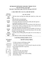
When comparing the mRNA decay time difference of
hap 1 and 2, none of the time differences for the four upstream fragments were significant. However, regarding the
time difference 0 to 24 hrs for the most downstream fragment, our results revealed that the 3’ end of hap 1 mRNA
was significantly more slowly degraded than that of hap 2
(P = 0.03) (Table 1).
mRNA secondary structure prediction and codon usage
analysis
Analysis of the predicted mRNA secondary structure
showed marked differences between the two haplotype
alleles (see Additional file 1: Figure S1). Codon usage analysis showed that most SNPs introduced changes from frequently to more rarely used codons or vise versa. These
effects were most pronounced for rs2229959, rs1044396
and rs1044397 (see Table 2).
Discussion
Taken together, our experimental data show that the
CHRNA4 haplotype alleles exert different functional
effects on mRNA stability as well as on receptor sensitivity including reversal of receptor sensitivity between
low and high ACh concentrations. Furthermore, in
silico analysis predict that the haplotype alleles also
differ with respect to codon usage and mRNA secondary structure. The experiments were conducted using
clones that contained identical fragments from the
CHRNA4 coding region, the only differences between
the clones being the respective alleles of the five SNPs
composing the haplotype. Thus the variation observed
in both ACh sensitivity and mRNA stability should be
attributable to the SNPs within the haplotype. Our results therefore strongly suggest that one or more of
the synonymous SNPs that constitute the haplotype
are functionally relevant. Such a conclusion would not
be too surprising, given that several examples exist in
which silent SNPs have been found to modulate gene
function, for example by altering mRNA stability, translation efficiency or protein conformation [30-32].
Page 3 of 7
Various mechanisms could explain the distinctive doseresponse curves of the two haplotypes. It is a possibility
that the observed changes in mRNA stability are one
of the mechanisms that contribute to the haplotypedependent differences in ACh sensitivity. One explanation could be that the altered mRNA stability may
lead to an increased translation rate of CHRNA4 mRNA
carrying the more stable haplotype 1. Such an increased
translation rate would expand the amount of α4 subunit
protein in hap 1 carriers while the amount of β2 subunit
protein would remain constant. This in turn could alter
the nAChRs stoichiometry so that more (α4)3(β2)2 than
(α4)2(β2)3 receptor subtypes are assembled. Such changes
in stoichiometry are a factor known to influence several
functional receptor characteristics and to increase receptor affinity [33,34]. It is also possible that, apart from
mRNA stability, additional mechanisms are responsible
for the observed differences in agonist sensitivity. For example, changes in the mRNA sequence are known to
affect its folding which in turn can influence the efficiency
and speed of protein synthesis [35]. This mechanism
would also be able to affect the ratio of α4 versus β2
subunits within the mature nAChR. Furthermore, codon
bias is discussed as mechanism for a gene expression
regulation because it has been observed that genes with
lower expression levels prefer codons which are recognized by tRNAs with lower gene copy numbers [36].
Another factor that might play a role would be codon
bias at the ribosome. It is assumed that the speed at
which a given mRNA is decoded at the ribosomes largely
depends on the availability of individual tRNA molecules.
However, most amino acids can be encoded by more than
one base pair triplet, and there are significant differences
with respect to the frequency with which individual codons occur in genes. In fact, synonymous codons are used
at nonrandom frequencies, a phenomenon termed codon
usage bias. Such differences in codon usage are not only
found between species, but in some examples also have
been described for different tissues from the same individual [37]. Both codon usage and tRNA gene numbers
evolved together, and, consequently, tRNAs that recognize
frequently used codons are usually more abundant at the
ribosome and are therefore more readily available for
translation [37,38]. Thus a silent SNP changing a frequently used codon into a rarer one can slow down the
speed of mRNA translation. In fact, synonymous codon
usage is recognized as the primary cause of non-uniform
translation rates, a mechanism known to cause for differential maturation and folding of nascent polypeptides
[39]. These differences in polypeptide processing are
possible because the time newly synthesized polypeptides spend at the ribosome is used to introduce various
modifications that are important for protein folding, stability, and interaction with binding partners [40]. The
Eggert et al. BMC Genetics (2015) 16:46
Table 1 Delta Cq mean values and p-values for qPCR fragments 1 to 5
mRNA decay
qPCR fragment
time difference
1
Δ Cq mean
2
3
4
5
p-value Δ Cq mean
p-value Δ Cq mean
p-value Δ Cq mean
p-value Δ Cq mean
p-value
hap 1
hap 2
hap1/2 hap 1
hap 2
hap1/2 hap 1
hap 2
hap1/2 hap 1
hap 2
hap1/2 hap 1
hap 2
hap1/2
0 to 3 hrs
1.16
0.29
0.242
1.91
0.09
0.0291
1.61
0.26
0.066
1.07
-0.07
0.08
1.17
0.37
0.436
0 to 6 hrs
2.54
0.403
3.34
0.313
3.04
0.546
2.23
0.503
3.69
(0.49-4.58)
(0.63-3.27)
(0.79-5.89)
(0.77-4.04)
(0.43-5.65)
(0.75-4.21)
(0.26-4.19)
(0.29-3.11)
(0.72-6.66)
(0.71-5.97)
0 to 24 hrs
5.74
5.64
0.918
6.18
6.24
0.952
5.06
4.77
0.778
5.21
4.77
0.679
8.16
10.73
(3.86-7.62)
(3.22-8.06)
(4.02-8.33)
(3.84-8.63)
(2.79-7.34)
(2.55-7.00)
(3.55-6.87)
(2.23-7.32)
(-0.72 - 3.04) (-1.57 -2.15)
1.95
(-0.31 -4.12) (-1.77 -1.94)
2.41
(-0.73 -3.95) (-1.61 -2.13)
2.48
(-0.70 -2.83) (-1.66 -1.51)
1.7
(-1.48 -3.81) (-1.73 -2.47)
3.34
0.821
0.0362
(5.42-10.90) (6.03-13.44)
Δ Cq mean, delta Cq mean: difference of the mean quantification cycle; confidence intervals are given in brackets.
1
confidence interval for delta Cq-mean values of hap1 and hap2 are not significant.
2
confidence interval for delta Cq-mean values of hap1 and hap2 are significant.
Page 4 of 7
Eggert et al. BMC Genetics (2015) 16:46
Page 5 of 7
Table 2 Changes of codon usage frequencies within the
haplotype
dbSNP ID
amino acid
positions
Alleles (hap
1/hap 2)
Codon usage frequencies
(hap 1/hap 2)
rs1044393
D213
GAT/GAC
21,8/25,1
rs1044394
C226
TGT/TGC
10,6/12,6
rs2229959
P403
CCG/CCT
6,9/17,5
rs2229960
C409
TGT/TGC
10,6/12,6
rs1044396
S543
AGC/AGT
19,5/12,1
rs1044397
A553
G/A
7,4/15,8
Data source for codon usage: />
silent SNPs that constitute the CHRNA4 haplotype
introduce several changes from frequent to rarer used
codons (or vise versa, see Table 2) and could therefore
alter, by the above discussed mechanisms, functional
characteristics of the nAChR such as stoichiometry,
surface expression or function. Pathomechanisms like
this have already been reported for other silent SNPs
associated with human disorders [31,35,41].
It appears remarkable that, within our experimental
setting, mRNA stability was only altered in the downstream (3’) region of the cloned CHRNA4 coding region
fragment. These observations suggest that it is the SNPs
in the 3’ part of the haplotype that are able to alter
mRNA stability. Interestingly, the 3’ end of the CHRNA4
haplotype harbors the two silent SNPs that have most
consistently shown association with clinical phenotypes.
For example, recent work from several groups including our own reported association between rs1044396/
rs1044397 and endophenotypes of schizophrenia as well
as nicotine addiction. Both SNPs are significantly associated with cognitive endophenotypes such as brain activation (N100-amplitude – in particular in prefrontal cortex)
during selective-attention-requiring tasks [15-19,21,23-26].
With a minor allele frequency above 0.45 both SNPs would
be common enough to contribute considerably to the
inter-individual variability in the processing of cognitive
tasks, addictive behavior and psychiatric disorders within
the general population. Additional studies are needed to
shed light on the complex interactions between these silent nAChR variants, differences in nAChR function, and
the inter-individual variability of neurological and behavioral traits in humans.
Conclusions
Our experimental and in silico data demonstrate that the
complementary alleles of the CHRNA4 exon 5 haplotype
differ with respect to mRNA stability, codon usage, and
agonist sensitivity. These results render it possible that
one or more of the haplotype-constituting SNPs are
causative for the previously reported associations with
neurological and behavioral phenotypes.
Materials and Methods
Receptor sensitivity analysis
The cDNAs with either one of the two complementary
CHRNA4 haplotypes and with the CHRNB2 wild type
sequence were injected in Xenopus oocytes in equal
amounts and the electrophysiological properties of the
α4β2 nAChR channel were determined using a twoelectrode voltage clamp technique (HiClamp, Multichannel
System®, Reutlingen Germany) and applying different
concentrations of acetylcholine (ACh). Concentrationactivation curves were fitted using a Hill equation in
the form Y = 1/1 + (EC50/ x)^ nH(1) where: y = the fraction of evoked current, EC50 = concentration for 50%
activation of the high affinity, nH = the apparent cooperativity for the high affinity, x = agonist concentration.
Concentration-inhibition curves are fit with a comparable equation Y = 1/1 + (x/IC50)^nH(2) where: y = the
fraction of remaining current, IC50 = concentration for
50% inhibition, nH = the apparent cooperativity, x = antagonist concentration.
mRNA stability testing
The Tet-Off® advanced inducible gene expression system
was purchased from Clontech (Saint-Germain-en-Laye,
France). The coding sequence of CHRNA4 hap 1, respectively hap 2 was obtained by PCR amplification of human
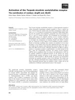
Figure 2 Schematic representation of the CHRNA4 gene. The positions of SNPs that constitute the CHRNA4 haplotype (see main text) are indicated
above, the fragments used for mRNA stability testing below the transcript.
Eggert et al. BMC Genetics (2015) 16:46
DNA. After KpnI and EcorV digestion of the pTRE-TightBI-AcGFP1 vector, the coding sequence of CHRNA4 hap
1, respectively hap 2 (1884 nt) was ligated into the multiple cloning site of pTRE-Tight-BI-AcGFP1 downstream
of the doxycycline-dependent promoter. The resulting
construct had the following structure (origin of fragments given in brackets): (pTRE-Tight) …GCTCGGTAC
(pTRACER) CGAGCTCGGATCCA (CHRNA4 uncoding)
CTAGTAGTGCGCC (CHRNA4 coding) ATG…TAG
(CHRNA4 uncoding) GAATAG (pTRACER) GAATTC
TGCAGAT (pTRE-Tight) ATCTC… After cloning the
inserts were confirmed by sequencing. Culturing of
Tet-Off® human embryonic kidney cells (HEK) 293
(Clontech, Saint-Germain-en-Laye, France) was performed using standard protocols. HEK 293 cells were
transfected with 10 ng plasmid pTRE-Tight-BI-AcGFP1
containing the coding sequence of CHRNA4 haplotype 1
and 2, respectively, using 3 μl of TransIT ®-LT1 Transfection Reagent (MoBiTec GmbH, Gưttingen, Germany) with
24 h transfection prior to medium change and addition of
1 μg doxycycline. RNA was extracted after 0; 3; 6 and
24 hours of doxycycline incubation by using QIAshredder
and RNeasy kit, including DNase treatment of 10 min
in solution, according to the manufacturer’s protocol
(Qiagen, Hilden, Germany). Real-time PCR was performed
targeting five fragments of the CHRNA4 coding sequence
(primer sequences: 1 F GCTCATTGACGTGGATGAGA,
1R CCCGTCAGCATTGTTGTAGA, 2 F GCTGGACTT
CTGGGAGAGTG, 2R AGGGGATGATGAGGTTGATG,
3 F TGCTCATCACCGAGATCATC, 3R ATGACGATG
GACAGGGTGAC, 5 F AAGGAGCCCTCTTCGGTGTC,
5R CTTCGGCCTTCAGGTGGTCT, 5 F GGCTGGCAT
GATCTAGGAAT, 5R GGGAGGTGTGGGAGGTTTTT,
AcGFP_F ATGATGTATCGCCCTCGAAC, AcGFP R CA
CATGAAGCAGCACGACTT) (Figure 2). Amplification
efficiency and test linearity (correlation coefficient R2)
were assessed for each primer pair. The reactions were
carried out in the Mini Opticon CFD-3120 cycler (Bio-Rad,
Munich, Germany). All experiments were repeated independently three times with triplicate biological and triplicate technical samples (nine experiments each in total).
Statistical analysis was performed with program R to compare the haplotype 1 and 2 RNA degradation rate for each
target fragment. A p value of p < 0.05 was considered statistically significant.
Prediction of mRNA secondary structure and codon usage
Changes in the minimum free energy (MFE) secondary
structure caused by the haplotype alleles were predicted
by the use of the RNA fold web server, Vienna RNA
package ( />A prediction software was employed for the codon usage
analysis ( />
Page 6 of 7
Additional file
Additional file 1: Figure S1. Minimum free energy structure of CHRNA4
haplotype mRNAs. The minimum free energy secondary structures for both
CHRNA4 haplotypes were calculated from base-pairing probabilities, using
the share ware program RNAfold web server ( The prediction shows the optimal secondary structure [42].
Abbreviations
ACh: Acetylcholine; ADNFLE: Autosomal dominant nocturnal frontal lobe
epilepsy; CHRNA4: Nicotinic acetylcholine receptor alpha 4 subunit gene;
CHRNB2: Nicotinic acetylcholine receptor beta 2 subunit gene;
fMRI: Functional magnetic resonance imaging; nAChRs: Nicotinic
acetylcholine receptors; SNPs: Single nucleotide polymorphisms.
Competing interests
The authors declare that they have no financial or non-financial competing
interests.
Author contributions
OST and DB planned the study and analyzed the data. JCH conducted the
electrophysiological experiments. ME and MW performed the molecular
genetic experiments. OST, DB and GW wrote the manuscript. All authors
read and approved the final manuscript.
Acknowledgements
The study was funded by the Deutsche Forschungsgemeinschaft (DFG SPP
1226, STE 1651/1-3 and BE 3834/1-2).
Author details
Marlene Eggert, Institute of Human Genetics, Ludwig-Maximilians-University
Hospital, 80336 Munich, Germany. 2Georg Winterer, Experimental and Clinical
Research Center (ECRC), Charité – University Medicine Berlin, Berlin, Germany.
3
Mario Wanischeck, Institute of Human Genetics,
Ludwig-Maximilians-University Hospital, 80336 Munich, Germany.
4
Jean-Charles Hoda, SwissCheckUp SA, 1400 Yverdon-Les-Bains, Switzerland.
5
Daniel Bertrand, HiQScreen, 1222 Vésenaz, Geneva, Switzerland. 6Ortrud K
Steinlein, Institute of Human Genetics, Ludwig-Maximilians-University
Hospital, 80336 Munich, Germany.
1
Received: 18 December 2014 Accepted: 22 April 2015
References
1. Grottick AJ, Higgins GA. Effect of subtype selective nicotinic compounds on
attention as assessed by the five-choice serial reaction time task. Behav
Brain Res. 2000;117(1-2):197–208.
2. Grottick AJ, Wyler R, Higgins GA. A study of the nicotinic agonist SIB-1553A
on locomotion, and attention as measured by the five-choice serial reaction
time task. Pharmacol Biochem Be. 2001;70(4):505–13.
3. Levin ED, McClernon FJ, Rezvani AH. Nicotinic effects on cognitive function:
behavioral characterization, pharmacological specification, and anatomic
localization. Psychopharmacology. 2006;184(3-4):523–39.
4. Wildeboer KM, Stevens KE. Stimulation of the alpha 4 beta 2 nicotinic
receptor by 5-I A-85380 improves auditory gating in DBA/2 mice. Brain Res.
2008;1224:29–36.
5. Hellstrom-Lindahl E, Mousavi M, Zhang X, Ravid R, Nordberg A. Regional
distribution of nicotinic receptor subunit mRNAs in human brain: comparison
between Alzheimer and normal brain. Mol Brain Res. 1999;66(1-2):94–103.
6. Burghaus L, Schutz U, Krempel U, de Vos RAI, Steur ENHJ, Wevers A, et al.
Quantitative assessment of nicotinic acetylcholine receptor proteins in the
cerebral cortex of Alzheimer patients. Mol Brain Res. 2000;76(2):385–8.
7. Rucktooa P, Haseler CA, van Elk R, Smit AB, Gallagher T, Sixma TK. Structural
characterization of binding mode of smoking cessation drugs to Nicotinic
Acetylcholine receptors through study of Ligand complexes with
Acetylcholine-binding protein. J Biol Chem. 2012;287(28):23283–93.
8. Teaktong T, Graham AJ, Johnson M, Court JA, Perry EK. Selective changes in
nicotinic acetylcholine receptor subtypes related to tobacco smoking: an
immunohistochemical study. Neuropath Appl Neuro. 2004;30(3):243–54.
Eggert et al. BMC Genetics (2015) 16:46
9.
10.
11.
12.
13.
14.
15.
16.
17.
18.
19.
20.
21.
22.
23.
24.
25.
26.
27.
28.
29.
Wevers A, Burghaus L, Moser N, Witter B, Steinlein OK, Schutz U, et al.
Expression of nicotinic acetylcholine receptors in Alzheimer's disease:
postmortem investigations and experimental approaches. Behav Brain Res.
2000;113(1-2):207–15.
Mobascher A, Winterer G. The molecular and cellular neurobiology of
nicotine abuse in schizophrenia. Pharmacopsychiatry. 2008;41:S51–9.
Steinlein OK, Mulley JC, Propping P, Wallace RH, Phillips HA, Sutherland GR,
et al. A Missense mutation in the neuronal nicotinic Acetylcholine-receptor
alpha-4 subunit is associated with Autosomal-dominant nocturnal frontallobe Epilepsy. Nat Genet. 1995;11(2):201–3.
Magnusson A, Stordal E, Brodtkorb E, Steinlein O. Schizophrenia, psychotic
illness and other psychiatric symptoms in families with autosomal dominant
nocturnal frontal lobe epilepsy caused by different mutations. Psychiat
Genet. 2003;13(2):91–5.
Bertrand D, Elmslie F, Hughes E, Trounce J, Sander T, Bertrand S, et al. The
CHRNB2 mutation I312M is associated with epilepsy and distinct memory
deficits. Neurobiol Dis. 2005;20(3):799–804.
Bertrand S, Weiland S, Berkovic SF, Steinlein OK, Bertrand D. Properties of
neuronal nicotinic acetylcholine receptor mutants from humans suffering
from autosomal dominant nocturnal frontal lobe epilepsy. Brit J Pharmacol.
1998;125(4):751–60.
Winterer G, Musso F, Konrad A, Vucurevic G, Stoeter P, Sander T, et al.
Association of attentional network function with exon 5 variations of the
CHRNA4 gene. Hum Mol Genet. 2007;16(18):2165–74.
Parasuraman R, Greenwood PM, Kumar R, Fossella J. Beyond heritability Neurotransmitter genes differentially modulate visuospatial attention and
working memory. Psychol Sci. 2005;16(3):200–7.
Espeseth T, Endestad T, Rootwelt H, Reinvang I. Nicotine receptor gene
CHRNA4 modulates early event-related potentials in auditory and visual
oddball target detection tasks. Neuroscience. 2007;147(4):974–85.
Greenwood PM, Fossella JA, Parasuraman R. Specificity of the effect of a
nicotinic receptor polymorphism on individual differences in visuospatial
attention. J Cognitive Neurosci. 2005;17(10):1611–20.
Greenwood PM, Lin MK, Sundararajan R, Fryxell KJ, Parasuraman R. Synergistic
effects of genetic variation in nicotinic and muscarinic receptors on visual
attention but not working memory. Proc Natl Acad Sci U S A. 2009;106(9):3633–8.
Greenwood PM, Sundararajan R, Lin MK, Kumar R, Fryxell KJ, Parasuraman R.
Both a Nicotinic single Nucleotide Polymorphism (SNP) and a noradrenergic
snp modulate working memory performance when attention is manipulated.
J Cognitive Neurosci. 2009;21(11):2139–53.
Reinvang I, Lundervold AJ, Rootwelt H, Wehling E, Espeseth T. Individual
variation in a cholinergic receptor gene modulates attention. Neurosci Lett.
2009;453(3):131–4.
Markett SA, Montag C, Reuter M. The association between dopamine DRD2
polymorphisms and working memory capacity is modulated by a functional
Polymorphism on the nicotinic receptor gene CHRNA4. J Cognitive
Neurosci. 2010;22(9):1944–54.
Markett S, Montag C, Walter NT, Reuter M. Evidence for the modality
independence of the genetic epistasis between the dopaminergic and
cholinergic system on working memory capacity. Eur Neuropsychopharm.
2011;21(2):216–20.
Markett S, Reuter M, Montag C, Weber B. The dopamine D2 receptor gene
DRD2 and the nicotinic acetylcholine receptor gene CHRNA4 interact on
striatal gray matter volume: evidence from a genetic imaging study.
Neuroimage. 2013;64:167–72.
Giessing C, Neber T, Thiel CM. Genetic variation in nicotinic receptors affects
brain networks involved in reorienting attention. Neuroimage. 2012;59(1):831–9.
Feng Y, Niu TH, Xing HX, Xu X, Chen CZ, Peng SJ, et al. A common
haplotype of the nicotine acetylcholine receptor alpha 4 subunit gene is
associated with vulnerability to nicotine addiction in men. Am J Hum
Genet. 2004;75(1):112–21.
Li MD, Beuten J, Ma JZ, Payne TJ, Lou XY, Garcia V, et al. Ethnic- and genderspecific association of the nicotinic acetylcholine receptor alpha 4 subunit gene
(CHRNA4) with nicotine dependence. Hum Mol Genet. 2005;14(9):1211–9.
Breitling LP, Dahmen N, Mittelstrass K, Rujescu D, Gallinat J, Fehr C, et al.
Association of nicotinic acetylcholine receptor subunit alpha 4
polymorphisms with nicotine dependence in 5500 Germans.
Pharmacogenomics J. 2009;9(4):219–24.
Kamens HM, Corley RP, McQueen MB, Stallings MC, Hopfer CJ, Crowley TJ,
et al. Nominal association with CHRNA4 variants and nicotine dependence.
Genes Brain Behav. 2013;12(3):297–304.
Page 7 of 7
30. Nackley AG, Shabalina SA, Tchivileva IE, Satterfield K, Korchynskyi O, Makarov SS,
et al. Human catechol-O-methyltransferase haplotypes modulate protein
expression by altering mRNA secondary structure. Science. 2006;314(5807):1930–3.
31. Kimchi-Sarfaty C. A 'silent' polymorphism in the MDR1 gene changes
substrate specificity (vol 315, pg 525, 2007). Science. 2007;318(5855):1382–3.
32. Tsai CJ, Sauna ZE, Kimchi-Sarfaty C, Ambudkar SV, Gottesman MM, Nussinov
R. Synonymous mutations and ribosome stalling can lead to altered folding
pathways and distinct Minima. J Mol Biol. 2008;383(2):281–91.
33. Krashia P, Moroni M, Broadbent S, Hofmann G, Kracun S, Beato M, et al.
Human alpha 3 beta 4 neuronal nicotinic receptors show different
stoichiometry if they are expressed in xenopus oocytes or Mammalian
HEK293 cells. Plos One. 2010;5(10):e13611.
34. d'Incamps BL, Ascher P. High affinity and low affinity heteromeric
nicotinic acetylcholine receptors at central synapses. J Physiol Lond.
2014;592(19):4131–6.
35. Bartoszewski RA, Jablonsky M, Bartoszewska S, Stevenson L, Dai Q, Kappes J,
et al. a synonymous single nucleotide polymorphism in delta F508 cftr
alters the secondary structure of the mRNA and the expression of the
mutant protein. J Biol Chem. 2010;285(37):28741–8.
36. Lavner Y, Kotlar D. Codon bias as a factor in regulating expression via
translation rate in the human genome. Gene. 2005;345(1):127–38.
37. Dittmar KA, Goodenbour JM, Pan T. Tissue-specific differences in human
transfer RNA expression. Plos Genet. 2006;2(12):2107–15.
38. Novoa EM, de Pouplana LR. Speeding with control: codon usage, tRNAs,
and ribosomes. Trends Genet. 2012;28(11):574–81.
39. Lavner Y, Kotlar D. Codon bias as a factor in regulating expression via
translation rate in the human genome. Gene. 2005;345(1):127–38.
40. Giglione C, Fieulaine S, Meinnel T. Cotranslational processing mechanisms:
towards a dynamic 3D model. Trends Biochem Sci. 2009;34(8):417–26.
41. Jacobo SMP, DeAngelis MM, Kim IK, Kazlauskas A. Age-related macular
degeneration-associated silent polymorphisms in HtrA1 impair its ability to
antagonize insulin-like growth factor 1. Mol Cell Biol. 2013;33(10):1976–90.
42. Gruber AR, Lorenz R, Bernhart SH, Neuboock R, Hofacker IL. The Vienna RNA
websuite. Nucleic Acids Res. 2008;36:W70–4.
Submit your next manuscript to BioMed Central
and take full advantage of:
• Convenient online submission
• Thorough peer review
• No space constraints or color figure charges
• Immediate publication on acceptance
• Inclusion in PubMed, CAS, Scopus and Google Scholar
• Research which is freely available for redistribution
Submit your manuscript at
www.biomedcentral.com/submit