Increased glycolipid storage produced by the inheritance of a complex intronic haplotype in the α-galactosidase A (GLA) gene
Bạn đang xem bản rút gọn của tài liệu. Xem và tải ngay bản đầy đủ của tài liệu tại đây (2.23 MB, 13 trang )
Gervas-Arruga et al. BMC Genetics (2015) 16:109
DOI 10.1186/s12863-015-0267-z
RESEARCH ARTICLE
Open Access
Increased glycolipid storage produced by
the inheritance of a complex intronic
haplotype in the α-galactosidase A (GLA)
gene
Javier Gervas-Arruga1,2,3,4*, Jorge J. Cebolla2,3,4, Pilar Irun1,2,3,4, Javier Perez-Lopez5, Luis Plaza6, Jose C. Roche7,
Jose L. Capablo7, Jose C. Rodriguez-Rey5, Miguel Pocovi3,4 and Pilar Giraldo1,2,3
Abstract
Background: Accumulation of galactosphingolipids is a general characteristic of Fabry disease, a lysosomal storage
disorder caused by the deficient activity of α-galactosidase A encoded by the GLA gene. Although many polymorphic
GLA haplotypes have been described, it is still unclear whether some of these variants are causative of disease
symptoms. We report the study of an inheritance of a complex intronic haplotype (CIH) (c.-10C > T, c.369 + 990C > A,
c.370-81_370-77delCAGCC, c.640-16A > G, c.1000-22C > T) within the GLA gene associated with Fabry-like symptoms
and galactosphingolipid accumulation.
We analysed α-Gal A activity in plasma, leukocytes and skin fibroblasts in patients, and measured accumulation of
galactosphingolipids by enzymatic methods and immunofluorescence techniques. Additionally, we evaluated GLA
expression using quantitative PCR, EMSA, and cDNA cloning.
Results: CIH carriers had an altered GLA expression pattern, although most of the carriers had high residual enzyme
activity in plasma, leukocytes and in skin fibroblasts. Nonetheless, CIH carriers had significant galactosphingolipid
accumulation in fibroblasts in comparison with controls, and also glycolipid deposits in renal tubules and glomeruli.
EMSA assays indicated that the c.-10C > T variant in the promoter affected a nuclear protein binding site.
Conclusions: Thus, inheritance of the CIH caused an mRNA deregulation altering the GLA expression pattern,
producing a tissue glycolipid storage.
Keywords: GLA, Fabry disease, Haplotypes, Galactosphingolipids, α-galactosidase A
Background
α-galactosidase A (α-Gal A, EC3.2.1.22) is a lysosomal enzyme that hydrolyses the terminal α-galactosyl moieties
from glycolipids. α-Gal A is encoded by the GLA gene and
mutations in this gene causes deficiency or absence of the
enzyme, resulting in Fabry disease (FD) (OMIM 301500),
an X-linked inherited lysosomal storage disorder. This disease leads to accumulation of globotriaosylcermide (Gb3),
globotriaosylsphingosine (lyso-Gb3), galabiosylceramide
* Correspondence:
1
Centro de Investigación Biomédica en Red de Enfermedades Raras
(CIBERER), Zaragoza, Spain
2
Translational Research Unit, Instituto de Investigación Sanitaria Aragón (IIS
Aragón), Miguel Servet University Hospital, Zaragoza, Spain
Full list of author information is available at the end of the article
(Ga2) and neutral glycosphingolipids in lysosomes of
several tissues, mainly in the endothelium of the
vascular tree [1]. Depending on the GLA mutation
and when manifestations initially occur, FD is classified as late-onset or classic phenotype. In males with
no, or reduced, α-Gal A enzyme activity, clinical
manifestations of the classic form include acroparesthesias, angiokeratomas, hypohidrosis, corneal and
lenticular opacities, cardiac dysfunction and brain and
renal involvement with proteinuria [2]. Heterozygous
females can either be asymptomatic, due to random
X-chromosomal inactivation [3], or can develop the
classic phenotype [1]. Because of the nonspecific
nature of its clinical manifestations, FD often remains
© 2015 Gervas-Arruga et al. Open Access This article is distributed under the terms of the Creative Commons Attribution 4.0
International License ( which permits unrestricted use, distribution, and
reproduction in any medium, provided you give appropriate credit to the original author(s) and the source, provide a link to
the Creative Commons license, and indicate if changes were made. The Creative Commons Public Domain Dedication waiver
( applies to the data made available in this article, unless otherwise stated.
Gervas-Arruga et al. BMC Genetics (2015) 16:109
undiagnosed in concordance with the prevalence calculated in some studies [4, 5]; however, early treatment is
essential to avoid significant disease progression [6, 7].
Over 700 GLA mutations, including missense and nonsense mutations, rearrangements, and splicing defects, have
been identified as causing FD [8] (.
ac.uk/ac/index.php). GLA [ENSG:00000102393] has seven
splice variants () and aberrant splicing accounts for ~5 % of mutations in FD (reported to the
Human Gene Mutation Database; .
ac.uk), but information on pre-mRNA splicing is only available for a limited number of patients. Exonic mutations
may also alter splicing, but are not easily recognized [9].
Many polymorphic GLA variants have been described, but
it is unclear if haplotypes formed by combinations of these
variants correlate with FD. A complex intronic haplotype
(CIH) within the GLA gene (c.-10C > T, c.640-16A > G,
c.1000-22C > T) is associated with early occurrence of
small-fibre neuropathy [10]. A screening study found
a second CIH (c.-10C > T, c.370-81_370-77delCAGCC,
c.640-16A > G, c.1000-22C > T) in 8.9 % of subjects
with FD symptoms [11]; associated with hypertrophic
heart disease [12] and, additionally, an analysis of the
male control population of a suspected FD female
index case permitted the identification of seven different GLA haplotypes [13]. Finally, a recent study described a large family with four male/female carriers
of a CIH who developed a classical phenotype with a
mild renal and neurological involvement [14]. Some
of these intronic variants have been reported as polymorphic variants in general population [15–17].
We performed a functional characterization of a
large family with a CIH in the GLA gene and investigated the molecular pathological mechanisms associated with different FD-related clinical manifestations.
We demonstrate that the inheritance of this CIH
causes GLA gene deregulation and glycolipid storage.
Methods
Patients
We studied a family with different FD-related clinical
manifestations. This family carried a CIH (c.-10C > T
[rs2071225], c.369 + 990C > A [rs1023431], c.370-81_37077delCAGCC [rs5903184], c.640-16A > G [rs2071397],
c.1000-22C > T [rs2071228]). The family consisted of 15
individuals (6 heterozygous and 4 hemizygous for CIH)
aged between 7 and 72 years. Written informed consent
was obtained from all patients including those from
the parents on the behalf of the minors involved in
our study. The study was approved by the Ethics
Committee of Aragon (CEICA) and was conducted in
accordance with the Declaration of Helsinki of 1975,
as revised in 2008.
Page 2 of 13
Reagents
The recombinant enzyme used was agalsidase alfa
(REPLAGAL) from Shire pharmaceuticals. As a source
of enzyme, we used the residual amounts of the
reconstituted recombinant enzyme prepared for the
treatment of FD patients. TNFα was purchased from
Miltenyi Biotec. The pharmacological chaperone, DGJ (1deoxygalactonojirimycin hydrochloride), was purchased
from Santa Cruz Biotechnology. The anti-human CD77
(Gb3) primary rat monoclonal antibody used for immunofluorescence analysis was purchased from Biorbyt; the
anti-human CD17 (lactosylceramide) mouse monoclonal
antibody was from Santa Cruz Biotechnology and the
anti-human LAMP1 rabbit monoclonal antibody was
from Sigma-Aldrich. Secondary conjugated antibodies
were Alexa Fluor 546 goat anti-rabbit IgG, Alexa Fluor
488 goat anti-rat IgG and Alexa Fluor 488 goat antimouse IgG, all from Life Technologies.
α-Gal A activity assay
The activity of α-Gal A was determined in plasma,
lysed leukocytes and lysed fibroblasts (in triplicate)
with a fluorometric assay using the artificial substrate
4-methylumbelliferyl (MU)-α-D-galactopyranoside (SigmaAldrich); for the assay of cell extracts, N-acetyl-Dgalactosamine (Sigma-Aldrich) was used to inhibit
α-galactosidase B (α-Gal B) [18]. The fluorescence of released 4-MU was measured in two replicates at an excitation wavelength of 366 nm and emission wavelength of
445 nm in a fluorometer (Perkin Elmer LS-45).
Fibroblast culture
Human skin fibroblasts were cultured in 175 cm2 flasks
in Dulbecco’s modified Eagle’s medium (DMEM) supplemented with GlutaMAX, 10 % heat-inactivated fetal
bovine serum (FBS), 0.5 % β-amphotericin (250 μg/mL)
and the antibiotics streptomycin (100 mg/L) and penicillin (100 U/L). Cells were maintained at 37 °C in a 5 %
CO2 atmosphere and the medium was replenished every
72 h. After reaching confluence, cells were kept
quiescent for 5 days, washed with PBS and harvested
by trypsin (0.05 % trypsin/EDTA for 5 min). All
reagents were provided by Gibco-Invitrogen. Cell suspensions were pelleted by centrifugation at 290×g for
5 min. After removing the supernatant, cells were
washed twice with PBS and stored at −20 °C for
further analysis.
Cell culture model of lysosomal storage dysregulation
Fibroblasts were cultured in DMEM with 1 % inactivated
FBS in 6-well culture plates (160, 000 cells/ 9.5 cm2).
Twenty-four hours before experimentation, cells were
activated with 0.1 nM TNFα for 16 h. After activation, the
medium was renewed and, where indicated, cells were
Gervas-Arruga et al. BMC Genetics (2015) 16:109
additionally incubated with DGJ (500 μM) for 24 h to
inhibit endogenous α-galactosidase activity [19] or with
agalsidase alfa, by adding 3 ml of the enzyme solution
(1.32 μg of enzyme per ml; final activity 3.8 μmol MU
mg−1.h−1) [20], to analyze galactolipid degradation as
an endogenous control. Cell cultures were incubated
for different periods of time (0, 16 and 24 h) and at
least two 9.5 cm2 wells were analyzed for each treatment combination.
Quantification of galactosphingolipids
Determination of galactosphingolipids using galactose
oxidase was carried out in cell pellets from lysed leukocytes and fibroblasts previously suspended in 1 % sodium
taurocholate (Sigma-Aldrich) and disrupted by combination of ultrasound short pulse (VibraCell) and timing-out
on ice. Samples were kept on ice during the procedure.
The lysate was centrifuged at 112×g for 10 min at 4 °C to
eliminate cell debris and 50 μl of supernatant was used for
quantification. Measurement of galactosphingolipids was
performed using the Amplex Red Galactose/Galactose
Oxidase Assay Kit (Invitrogen).
Immunofluorescence analysis
Immunocytochemistry was performed to examine the
distribution of CD77, CD17 and LAMP1. Fibroblasts
grown on coverslips were fixed with 4 % paraformaldehyde for CD77/LAMP1 analysis and with methanol for
CD17/LAMP1 localization, permeabilized with 0.1 %
saponin and blocked with 0.1 % saponin 5 % BSA diluted
in PBS. Cells were incubated with primary antibodies
(CD77 diluted to 50 μg/ml, CD17 diluted to 2 μg/ml and
LAMP1 diluted to 0.2 μg/ml) overnight at 4 °C and then
conjugated secondary antibodies diluted to 0.1 μg/ml
were added and were incubated for 20 min at 25 °C.
Coverslips were mounted on slides and covered with
5 μl of DAPI-Mowiol medium (Life Technologies/
Calbiochem). All samples were examined with an Olympus FluoView FV10i confocal microscope under identical
conditions. We used 405 nm, 473 nm and 635 nm
excitation lasers, which were switched on separately to reduce crosstalk of the two fluorochromes. A threshold was
applied to the images to exclude ∼ 99 % of the signal found
in control images. Pixel and cell surface fluorescence
quantification was done using FV10-ASW 3.1 software
from Olympus.
Molecular GLA analysis
Genomic DNA was isolated from whole blood by standard procedures. The entire GLA gene (g.101409127g.101397726) including promoter, exons and introns,
was amplified by PCR using the primers described in
Additional file 1. Amplicons were purified and sequenced in an automated DNA sequencer (3500XL
Page 3 of 13
genetic analyser, Applied Byosistems). Sequences were
compared with the genomic GLA reference sequence
[ENSG:00000102393].
Multiplex Ligand Probe Amplification (MLPA) GLA analysis
Patients F1.1, F1.4 and F1.10 were analysed by
MLPA technique in order to find any pathological
rearrangement in GLA gene. The details of the
MLPA probe target regions and test methodology are
described by MRC Holland ().
SALSA MLPA P159 GLA probemix, version A2
(MRC Holland, Netherlands) was used and data were
normalized using 3 healthy controls matched by age
and sex.
RNA isolation, cDNA synthesis, small RNA cloning, and
quantitative real-time PCR
Total RNA was isolated from patients and control
peripheral blood samples using the PAXgene Blood
RNA Kit (PreAnalytiX). RNA integrity was assessed
by agarose gel electrophoresis and concentration was
determined in a NanoVue 4282 V1.7.3 Spectrophotometer (GE Healthcare). Total RNA (1 μg) was
reverse transcribed in triplicate 20 μL reactions using
the SuperScript II reverse transcriptase Kit (200 units)
and Oligo(dT)12–18 (500 μg/mL) (Invitrogen). The RTPCR profile was: 10 min at 42 °C, 45 min at 60 °C,
and 5 min at 95 °C.
To identify splicing variants, the entire GLA mRNA
was amplified by PCR using 1 μL cDNA and the
primers described in Additional file 1. The amplification conditions were: 2 min at 94 °C followed by
40 cycles of 20 s at 94 °C, 30 s at 57 °C, and 30 s at
72 °C, and a final extension of 72 °C for 4 min.
Amplicons were diluted and nested PCR was performed to amplify the GLA mRNA in two parts.
Both amplicons were purified with ExoSap-IT (GE
Healthcare) and sequenced.
A fragment of the 3′ region of intron 6 (g.101398149g.101398099) was amplified from cDNA of CIH carriers
by nested PCR using miScript PCR system and miScript
Primer Assays (Qiagen) trying to identify small RNA
fragments. The product was cloned into the pGEM-T
Easy Vector (Promega) and purified with the PureLink
Quick Plasmid Miniprep Kit (Invitrogen). Inserts were
amplified by PCR with T7 and SP6 universal primers,
and products were sequenced in forward and reverse
directions.
Quantification of GLA mRNA was performed in
triplicate samples from each patient using a quantitative real-time PCR (qPCR) method based on TaqMan
technology (Applied Biosystems). Probe and primers
for wild-type GLA (ENST00000218516) and GLA-M
(cloned fragment) are listed in Additional file 1. qPCR
Gervas-Arruga et al. BMC Genetics (2015) 16:109
was performed with the ABI Prism 7000 Sequence
Detector (PE Applied Biosystems) using 20 ng of
cDNA in a reaction mixture containing 10 μl Universal Master Mix without amperase, 300 nmol of each
primer and 200 nmol of probe (Additional file 1).
Conditions for qPCR were: 95 °C for 10 min followed
by 40 cycles of 95 °C for 15 s and 60 °C for 1 min.
We used efficiency-corrected gene expression measurements [21].
Electrophoretic mobility shift assay (EMSA)
IRDye680-labelled double-stranded oligonucleotides with
the sequence GCTGTCCGGT[C/T]ACCGTGACAA were
purchased from LICOR Biosciences (Lincoln, NE,
USA). The preparation of nuclear extracts and the electrophoresis procedures have been described previously [22].
EMSA results were analysed and quantified using the
Odyssey Infrared Imaging System (LICOR Biosciences).
For competition assays, an excess of unlabelled T-allele
oligonucleotide was added to the mix prior to the addition
of the labelled oligonucleotides. The inverse of band intensity was plotted against the excess of unlabelled oligonucleotide and the slope of the resulting straight line
indicated the affinity of each allele for the proteins in the
nuclear extract.
Splice-site score (SSS)
Splice mutations were analysed using the SSPNN program ( and
a splice-site score (SSS) was obtained.
Statistical analysis
Statistical analysis was carried out using the SPSS
software package (IBM). Normality of the distribution
Page 4 of 13
of variables was analysed by the Kolmogorov-Smirnov
test, and mean comparison by the parametric t-test
and one way ANOVA. Differences with p ≤ 0.05 were
considered statistically significant.
Results
Patients
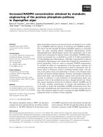
The family pedigree is shown in Fig. 1. The index case
(F1.1) was a 43-year-old female, with clinical records of
acroparesthesias from adolescence, hypohidrosis, and
reiterated episodes of abdominal pain with alternating
phases of diarrhoea with constipation. At 40 years of age
was referring several episodes of pain chest that requiring
emergency care. At 41 years of age she was hospitalized
by a new episode of chest pain and was diagnosed with
anteroseptal myocardial infarction, requiring the implantation of three stents. The evaluation of hearth function by
MRI showed residual fibrous scar by myocardial infarction
in the territory of the anterior and apex segments. LV
slightly dilated with decreased LVEF (48.4 %) and
generalized hypokinesia. Valvular function with MI
moderate to severe and moderate AI. The cardiologic
diagnosis suspicion was valvular and ischemic hearth
variant secondary to Fabry disease. Complete haematological, neurological, and ophthalmological examinations
were performed, and renal function was measured and
determined to be normal. It discard associate factors of
hypercoagulability status. Peripheral nervous conduction
was evaluated by quantitative sensory test (QST) [23],
which revealed abnormalities in the small fiber conduction
compared with normal population. Physical examination
indicated that the patient had absence of hemangiokeratomas, nodes or visceral enlargement, and no cardiovascular
risks such as high plasmatic cholesterol, low c-HDL levels
Fig. 1 Pedigree of the family and complex intronic haplotype (c.-10C > T, c.369 + 990C > A, c.370-81_370-77delCAGCC, c.640-16A > G, c.1000-22C > T)
carriers. Index case is indicated with an arrow
Gervas-Arruga et al. BMC Genetics (2015) 16:109
or hypertension. Genetic analysis revealed a CIH in the
GLA gene: IVSO-10C > T (c.-10C > T) within the promoter region, intron 2 IVS2 + 990C > A (c.369 + 990 C >
A); IVS2-76_80del 5 (c.370-77_81 del CAGCC), intron 4
IVS4-16A > G (c.640-16A > G) and intron 6 IVS6-22C > T
(c.1000-22C > T). The same haplotype was identified in
the patient’s mother (F1.8) and daughter (F1.5), in four of
her brothers (F1.2, F1.3, F1.4, and F1.10) and one of her
two sisters (F1.7), as well as in the daughters (F1.6, F1.9)
of two of the affected brothers. MLPA assays did not
reveal any copy number differences in patients F1.1, F1.4
and F1.10.
At age 52 years, the first brother (F1.3) was referred
for examination to exclude malignant monoclonal gammopathy IgG-kappa. At age 40 years, he underwent
vertebral fixation surgery because of severe back pain.
He reported acroparesthesia, heat intolerance, and hypohidrosis since childhood, as well as shortness of breath
and inability to perform any physical activity. This
patient was hemizygous for the CIH.
The second brother (F1.10), hemizygous for the CIH,
was 49 years old. His main symptoms were related to left
ventricular hypertrophy and valvular aortic stenosis
associated with severe acral pain that demanded continuous intake of analgesic drugs.
The third brother (F1.13) did not accept to be a part
of the study and the fourth brother was deceased.
The fifth brother (F1.4) was referred for hearing loss
and mild acroparesthesia at 41 years of age. Renal
involvement was characterised by microalbuminuria
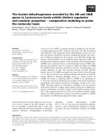
(34 mg/24 h) and glycolipid deposits in renal tubules
and glomeruli (Fig. 2). It is important to note that this
patient did not use chloroquine or amiodarone-like
drugs. His 7-year-old daughter (F1.6) had microalbuminuria (29 mg/24 h), and was heterozygous for the CIH.
The sixth brother (F1.2) was 39 years old, hemizygous
for the CIH, and was referred for acroparesthesias,
Page 5 of 13
bilateral hearing loss, heat intolerance and hypohidrosis,
but no cardiac abnormalities were observed.
One sister (F1.7) was 37 years old and a carrier of the
CIH. She had a previous history of tachyarrhythmias, and
since childhood she had acroparesthesias, hypohidrosis
and heat intolerance. Her mother (F1.8) presented no
clinical manifestations.
α-Gal A activity in plasma, leukocytes and fibroblasts
Enzymatic activity was measured in plasma, lysed leukocytes and fibroblasts from CIH carriers and normal
healthy controls. The individual values from leukocytes
and plasma are presented in Table 1. The mean value of
lysed leukocyte activity in the control group (n = 27) was
(mean ± SD) 58.1 ± 26.6 nmol/mg protein/h. Significantly lower levels were found in the CIH group (n = 8),
46.25 ± 9.66 nmol/mg protein/h (Fig. 3a; p ≤ 0.05).
The mean value of plasma activity in the control
group (n = 33) was (mean ± SD) 20.8 ± 12.5 nmol/mL/h,
whereas the mean value for the CIH group (n = 9) was
19.5 ± 10.3 nmol/mL/h. There was no significant difference between the plasma activity in control and CIH
group (Fig. 3a). Enzymatic activity was also measured in
cultured skin fibroblasts from controls and from patients
F1.1, F1.3, F1.4 and F1.10. The activity measurements in
the patients’ fibroblasts were not significantly different to
controls, although there was a trend for reduction in patient F1.10 and fibroblasts from patient F1.3 had
significantly higher levels of α-Gal A (Fig. 3b).
In vitro model of lysosomal storage disease
In order to accurately determine galactosphingolipid
levels, we first used wild-type fibroblasts to establish a
model of lysosomal storage dysregulation. Fibroblasts
were treated or not with TNFα (16 h) to activate a
proinflammatory response, followed by a medium
change without TNFα, and culturing was then continued
Fig. 2 Cytoplasmic vacuolation observed in a podocyte by light microscopy with a Masson’s trichrome and b Haematoxylin and eosin staining.
c Myelin-like structures in a podocyte, with concentric lamellated ultra-structural appearance. (Electron Microscopy). d Focal areas of podocyte
effacement; amorphous myelin-like structures are visible in glomerular parietal epithelial cells and in endothelial cells (Electron Microscopy).
e Myelin-like structures parallel with zebra-like body appearance (Electron Microscopy)
ID
SEX
Age
Variants
Clinical Manifestations
CIH
galactosphingolipids
galactosphingolipids
α-Gal A
α-Gal A
mRNA
mRNA
Leukocytes
Fibroblasts
Leukocytes
Plasma
GLA
GLA-M
RQ
(nmol/mg protein)
(nmol/mg protein)
(nmol/mg protein/h)
(nmol/mL/h)
RQ
F1.1
f
43
Het
Ischemic heart disease, Gastro- intestinal,
Fine fiber alterations
0.62
0.83
63
23
0.48*
0.85
F1.2
m
39
Hemi
Acroparesthesias, Heat intolerance,
Hypohidrosis, Hearing loss
0.13
N/A
57
19
0.41
5.47
F1.3
m
53
Hemi
Acroparesthesias, Heat intolerance,
Hypohidrosis
0.11
0.80
38
14
0.36*
0.84
F1.4
m
41
Hemi
Hearing loss, Microalbuminuria,
Renal deposits
0.11
1.12
37
15
0.88
2.56
F1.5
f
20
Het
N/A
0.13
N/A
42
37
N/A
N/A
F1.6
f
7
Het
Microalbuminuria
N/A
N/A
N/A
9
N/A
N/A
F1.7
f
37
Het
Acropaesthesias, Microalbumminuria,
Hypohidrosis, Tachyarrhythmias
0.06
N/A
43
15
1.06
1.51
F1.8
f
72
Het
N/A
0.27
N/A
51
35
1.12
0.90
F1.10
m
49
Hemi
Left Ventricular Hypertrophy
1.93
0.93
39
9
0.02**
117.76**
Gervas-Arruga et al. BMC Genetics (2015) 16:109
Table 1 Clinical manifestations; α-Gal A activity in plasma and leukocytes, galactosphignolipid concentrations and GLA mRNA expression
CIH = c.-10C > T [rs2071225], c.369 + 990C > A [rs1023431], c.370-81_370-77delCAGCC [rs5903184], c.640-16A > G [rs2071397], c.1000-22C > T [rs2071228]. The mean ± SD normal concentration of leukocytes
galactosphingolipids is (n = 17) 0.33 ± 0.3 (nmol/mg protein). The mean ± SD normal concentration of fibroblasts galactosphingolipids is (n = 5) 0.63 ± 0.12 (nmol/mg protein). The normal mean ± SD of α-Gal A leukocyte
activity in our assay is (n = 27) 58.1 ± 26.6 (nmol/mg protein/hour) and normal mean ± SD of α-Gal A plasma activity is (n = 33) 20.8 ± 22.03 (nmol/mL/ hour). * = p < 0.05, ** = p < 0.001. N/A = Not applicable
Page 6 of 13
Gervas-Arruga et al. BMC Genetics (2015) 16:109
Page 7 of 13
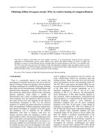
further increased to 150 fold after 24 h (Fig. 4c). Cotreatment with DGJ further increased CD77 staining
and this was dramatically reduced upon exposure to
agalsidase alfa (Fig. 4c). A similar analysis was performed with an antibody to lactosylceramide (CD17).
α-Gal A catalyzes the hydrolysis of Gb3 to lactosylceramide
and α-galactose. Lactosylceramide acumulation was decreased upon exposure of fibroblasts to TNFα and DGJ.
Agalsidase alfa treatment increased lactosylceramide accumulation as demonstrated by increased CD17 fluorescence,
corroborating the CD77 results (Fig. 4c). Collectively, these
results show that galactosphingolipids can be precisely
measured in cell cultures using this assay and immunocytochemistry is a useful technique to assess lipid storage.
Quantification of galactosphingolipids in leukocytes and
human fibroblasts
Fig. 3 α-Gal enzyme activity. a Enzymatic activity in lysed leukocytes
(nmol/mg protein/hour) and plasma (nmol/mL/hour), represented as
mean ± SEM. b Enzymatic activity in lysed fibroblasts (nmol/mg
protein/hour) represented as mean ± SEM, n = 3 *p ≤ 0.05
for a further 8 h. Accumulation of galactosphingolipids
was measured by galactose oxidase assay at 16 h and
24 h has described. TNFα treatment resulted in an increase in galactosphingolipids in wild-type fibroblasts
compared with control samples (without TNFα treatment) (Fig. 4a). Notably, this increase was accentuated by
co-treatment of fibroblasts with TNFα and DGJ, used at a
concentration which inhibits endogenous α-Gal A activity
[19] (Fig. 4a). Moreover, addition of recombinant agalsidase alfa significantly decreased the level of endogenous
galactosphingolipids in TNFα-treated fibroblasts with respect to control cells (Fig. 4a). To corroborate these results, immunofluorescence microscopy was performed to
evaluate the presence of Gb3 (CD77) using an antibody to
CD77 (Fig. 4b). In good agreement with the biochemical
assay, immunofluorescence staining of fibroblasts exposed
to TNFα demonstrated an increase in CD77, which colocalized with the lysosomal membrane marker LAMP1.
Additionally, Gb3 signal intensity was increased upon
exposure of fibroblasts to TNFα and DGJ (Fig. 4b). Superimposition of CD77/LAMP1 images revealed a significant
degree of overlap (Fig. 4b). As anticipated, agalsidase alfa
treatment reduced Gb3 accumulation as demonstrated by
decreased CD77 fluorescence, without affecting LAMP1
staining (Fig. 4b). Quantification of overlapped pixels of
confocal images revealed that TNFα treatment resulted in
a 50 fold increase in CD77 staining in fibroblasts, which
Quantification of galactosphingolipids was performed in
leukocytes and fibroblasts from CIH carriers and controls.
The individual values of leukocyte galactosphingolipids
are presented in Table 1. The mean value for the control
group (n = 17) was (mean ± SD) 0.33 ± 0.30 nmol/mg
protein and in the CIH group (n = 8) 0.42 ± 0.63 nmol/mg
protein. Galactosphingolipids were also measured in
cultured fibroblasts from CIH patients. After 5 days of
quiescence, in all cases galactosphingolipid levels in
patient fibroblasts were greater than in equivalent control
cell lines, which was significant for patients F1.1, F1.4 and
F1.10 (Fig. 5a). Interestingly, in patients F1.3 and F1.4,
leukocyte levels were lower than controls, whereas in
equivalent fibroblasts the levels were higher. The origin of
these differences may reflect distinct GLA expression
states of different cells. Ferreira et al., suggest that the
GLA 5’UTR polymorphysms are a possible modulators of
GLA expression varying among different cell types [24].
Atypical FD variants are often not associated with
increased lyso-Gb3 levels, although biopsies of affected
organs revealed lamellar inclusion bodies characteristic for
FD [25]. Consistent with the increase in galactosphingolipids, immunostaining of fibroblasts from patient
F1.4 and F1.10 revealed elevated levels of Gb3, which
were further increased after exposure to TNFα (Fig. 5b).
Additionally, immunofluorescence revealed that galactolipids were mostly confined to cytosolic and membrane
compartments (Fig. 5b). Quantification of CD77/LAMP1
lysosomal co-localization revealed an increase in
CD77 staining in fibroblasts from patients F1.4 and
F1.10, which was significant for patient F1.10 relative
to control values (Fig. 5c).
cDNA analysis
No sequence changes were discovered after sequencing nested PCR products of the two GLA
Gervas-Arruga et al. BMC Genetics (2015) 16:109
A
Page 8 of 13
B
C
D
Fig. 4 Model of FD substrate accumulation in vitro. a Galactosphingolipid variation rate in wild-type fibroblasts under different conditions.
Galactosphingolipids concentration was measured as described (n = 3) and changes in galactosphingolipids levels are represented. b Representative
images of in vitro FD model. Control fibroblasts were stained with CD77 (Gb3), green and LAMP1, red. CD77 and LAMP1 merged appear
as orange/yellow. Nuclei were stained with DAPI, blue. b.1) Control 24 h; b.2) Control + TNFα 24 h; b.3) Control + TNFα 24 h + DGJ;
b.4) Control + TNFα 24 h + agalsidase alfa. c Quantification of CD77/LAMP1 (orange/yellow) fluorescence rate (n = 3). d Quantification of
CD17/LAMP1 (orange/yellow) fluorescence rate (n = 3)
amplicons in comparison with the reference sequence
(data not shown).
Small RNA cloning
Sequencing of the 3′ region of intron 6 insert in all
CIH carriers revealed the presence of one cDNA fragment formed by a 49-bp portion of intron 6 and exon 7
cloned only in patient F1.4. The genomic coordinates
of this exon 7 extension are: ChrX(GRCh38):
g.101398050-g. 101397803 :-1.
Quantitative PCR of the GLA transcripts
Relative quantification (RQ) of wild-type (wt) GLA and
GLA-M mRNA (3′ region of intron 6 cloned fragment)
from all carriers and controls (n = 8; 50 % females) was
performed by real-time PCR and the GLA/GLA-M expression profiles were compared between control group
and carriers. Reduced wt GLA expression was found in
all hemizygous carriers and in the heterozygous proband
F1.1, and was significant in carriers F1.3, F1.10 and F1.1
(Table 1). The relative expression of GLA-M was
increased in carriers F1.2, F1.4 and F1.7, and significantly in patient F1.10 compared with controls (Table 1).
The heterozygous group for CIH presented higher RQ
values for GLA wt expression in comparison with the
hemizygous group (0.89 vs 0.41).
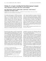
Electrophoretic mobility shift assay (EMSA)
To investigate whether the C > T change in the GLA
gene promoter variant c.-10C > T impacted the binding
capacity of possible transcription factors, EMSA was
performed with specific primers for each variant. The
results demonstrated marked differences in the affinity
of nuclear proteins between the two alleles, with allele C
having a greater ability to bind nuclear proteins (Fig. 6a).
To confirm this difference, we performed a competitive
Gervas-Arruga et al. BMC Genetics (2015) 16:109
A
Page 9 of 13
B
C
Fig. 5 Accumulation of galactosphingolipids in fibroblast lysates. a Biochemical quantification after 5 days of culture represented as mean ± SEM.
(n = 3) *p ≤ 0.05 **p ≤ 0.001. b Quantification of Gb3 (CD77) confocal images represented as mean ± SD (n = 3) b.1) patient F1.10.*p ≤ 0.05; (+)TNFα vs.
(−)TNFα and #p ≤ 0.05; Control vs. F1.10., b.2) Patient F1.4. c Immunocytochemistry of CD77 expression in fibroblasts from c.1) control, c.2)
patient F1.4 and c.3) patient F1.10 after 16 h TNFα activation. Nuclei were stained with DAPI, blue
binding EMSA using increasing amounts of an unlabelled
oligonucleotide corresponding to the T allele. The
results indicated that the T allele was more easily
displaced from the protein-DNA complex (0.003 vs
0.001) (Fig. 6b). Therefore, the C > T substitution resulted in decreased protein binding capacity of the fragment. In an attempt to identify the transcription factors
present in the complex, we scanned sequences surrounding the SNP c.-10C > T with MatInspector (Genomatix) [26]. The results of this analysis indicated that
the mutation could affect the binding sites for small
nuclear RNA (snRNA)-activating protein complex
(SNAP-C), doublesex/mab-3 related (DMRT), and X
box-binding factors.
Splice-site score (SSS)
We analysed the variants of the CIH using SSPNN software. Changes in GLA splicing with CIH were observed
only in c.370-81_370-77delCAGCC and c.640-16A > G
variants. The c.370-81_370-77delCAGCC variant resulted in the disappearance of a possible acceptor site
in IVS2-78, with a SSS of 0.80. The SSS for a normal
acceptor site in intron 4 (IVS4-1) was raised from
0.58 to 0.62 with the c.640-16A > G variant. Although
the SSS were minimal, the results indicated that the
co-segregation of CIH variants may cause an abnormal splice pattern.
Discussion
Detection of mutations in the GLA gene is essential to
support clinical diagnosis of FD. Mutations in intronic regions can alter the GLA gene expression pattern in a manner related to different disease phenotypes and clinical
manifestations [4, 5]. Intronic GLA variants often remain
unidentified because these regions are not routinely evaluated by gene sequencing; consequently, the prevalence of
FD may be underestimated [27, 28].
Gervas-Arruga et al. BMC Genetics (2015) 16:109
Page 10 of 13
Fig. 6 EMSA analysis. a EMSA carried out with probes containing the C or T allele for the IVSO-10C > T variant in the GLA gene promoter. b The
inverse of band densities from the EMSA plotted against the excess of cold allele T oligonucleotides, showing that allele T (slope = 0.003) was more
easily displaced from the complex than allele C (slope = 0.001)
In the present study, we sequenced the entire GLA gene
from genomic DNA of the family members and we used
MLPA in an attempt to find alterations that could explain
the FD-like characteristics observed. We identified a
complex haplotype consisting of five intronic variants
(c.-10C > T, c.369 + 990C > A, c.370-81_370-77delCAGCC,
c.640-16A > G, c.1000-22C > T). In one study, 12 % of 740
subjects with clinically suspected FD showed polymorphisms in the GLA promoter region; of these, 99 % had
simultaneous polymorphisms throughout the gene, and
CIH formed by four of five variants observed in our study
occurred in 9 % of these cases [11]. In a second study, Ferri
and colleagues [13] identified five GLA haplotypes in
non-coding regions in 67 female probands with FD
manifestations. The most frequent of these was the CIH
formed by four variants (13.4 %). Previous reports have
found c.-10C > T, c.370-81_370-77delCAGCC, c.64016A > G and c.1000-22C > T variants to be associated
with different clinical manifestations including mild
renal, neurological [10] and cardiac disorders [12]. It is
important to know that depending on the sequencing
design, these haplotypes might be the same type. We
found FD-like symptoms (renal, cardiac and neurological
involvement) associated with the family in our study
(Table 1). In accordance with our results Apeland et al.,
described two unrelated families, one of them carrier of
c.640-16A > G and c.1000-22C > T intronic variants,
presenting a cardiomyopathy mimicking FD with normal enzymatic activity values and renal and cardiac
deposits without accumulation of glycolipids in urine
or plasma. In two patients, a 100-fold increase in Gb3
was observed in cardiac biopsies. Exon sequencing
failed to detect heterozygosity in potential candidate
genes [29].
We examined the c.-10C > T variant located in the
GLA gene promoter region, which may be codominantly associated with a relatively decreased GLA
expression at the level of transcription and/or translation
[30]. By EMSA, we found that the T allele reduced the
affinity of the nuclear protein binding site. A computer
analysis using MatInspector showed that this region is a
possible binding site for three families of transcription
factors: SNAP-C, X box-binding factors (XBBF) and
DMRT. SNAP-C binds to Oct-1 and TATA binding
proteins (TBP), which are activators of snRNA and RNA
polymerases, respectively [31]. X box-Protein 1 (XBP1)
becomes initiated during the endoplasmic reticulum
(ER) stress response [32]. In humans, the DMRT gene
family encodes transcription factors that are related to
the Drosophila double sex proteins [33]. Unfortunately,
no tested antibodies are currently available to perform a
supershift assay.
The c.-10 C > T variant is situated in a CpG island region
( DNA methylation
Gervas-Arruga et al. BMC Genetics (2015) 16:109
is a well-recognized epigenetic modifier in the control of
gene expression. This reversible DNA modification takes
place almost exclusively at cytosine residues that are associated with guanosine in CpG doublets, and mediates control of transcription through chromatin remodelling.
This modification is widely implicated in various
biological processes including X-inactivation, the regulation of tissue- and development-specific gene expression, foreign DNA inactivation and genomic imprinting
[34]. FD symptoms exhibited by females carrying the T
allele could partially depend on the methylation state of
the C allele. Indeed, Bono and co-workers [11] reported a
relationship between FD symptoms and polymorphisms in
the promoter region. Future studies on the methylation
states of the promoter region may provide more clues on
these epigenetic effects in relation to phenotype.
We found low levels of wild-type transcript in some
patients in agreement with previous reports [28, 35].
GLA-M transcript levels (3′ region of the intron 6
cloned fragment) were also altered with respect to their
controls in most cases. The index case of this family
(F1.1), a female heterozygous for CIH, presented significantly lower levels of wild-type transcript, whereas GLAM expression was slightly reduced but not significantly
different to control. Although she presented a cardiac
phenotype, her leukocyte and plasma enzyme activities
were not decreased. Higher residual activity is often found
in atypical male patients who do not show the classic
phenotype and have later onset of symptoms [36, 37],
including cardiac [35] and renal variants [38]. It is possible
that GLA expression in other tissues may be different. The
ratio of the alternatively spliced transcript produced by
another intronic variant, IVS4 + 919G > A mutation, to
total α-Gal A mRNA, is higher in human muscle and lung
tissues [27]. In the case of this studied family, the CIH
only produced an altered GLA expression profile, presumably resulting in a late-onset FD-like phenotype. The
differences observed in the qPCR assay may coincide with
the SSS data variations for c.370-81_370-77delCAGCC
and c.640-16A > G variants, the EMSA assay results for c.10 C > T and also with the cDNA cloned fragment formed
by a 49-bp segment of 3′ intron 6 and exon 7 in patient
F1.4. However, PCR and sequencing did not reveal any
products of splicing variants due to c.370-81_370-77delCAGCC or c.640-16A > G. It is possible that these transcripts are degraded by nonsense-mediated mRNA decay.
In accord with the EMSA results, the expression levels of
heterozygous CIH patients were approximately 50 %
higher than in hemizygous carriers. Enzymatic activity was
measured in plasma, lysed leukocytes and fibroblasts from
CIH carriers and normal healthy controls. The mean value
of lysed leukocyte activity in the CIH group was significantly lower (~20 %) but there was no significant
difference between the plasma and fibroblast activity
Page 11 of 13
in control and CIH group. The expression is reduced
in leukocytes reducing the enzyme activity but no the
enzyme activity in other tissues like plasma supporting previous studies [30]. This effect may contribute
to the glycolipid alteration and therefore may develop
the clinical findings.
The levels of storage products in urine and plasma are
elevated in most, but not all, FD patients. The demonstration of increased storage product levels is very useful
in making a diagnosis in many cases and also for treatment monitoring. The predominant storage product in
FD is Gb3, but other storage products such as Ga2 or
lyso- Gb3 may also accumulate. Consequently, significant
differences in the Gb3 and Ga2 isoform profiles in urinary
sediment were found amongst young, adult and atypical
hemizygotes and heterozygotes using a combination of
MALDI-TOF MS and tandem MS [39]. Additionally,
increased sphingolipid storage in skin fibroblasts from
patients has been described previously [40]. Therefore, we
used an enzymatic fluorometric technique to quantify all
galactosphingolipids in samples obtained from controls
and CIH carriers in lysed leukocytes and skin fibroblasts.
Galactose is a component of the headgroup of many
glycolipids. Galactose oxidase specifically oxidizes the C-6
hydroxymethyl group of free galactose as well as all galactosyl derivatives, such as Gb3, lyso- Gb3 and Ga2, carrying
a galactose residue in the terminal position. Bile acids, for
example sodium taurocholate, do not alter the kinetics of
galactose oxidase [41]. The enzymatic method to detect
urinary Gb3, showed a good recovery and comparability
with a previously validated HPLC method [42]. Importantly, we validated this assay using a synthetic model of
lysosomal storage in fibroblasts activated with TNFα and
corroborated these findings with confocal microscopy
quantification of CD77 (Gb3)/CD17(Lactosylceramide)/
LAMP1. Confocal microscopy revealed that galactolipids
were mostly confined to cytosolic and membrane compartments (Fig. 5b) in concordance with several studies
that demonstrated that Gb3 in FD is not only present in
lysosomes, but rather widely distributed in other cellular
structures [43]. In vitro studies in skin fibroblasts showed
that CIH carriers accumulated galactosphingolipids
significantly after 5 days culture, in the range between 30
and 80 % in comparison with control samples. Fibroblasts
from a male patient (F1.4) with a renal phenotype
and glycolipid deposits demonstrated by renal biopsy,
accumulated approximately 50 % more substrate compared with the F1.1 patient, a heterozygous female
with a cardiac phenotype. In concordance with our
results is important to remark that Namdar et al.
demonstrated that vasculopathy in FD is directly
caused by intracellular Gb3 accumulation while deficiency of GLA alone does not cause any deregulation
of key vasoactive mediators [44].
Gervas-Arruga et al. BMC Genetics (2015) 16:109
Most of the carriers had high in vitro residual enzyme
activity in plasma, leukocytes and cultured fibroblasts;
however, CIH carriers had significant galactosphingolipid
accumulation in fibroblasts in comparison with controls.
Presumably, because the enzyme structure is not altered,
only the GLA structural regulatory mechanism was affected
by inheritance of the CIH, leading to GLA activationdependent accumulation of substrates, influenced perhaps
by environmental factors such as the proinflammatory state
of the patient.
Conclusions
CIH carriers showed a wide variation in residual enzymatic activities in leukocytes, plasma, and fibroblasts, but
generally activity was normal. In contrast, galactosphingolipid accumulation was in the main significantly greater in
fibroblasts compared with controls. Position −10 in the
GLA promoter is a putative nuclear protein binding site
situated in the CpG island region, acting as a gene regulatory zone. The inheritance of the co-segregated CIH
variants alters the GLA expression pattern, producing a
tissue glycolipid storage disorder.
The genetic analysis of the entire GLA gene sequence
and MLPA, the study of GLA expression and glycolipid
quantification in relation to FD clinical manifestations can
be extremely helpful as tools for FD-related diagnosis.
Further studies are needed to elucidate how the
inheritance of complex intronic haplotypes are implicated in the GLA regulatory mechanisms and therefore,
the glycolipid metabolism alteration.
Additional file
Additional file 1: Table S- Sequences of primers and probes.
(DOCX 21 kb)
Abbreviations
GLA: Alfa galactosidase gene; FD: Fabry disease; Gb3: Globotriaosylcermide;
Lyso-Gb3: Globotriaosylsphingosine; Ga2: Galabiosylceramide; α-Gal A:
α-galactosidase A; CIH: Complex intronic haplotype; TNFα: Tumor necrosis
factor alpha; LAMP1: Lysosomal-associated membrane protein 1; DGJ:
1-deoxygalactonojirimycin hydrochloride; MU: Methylumbelliferyl;
PCR: Polymerase chain reaction; RT-PCR: Reverse transcription polymerase
chain reaction; MLPA: Multiplex ligand probe amplification; FBS: Fetal bovine
serum; DMEM: Dulbecco’s modified Eagle’s medium; qPCR: Quantitative
real-time PCR; EMSA: Electrophoretic mobility shift assay; SSS: Splice-site
score; MRI: Magnetic resonance image; LV: Left ventricle; LVEF: Left
ventricular ejection fraction; MI: Mitral insufficiency; AI: Aortic insufficiency;
QST: Quantitative sensory test; RQ: Relative quantification; Wt: Wild type;
SNP: Single nucleotide polymorphism; snRNA: Small nuclear RNA; SNAP-C: Small
nuclear RNA activating protein complex; XBBF: X box-binding factors;
DMRT: Doublesex/mab-3 related; TBP: TATA binding proteins.
Competing interests
The authors declare that they have no competing interests.
Authors’ contributions
JGA, JCRR and MP conceived and designed the experiments. JGA, JJC, PI and
JPL performed the experiments. JGA carried out statistical analysis. LP carried
out the renal biopsy analyzes. JLC, JCR and PG carried out the clinical
Page 12 of 13
evaluation. JLC, JCR, JCRR, MP and PG contributed reagents/materials/analysis
tools. JGA drafted the manuscript. JGA, JJC, JCRR, MP and PG carried out the
interpretation of data. JGA, MP and PG participated in the design and
coordination of the study. All authors read and approved the final
manuscript.
Acknowledgements
The authors gratefully thank Cesar Vallejo for technical support; Dr. Erika
Fernandez-Vizarra for critical revision of the manuscript and Dr. Gracia
Mendoza for immunofluorescence support. This study was supported by
grant FIS (PI 09/02556) and by the Centro de Investigación Biomédica en
Red de Enfermedades Raras (CIBERER), an initiative of the ISCIII.
Author details
1
Centro de Investigación Biomédica en Red de Enfermedades Raras
(CIBERER), Zaragoza, Spain. 2Translational Research Unit, Instituto de
Investigación Sanitaria Aragón (IIS Aragón), Miguel Servet University Hospital,
Zaragoza, Spain. 3Instituto de Investigación Sanitaria Aragón (IIS Aragón),
Zaragoza, Spain. 4Biochemistry and Molecular and Cellular Biology
Department, Universidad de Zaragoza, Zaragoza, Spain. 5Molecular Biology
Department, Cantabria University and IFIMAV, Santander, Spain. 6Anatomic
Pathology Department, Miguel Servet University Hospital, Zaragoza, Spain.
7
Neurology Department, Miguel Servet University Hospital, Zaragoza, Spain.
Received: 9 April 2015 Accepted: 25 August 2015
References
1. Desnick R, Ioannou Y, Eng C. Fabry disease: a-galactosidase A deficiency. In:
Scriver C, editor. Metabolic and molecular bases of inherited disease. New
York: McGraw-Hill; 2001. p. 3733–74.
2. Mehta A, Ricci R, Widmer U, Dehout F, Garcia de Lorenzo A, Kampmann C,
et al. Fabry disease defined: baseline clinical manifestations of 366 patients
in the Fabry Outcome Survey. Eur J Clin Invest. 2004;34(3):236-42.
3. Lyon MF. Gene action in the X-chromosome of the mouse (Mus musculus L.).
Nature. 1961;190:372–3.
4. Spada M, Pagliardini S, Yasuda M, Tukel T, Thiagarajan G, Sakuraba H, et al.
High incidence of later-onset fabry disease revealed by newborn screening.
Am J Hum Genet. 2006;79(1):31-40.
5. Hwu WL, Chien YH, Lee NC, Chiang SC, Dobrovolny R, Huang AC, et al.
Newborn screening for Fabry disease in Taiwan reveals a high incidence of the
later-onset GLA mutation c.936+919G>A (IVS4+919G>A). Hum Mutat.
2009;30(10):1397-405.
6. Desnick RJ, Brady R, Barranger J, Collins AJ, Germain DP, Goldman M, et al.
Fabry disease, an under-recognized multisystemic disorder: expert
recommendations for diagnosis, management, and enzyme replacement
therapy. Ann Intern Med. 2003;138(4):338-46.
7. Eng CM, Germain DP, Banikazemi M, Warnock DG, Wanner C, Hopkin RJ,
et al. Fabry disease: guidelines for the evaluation and management of
multi-organ system involvement. Genet Med. 2006;8(9):539-48.
8. Stenson PD, Ball EV, Mort M, Phillips AD, Shiel JA, Thomas NS, et al. Human
Gene Mutation Database (HGMD): 2003 update. Hum Mutat. 2003;21(6):577-81.
9. Lai LW, Whitehair O, Wu MJ, O'Meara M, Lien YH. Analysis of splice-site
mutations of the alpha-galactosidase A gene in Fabry disease. Clin Genet.
2003;63(6):476-82.
10. Tanislav C, Kaps M, Rolfs A, Bottcher T, Lackner K, Paschke E, et al.
Frequency of Fabry disease in patients with small-fibre neuropathy of
unknown aetiology: a pilot study. Eur J Neurol. 2011;18(4):631-6.
11. Bono C, Nuzzo D, Albeggiani G, Zizzo C, Francofonte D, Iemolo F, et al.
Genetic screening of Fabry patients with EcoTILLING and HRM technology.
BMC Res Notes. 2011;4:323.
12. Pisani A, Imbriaco M, Zizzo C, Albeggiani G, Colomba P, Alessandro R, et al. A
classical phenotype of Anderson-Fabry disease in a female patient with intronic
mutations of the GLA gene: a case report. BMC Cardiovasc Disord. 2012;12:39.
13. Ferri L, Guido C, la Marca G, Malvagia S, Cavicchi C, Fiumara A, et al. Fabry
disease: polymorphic haplotypes and a novel missense mutation in the GLA
gene. Clin Genet. 2012;81(3):224-33.
14. Tuttolomondo A, Duro G, Pecoraro R, Simonetta I, Miceli S, Colomba P, et al.
A family with various symptomatology suggestive of Anderson-Fabry
disease and a genetic polymorphism of alpha galactosidase A gene.
Clin Biochem. 2015;48(1-2):55-62.
Gervas-Arruga et al. BMC Genetics (2015) 16:109
15. Oliveira JP, Ferreira S, Barcelo J, Gaspar P, Carvalho F, Sa Miranda MC, et al.
Effect of single-nucleotide polymorphisms of the 5' untranslated region of
the human alpha-galactosidase gene on enzyme activity, and their
frequencies in Portuguese caucasians. J Inherit Metab Dis. 2008;31
Suppl 2:S247-53.
16. Davies JP, Winchester BG, Malcolm S. Sequence variations in the first exon
of alpha-galactosidase A. J Med Genet. 1993;30(8):658–63.
17. Bekri S, Enica A, Ghafari T, Plaza G, Champenois I, Choukroun G, et al. Fabry
disease in patients with end-stage renal failure: the potential benefits of
screening. Nephron Clin Pract. 2005;101(1):c33-8.
18. Mayes JS, Scheerer JB, Sifers RN, Donaldson ML. Differential assay for
lysosomal alpha-galactosidases in human tissues and its application to
Fabry's disease. Clin Chim Acta. 1981;112(2):247-51.
19. Hsu J, Serrano D, Bhowmick T, Kumar K, Shen Y, Kuo YC, et al.
Enhanced endothelial delivery and biochemical effects of alphagalactosidase by ICAM-1-targeted nanocarriers for Fabry disease. J
Control Release. 149. Netherlands: 2010 Elsevier B.V; 2011. p. 323-31.
20. Keslová-Veselíková J, Hůlková H, Dobrovolný R, Asfaw B, Poupetová H, Berná
L, et al. Replacement of alpha-galactosidase A in Fabry disease: effect on
fibroblast cultures compared with biopsied tissues of treated patients.
Virchows Arch. 2008;452(6):651-65.
21. Pfaffl M. Relative quantification. In: Dorak T, editor. Real-time PCR. La Jolla,
CA: International University Line; 2006. p. 63–82.
22. Riancho JA, Vazquez L, Garcia-Perez MA, Sainz J, Olmos JM, Hernandez JL,
et al. Association of ACACB polymorphisms with obesity and diabetes.
Mol Genet Metab. 2011;104(4):670-6.
23. Rolke R, Baron R, Maier C, Tolle TR, Treede RD, Beyer A, et al. Quantitative
sensory testing in the German Research Network on Neuropathic Pain (DFNS):
standardized protocol and reference values. Pain. 2006;123(3):231-43.
24. Ferreira, S., C. Reguenga, and J.P. Oliveira, The Modulatory Effects of the
Polymorphisms in GLA 5'-Untranslated Region Upon Gene Expression Are
Cell-Type Specific. JIMD Rep, 2015.
25. Mitobe S, Togawa T, Tsukimura T, Kodama T, Tanaka T, Doi K, et al. Mutant
alpha-galactosidase A with M296I does not cause elevation of the plasma
globotriaosylsphingosine level. Mol Genet Metab. 2012;107(3):623-6.
26. Quandt K, Frech K, Karas H, Wingender E, Werner T. MatInd and MatInspector:
new fast and versatile tools for detection of consensus matches in nucleotide
sequence data. Nucleic Acids Res. 1995;23(23):4878-84.
27. Ishii S, Nakao S, Minamikawa-Tachino R, Desnick RJ, Fan JQ. Alternative
splicing in the alpha-galactosidase A gene: increased exon inclusion results
in the Fabry cardiac phenotype. Am J Hum Genet. 2002;70(4):994-1002.
28. Filoni C, Caciotti A, Carraresi L, Donati MA, Mignani R, Parini R, et al.
Unbalanced GLA mRNAs ratio quantified by real-time PCR in Fabry patients'
fibroblasts results in Fabry disease. Eur J Hum Genet. 2008;16(11):1311-7.
29. Apelland T, Gude E, Strøm EH, Gullestad L, Eiklid KL, Månsson JE, et al.
Familial globotriaosylceramide-associated cardiomyopathy mimicking Fabry
disease. Heart. 2014;100(22):1793-8.
30. Oliveira JP, Ferreira S, Reguenga C, Carvalho F, Mansson JE. The g.1170C>T
polymorphism of the 5' untranslated region of the human alphagalactosidase gene is associated with decreased enzyme expression–
evidence from a family study. J Inherit Metab Dis. 2008;31 Suppl 2:S405-13.
31. Sadowski CL, Henry RW, Kobayashi R, Hernandez N. The SNAP45 subunit of
the small nuclear RNA (snRNA) activating protein complex is required for
RNA polymerase II and III snRNA gene transcription and interacts with the
TATA box binding protein. Proc Natl Acad Sci U S A. 1996;93(9):4289-93.
32. Iwakoshi NN, Lee AH, Glimcher LH. The X-box binding protein-1
transcription factor is required for plasma cell differentiation and the
unfolded protein response. Immunol Rev. 2003;194:29–38.
33. Hong CS, Park BY, Saint-Jeannet JP. The function of Dmrt genes in
vertebrate development: it is not just about sex. Dev Biol. 2007;310(1):1–9.
34. Robertson KD, Wolffe AP. DNA methylation in health and disease. Nat Rev
Genet. 2000;1(1):11–9.
35. Nakao S, Takenaka T, Maeda M, Kodama C, Tanaka A, Tahara M, et al. An
atypical variant of Fabry's disease in men with left ventricular hypertrophy.
N Engl J Med. 1995;333(5):288-93.
36. Ishii S, Chang HH, Kawasaki K, Yasuda K, Wu HL, Garman SC, et al. Mutant
alpha-galactosidase A enzymes identified in Fabry disease patients with
residual enzyme activity: biochemical characterization and restoration of
normal intracellular processing by 1-deoxygalactonojirimycin. Biochem J.
406. England2007. p. 285-95.
Page 13 of 13
37. Winchester B, Young E. Biochemical and genetic diagnosis of Fabry disease.
In: Mehta A, Beck M, Sunder-Plassmann G, editors. Fabry disease:
perspectives from 5 years ofFOS. Oxford: Pharmagenesis; 2006. p. 169–81.
38. Nakao S, Kodama C, Takenaka T, Tanaka A, Yasumoto Y, Yoshida A, et al.
Fabry disease: detection of undiagnosed hemodialysis patients and
identification of a "renal variant" phenotype. Kidney Int. 64. United
States2003. p. 801-7.
39. Touboul D, Roy S, Germain DP, Baillet A, Brion F, Prognon P, et al. Fast
fingerprinting by MALDI-TOF mass spectrometry of urinary sediment
glycosphingolipids in Fabry disease. Anal Bioanal Chem. 2005;382(5):1209-16.
40. Kanekura T, Fukushige T, Kanda A, Tsuyama S, Murata F, Sakuraba H, et al.
Immunoelectron-microscopic detection of globotriaosylceramide
accumulated in the skin of patients with Fabry disease. Br J Dermatol.
2005;153(3):544-8.
41. Fortelius M, Mattjus P. Galactose oxidase action on galactose containing
glycolipids–a fluorescence method. Chem Phys Lipids. 2006;142(1–2):103–10.
42. Rozenfeld PA, De Francesco NP, Borrajo GJ, Ceci R, Fossati CA. An easy
and sensitive method for determination of globotriaosylceramide (Gb3)
from urinary sediment: utility for Fabry disease diagnosis and treatment
monitoring. Clin Chim Acta. 403. Netherlands2009. p. 194-7.
43. Askari H, Kaneski CR, Semino-Mora C, Desai P, Ang A, Kleiner DE, et al.
Cellular and tissue localization of globotriaosylceramide in Fabry disease.
Virchows Arch. 2007;451(4):823-34.
44. Namdar M, Gebhard C, Studiger R, Shi Y, Mocharla P, Schmied C, et al.
Globotriaosylsphingosine accumulation and not alpha-galactosidase-A
deficiency causes endothelial dysfunction in Fabry disease. PLoS One. 7.
United States2012. p. e36373.
Submit your next manuscript to BioMed Central
and take full advantage of:
• Convenient online submission
• Thorough peer review
• No space constraints or color figure charges
• Immediate publication on acceptance
• Inclusion in PubMed, CAS, Scopus and Google Scholar
• Research which is freely available for redistribution
Submit your manuscript at
www.biomedcentral.com/submit