Genetic characterization of african swine fever virus (asfv) isolated in some northern provinces of vietnam in 2020
Bạn đang xem bản rút gọn của tài liệu. Xem và tải ngay bản đầy đủ của tài liệu tại đây (1.7 MB, 60 trang )
VIETNAM NATIONAL UNIVERSITY OF AGRICULTURE
FACULTY OF BIOTECHNOLOGY
-------------***-------------
UNDERGRADUATE THESIS
TITLE:
GENETIC CHARACTERIZATION OF AFRICAN
SWINE FEVER VIRUS (ASFV) ISOLATED IN SOME
NORTHERN PROVINCES OF VIETNAM IN 2020
HANOI– 2021
VIETNAM NATIONAL UNIVERSITY OF AGRICULTURE
FACULTY OF BIOTECHNOLOGY
-------------***-------------
UNDERGRADUATE THESIS
TITLE:
GENETIC CHARACTERIZATION OF AFRICAN
SWINE FEVER VIRUS (ASFV) ISOLATED IN SOME
NORTHERN PROVINCES OF VIETNAM IN 2020
Student
:
LE QUANG DAI
Class
:
K61CNSHE
Major
:
Biotechnology
Supervisors
:
Assoc. Prof. LE VAN PHAN
Dr. DINH TRUONG SON
HANOI – 2021
COMMITMENT
I hereby declare this is my research. The data and results were mentioned in this
thesis is true and has not been used and published in the thesis, dissertations and
scientific works ever before.
I hereby declare that the cited information in this thesis has been made of the
source, ensuring cited as prescribed.
I bear full responsibility for these reassurances.
Hanoi, January 15th, 2021
Student
Le Quang Dai
i
ACKNOWLEDGEMENT
First and foremost, I have to thank my research supervisors, Assoc. Prof Le Van
Phan and Dr. Dinh Truong Son. Without their assistance and dedicated involvement in
every step throughout the process, this thesis would have never been accomplished. I
would like to thank you very much for your support and understanding over these past
four years.
I would like to honestly thanks to the teachers of the Faculty of Biotechnology
for conveyed and taught me the important knowledge which has been the basis for
future research and further career at Vietnam National University of Agriculture
(VNUA). In addition, I send sincerely thanking the teachers and dear friends.
In July 2020, I went to Virology Laboratory of Key Laboratory of Veterinary
Biotechnology for six months to study with MSc. Nguyen Van Tam, MSc. Nguyen Thi
Thu Huyen, DMc. Trinh Thi Bich Ngoc and DMc. Vu Xuan Dang. My time at
Virology Laboratory has been highly productive and working with Mr. Dang was an
extraordinary experience. Also, Ms. Huyen, Ms. Ngoc and Mr. Tam are really helpful
and whole-hearted to lead me in the laboratory and do my experiments.
Most importantly, none of this could have happened without my family. My
parent and my brother - it would be an understatement to say that, as a family, we have
experienced some ups and downs in the past four years. This dissertation stands as a
testament to your unconditional love and encouragement.
Ha Noi, 29 January, 2021
Student
Le Quang Dai
ii
TABLE OF CONTENTS
COMMITMENT .............................................................................................................. i
ACKNOWLEDGEMENT ............................................................................................... ii
TABLE OF CONTENTS ............................................................................................... iii
LIST OF TABLES .......................................................................................................... v
LIST OF ABBREVIATIONS ....................................................................................... vii
ABSTRACT ................................................................................................................... ix
TÓM TẮT........................................................................................................................ x
PART 1. INTRODUCTION ............................................................................................ 1
1.1.
Introduction .......................................................................................................... 1
1.2.
Purposes and requirements ................................................................................... 3
1.2.1. Purpose ................................................................................................................. 3
1.2.2. Requirements ........................................................................................................ 3
PART 2. LITERATURE REVIEW ................................................................................. 4
2.1.
Overview of African swine fever virus ................................................................ 4
2.1.1. Introduction .......................................................................................................... 4
2.1.2. Geographical Distribution and Virus Isolates ...................................................... 4
2.1.3. Origin and transmission of the African Swine Fever Virus ................................. 6
2.1.4. Viral entry mechanism ......................................................................................... 7
2.2.
Roles of B646L/p72 and central variable region (CVR) of B602L genes in
viral entry mechanism .......................................................................................... 8
2.3.
Review of molecular techniques to assess genetic diversity .............................. 10
PART 3. MATERIAL AND METHOD ....................................................................... 12
3.1.
Time and place of study ..................................................................................... 12
3.2.
Research subject ................................................................................................. 12
3.3.
Material............................................................................................................... 12
3.3.1. Chemicals used in research ................................................................................ 12
3.3.2. Equipment........................................................................................................... 12
3.4.
Methods .............................................................................................................. 12
3.4.1. Sample collection ............................................................................................... 12
iii
3.4.2. Sample preparation ............................................................................................. 15
3.4.3. Viral DNA extraction ......................................................................................... 15
3.4.4. Amplification of B646L C-terminus and central variable region (CVR) of
B602L ................................................................................................................. 17
3.5.
Agarose gel electrophoresis................................................................................ 19
3.6.
B646L C-terminus and central variable region (CVR) of B602L sequencing ... 20
3.7.
Processing raw sequencing data ......................................................................... 20
3.8.
Genetic characterization ..................................................................................... 20
3.8.1. DNA sequence and phylogenetic analyses based on ASFVs‘ B646L Cterminus homologous sequences ........................................................................ 20
3.8.2. Deducing tandem repeat sequences (TRSs) from central variable regions
(CVRs) of B602L ............................................................................................... 22
PART 4. RESULTS AND DISCUSSION .................................................................... 23
4.1.
Sample analysis .................................................................................................. 23
4.2.
Detection of the ASFV genome ......................................................................... 24
4.3.
Nucleotide sequence of p72 (B646L) gene ........................................................ 25
4.4.
Phylogenic tree analysis of p72 (B646L) gene .................................................. 27
4.5.
Investigation on an intra-genotypic scale by TRSs located in the CVR
within the B602L gene of ASFV sampled from Northern Vietnam
provinces and beyond Vietnam‘s boundary ....................................................... 29
4.6.
Discussion........................................................................................................... 33
PART 5. CONCLUSION AND SUGGESTION .......................................................... 38
5.1.
Conclusion .......................................................................................................... 38
5.2.
Suggestion .......................................................................................................... 38
REFERENCES .............................................................................................................. 39
APPENDIX ................................................................................................................... 46
iv
LIST OF TABLES
Table 2.1. ASF virus isolates ........................................................................................... 6
Table 3.1. Sample used in this study .............................................................................14
Table 3.2. Kit contents ...................................................................................................16
Table 3.3. PCR sample preparation ...............................................................................18
Table 3.4. Primers used in this study.............................................................................18
Table 3.5. PCR cycling conditions for C-terminal region of P72 and CVR region of
B602L amplification with modification of MyTaq Mix™ (Bioline Reagents Ltd.,
United Kingdom) manual instruction ............................................................................19
Table 4.1. Information about collected samples............................................................ 23
Table 4.2. Information about representative query sequence ........................................26
Table 4.3. Reference strains included on the phylogenetic tree characterizing some p72
genotypes .......................................................................................................................27
Table 4.4. List of alphabetic characters encoding each set of tetramer .......................29
Table 4.5. Tetrameric tandem repeat sequences (TRSs) of central variable region
(CVR) of B602L gene obtained from Vietnam ASFV variants and retrieved from
public sources of the same Vietnam ASFV strains‘ or diverse p72 genotypes.............31
v
LIST OF FIGURES
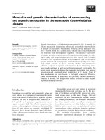
Figure 2.1. Three main stage of PCR reaction and their cycles ....................................11
Figure 3.1. Map showing sites of sample collection in Northern Vietnam ...................13
Figure 3.2. Kit box.........................................................................................................16
Figure 4.1. Gel agarose electrophoresis separating the PCR products of 478bp-size
B646L gene in different sample types and origins. M: marker .....................................24
Figure 4.2. Gel agarose electrophoresis separating the PCR products of 665bp-size
B602L gene in different sample types and origins. M: marker .....................................25
Figure 4.3. p72 (B646L) gene sequences results of five northern provinces. Conserved
regions between comparable sequences were identified as dots while variations were
represented by nucleotide letters. Nucleic acid bases were colour-coded: A (green), T
(red), G (black) and C (blue). The graphical view was produced by BioEdit version
7.2.5 ( ..............................................26
Figure 4.4. Phylogenetic tree of p72 (partial gene nucleotide sequences) of ASF
isolates between Vietnamese strains (black circle symbol) and Korea strain (red square
symbol) and other countries .......................................................................................... 28
vi
LIST OF ABBREVIATIONS
Abbreviations
Definitions
A
Adenine
ASF
African Swine Fever
ASFV
African Swine Fever Virus
µl
Microlitre
BLAST
Basic Local Alignment Search Tool
bp
Base pairs
C
Cytosine
CSF
Classical Swine Fever
CSFV
Classical Swine Fever Virus
CVR
Central variable region
d.s
Direct submission
ds DNA
Double- stranded DNA
DTLCP
Dịch tả lợn châu Phi
E
Expectation
EDTA
Ethylenediaminetetraacetic acid
ER
Endoplasmic reticulum
FAO
Food and Agriculture Organization
FMDV
Foot- and- Mouth Disease Virus
G
Guanine
IUPAC
International Union of Pure and Applied Chemistry
JC
Jukes Cantor
MEGA
Molecular Evolutionary Genetics Analysis
n.g
Not given
NCBI
National Center for Biotechnology Information
NIVR
National Institute of Veterinary Research
NJ
Neighbor Joining
OIE
World Organization for Animal Health
PAM
Pulmonary alveolar macrophage
vii
PBS
Phosphate buffered saline
PCR
Polymerase chain reaction
PEDV
Porcine Epidemic Diarrhea Virus
PRRS
Porcine Reproductive and Respiratory Syndrome
PRRSV
Porcine Reproductive and Respiratory Syndrome Virus
RT
Room temperature
RT- PCR
Real- time PCR
STR
Short tandem repeat
T
Thymine
TBE
Tris/ Borate/ EDTA
TE
Tris/ EDTA
TRS
Tandem repeat sequence
UPGMA
Unweighted Pair- Group Method with Arithmetic
VNUA
Vietnam National University of Agriculture
MUSCLE
Multiple sequence comparison by log- expectation
viii
ABSTRACT
In 1921, beginning with the first identified case in Kenya, Africa, African
Swine Fever (ASF), has followed its path to nearby areas, Portugal (1957, 1960-1994,
1999), Spain (1960-1995), France (1964), Italy (1967, 1969, 1993), etc.
The virus transmission started with the trading of infected pork products, swine fever
disease has made a huge step to becoming a multi-continental disease and has infected
many countries/regions such as: South America, Brazil (1978), Lisboa (1957, 1960),
Asia. Until August 2018, China announced the first ASF-infected case and in early
February 2019 in the Thai Binh and Hung Yen provinces, Vietnam. With a national
herd of approximately 31 million, the pig value chain in Vietnam has developed
immensely, providing pork and pig-derived products for not only local consumers but
also the world export trade. However, the country pig industry has been witnessing
several outbreaks of African Swine Fever (ASF), a fatal contagious disease with a
mortality rate approaching 100% in domestic pigs. African swine fever virus is a virus
with a large structure, double-stranded DNA, is the only member of Asfarviridae
family, Asfivirus genus. The swine fever disease is very dangerous, difficult to control
since this is the first time it exists in Vietnam, alongside no available vaccinations and
is considered a threat to the livestock industry. In this research, infected pig samples
which have symptoms such as high fever, hemorrhage, ... are collected from 5
Northern provinces of Vietnam including Lao Cai, Bac Giang, Hung Yen, Thai Binh
and Hanoi in 2020 and were characterized via inter and intra-genotyping methods
using their sequenced genomic portions: B646L (p72) C-terminus and the central
variable region (CVR) of B602L genes, respectively.
Keyword: ASF, ASFV, African swine fever, Asfarviridae, p72 (B646L), B602L
ix
TÓM TẮT
Năm 1921, bắt nguồn từ lần đầu tiên được xác nhận ở Kenya, Châu Phi, dịch tả
lợn Châu Phi, hay còn được gọi là African Swine Fever Virus (ASFV), đã lây lan sang
các vùng lân cận, Bồ Đào Nha (1957, 1960-1994, 1999), Tây Ban Nha (1960-1995),
Pháp (1964), Ý (1967, 1969, 1993),… Bằng sự lây lan qua thịt lợn bị nhiễm bệnh, dịch
tả đã có một bước nhảy lớn trong việc lây lan ra các lục địa khác như Nam Mĩ, Brazil
(1978), Lisboa (1957, 1960), châu Á. Đến tháng 8, 2018, Trung Quốc xác nhận ca
nhiễm bệnh đầu tiên và mới đây là vào đầu tháng 2, 2019 tại Thái Bình và Hưng Yên,
Việt Nam. Với tổng đàn lợn quốc gia lên tới khoảng 31 triệu con, chuỗi giá trị cung
cấp lợn tại Việt Nam đã và đang phát triển mạnh mẽ, cung cấp thịt lợn và các sản
phẩm từ lợn không chỉ riêng cho thị trường nội địa mà cịn đáp ứng nhu cầu xuất khẩu
tồn cầu. Tuy nhiên, ngành công nghiệp lợn tại Việt Nam đã phải đương đầu với rất
nhiều đợt bùng phát dịch tả lợn châu Phi (DTLCP), một căn bệnh truyền nhiễm với tỉ
lệ tử vong cao lên tới 100% ở lợn nhà. Vi-rút tả lợn châu Phi có kích thước lớn, DNA
mạch đơi, là thành viên duy nhất của họ Asfarviridae, chi Asfivirus. Đây là bệnh mang
tính chất nguy hiểm, khó nắm bắt do là lần đầu tiên xuất hiện tại Việt Nam, đi kèm
chưa có vắc-xin chữa bệnh và là mối đe dọa đối với ngành chăn nuôi. Trong nghiên
cứu này, mẫu bệnh phẩm của lợn xuất hiện các triệu chứng sốt, xuất huyết, tiêu chảy,
... được thu thập từ 5 tỉnh miền Bắc Việt Nam bao gồm Lào Cai, Bắc Giang, Hưng
Yên, Thái Bình và Hà Nội trong năm 2020 và tiến hành nghiên cứu xác định đặc điểm
gen B646L (protein p72 đầu Carbon) và vùng trung tâm biến đổi (CVR) của gen
B602L.
x
PART 1: INTRODUCTION
1.1. Introduction
Vietnam, together with other Asian countries are chief pig production suppliers for
the global citizens with the herd population making up over a half of the world‘s
(Kedkovid et al., 2020). The increasing rate in pork consumption is proportional to
exponential human population growth, making pig business a prosperous sector.
However, this comes with risks from newly developed swine diseases, as freedom of
trade across national borders facilitates a great variety of animal-source foodassociated diseases to spread rapidly.
Among these, viral diseases caused by Classical Swine Fever Virus (CSFV),
Porcine Reproductive and Respiratory Syndrome Virus (PRRSV), Porcine Epidemic
Diarrhea Virus (PEDV), Foot-and-Mouth Disease Virus (FMDV) have been constantly
circulating in pig-raising countries in recent years, posing serious threats to the global
swine health and food security (Kedkovid et al., 2020). Vaccination strategy still yet
takes control of their transmission to some extent (Blome et al., 2017; Hu and Zhang,
2014; Gerdts and Zakhartchouk, 2017; USDA Factsheet, 2018). Nevertheless, the
scenarios are dramatically worse when there are no effective vaccines or curative
treatments available for highly lethal epizootic such as African Swine Fever (ASF)
(Sánchez et al., 2019). With 100% mortality reported in Sus scrofa in the starting
Kenya, East Africa outbreak, ASF has devastatingly disseminated outside its African
confinement over the 100-year-period and Asian countries including Vietnam are its
new targets (Eustace Montgomery, 1921; GARA Gap Analysis Report, 2018; Kedkovid
et al., 2020; Le et al., 2019). The epidemiological situation during the 10th to 23th of July
2020 notified 12 (partial) Asian nations that were harbouring the virus: China, India,
Indonesia, Korea, Laos, Myanmar, Papua New Guinea, Philippines, Russia, TimorLeste and Vietnam. Up to now, Vietnam has accounted for 1,472 over 7,030 ASF
outbreaks currently occurring worldwide (OIE Report No49: July 10-23, 2020).
ASF is a highly contagious hemorrhagic viral disease developed in domestic and
wild pigs by African Swine Fever Virus (ASFV) (Costard et al., 2009). This linearly
sizable (170-193 kilobase pairs) double-stranded DNA (dsDNA) virus is the only
member of Asfivirus genus, Asfarviridae family (King et al., 2012). Indeed, being a
1
DNA arbovirus complicates the transmission pathways of the disease (Villiers et al.,
2010). To specify, the direct route includes four types: a sylvatic cycle involving
Ornithodoros soft ticks and wild suid species (Sus scrofa, Phacochoerus africanus and
Potamochoerus porcus), a cycle combining the former two with domestic pigs, a soft
ticks-domestic pigs cycle and an intra-domestic pigs cycle (Jori and Bastos, 2009;
Zimmerman et al., 2012). The latter occurs predominantly in Vietnam, where poor
biosecurity of back-yard farms and the preference of wet market trading aid in rapid
disease spread through indirect contact with contaminated pigswill or fomites (Nguyen
et al., 2014; Kedkovid et al., 2020).
Infected pigs have a wide range of clinical indications varying from peracute, acute,
subacute to chronic determined by viral virulence, dose, route of exposure and pig
species (Zimmerman et al., 2012). Pigs suffering from peracute to acute forms show
three aspects: higher virulent strains infection, shorter survival time, more severe
symptoms like high fever, purple discoloration of the skin, multiple haemorrhages,
respiratory distress, incoordination; which are in stark contrast to clinical implications
in subacute to chronic pigs (Sánchez-Vizcaíno et al., 2015).
It is insufficient to draw an ASF conclusion simply based on just clinical or postmortem examination since haemorrhagic patterns are seen in subjected suids can be
perceived as implication of Classical Swine Fever (CSF) or bacterial septicaemias
(OIE Terrestial Manual, Chapter 3.8.1, 2019). In order to opt for ASFV, an appropriate
scheme of viral diagnostic tests should be launched according to the epidemiologist‘s
purposes (e.g., certificating animal movement, enacting eradication policies,
confirming clinical cases), thus, evaluation on viral presence or immune response
mounted after ASFV infection could be done. In particular, polymerase chain reaction
(PCR) has been well known for the most sensitive, specific ASFV detection technique
with high throughput applications. It compensates for other techniques‘ limitations
such as non-haemadsorbing phenotype, which is the outcome of no CD2v protein
(EP402R) expression (Dixon et al., 2004), viral culture inconformity, low virulent
viruses, by the capability to amplify viral genome even though suspected samples in
poor preservation (OIE Terrestial Manual, Chapter 3.8.1, 2019).
2
Therefore, I carry out this study to determine the genetic characterization of African
swine fever virus with the name : “Genetic characterization of African swine fever
virus (ASFV) isolated in some northern provinces of Vietnam in 2020”.
1.2. Purposes and requirements
1.2.1. Purpose
The aim of the present study was to compare and provide the data of genetic
characterization, especially two protein structures p72 and central variable region
(CVR) of B602L genes of African swine fever virus strain circulating in some
northern provinces of Vietnam.
1.2.2. Requirements
- Optimization of PCR.
- Determination of genetic characterization of African swine fever virus.
- Construction of phylogenetic tree based on p72 (B646L) sequences.
3
PART 2: LITERATURE REVIEW
2.1. Overview of African swine fever virus
2.1.1. Introduction
African swine fever virus (ASFV) is a large, double-stranded DNA and
icosahedral shaped virus. The virus has become a huge threat for local, national and
worldwide agriculture department due to its high mortality rate in domestic pigs and
wild boar, up to 100% with highly virulence genotype II strain Georgia 2007/1
(Chapman et al., 2011). It showed no symptom or similar symptoms to other diseases
in domestic pigs, warthogs (Phacochoerus aethiopicus), which makes it become more
difficult to detect and propose a rapid and safe treatment (Parker et al., 1969;
Thompson et al., 1980; Anderson et al., 1998). ASFV is a unique member of
Asfarviridae family (Virus Taxonomy, 8th report of ICTV, pp. 135-143, 2005).
According to Kleiboeker et al, 1999 and Anderson et al., 1998, ASFV is known as the
only arthropods-transmitted, soft stick of the Ornithodoros genus, DNA virus. Soft
tick Ornithodoros moubata and Ornithodoros erraticus are parts of the sylvatic
transmission cycle of the ASFV in Africa and Europe, respectively. The genome of
different ASFV isolates could be ranged from 170 to 190 Kbp. The viral site of
replication is primarily cytoplasmic but some research suggested that viral DNA
synthesis could be in the nucleus (García-Beato et al., 1992; Rojo et al., 1999). The
main targets of ASFV are macrophages and monocytes, B and T lymphocytes did not
show any evidence as a site of viral replication (Minguez et al., 1988; GomezVillamandos et al., 1995). The ASFV has icosahedral shape which contains four
layers: the central nucleoid, the core shell, the inner envelope and the icosahedral
caspid. The ASFV genomic DNA is surrounded by the nucleoid which is enveloped by
a matrix shell. The matrix shell is coated by the inner lipid envelope and the outermost
layer, the caspid (Salas and Andrés, 2013).
2.1.2. Geographical Distribution and Virus Isolates
ASF was first recognized as a distinct disease in 1910 when domestic pigs were
introduced in Kenya, where they could come in contact with wild swine. The classical
papers by MONTGOMERY (1921) described ASF as a peracute disease characterized
4
by fever, cyanosis, hemorrhages of the internal organs, and a lethality close to 100%.
MONTGOMERY recognized the viral nature of the disease, its likely transmission
from wild swine which probably acted as virus carriers, and the lack of protection by
passive immunization.
In 1957 the virus was found for the first time outside Africa, in Portugal, where
the disease was apparently eradicated (MANSO RIBEIRO et al. 1958). In 1960, ASF
reappeared or was reintroduced in Portugal (MANSO RIBEIRO and ROSA
AZEVEDO 1961) and spread to Spain (POLO JOVER and SANCHEZ BOTIJA
1961). In the 1960s and early 1970s the virus was found in Madeira (ANONYMOUS
1977), France (LARENAUDIE et al. 1964; GAYOT et al. 1974), Italy
(MAZZARACCHIO 1968), and Cuba (OROPESA 1971). In 1977 there were in
Portugal and Spain a number of ASF outbreaks, larger than in previous years, and in
1978 the disease appeared in Malta (WILKINSON et al. 1980), Sardinia (CONTINI et
al. 1982), Brazil (MEBUS et al. 1978; DE PAULA LYRA 1982), the Dominican
Republic (MEBUS et al. 1978; RIVERA 1982), and, in 1979, in Haiti. Early in 1980
ASF reappeared or was reintroduced in Cuba (SIMEON 1982). Table 1.1 gives a list
of ASF virus isolates.
5
Table 2.1. ASF virus isolates
Namea
Isolation country
Reference
Gasson 50
Kenya
GREIG et al. (1967)
Spencer 51
South Africa
GREIG et al. (1967)
Hinde-I 54
Kenya
HAMMOND and DETRAY (1955)
Hinde-II 59
Kenya
COLGROVE et al. (1969)
Uganda 59
Uganda
DETRAY (1960)
Tengani 61
Malawi
COX and HESS (1962)
Mozambique 64
Mozambique
VIGARIO et al. (1974) and personal
African isolate
communication
Katanga 67
Zaire
VIGARIO et al. (1974) and personal
communication
Kirawira 70
Tanzania
GREIG and PLOWRIGHT (1970)
Angola 72
Angola
VIGARIO et al. (1974) and personal
communication
Lillie 73
South Africa
PINI and WAGENAR (1974)
Cameroon 82
Cameroon
WESLEY and TUTHILL (1984)
Lisbon 57
Portugal
MANSO RIBEIRO et al. (1958)
Lisbon 60
Portugal
MANSO
RIBEIRO
and
ROSA
AZEVEDO (1961)
Perpignan-32 64
France
HAAG et al. (1965)
Rome 67
Italy
MAZZARACCHIO (1968)
Havana 71
Cuba
OROPESA (1971)
Rio 78
Brazil
MEBUS et al. (1978)
Guantanamo 80
Cuba
SIMEON (1982)
a
: The number following the name of the virus isolate indicates the isolation year
2.1.3. Origin and transmission of the African Swine Fever Virus
The first case of African Swine Fever (ASF) was recorded in 1921 in Kenya
(Montgomery, 1921). Located in the south of the Sahara desert, the disease level is
6
mostly endemic in many African countries. The disease is considered eradicated in
Europe, until 2007, ASF made its way to the Italian island of Sardinia, which was the
only European area still infected with ASF (Mur et al., 2016). In the same year, 2007,
the disease took a huge step in spreading to other continents, Asia and Europe, when
ASF-infected cases were reported in Georgia (Rowlands et al., 2008; Gallardo et al.,
2014). From there, neighboring countries such as Azerbaijan, Armenia, Russia and
later, Ukraine, Belarus, and Moldova contacted the disease. In 2014, Baltic countries
(Latvia, Estonia, Lithuania) and Poland reported the first cases of ASF in each country.
In 2017, ASF virus spread to the West, ASF-infected wild boar and domestic pigs
were recorded in the Czech Republic and Romania, respectively (Olesen et al., 2018).
According to The World Organization for Animal Health (OIE) report, ASF has
infected wild boar in Hungary (April, 2018), Belgium (September 2018) and Bulgaria
(October 2018). Unfortunately, in August 2018, the virus made its way further to the
East Asia, China announced the first case of ASF. This has been a big impact for this
country since China is the largest pork-producing country and this would affect the
local and worldwide pig industry (Li and Tian, 2018). Vietnam reported the first case
of ASF in February 2019 (OIE report) and has 30 infected-provinces since then.
2.1.4. Viral entry mechanism
The viral entry mechanisms of ASFV have still remained unclear. In general,
ASFV chooses the endocytosis or internalization pathway to overcome the immune
system, hijacks the cells for viral replication. Viral endocytosis process requires the
activation of specific cell signaling pathways, which are initiated by virus-cell
interactions. By the observation of transmission electron microscopy (TEM) showed
that viral particles enter through the cell membrane by internalizing; but, such an
increase in vacuolar pH would inhibit the infection by using lysosomotropic drugs
(Geraldes and Valdeira, 1985; Acalmí et al., 1989). The cycle of infection commences
by the viral adsorption and entry into the host cell. Some early researches have defined
this process as a low pH- and temperature-dependent process in Vero cells and porcine
macrophages (Geraldes and Valdeira, 1985; Acalmí et al., 1989, 1990). While there is
still a lack of researches on the receptor(s) of the virus and ASFV also showed a poor
cell tropism behavior, this suggests that specific receptor(s) on macrophages is
7
required for the infection process. Numerous researches supported the receptordependent mechanism of viral entry process such as clathrin-mediated dynamindependent endocytosis (Hernaez and Alonso., 2010; Galindo et al., 2015). However,
the expression of the CD163 scavenger receptor, which can be called as a hallmark of
macrophage maturation revealed the idea of a monoclonal antibody treatment by
blocking this receptor, the infection of primary alveolar macrophages is also blocked
(Sánchez-Torres et al., 2003). However, it depends on the viral strain, the blocking of
CD163 scavenger receptor is not needed for the infection process of strain Georgia
2007/1 (P.L et al., 2017). Other research showed that the virus uses different pathways
to enter the target host cell such as phagocytosis (Basta et al., 2010) or
macropinocytosis (Sánchez et al, 2012).
2.2. Roles of B646L/p72 and central variable region (CVR) of B602L genes in
viral entry mechanism
B646L/p72 protein is a major caspid protein of the African swine fever virus
(ASFV) genes encoding ASFV structural proteins or those related to morphogenesis
have been sequenced to classify the viral strains corresponding to specific genomic
region varieties (Dixon et al., 2013). To clarify, by comparing partial sequence of
B646L gene at its C-terminus, ASFV strains circulating on the globe have been typed
into 24 distinct genotypes (Boshoff et al., 2007; Quembo et al., 2018). This gene
encodes for major capsid protein (MCP) p72 (VP72) which primarily builds up the
icosahedral capsid of this multilayered virion (Cobbold and Wileman, 1998). The most
diversity patterns consisting of I-XXII genotypes are persistent in eastern and southern
African‘s sylvatic cycle (Boshoff et al., 2007). Whereas, genotype I was dominant
within the domestic pig population of central and western Africa and then spread to
Portugal, beginning its first entry to European region (Gallardo et al., 2011; Lubisi et
al., 2005; Nix et al., 2006). Worthy of note, prior occurrence in south-eastern Africa,
genotype II reemerged in Georgia in 2007 and dispersed to neighboring countries in
the Caucasus (e.g., Russia) (Chapman et al., 2011; Gogin et al., 2013). Up until 2 years
ago, it first introduction in China lead to a cascade of affected Asian fellows including
Vietnam and Korea (Zhou et al., 2018; Le et al., 2019; Kim et al., 2020). In addition to
p72, its assistant, helping with the capsid folding process - a chaperone-coding B602L
(Dixon et al., 2004) is examined closely for its central variable region (CVR) to
8
determine the tandem repeat sequences (TRSs), thus this variation further resolve
indistinguishable strains within a genotype (Bastos et al., 2004; Boshoff et al., 2007;
Nix et al., 2006; Gallardo et al., 2009). A two-parallel-steps, which were fragment size
analysis and sequencing, classified Europe, the Caribbean, middle and lower regions
of Africa-circulating ASFV strains recognized as p72 genotype I, VIII and X into 17
and 31 sub-groups respectively according to the size and number of B602L amino acid
tetramer variation (Nix et al., 2006). Surveillance of epidemiological links, for
example naïve Madagascar prone to ASF introduced from Mozambique in 1998 or the
prolonged and transboundary spread of ASFV from Zambia (1988) to Mozambique
(1998), via comparison of B602L CVR tandem repeats corresponding to individual
four p72 genotype ASFV variants: II, V, VI and VIII, uncovered CVR lineages with
regard to their variation in size and tetramer composition (Bastos et al., 2004). Genetic
characteristics of these antigenic immunodeterminants not only provides valuable data
for tracking virus spread based on their evolutionary relatedness but also piles up the
virulence knowledge at the molecular scale for better accelerate the development of
effective vaccines (OIE Terrestial Manual, Chapter 3.8.1, 2019; Rock, 2017).
Molecular phylogenetics has paved the way for genotyping to deduce historical
field strain relationships by comparing DNA or protein sequences (Baldauf, 2003). As
information about ancient history of molecular evolution is unattainable, geneticists
could only make tree estimation based on two primary approaches: algorithmic and
tree-searching methods (Hall, 2018). When reconstructing p72 genotype phylogram of
various ASFV strains, different methods could generate the same tree topology or
different ones depending on degree of data robustness (Bastos et al., 2003; Boshoff et
al., 2007). The reliable data input for phylogenetic-tree program builders could be
acquired by processing raw sequence data properly and performing exhaustive
multiple sequence alignment with computational methods, for instance, ClustalW
(Thompson et al., 1994), MUSCLE (Edgar, 2004a,b), GUIDANCE2 (Sela et al.,
2015). Obtaining these ascertainable alignment dataset, agreeable phylogenetic
analyses have been carried out to confirm the clustering of Vietnam ASFV strains with
those of the typical Caucasus‘s or recent China‘s outbreaks into the same subtree
genotype II by using the distance-based method, Neighbor-joining (NJ) (Le et al.,
2019; Tran et al., 2020).
9
2.3. Review of molecular techniques to assess genetic diversity
Deoxyribonucleic acid (DNA) extraction is the process by which DNA is
separated from proteins, membranes, and other cellular material contained in the cell
from which it is recovered. This extraction can be one of the most labor-intensive parts
of DNA analysis. Extraction methods may require an overnight incubation, may be a
protocol that can be completed in minutes or a couple of hours, or may be a recent
procedure that employs reagents for which this step can be skipped completely. DNA
extractions are a vital component of modern molecular biology endeavors. The ability
to extract and purify DNA is often the key starting point for a variety of downstream
experimental procedures, including polymerase chain reaction (PCR), vector cloning
and Southern blot hybridizations. The quality and integrity of the DNA obtained will
directly affect the reliability of all subsequent experiments (Cseke & Herdy, 2012;
Sambrook et al., 2001).
Gel electrophoresis is a laboratory method used to separate mixtures of DNA,
RNA, or proteins according to molecular size. In-gel electrophoresis, the molecules to
be separated are pushed by an electrical field through a gel that contains small pores.
The molecules travel through the pores in the gel at a speed that is inversely related to
their lengths. This means that a small DNA molecule will travel a greater distance
through the gel than will a larger DNA molecule. In particular, this field is applied
such that one end of the gel has a positive charge and the other end has a negative
charge. Because DNA and RNA are negatively-charged molecules, they will be pulled
toward the positively charged end of the gel. Proteins, however, are not negatively
charged; thus, when researchers want to separate proteins using gel electrophoresis,
they must first mix the proteins with a detergent called sodium dodecyl sulfate. This
treatment makes the proteins unfold into a linear shape and coats them with a negative
charge, which allows them to migrate toward the positive end of the gel and be
separated. Finally, after the DNA, RNA, or protein molecules have been separated
using gel electrophoresis, bands representing molecules of different sizes can be
detected. ()
Polymerase chain reaction (PCR) is a common laboratory technique used to
make many copies (millions or billions!) of a particular region of DNA. This DNA
region can be anything the experimenter is interested in. For example, it might be a
10
gene whose function a researcher wants to understand, or a genetic marker used by
forensic scientists to match crime scene DNA with suspects. Typically, the goal of
PCR is to make enough of the target DNA region that it can be analyzed or used in
some other way. For instance, DNA amplified by PCR may be sent for sequencing,
visualized by gel electrophoresis, or cloned into a plasmid for further experiments.
PCR is used in many areas of biology and medicine, including molecular biology
research, medical diagnostics, and even some branches of ecology. The components of
PCR cycler are DNA template, DNA polymerase (usually Taq polymerase), DNA
forward and reverse primer, deoxynucleoside triphosphates (dNTPs), a buffer solution,
bivalent cations ( usually Mg+ ) and monovalent cations (typically potassium (K) ions).
Figure 2.1. Three main stage of PCR reaction and their cycles
Source:
DNA sequencing is the process of determining the sequence of nucleotide bases
(As, Ts, Cs, and Gs) in a piece of DNA. DNA sequencing may be used to determine
the sequence of individual genes, larger genetic regions (i.e. clusters of genes or
operons), full chromosomes or entire genomes, of any organism. DNA sequencing is
also the most efficient way to sequence RNA or proteins (via their open reading
frames). In fact, DNA sequencing has become a key technology in many areas of
biology and other sciences such as medicine, forensics, or anthropology.
11
PART 3. MATERIAL AND METHOD
3.1. Time and place of study
- Time of study: The thesis was conducted from September, 2020 to February,
2021.
- Place of study: Virology Laboratory, Key Laboratory for Veterinary
Biotechnology, Faculty of Veterinary Medicine, Vietnam National University of
Agriculture.
3.2. Research subject
African swine fever virus
3.3. Material
3.3.1. Chemicals used in research
Ethanol was purchased from EMD Millipore Corporation (Germany), Redsafer
was purchased from iNtRON Biotechnology (USA), CVR and p72 primers were
purchased from Primer Diagnosis Healthcare Bio (Vietnam), Phosphate Buffered
Saline and Tris/Borate/EDTA were purchased from Bio Basic Company (USA),
TopVision Agarose was purchased from Thermo Scientific (USA).
3.3.2. Equipment
Vortexer was purchased from Cleaver Scientific Ltd (United Kingdom), Viral
Gene- spinTM Viral DNA/RNA Extraction kit from iNtRON Biotechnology, Mini
Centrifuge were purchased from Nippon Genetics EUROPE (Germany), Mupid One
Electrophoresis System (Advance) and PCR Thermal Cycler DiceTM Touch from
Takara Bio Inc. (USA), BioDoc-It UVP 1-Door Imaging System from Cole Parmer
(USA) and iNtRON's Maxime PCR PreMix Kit from iNtRON Biotechnology (USA).
3.4. Methods
3.4.1. Sample collection
The samples used in this study, including porcine spleens, lymph nodes, whole
blood, and serum were kindly provided by the Key Laboratory of Veterinary
Biotechnology, College of Veterinary Medicine, Vietnam National University of
Agriculture (VNUA). Blood samples and organ specimens originated from suspected
or dead pigs, which exhibited the clinical signs of ASF such as highly hemorrhagic
12
fever, ataxia, emaciation, dyspnea, vomiting, coughing, nasal and/or ocular discharge,
etc. These domestic pigs were under necropsy and their organ and blood samples were
collected for ASFV detection by official veterinarians only because all animal
holdings follow strict biosecurity measures imposed by the Vietnam Department of
Animal Health, thus limiting the entry of strangers. At necropsy, pathological lesions
characterized as splenomegaly, purplish black spleen and severe pulmonary edema
and/or haemorrhage. The collections were carried out in Northern Vietnamese farms
located in Lao Cai, Bac Giang, Hung Yen, Thai Binh and Hanoi. Organ specimens and
blood samples were held in -80℃ condition for long-term storage. Sample data are
summarized in table 3.1
Figure 3.1. Map showing sites of sample collection in Northern Vietnam
13