Báo cáo hóa học: " Correlation of pharmacodynamic activity, pharmacokinetics, and anti-product antibody responses to anti-IL-21R antibody therapeutics following IV administration to cynomolgus monkeys'''' pot
Bạn đang xem bản rút gọn của tài liệu. Xem và tải ngay bản đầy đủ của tài liệu tại đây (662.05 KB, 14 trang )
Vugmeyster et al. Journal of Translational Medicine 2010, 8:41
/>Open Access
RESEARCH
BioMed Central
© 2010 Vugmeyster et al; licensee BioMed Central Ltd. This is an Open Access article distributed under the terms of the Creative Com-
mons Attribution License ( which permits unrestricted use, distribution, and reproduc-
tion in any medium, provided the original work is properly cited.
Research
Correlation of pharmacodynamic activity,
pharmacokinetics, and anti-product antibody
responses to anti-IL-21R antibody therapeutics
following IV administration to cynomolgus
monkeys
Yulia Vugmeyster*, Scott Allen, Pamela Szklut, Andrea Bree, Mark Ryan, Margery Ma, Vikki Spaulding, Deborah Young,
Heath Guay, Laird Bloom, Michael W Leach, Margot O'Toole and Karissa Adkins
Abstract
Background: Anti-IL-21R antibodies are potential therapeutics for the treatment of autoimmune diseases. This study
evaluated correlations between the pharmacodynamic (PD) activity, pharmacokinetics, and anti-product antibody
responses of human anti-IL-21R antibodies Ab-01 and Ab-02 following IV administration to cynomolgus monkeys.
Methods: The PD assay was based on the ability of recombinant human IL-21 (rhuIL-21) to induce expression of the IL-
2RA gene in cynomolgus monkey whole blood samples ex vivo. Monkeys screened for responsiveness to rhuIL-21
stimulation using the PD assay, were given a single 10 mg/kg IV dosage of Ab-01, Ab-02, or a control antibody (3/
group), and blood samples were evaluated for PD activity (inhibition of IL-2RA expression) for up to 148 days. Anti-IL-
21R antibody concentrations and anti-product antibody responses were measured in serum using immunoassays and
flow cytometry.
Results: Following IV administration of Ab-01 and Ab-02 to cynomolgus monkeys, PD activity was observed as early as
5 minutes (first time point sampled). This PD activity had good correlation with the serum concentrations and anti-
product antibody responses throughout the study. The mean terminal half-life (t
1/2
) was ~10.6 and 2.3 days for Ab-01
and Ab-02, respectively. PD activity was lost at ~5-13 weeks for Ab-01 and at ~2 weeks for Ab-02, when serum
concentrations were relatively low. The estimated minimum concentrations needed to maintain PD activity were ~4-6
nM for Ab-01 and ~2.5 nM for Ab-02, and were consistent with the respective K
D
values for binding to human IL-21R.
For Ab-01, there was noticeable inter-animal variability in t
1/2
values (~6-14 days) and the resulting PD profiles, which
correlated with the onset of anti-product antibody formation. While all three Ab-01-dosed animals were positive for
anti-Ab-01 antibodies, only one monkey (with the shortest t
1/2
and the earliest loss of PD activity) had evidence of
neutralizing anti-Ab-01 antibodies. All three Ab-02-dosed monkeys developed neutralizing anti-Ab-02 antibodies.
Conclusions: For anti-IL-21R antibodies Ab-01 and Ab-02, there was good correlation between PD activity and PK
profiles following IV administration to cynomolgus monkeys. Compared with Ab-01, Ab-02 was eliminated markedly
faster from the circulation, which correlated with a shorter duration of PD activity.
Background
Interleukin 21 (IL-21) is a type I cytokine that is produced
by activated CD4+ T cells and natural killer (NK) T cells
[1-4]. IL-21 signals via the IL-21 receptor (IL-21R), which
is comprised of the high affinity alpha IL-21R chain and
the common gamma chain [5]. The common gamma
chain is also a part of the receptor complex for other
cytokines, such as interleukins 2, 4, 7, 9, and 15. Engage-
ment of IL-21R by IL-21 leads to signaling via the Janus
* Correspondence:
1
Pfizer, Inc., Andover, MA, 01810, USA
Full list of author information is available at the end of the article
Vugmeyster et al. Journal of Translational Medicine 2010, 8:41
/>Page 2 of 14
kinase/signal transducer and activator of transcription
(JAK/STAT) pathway (reviewed in [3,4]). IL-21R is
expressed by a number of cell types, including lymphoid
cells (such as T, B, NK, and NKT cells), fibroblasts, kerati-
nocytes, and intestinal epithelial cells [4,6-9]. IL-21/IL-
21R signaling induces expression of multiple immune
function-related genes and results in pleiotropic effects
on the immune system. IL-21 promotes B cell activation
and antibody production and is also an important growth
factor for the TH17 lymphocyte subset, commonly asso-
ciated with chronic inflammation [3,4,10,11]. IL-21 can
also promote differentiation of NK cells and cells of the
granulocyte and macrophage lineage, as well as enhance
function of CD8+ T cells and NK T cells. Treatment of
mice with an IL-21R-Fc fusion protein reduced disease
markers in mouse models of systemic lupus erythemato-
sus, rheumatoid arthritis, and inflammatory bowel dis-
ease [11-13]. Thus, selective neutralization of the IL-21/
IL-21R signaling pathway is a promising approach for the
treatment of a variety of autoimmune diseases.
Ab-01 and Ab-02 are human neutralizing anti-IL-21R
antibodies generated by phage display technology. Ab-01
and Ab-02 bind to the same epitope on the human IL-
21R, but differ in K
D
values for the human IL-21R (~2 and
0.4 nM, respectively) [14,15]. This difference in K
D
values
for human IL-21R between the two human anti-IL-21R
antibodies is primarily driven by the slower k
off
rate con-
stant for Ab-02. The binding affinities of Ab-01 and Ab-
02 to cynomolgus monkey IL-21R are similar to the
respective values for human IL-21R. To support preclini-
cal development of Ab-01 and Ab-02, pharmacokinetic
(PK) profiles of Ab-01 and Ab-02 were evaluated in cyno-
molgus monkeys [14]. These initial PK studies in cyno-
molgus monkeys indicated that Ab-02 was cleared from
the blood markedly faster compared to Ab-01 following a
single IV administration. However, because of the high
affinity of Ab-02 for its target and slow k
off
rate, the possi-
bility that pharmacodynamic (PD) activity of Ab-02 per-
sisted beyond disappearance of drug from the circulation
could not be excluded.
The study presented in this manuscript was conducted
to monitor the PD activity of Ab-01 and Ab-02 in cyno-
molgus monkeys following IV administration, and to cor-
relate PD activity with serum concentrations of these
antibodies and the presence of an anti-product antibody
response. The PD assay used in this study was based on
the ability of recombinant human IL-21 (rhuIL-21) to
induce expression of interleukin-2 receptor alpha (IL-
2RA), IL-21R, perforin (PRF1), granzyme B (GZMB),
and/or interleukin 6 (IL-6) in cynomolgus monkey whole
blood samples ex vivo (Arai et al, manuscript in prepara-
tion). Prior to conducting the in vivo study, inter-animal
variability in responsiveness to ex vivo rhuIL-21 stimula-
tion for these five previously identified genes in blood
samples of untreated monkeys was examined to guide
animal selection for this study. Monkeys pre-screened in
the PD assay for responsiveness to rhuIL-21 stimulation
(based on the magnitude of IL-2RA expression), were
administered a single 10 mg/kg IV dosage of Ab-01, Ab-
02, or a control antibody (3/group), and whole blood
samples were evaluated for PD activity (i.e. inhibition of
rhuIL-21-induced expression of IL-2RA). Anti-IL-21R
antibody concentrations and anti-product antibody
responses were measured in serum using immunoassays
and flow cytometry.
Methods
Test Articles
Human anti-IL-21R antibodies (IgG1,λ) Ab-01 (clone
VL6, also referred to as ATR-107) and Ab-02 (clone VL9),
as well as a human anti-tetanus toxin IgG1 isotype con-
trol antibody were produced at Wyeth and formulated in
10 mM L-histidine, pH 6.0, containing 5% sucrose.
Animals
For the characterization of responsiveness to ex vivo
rhuIL-21 stimulation in the whole blood PD assay, 37
protein-naïve cynomolgus monkeys (13 males and 24
females housed at Wyeth Research, Andover MA and
Pearl River, NY, respectively) were used. Nine of these
monkeys (males, Andover, MA) were enrolled into the in
vivo study, based on the magnitude of IL-2RA gene
expression and their health status prior to dosing. Wyeth
Institutional Animal Care and Use Committees approved
all aspects of these experiments.
In vivo study design
Groups of 3 male protein-naive cynomolgus monkeys
were dosed with 10 mg/kg of Ab-01 (Group A), Ab-02
(Group B), or IgG control antibody (Group C). The dose
was administered intravenously (infusion rate of ~4 mL/
min) into the saphenous vein with a dose volume of 2.5
mL/kg.
Blood samples (~7.0 mL) for the determination of PD
activity (all three groups) were collected into tubes con-
taining sodium citrate as the anticoagulant. Blood sam-
ples (~3.0 mL) for the determination of serum Ab-01 or
Ab-02 concentrations and for the evaluation of anti-prod-
uct antibodies were collected into tubes without antico-
agulant, allowed to clot at room temperature for
approximately 15 minutes, and processed for serum by
centrifugation. The sample collection schedule is shown
in Table 1. Note that after day 50, additional sampling
time points were added for animals 1 and 3 in the Ab-01
group (Group A) to demonstrate reversibility of PD activ-
ity, as these animals still had suppression of rhuIL-21-
induced IL-2RA stimulation at day 50.
Vugmeyster et al. Journal of Translational Medicine 2010, 8:41
/>Page 3 of 14
Ex vivo whole blood assay
Whole blood samples collected from the male monkeys
(Andover, MA) were placed in sterile, nuclease-free, 2 mL
micro-centrifuge tubes (Axygen, Union City, CA) and
treated with vehicle (10 mM L-histidine, 5% sucrose), 50
ng/mL rhuIL-21, 50 ng/mL rhuIL-21 with 30 nM IgG
control antibody, or 50 ng/mL rhuIL-21 with 30 nM anti-
IL-21R antibody (Ab-01 or Ab-02, as specified in Results)
for 4 hrs at 37°C on a platform shaker. Whole blood sam-
ples collected from the female monkeys (Pearl River, NY)
were treated with either vehicle or 20 ng/mL rhuIL-21.
Peripheral blood mononuclear cells (PBMCs) in the
blood samples were isolated using Ficoll methods accord-
ing to manufacturer's instructions (GE Healthcare, Pis-
cataway, NJ) and washed once in PBS.
RNA isolation was performed using the RiboPure™-
Blood Kit (Applied Biosystems, Foster City, CA;, males)
or RNeasy kit (Qiagen, Valencia, CA; females) according
to manufacturer's instructions. RNA yield was deter-
mined using a NanoDrop 1000A spectrophotometer
(NanoDrop, Wilmington, DE) and RNA quality was
assessed using a 2100 Bioanalyzer (Agilent, Santa Clara,
CA). RNA concentration was adjusted to 28 ng/μL
(males) or 20 ng/μL (females). For RNA from the male
monkeys, synthesis of cDNA was performed using a High
Capacity cDNA Reverse Transcription Kit (Applied Bio-
systems) according to manufacturer's instructions with
700 ng of RNA, and gene expression analysis was per-
formed using a Wyeth custom TLDA card (Applied Bio-
systems) designed for detection of cynomolgus monkey
genes. Each cDNA synthesis reaction was mixed with
TaqMan
®
2× PCR Master Mix (Applied Biosystems), and
100 μL was loaded onto a TLDA card. TLDA cards were
processed according to manufacturer instructions and
amplification was performed using an ABI Prism
®
7900HT Sequence Detection System. Cycling parameters
used for each run were as follows: 50°C for 2 min, 95°C
for 10 min, and 40 cycles of 95°C for 15 sec followed by
60°C for 1 min. Cycle thresholds (C
T
) were calculated
using Sequence Detection Software (version 2.3, Applied
Biosystems). For RNA from the female monkeys, TaqMan
quantitative RT-PCR for IL-2RA only was performed
using pre-qualified primers and probes to IL-2RA
(Applied Biosystems; same IL-2RA primers and probes as
those in custom TLDA). For both male and female mon-
keys, the relative quantification (RQ) of gene expression
was then calculated using the delta delta Ct (ΔΔCt)
method where RQ = 2
-ΔΔCt
[16]. Zinc finger protein 592
(ZNF592, males) or protein kinase G-1 (PKG1, females)
was used as the endogenous control, and the vehicle con-
trol sample was used as the calibrator for RQ calcula-
tions. Samples with RQ values greater or equal to 1.5
were considered to have gene expression higher than the
corresponding vehicle control sample.
Statistical analysis
RQ values for IL-2RA expression obtained at baseline
from 37 monkeys were log-transformed and the distribu-
tion of the RQ and log [RQ] (i.e. ΔΔCt) values were tested
in the Shapiro-Wilk and D'Agostino & Pearson normality
Table 1: In vivo study design and sample collection in male cynomolgus monkeys
Group (Dose)
Animal #
Time points
(days)
Sample collection
a
A; Ab-01
(10 mg/kg, IV)
Animals 1-3
-13, 1(pre-
b
and 5 min post-dose), 2, 8, 15, 22, 36, 50 Animals 1-3
71, 92
c
Animal 3
92, 106, 113, 134, 148
c
Animal 1
B; Ab-02
(10 mg/kg, IV)
Animals 4-6
-13, 1(pre- and 5 min post-dose), 2, 8, 15, 22, 36 Animals 4-6
C; IgG control
(10 mg/kg, IV)
Animals 7-9
-13, 1(pre- and 5 min post-dose), 2, 8, 15, 22, 36 Animals 7-9
a. For Groups A and B, serum was collected to assay for test article concentrations and anti-product antibodies, and whole blood was collected
for the ex vivo PD assay. For Group C, only whole blood samples were collected.
b. For animal 1, pre-dose day 1 samples were not collected.
c. Following PD analysis at day 50, additional sampling time points were included to demonstrate reversibility of PD activity, as these animals
still had suppression of rhuIL-21 stimulation at day 50.
Vugmeyster et al. Journal of Translational Medicine 2010, 8:41
/>Page 4 of 14
tests (. The normality hypothesis was rejected for the RQ
distribution (p < 0.05) but not for the log [RQ] distribu-
tion (p = 0.16 for the Shapiro Wilk test and p = 0.48 for
the D'Agostino & Pearson test). The log-transformed RQ
values were fitted into normal distribution (R
2
= 0.69).
For comparison of the rhuIL-21-induced gene expres-
sion changes in the presence of anti-IL-21R or isotype
control antibodies in untreated male monkeys (Figure
1A), Dunnett's test was performed on the log-trans-
formed RQ values with rhuIL-21 treatment alone as the
control group (p < 0.001 for rhuIL-21/Ab-02 treatment
and p > 0.05 for rhuIL-21/IgG treatment).
GraphPad Prizm 5 software package (GraphPad Soft-
ware Inc, San Diego, CA) was used for all statistical anal-
yses.
Enzyme linked immunosorbent assays (ELISA) for
determination of Ab-01 and Ab-02 serum concentrations
The test articles in serum samples (in duplicate) were
captured onto a microtiter plate that was coated with
His
6
-tagged rhuIL-21 receptor. The bound anti-IL-21R
antibody was detected with a mouse monoclonal anti-
body to human IgG conjugated to horseradish peroxi-
dase.
The enzyme substrate, 3, 3', 5, 5'-tetramethylbenzidine
(TMB), was used to produce a colored end-product to
visualize the bound anti-IL-21R antibody. Optical densi-
ties (OD) were measured at 450 nm. Sample concentra-
tions were determined by interpolation from a calibration
curve that was fit using a 4 parameter logistic equation
(Softmax Pro, version 4.3.1, Molecular Devices and Wat-
son LIMS, version 7.0.0.01, Thermo Electron Corpora-
tion). The lower limit of quantitation (LLOQ) was 30.0
ng/mL.
Pharmacokinetic calculations
Because of the relatively large sample volume required for
the PD assay and limitations on blood volumes that could
be collected from each individual cynomolgus monkey,
extensive serum sampling required for determination of a
complete set of PK parameters could not be performed.
The only PK parameter that could be calculated under
the sampling scheme employed in this study was the
elimination half-life (t
1/2
). The apparent t
1/2
was deter-
mined for each individual animal using a non-compart-
mental analysis module (Model 202) of the
pharmacokinetic software package WinNonlin, ver. 5.1
(Pharsight, Mountain View, CA). The slope of the appar-
ent terminal phase was estimated by log-linear regression
using at least 3 data points and the terminal rate constant
(λ) was derived from the slope. The apparent elimination
half-life (t
1/2
) was calculated as 0.693/λ.
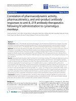
Figure 1 Distribution of Relative Quantification (RQ) values for IL-
2RA gene expression in whole blood of cynomolgus monkeys fol-
lowing ex vivo stimulation with rhuIL-21. Whole blood samples
were obtained from 37 protein naive cynomolgus monkeys and stim-
ulated ex vivo with rhuIL-21. IL-2RA gene expression was analyzed, as
described in Materials and Methods. Relative quantification (RQ) of IL-
2RA gene expression was performed using a vehicle control sample, as
the calibrator. To confirm that rhuIL-21-induced gene expression was
dependent on engagement of cynomolgus monkey IL-21R, separate
whole blood aliquots (for six monkeys) were stimulated simultaneous-
ly with either rhuIL-21 and Ab-02 (squares) or with rhuIL-21 and control
IgG (triangles), and individual animal and median (solid lines) RQ values
were compared with those obtained for rhuIL-21 stimulation alone
(circles; A). Histogram for the RQ values (B) and log
2
-transformed RQ
values (C) for 37 monkeys, as well as the fitting of these values into a
Gaussian distribution (solid line) are shown. RQ
cutoff
= 2.3 and is the
minimum RQ required for the enrolment into the in vivo study.
log
2
{IL-2R$
$
RQ}
0.1 0.5 0.9 1.3 1.7 2.1 2.5 2.9 3.3
Number of animals
0
2
4
6
8
10
12
14
16
18
Observed
Fitted
log
2
(RQcutoff)
IL-2R
$
RQ
1.5 2.5 3.5 4.5 5.5 6.5 7.5 8.5
Number of animals
0
2
4
6
8
10
12
14
16
18
Observed
RQcutoff
A
C
IL-21
IL-21/IgG
IL-21/Ab-02
0
2
4
6
IL-2RA RQ
B
Vugmeyster et al. Journal of Translational Medicine 2010, 8:41
/>Page 5 of 14
Electrochemiluminescent paramagnetic bead assay for
detection of anti-Ab-01 antibodies in serum
Serum samples (in duplicate) were co-incubated with
biotinylated-Ab-01 and ruthenylated- Ab-01 overnight.
After incubation with streptavidin-coated paramagnetic
beads, the mixture and the plate were placed in the BioV-
eris M Series 384 Analyzer 2004 (BioVeris Corporation,
Washington, DC), a magnet was applied, and unbound
reactants were washed away. The emitted light was mea-
sured by photo detectors with the read out in response
units (RU). Positive and negative control serum samples
were included on each plate to monitor assay perfor-
mance. The negative control serum samples were also
used to determine the cutpoint RU, which was defined as
twice the mean RU of the negative control. Samples were
initially tested in a screening format at dilutions of 1:25
and 1:75. Samples generating an RU greater than or equal
to the cutpoint RU were considered positive. Positive
samples were reanalyzed in a full dilution series to deter-
mine the titer (the dilution that would generate an RU
equal to the cutpoint RU). For positive samples, the log of
the titer was reported. The minimum required dilution
was 1:25 and the limit of detection was 1.40 (the log of
25). Therefore, negative samples were designated as
<1.40. This assay detects both neutralizing and non-neu-
tralizing anti-Ab-01 antibodies.
Flow cytometry assay for detection of neutralizing anti-Ab-
02 antibodies in serum
TF-1 and TF-1/huIL-21R (TF-1 cells transfected with
huIL-21R; Wyeth) were grown in RPMI media containing
25 ng/ml huGMCSF (R&D Systems, Inc., Minneapolis,
MN). Confluent cell cultures were centrifuged at 300 g
for 10 min, resuspended in OptiMEM serum free
medium (Invitrogen, Carlsbad, CA) at 10
6
cells/mL, and
incubated at 37°C for 2 hours. The cells were then washed
in cold PBS/0.5%BSA, re-suspended in ice-cold PBS buf-
fer, and kept on ice until staining. To determine the EC
50
for Ab-02-biotin binding to TF-1/huIL-21R cells, both
the parental TF-1 and the TF-1/huIL-21R cells (10
5
cells
per test) were incubated with either Ab-02-biotin, or IgG-
biotin control using serial 3-fold dilutions (range = 16-
0.0002 μg/mL) on ice for 30 minutes, washed in PBS/
0.5%BSA, and then incubated with streptavidin-allophy-
cocyanin (APC; Invitrogen). Geometric mean fluorescent
intensities ("GMFI") of the APC channel peaks was col-
lected on an LSRII flow cytometer (BD Biosciences, San
Jose, CA) and analyzed using Flowjo 8.3.3 software (Tree
Star, Inc., Ashland, OR). Linear regression analysis of the
plots was performed using Prism 4 for Macintosh v4.0b
(GraphPad Software, Inc.).
The minimum required dilution (MRD) for testing
serum samples in this assay was determined to be 1:6 in
PBS/0.5%BSA. To test for inhibition of Ab-02-biotin to
TF-1/huIL-21R cells (i.e. for the presence of neutralizing
activity), TF-1/huIL-21R cells were pre-incubated with
sera from anti-IL-21R-dosed monkeys (using a 3 fold
dilution series starting at the MRD), stained with an anti-
IL-21R-biotin (at the estimated EC
50
concentration),
washed in PBS/0.5%BSA, stained with streptavidin-APC,
and analyzed for GMFI as described above. Each serum
sample was run in duplicate in two individual experi-
ments, and the average GMFI value for the 4 replicates
was obtained for each dilution point. The relative GMFI
value for each serum sample for each dilution point was
calculated using the formula [100%* average GMFI/aver-
age GMFI pre-dose]. A sample was considered positive if
the relative GMFI value was less than or equal to 80% at
the MRD. For positive samples, the log titer was calcu-
lated as the log [reciprocal dilution that would generate
relative GMFI >80%]. Based on the MRD, log titers for
negative samples were reported as <0.78 (log 6).
Results
Characterization of responsiveness of cynomolgus monkey
whole blood to ex vivo rhuIL-21 stimulation using a PD
assay
Prior to conducting the in vivo study, inter-animal vari-
ability in responsiveness to ex vivo rhuIL-21 stimulation
for five previously identified genes (Arai et al, manuscript
in preparation) in blood samples of untreated cynomol-
gus monkeys was examined. Gene expression changes
(relative to vehicle control) for IL-2RA, IL-21R, PRF1,
GZMB, and/or IL-6 following ex vivo stimulation of
whole blood samples with rhuIL-21 for thirteen monkeys
are shown in Table 2. In this assay, gene expression was
quantified using relative quantification (RQ) units, as
described in Materials and Methods. Not all animals had
induction of gene expression of all of the above genes and
there was noticeable inter-animal variability in the RQ
values for all genes. Samples with RQ values greater or
equal to 1.5 were considered to have gene expression
higher than the corresponding vehicle control sample. IL-
2RA was determined to have the largest magnitude (high-
est mean RQ) and most consistent change (highest per-
centage of animals that had RQ >1.5) in rhuIL-21-
induced gene expression of the genes evaluated, and was
therefore considered the best single gene for assessing PD
activity of the anti-IL-21R antibodies.
To confirm that rhuIL-21-induced gene expression was
dependent on engagement of cynomolgus monkey IL-
21R, whole blood samples from six monkeys known to be
responsive to rhuIL-21 were incubated simultaneously
with rhuIL-21 and the IL-21R neutralizing antibody Ab-
02 (30 nM). As expected, ex vivo addition of Ab-02 anti-
body simultaneously with rhuIL-21, completely inhibited
(p < 0.001) rhuIL-21-induced gene expression changes in
the whole blood assay (i.e. RQ value < 1.5; Figure 1A).
Vugmeyster et al. Journal of Translational Medicine 2010, 8:41
/>Page 6 of 14
To obtain a larger number of samples for characteriza-
tion of the distribution of the IL-2RA response to rhuIL-
21 in the ex vivo assay, blood samples were collected from
24 additional female cynomolgus monkeys, stimulated ex
vivo with rhuIL-21, and analyzed for IL-2RA gene expres-
sion (in RQ units) using quantitative RT-PCR (IL-21R,
PRF1, GZMB, and IL-6 expression were not analyzed for
these monkeys). There were no noticeable differences in
IL-2RA RQ distribution between male and female mon-
keys (median ± SD RQ values of 3.8 ± 1.7 and 3.0 ± 1.9,
respectively), and subsequent analysis of IL-21RA RQ
distribution was performed using a combined data set (n
= 37). All cynomolgus monkeys tested had IL-2RA RQ
values greater or equal to1.5 following ex vivo stimulation
with rhuIL-21. The median IL-2RA RQ value (n = 37) was
3.2 with a range of 1.5 to 8.1. The distribution of the IL-
2RA RQ values and log transformation (log2) of the RQ
values obtained in the ex vivo assay are shown in Figure
1B and 1C, respectively. The distribution of IL-2RA RQ
values appeared approximately lognormal, based on the
normality tests described in Materials and Methods.
The minimum RQ value for IL-2RA gene expression in
the ex vivo PD assay required for the inclusion into the in
vivo PD study of Ab-01 and Ab-02 in cynomolgus mon-
keys (RQ
cutoff
) was defined as 2.3 using the formula: log
[RQ
cutoff
] = mean of the log-transformed RQ values -
standard deviation of the log-transformed RQ values.
Approximately 81% of monkeys tested (30 of 37) had RQ
values greater than 2.3 and were considered to be good
responders in the ex vivo PD assay.
Nine male monkeys that were determined to be good
responders in the ex vivo PD assay, were administered a
single 10 mg/kg IV dosage of Ab-01, Ab-02, or a control
antibody (3/group), and monitored for PD activity, serum
concentration, and anti-product antibody responses at
the time points shown in Table 1.
Serum concentrations of Ab-01 and Ab-02 in cynomolgus
monkeys
Initial PK studies in cynomolgus monkeys demonstrated
that following single IV administration, Ab-02 was
cleared markedly faster compared to Ab-01 [14]. In the
PD study presented here, extensive serum sampling
required for determination of a complete set of PK
parameters could not be performed because of the rela-
tively large sample volume required for the PD assay and
limitations on blood volumes that could be collected
from each individual cynomolgus monkey. Samples for
determination of anti-IL-21R serum concentrations were
taken only at those time points at which PD activity was
assessed to enable correlation between the serum con-
centrations and PD activity for each individual animal.
Thus, only elimination half-life (t
1/2
) was calculated based
on the terminal phases of serum concentration-time pro-
files.
Following a single 10 mg/kg IV dose, Ab-01 was elimi-
nated slowly from cynomolgus monkeys, with a mean
apparent terminal half-life (t
1/2
) of ~10.6 ± 3.92 days
(Table 3). Up to day 22, Ab-01 serum concentrations were
very similar between all three Ab-01 dosed animals (Fig-
ure 2). However, at day 36 and later time points, Ab-01
serum concentrations in animal 2 declined rapidly (to
~0.6 μg/mL) compared with those for animals 1 and 3 (to
~2 μg/mL). At day 50, animal 2 had no detectable serum
Ab-01 concentration(less than LOQ of 30 ng/mL), while
animals 1 and 3 had Ab-01 serum concentrations of ~0.9-
Table 2: Relative quantification (RQ) of gene expression induced by ex vivo addition of rhuIL-21 to whole blood obtained
from male cynomolgus monkeys
ANIMAL # IL-2RA IL-21R PRF1 GZMB IL-6
1 2.8 2.5 2.8 2.0 4.2
2 3.4 2.3 1.2 1.1 1.7
3 5.5 2.9 2.5 1.4 3.6
4 4.1 2.1 2.5 1.1 4.9
5 5.3 3.2 2.1 2.1 4.1
6 2.8 1.8 1.8 2.0 0.5
7 6.3 1.9 2.4 1.9 2.6
8 2.8 1.8 1.6 1.6 6.7
9 3.8 2.0 1.9 2.2 1.2
10 2.9 1.8 0.7 1.0 0.9
11 7.7 3.4 2.6 2.2 2.8
12 4.5 3.6 1.4 1.5 1.1
13 2.1 1.5 1.1 1.2 1.4
Vugmeyster et al. Journal of Translational Medicine 2010, 8:41
/>Page 7 of 14
1 μg/mL. Thus, the estimated t
1/2
of Ab-01 was shorter
for animal 2 (~6.2 days) compared to that for animals 1
and 3 (~12 and 14 days, respectively).
As expected based on the initial PK studies [14], after a
single 10 mg/kg IV dose, the serum concentrations of Ab-
02 declined much faster than those of Ab-01 (Figure 2).
Note that test article concentrations in sera were mea-
sured at all the time points at which PD activity was
assessed (as shown in Table 1). However, the data points
with serum concentrations below the LOQ (30 ng/mL)
are not shown in Figures 2, 3 and 4. All three Ab-02-
dosed monkeys had similar concentration time-profiles
and apparent t
1/2
values (Table 3), with serum concentra-
tions declining to relatively low levels at day 15 (<0.4 μg/
mL) and to less than the LOQ at day 22. The estimated
mean t
1/2
of Ab-02 was 2.3 ± 0.16 days. Ab-01 and Ab-02
concentrations started to diverge as early as 24 hrs post-
dose and Ab-02 concentrations were more than 10-fold
lower than Ab-01 concentrations at the one week time
point. These data confirmed observations from the ear-
lier PK studies [14], in which Ab-02 had ~5-7 fold faster
CL, compared to Ab-01 (i.e. 500-700% of the Ab-01
value).
Pharmacodynamic response
For all nine monkeys enrolled into this study, each pre-
dose and post-dose blood sample was divided into four
1.5 mL aliquots. The first and second aliquots were
treated with either rhuIL-21 or vehicle (a calibrator for
RQ calculations), and were used to assess whether circu-
lating test article affected ex vivo rhuIL-21-induced IL-
2RA gene expression (i.e. demonstrated PD activity). The
third aliquot was treated with rhuIL-21 and an anti-IL-
21R antibody (30 nM), and the fourth aliquot was treated
with rhuIL-21 and an IgG control antibody (negative con-
trol for the anti-IL-21R antibody). The third and fourth
aliquots were used to assess whether inhibition of rhuIL-
21-induced IL-2RA gene expression by circulating test
article in a given post-dose sample was complete (for time
points at which PD activity was observed), to assess
whether the return of rhuIL-21-induced gene expression
was mediated through the IL-21R (for time points at
which PD activity was lost), and to monitor for the pres-
ence of neutralizing anti-product antibodies.
For Ab-01, complete inhibition of rhuIL-21-induced IL-
2RA gene expression (IL-2RA RQ <1.5) persisted until at
least day 22 for animal 2 and at least day 50 for animals 1
and 3, when serum Ab-01 concentrations were at or
above 0.9 μg/mL (6 nM) for all three monkeys (Table 3
and Figure 3, data points with serum concentrations
below the LOQ of 30 ng/mL are not shown). Ex vivo
rhuIL-21-induced IL-2RA expression returned to pre-
dose values (i.e. PD activity was lost) at day 92 for animals
1 and 3, coincident with the time points at which serum
concentrations of the test articles were <LOQ (Figure 3).
For animal 2, PD activity was lost at day 36, when serum
Ab-01 concentration had declined to a relatively low level
of ~4 nM (0.6 μg/mL). For all time points examined in
this study, the PD activity of Ab-01 appeared to be all or
none, such that there was typically either complete inhi-
bition of rhuIL-21-induced IL-2RA gene expression (RQ
< 1.5), or a lack of inhibition (RQ similar to that in the
corresponding pre-dose sample). A partial PD response
was difficult to differentiate because of the intra-animal
variability observed in IL-2RA RQ values. It is possible
that data points with partial PD response for Ab-01
would have been observed if additional sampling time
points were collected. The minimum concentration that
was needed to maintain minimum PD activity of Ab-01
(C
min
) could not be precisely estimated, but is likely to be
~4-6 nM.
For Ab-02, PD activity persisted until at least day 8 (RQ
< 1.5), when serum Ab-02 concentrations were at or
above 1.3 μg/mL (Figure 4 and Table 3). PD activity was
lost (RQ > 1.5) at day 15 for all three monkeys. In blood
samples obtained on day 15 from animals 5 and 6, IL-2RA
RQ values appeared similar to the corresponding pre-
dose values (i.e. complete loss of PD activity) and serum
Ab-02 concentrations were less or equal to 0.26 μg/mL
(1.7 nM). There was an apparent partial PD response in
blood samples obtained on day 15 from animal 4, as the
observed IL-2RA RQ value of 2.7 was less than that at
pre-dose (RQ = 4.8) and at the subsequent day 22 time-
Figure 2 Serum concentrations following a single 10 mg/kg IV
dosage of anti-IL-21R antibody Ab-01 or Ab-02 to cynomolgus
monkeys. Serum samples were collected up to 148 days for Ab-01
(filled symbols and solid lines) and up to 36 days for Ab-02 (open sym-
bols and dashed lines) post-dose. Ab-01 and Ab-02 serum concentra-
tions were measured by a specific ELISA, in which his-tagged rhuIL-21
and an anti-human Fc antibody were used as a capture and a detector,
respectively. Data points with serum concentrations below the LOQ of
30 ng/mL (after day 71 and day 15 for Ab-01 and Ab-02, respectively),
are not shown. an = animal number.
Time (days)
1 1529435771
Serum concentration (
P
g/mL)
0.1
1
10
100
Ab-01 an 1
Ab-01 an 2
Ab-01 an 3
Ab-02 an 4
Ab-02 an 5
Ab-02 an 6
Vugmeyster et al. Journal of Translational Medicine 2010, 8:41
/>Page 8 of 14
point (RQ = 5.3; Figure 4). Animal 4 also had a slightly
longer estimated t
1/2
and somewhat higher Ab-02 serum
concentration at day 15 (~2.5 nM), compared to animals
5 and 6. These data suggested that the C
min
of Ab-02 that
was needed to maintain PD activity was approximately
2.5 nM.
For the isotype control group, ex vivo added rhuIL-21
induced IL-2RA gene expression in whole blood samples
from all three monkeys at all time points, with noticeable
intra-animal variability in the IL-2RA RQ values (data not
shown).
Anti-product antibody response
At the first time point where loss of PD activity was
observed, ex vivo addition of an anti-IL-21R antibody
simultaneous with rhuIL-21 inhibited the induction of
IL-2RA gene expression (RQ < 1.5) in all Ab-01- and Ab-
02-dosed monkeys, indicating that the return of rhuIL-
21-induced gene expression was mediated through the
IL-21R and that neutralizing anti-IL-21R antibodies were
not present (Figure 3 and 4). Ex vivo addition of Ab-01
continued to demonstrate inhibitory activity at subse-
quent time points collected from animals 1 and 3. How-
ever, Ab-01 had no ex vivo inhibitory activity at the day 50
time point from animal 2 (Figure 3). Similarly, Ab-02 had
no ex vivo inhibitory activity at the day 22 and/or day 36
time points collected from all animals in the Ab-02 dosed
group (Figure 4). These data suggested that animal 2 in
the Ab-01 group and all three animals (4-6) in the Ab-02
group had developed neutralizing anti-product antibod-
ies.
The presence of neutralizing anti-Ab-02 antibodies in
Ab-02-dosed animals was confirmed using an orthogonal
flow cytometric (FACS)-based assay. In this assay, TF-1
cells transfected with human IL-21R (TF-1/hIL-21R)
were stained with Ab-02-biotin in the presence of serum
samples obtained from Ab-02-dosed monkeys. Streptavi-
din-APC was added to detect Ab-02-biotin binding to
TF-1/hIL-21R cells. All three Ab-02 dosed animals tested
positive in the FACS-based neutralizing antibody assay at
day 22 and day 36, with log titers ranging from 2.2 to 4.1
(Table 4).
Since only one of the Ab-01-dosed animals showed evi-
dence of neutralizing anti-Ab-01 antibodies in the ex vivo
IL-2RA gene expression assay, serum samples from Ab-
01-dosed monkeys were tested in an electrochemilumi-
nescent paramagnetic bead-based assay that detected
both neutralizing and non-neutralizing anti-Ab-01 anti-
bodies. In this assay, serum samples were co-incubated
with biotinylated- Ab-01 and ruthenylated- Ab-01,
streptavidin coated paramagnetic beads were added to
the mixture, and the emitted light was detected using
BioVeris technology. All three Ab-01 dosed monkeys
were positive for anti-Ab-01 antibodies in this assay, with
log titers ranging from 1.86 to 3.43 (Table 5). There was
significant inter-animal variability in the apparent onset
of anti-Ab-01 generation. The first serum sample that
was positive for anti-Ab-01 antibodies in the BioVeris-
based assay was obtained at day 134, 36, and 92 for ani-
mals 1, 2, and 3 respectively. Thus, among the three Ab-
01-dosed animals, animal 2 had the shortest t
1/2
and the
fastest onset and the highest titer of anti-Ab-01 antibody
Table 3: Peak and last detectable concentrations, and elimination half-life after a single 10 mg/kg IV dosage of Ab-01 or
Ab-02 to cynomolgus monkeys
Group Animal
C
peak
(μg/mL)
t
1/2
(days)
C
last
(μg/mL)
T
last
(days)
A (Ab-01) 1 200 12 0.91 50
21396.20.5636
3 153 14 0.37 71
Mean164110.6152
SD 32 3.9 0.27 18
B (Ab-02) 4 145 2.5 0.36 15
51552.30.2615
62012.10.2515
Mean 167 2.3 0.29 15
SD 30 0.16 0.06 0
C
peak
= Concentration at 5 minutes, the first sampling time point after IV administration; t
1/2
= elimination half-life; T
last
= last time point at
which the test article concentration was above the LOQ (30.0 ng/mL); C
last
= concentration at T
last
.
Vugmeyster et al. Journal of Translational Medicine 2010, 8:41
/>Page 9 of 14
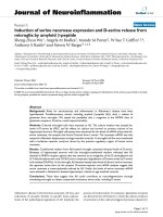
Figure 3 Correlation of serum concentrations, PD activity, and anti-product antibody responses following a single 10 mg/kg IV dosage of
anti-IL-21R antibody Ab-01 to cynomolgus monkeys. Each pre-dose (day -13 for animal 1 and day 1 for animals 2 and 3) and post-dose whole
blood sample was divided into four aliquots. The first and second aliquots were treated with either rhuIL-21 (filled circle) or vehicle (a calibrator for RQ
calculations), respectively, and were used to assess whether circulating test article affected ex vivo rhuIL-21-induced IL-2RA gene expression (i.e. PD
activity). The third aliquot (filled square) was treated with rhuIL-21 and Ab-01, except for day -13 samples for which Ab-02 was used. The fourth aliquot
(open triangle) was treated with rhuIL-21 and an IgG control antibody (negative control for the third treatment). Ab-01 serum concentrations (filled
diamonds) were measured by a specific ELISA up to days 148, 50, and 92 for animals 1, 2, and 3, respectively; data points with serum concentrations
below the LOQ (30 ng/mL) are not shown. Anti-Ab-01 antibodies (neutralizing and non-neutralizing) were assessed by bead-based immunoassay;
positive result indicated by "A".
-13 1 15 29 43 57 71 85 99 113 127 141 155
RQ
1
2
3
4
5
6
7
8
9
10
Concentration (
P
g/mL)
0.1
1
10
100
IL-21
IL-21 + IgG
IL-21 + anti-IL-21R Ab
Serum concentration of Ab-01
-13 1 15 29 43 57 71 85 99 113 127 141 155
RQ
1
2
3
4
5
6
7
8
9
10
Concentration (
P
g/mL)
0.1
1
10
100
Time (Days)
-13 1 15 29 43 57 71 85 99 113 127 141 155
RQ
1
2
3
4
5
6
7
8
9
10
Concentration (
P
g/mL)
0.1
1
10
100
A
A
A
A
A
A
Animal 1
Animal 2
Animal 3
Vugmeyster et al. Journal of Translational Medicine 2010, 8:41
/>Page 10 of 14
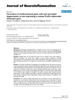
Figure 4 Correlation of serum concentrations, PD activity, and anti-product antibody responses following a single 10 mg/kg IV dosage of
anti-IL-21R antibody Ab-02 to cynomolgus monkeys. Each pre-dose (day 1) and post-dose whole blood sample was divided into four 1.5 mL ali-
quots. First and second aliquots were treated with either rhuIL-21 (filled circle) or vehicle (a calibrator for RQ calculations), respectively, and were used
to assess whether circulating test article affected ex vivo rhuIL-21-induced IL-2RA gene expression (i.e. PD activity). The third aliquot (filled square) was
treated with rhuIL-21 and Ab-02 and the fourth aliquot (open triangle) was treated with rhuIL-21 and an IgG control antibody (negative control for
the third treatment). Ab-02 serum concentrations (filled diamonds) were measured by a specific ELISA up to day 36. Data points with serum concen-
trations below the LOQ (30 ng/mL) are not shown. Neutralizing anti-Ab-02 antibodies were assessed by flow cytometry; positive result indicated by
"A
N
".
1 8 15 22 29 36
RQ
1
2
3
4
5
6
7
Concentration (
P
g/mL)
0.1
1
10
100
1 8 15 22 29 36
RQ
1
2
3
4
5
6
7
Concentration (
P
g/mL)
0.1
1
10
100
Time (Days)
1 8 15 22 29 36
RQ
1
2
3
4
5
6
7
Concentration (
P
g/mL)
0.1
1
10
100
A
N
A
N
A
N
A
N
A
N
A
N
Animal 4
Animal 5
Animal 6
Vugmeyster et al. Journal of Translational Medicine 2010, 8:41
/>Page 11 of 14
response. Animal 2 was also the only Ab-01-dosed mon-
key that showed evidence of neutralizing anti-Ab-01 anti-
body response in the ex vivo IL-2RA gene expression
assay, similar to all three Ab-02 dosed monkeys.
Discussion
The study presented in this manuscript was conducted to
monitor the PD activity of anti-IL-21R antibodies Ab-01
and Ab-02 in cynomolgus monkeys following IV admin-
istration and to correlate PD activity with serum concen-
trations of these antibodies and the presence of an anti-
product antibody response. Since Ab-02 had slower k
off
rate leading to a lower K
D
value for in vitro binding to
human IL-21R (~0.4 nM) compared to Ab-01 (~2 nM)
[14,15]; this study also explored whether improvement in
target binding affinity in vitro translated into an
improved PK-PD profile in primates for Ab-02.
Prior to conducting the in vivo PD study of anti-IL-21R
antibodies in cynomolgus monkeys, a single gene, IL-
2RA, was identified for assessing PD activity, and inter-
animal variability in IL-2RA gene expression was charac-
terized in monkeys. The distribution of IL-2RA RQ val-
ues in response to rhuIL-21 stimulation appeared
approximately lognormal, which was similar to the distri-
bution of RQ values for genes upregulated in disease con-
ditions or to the distribution of ΔCt values of immune
response genes in blood obtained from healthy human
donors [17,18]. Based on the statistical analysis of the IL-
2RA RQ values in the 37 monkeys tested, the good
Table 4: Formation of neutralizing anti-Ab-02 antibodies (log Titer) after a single 10 mg/kg IV dosage of Ab-02 to
cynomolgus monkeys
TIME
(DAYS)
ANIMAL 4 ANIMAL 5 ANIMAL 6
Pre-dose Negative Negative Negative
15 Negative Negative Negative
22 4.12 2.7 2.2
36 2.2 2.7 2.7
Formation of neutralizing anti-Ab-02 was assessed using a flow cytometric assay, in which serum samples from Ab-02 dosed monkeys were
tested for inhibition (relative to pre-dose) of anti-IL-21R-biotin binding to TF-1 cells transfected with human IL-21R. Negative samples had no
inhibition at the minimum required dilution of 1:6 and had a log titer < 0.78.
Table 5: Formation of anti-Ab-01 antibodies (log Titer) after a single 10 mg/kg IV dosage of Ab-01 to male cynomolgus
monkeys
TIME
(DAYS)
ANIMAL 1 ANIMAL 2 ANIMAL 3
Pre-dose Negative Negative Negative
15 Negative Negative Negative
22 Negative Negative Negative
36 Negative 2.13 Negative
50 Negative 3.43 Negative
71 ND ND Negative
92 Negative ND 2.27
106 ND ND 2.79
113 Negative ND ND
134 1.86 ND ND
148 2.4 ND ND
"ND" = Not determined
Formation of anti-Ab-01 antibodies (neutralizing and non-neutralizing) was assessed using an electrochemiluminescent paramagnetic bead
assay, in which serum samples from Ab-01-dosed monkeys were tested for binding to biotinylated- Ab-01 and ruthenylated- Ab-01. Negative
samples had no binding at the minimum required dilution of 1:25 and had a log titer < 1.40.
Vugmeyster et al. Journal of Translational Medicine 2010, 8:41
/>Page 12 of 14
responders to rhuIL-21 were defined as those with the
RQ values ≥ 2.3 and the frequency of good responders
was estimated to be ~80%. Animals that were good
responders prior to the in vivo study also were good
responders at subsequent time points collected prior to
dosing. These data illustrate that for preclinical or clinical
studies that rely on ex vivo assays for assessing PD activ-
ity, potential heterogeneity in the ex vivo response needs
to be examined, characterized, and taken into account
during study design and animal or patient selection.
Following IV administration of anti-IL-21R antibodies
Ab-01 and Ab-02 to cynomolgus monkeys, there was a
good correlation between the PD activity (based on inhi-
bition of IL-2RA gene expression induced by ex vivo addi-
tion of rhuIL-21 to whole blood), serum concentrations,
and anti-product antibody responses. Complete inhibi-
tion of rhuIL-21-induced IL-2RA gene expression (PD
activity) was observed at the first time point, 5 minutes,
after dosing, for both Ab-01 and Ab-02. PD activity was
lost (i.e. rhuIL-21 induction of IL-2RA expression was
again observed) with the elimination of Ab-01 and Ab-02
from circulation. In agreement with earlier PK studies
[14], Ab-02 had faster elimination in monkeys compared
with Ab-01, with a mean apparent t
1/2
of 10.6 and 2.3 days
for Ab-01 and Ab-02, respectively. At the day 15 time
point, PD activity was completely or partially lost in all
three Ab-02-dosed monkeys, while all three monkeys in
the Ab-01 dose group had relatively high serum Ab-01
concentrations (~6.0-7.4 μg/mL) and full PD activity.
Thus, Ab-01 had a longer duration of PD activity and a
longer t
1/2
in cynomolgus monkeys.
All three Ab-01-dosed monkeys were positive for anti-
product antibodies, based on an assay that detected both
neutralizing and non-neutralizing anti-Ab-01 antibodies.
However, only one of three Ab-01-dosed animals had evi-
dence of neutralizing anti-Ab-01 antibodies in the IL-
2RA gene expression assay. There was significant inter-
animal variability in the apparent terminal serum half-life
of Ab-01 (~6 to 14 days), which appeared to correlate
with the onset and titer of anti-product antibody
response for the three Ab-01-dosed animals. Animal 2
had the shortest t
1/2
, the fastest onset, and highest titer of
anti-Ab-01 antibody response. Animal 2 was the only Ab-
01-dosed monkey that showed evidence of neutralizing
anti-Ab-01 response in the ex vivo IL-2RA gene expres-
sion assay, similar to all three Ab-02-dosed monkeys.
Animals 1 and 3 had noticeably longer t
1/2
and later onset
of anti-Ab-01 antibody responses.
Despite the variability in serum t
1/2
of Ab-01 between
the three Ab-01-dosed animals, there was a good correla-
tion between Ab-01 serum concentrations and PD activ-
ity, such that PD activity was observed in all three Ab-01-
dosed monkeys when serum concentrations of Ab-01
were at or above 0.9 μg/mL (~6 nM) and was lost in all
three monkeys when serum concentrations were < 0.6
ng/mL (~4 nM). The minimum serum concentration of
Ab-01 which was needed to maintain PD activity (C
min
)
could not be precisely determined from the data obtained
in this study, because Ab-01 PD activity appeared to be all
or none at the time points collected and there were not
enough time points taken in the lower concentration
range to fully evaluate the concentration-effect relation-
ship. However, the available concentration-effect data
discussed above suggested that the C
min
of Ab-01 was ~4-
6 nM.
All three of the Ab-02-dosed monkeys had similar Ab-
02 concentration-time profiles, onset of anti-product
antibody responses, and resulting PD-time profiles. All
three Ab-02-dosed monkeys were positive for neutraliz-
ing antibodies in the two orthogonal assays (gene expres-
sion and flow cytometry) at and after day 22. In blood
samples from two Ab-02-dosed monkeys, Ab-02 PD
activity was all or none at time points tested. One Ab-02-
dosed animal had apparent partial PD activity at one time
point (day 15), when serum Ab-02 concentrations were
~2.5 nM. Based on these data, C
min
needed to maintain
PD activity of Ab-02 was assumed to be approximately
2.5 nM. Additional studies would be needed to confirm
the C
min
for both Ab-01 and Ab-02. However, the prelimi-
nary C
min
estimates of ~4-6 nM for Ab-01 and ~2.5 nM
for Ab-02 were consistent with the K
D
values for Ab-01
and Ab-02 binding to human IL-21R (~2.0 and 0.5 nM,
respectively). Thus, Ab-02 had higher binding affinity for
human IL-21R and a lower estimated C
min
needed to
maintain PD activity, compared to Ab-01. However,
because of the fast elimination of Ab-02 from the circula-
tion, loss of PD activity occurred much faster in Ab-02-
dosed monkeys (day 15-22), compared to Ab-01 (not ear-
lier than day 36).
The mechanism of fast elimination of Ab-02 in mon-
keys is not known but is unlikely to be entirely target (IL-
21R)-mediated because it appeared non-saturable at a
relatively high dose level (100 mg/kg) [14]. While the dos-
age that is needed to saturate IL-21R binding sites in cyn-
omolgus monkeys or other species has not been defined,
it is very unlikely to exceed 100 mg/kg, based on reports
on other human antibodies directed against highly-
expressed cell surface receptors, including an antibody to
another type I cytokine receptor, IL-2R [19]. A single 2
mg/kg dosage of anti-IL-2R antibody to human subjects
resulted in complete saturation of IL-2R on peripheral
blood lymphocytes for ~45 days post dose, as long as the
serum anti-IL-2R Ab concentration were above 1 μg/mL
(~6 nM), which was similar to the C
min
needed to main-
tain PD activity of the anti-IL-21R antibodies Ab-01 and
Ab-02 in the ex vivo whole blood assay following a single
Vugmeyster et al. Journal of Translational Medicine 2010, 8:41
/>Page 13 of 14
10 mg/kg aose to cynomolgus monkeys. Differences in
Ab-01 and Ab-02 serum concentrations observed in cyn-
omolgus monkeys in this study and earlier PK studies are
not likely to be entirely explained by neutralizing anti-
product responses, because Ab-01 and Ab-02 serum con-
centrations started to diverge as early as 24 hrs post-dose
and Ab-02 concentrations were more than 10-fold lower
than Ab-01 concentrations at the one week time point.
Although total body clearance (CL) of Ab-01 and Ab-02
could not be accurately estimated in this study, the
observed serum concentration profiles were consistent
with the previously reported faster CL of Ab-01 (~5-7
mL/hr/kg) compared to that of Ab-02 (~1 mL/hr/kg)
[14]. Further studies are needed to delineate the mecha-
nism of fast clearance of Ab-02 in monkeys.
Finally, data presented in this report, suggested that for
anti-IL-21R antibody Ab-02, a lower K
D
value for target
(IL-21R) binding in the in vitro assay (~0.4 nM for Ab-02
versus ~2 nM for Ab-01; [15,16]) did not translate into an
improved PK-PD profile in primates, primarily due to dif-
ferences in pharmacokinetics between the two antibod-
ies. Thus, optimization of candidate anti-IL-21R
antibodies in in vitro systems may not be sufficient for
generation of therapeutic antibodies with improved PK-
PD profiles, and PK-PD studies in non-human primates
are recommended prior to first in human studies.
Conclusions
Following IV administration of anti-IL-21R antibodies
Ab-01 and Ab-02 to cynomolgus monkeys, there was
good correlation between the PD activity (based on IL-
2RA gene expression in ex vivo rhuIL-21-stimulated
whole blood), the respective serum concentration pro-
files, and anti-product antibody responses. Compared
with Ab-01, Ab-02 was eliminated markedly faster from
the circulation (shorter t
1/2
and faster clearance), which
correlated with a shorter duration of PD activity. Thus,
slower k
off
rate leading to a lower K
D
value for in vitro
binding to human IL-21R of Ab-02 (~0.4 nM) compared
to Ab-01 (~2 nM) did not translate into an improved PK-
PD profile in primates, primarily due to differences in
pharmacokinetics between the two antibodies. This study
exemplifies that detailed in vivo PK-PD studies in non-
human primates (such as those presented in this report)
are crucial for the selection of lead biotherapeutic candi-
dates for first-in-human clinical studies.
Competing interests
All authors are current or former employees and/or hold stocks or stock
options of Wyeth, Inc (currently Pfizer) at the time the manuscript was pre-
pared.
Authors' contributions
YV and KA drafted the manuscript. YV, KA, LB, HG, MO, AB, and DY carried out
study design, conduct and data interpretatin. ML critically reviewed the manu-
script and data interpretation. MO identified the biomarkers and devised the
PD assay strategy for this study. PS performed immunoassay analysis. SA, MM,
VS, performed gene expression analysis, and MR performed FACS analysis. All
authors have read and approved the final manuscript.
Acknowledgements
We acknowledge S. Jain and A. Weaver for the pilot work done to identify the
PD biomarkers; N. Duriga, C. Shea, B. Leary, and J. Emerson for technical help
with serum sample analysis; J, Ren and B. Ma for technical help with the PD
assay; F. Schlerman, W. McWilliams, and J. Phillips for help with monkey blood
sample collection; J. McClellan. J. Targ, K. Heveron, P. Giampa, A. Robak, J.
Zhang, J. Sanford, A. Root, R. Jackobek, K. Lam, S. Olland, R. Zollner and K. Lam
for expression and purification of antibodies.
Author Details
Pfizer, Inc., Andover, MA, 01810, USA
References
1. Coquet JM, Kyparissoudis K, Pellicci DG, Besra G, Berzins SP, Smyth MJ,
Godfrey DI: IL-21 is produced by NKT cells and modulates NKT cell
activation and cytokine production. J Immunol 2007, 178:2827-2834.
2. Parrish-Novak J, Dillon SR, Nelson A, Hammond A, Sprecher C, Gross JA,
Johnston J, Madden K, Xu W, West J, et al.: Interleukin 21 and its receptor
are involved in NK cell expansion and regulation of lymphocyte
function. Nature 2000, 408:57-63.
3. Ettinger R, Kuchen S, Lipsky PE: The role of IL-21 in regulating B-cell
function in health and disease. Immunol Rev 2008, 223:60-86.
4. Spolski R, Leonard WJ: Interleukin-21: basic biology and implications for
cancer and autoimmunity. Annu Rev Immunol 2008, 26:57-79.
5. Ozaki K, Kikly K, Michalovich D, Young PR, Leonard WJ: Cloning of a type I
cytokine receptor most related to the IL-2 receptor beta chain. Proc
Natl Acad Sci USA 2000, 97:11439-11444.
6. Caruso R, Fina D, Peluso I, Stolfi C, Fantini MC, Gioia V, Caprioli F, Del
Vecchio Blanco G, Paoluzi OA, Macdonald TT, et al.: A functional role for
interleukin-21 in promoting the synthesis of the T-cell
chemoattractant, MIP-3alpha, by gut epithelial cells. Gastroenterology
2007, 132:166-175.
7. Distler JH, Jungel A, Kowal-Bielecka O, Michel BA, Gay RE, Sprott H,
Matucci-Cerinic M, Chilla M, Reich K, Kalden JR, et al.: Expression of
interleukin-21 receptor in epidermis from patients with systemic
sclerosis. Arthritis Rheum 2005, 52:856-864.
8. Jungel A, Distler JH, Kurowska-Stolarska M, Seemayer CA, Seibl R, Forster A,
Michel BA, Gay RE, Emmrich F, Gay S, Distler O: Expression of interleukin-
21 receptor, but not interleukin-21, in synovial fibroblasts and synovial
macrophages of patients with rheumatoid arthritis. Arthritis Rheum
2004, 50:1468-1476.
9. Monteleone G, Caruso R, Fina D, Peluso I, Gioia V, Stolfi C, Fantini MC,
Caprioli F, Tersigni R, Alessandroni L, et al.: Control of matrix
metalloproteinase production in human intestinal fibroblasts by
interleukin 21. Gut 2006, 55:1774-1780.
10. Korn T, Bettelli E, Gao W, Awasthi A, Jager A, Strom TB, Oukka M, Kuchroo
VK: IL-21 initiates an alternative pathway to induce proinflammatory
T(H)17 cells. Nature 2007, 448:484-487.
11. Fina D, Sarra M, Fantini MC, Rizzo A, Caruso R, Caprioli F, Stolfi C, Cardolini I,
Dottori M, Boirivant M, et al.: Regulation of gut inflammation and th17
cell response by interleukin-21. Gastroenterology 2008, 134:1038-1048.
12. Herber D, Brown TP, Liang S, Young DA, Collins M, Dunussi-Joannopoulos
K: IL-21 has a pathogenic role in a lupus-prone mouse model and its
blockade with IL-21R.Fc reduces disease progression. J Immunol 2007,
178:3822-3830.
13. Young DA, Hegen M, Ma HL, Whitters MJ, Albert LM, Lowe L, Senices M,
Wu PW, Sibley B, Leathurby Y, et al.: Blockade of the interleukin-21/
interleukin-21 receptor pathway ameliorates disease in animal models
of rheumatoid arthritis. Arthritis Rheum 2007, 56:1152-1163.
14. Vugmeyster Y, Szklut P, Guay H, Abu-Qare A, Duriga N, Shea C, Emerson J,
Olland S, Young D, Bloom L: Pharmacokinetics of Anti-Il-21r Antibodies
in Mice and Monkeys [abstract]. AAPS J 2008, 2:3.
15. Vugmeyster Y, Guay H, Szklut P, Qian MD, Jin M, Widom A, Spaulding V,
Bennett F, Lowe L, Andreyeva T, Lowe D, Lane S, Thom G, Valge-Archer V,
Received: 10 November 2009 Accepted: 26 April 2010
Published: 26 April 2010
This article is available from: 2010 Vugmeyster et al; licensee BioMed Central Ltd. This is an Open Access article distributed under the terms of the Creative Commons Attribution License ( which permits unrestricted use, distribution, and reproduction in any medium, provided the original work is properly cited.Journal of Tr anslational Medi cine 2010, 8:41
Vugmeyster et al. Journal of Translational Medicine 2010, 8:41
/>Page 14 of 14
Gill D, Young D, Bloom L: In vitro potency, pharmacokinetic profiles, and
pharmacological activity of optimized anti-IL-21R antibodies in a
mouse model of lupus. MAbs 2010, 2:3.
16. Livak KJ, Schmittgen TD: Analysis of relative gene expression data using
real-time quantitative PCR and the 2(-Delta Delta C(T)) Method.
Methods 2001, 25:402-408.
17. McLoughlin K, Turteltaub K, Bankaitis-Davis D, Gerren R, Siconolfi L, Storm
K, Cheronis J, Trollinger D, Macejak D, Tryon V, Bevilacqua M: Limited
dynamic range of immune response gene expression observed in
healthy blood donors using RT-PCR. Mol Med 2006, 12:185-195.
18. Negaard HF, Iversen N, Bowitz-Lothe IM, Sandset PM, Steinsvik B,
Ostenstad B, Iversen PO: Increased bone marrow microvascular density
in haematological malignancies is associated with differential
regulation of angiogenic factors. Leukemia 2009, 23:162-169.
19. Vincenti F, Pace D, Birnbaum J, Lantz M: Pharmacokinetic and
pharmacodynamic studies of one or two doses of daclizumab in renal
transplantation. Am J Transplant 2003, 3:50-52.
doi: 10.1186/1479-5876-8-41
Cite this article as: Vugmeyster et al., Correlation of pharmacodynamic
activity, pharmacokinetics, and anti-product antibody responses to anti-IL-
21R antibody therapeutics following IV administration to cynomolgus mon-
keys Journal of Translational Medicine 2010, 8:41