Báo cáo hóa học: " Endotoxin and CD14 in the progression of biliary atresia" ppt
Bạn đang xem bản rút gọn của tài liệu. Xem và tải ngay bản đầy đủ của tài liệu tại đây (5.22 MB, 14 trang )
RESEARC H Open Access
Endotoxin and CD14 in the progression of
biliary atresia
Ming-Huei Chou
1,2
, Jiin-Haur Chuang
2,3
, Hock-Liew Eng
4
, Ching-Mei Chen
4
, Chiou-Huey Wang
5
, Chao-Long Chen
3
,
Tsun-Mei Lin
1,5,6*
Abstract
Background: Biliary atresia (BA) is a typical cholestatic neonatal disease, characterized by obliteration of intra- and/
or extra-hepatic bile ducts. However, the mechanisms contributing to the pathogenesis of BA remain uncertain.
Because of decreased bile flow, infectious complications and damaging endotoxemia occur frequently in patients
with BA. The aim of this study was to investigate endotoxin levels in patients with BA and the relation of these
levels with the expression of the endotoxin receptor, CD14.
Methods: The plasma levels of endotoxin and soluble CD14 were measured with a pyrochrome Limulus
amebocyte lysate assay and enzyme-linked immunosorbent assay in patients with early-stage BA when they
received the Kasai procedure (KP), in patients who were jaundice-free post-KP and followed-up at the outpatient
department, in patients with late-stage BA when they received liver transplantation, and in patients with
choledochal cysts. The correlation of CD14 expression with endotoxin levels in rats following common bile duct
ligation was investigated.
Results: The results demonstrated a significantly higher hepatic CD14 mRNA and soluble CD14 plasma levels in
patients with early-stage BA relative to those with late-stage BA. However, plasma endotoxin levels were significantly
higher in both the early and late stages of BA relative to controls. In rat model, the results demonstrated that both
endotoxin and CD14 levels were significantly increased in liver tissues of rats following bile duct ligation.
Conclusions: The significant increase in plasma endotoxin and soluble CD14 levels during BA implies a possible
involvement of endotoxin stimulated CD14 production by hepatocytes in the early stage of BA for removal of
endotoxin; whereas, endotoxin signaling likely induced liver injury and impaired soluble CD14 synthesis in the late
stages of BA.
Background
Biliary atresia (BA) is a typical cholestatic neonatal dis-
ease, characterized by obliteration of intra- and/or
extra-hepatic b ile ducts with repeated episodes of cho-
langitis and progressive liver fibrosis and cir rhosis [1-3].
However, the mechanisms contributing to the pathogen-
esis of BA remain uncertain. A decrease of bile flow to
the bowel may promote bacterial translocation to the
liver and increase endotoxin or lipopolysaccharide (LPS)
levels in the peripheral circulation [4]. LPS represent the
major component of the outer membrane of Gram-
negative bacteria and has been implicated in sepsis,
organ failure, and shock [5]. In experimental studies on
healthy animals, LPS is cleared from the circulation
within a few minutes of intravenous injection, and the
majority of LPS is traced to the liver [6,7]. In addition
to clearing LPS, the liver also responds to the presence
of LPS with production of cytokines and reactive oxygen
intermediates. Accumulating evidence suggests that both
endotoxins and pro-inflammatory cytokines participate
in liver damage during endotoxemia [8,9].
CD14 is a glycosylphosphatidylinositol-anchored LPS
receptor. It was first reported as a differentiation marker
expressed on the surface of macrophages, neutrophils,
and other myeloid lineage cells [10-13]. Human hepato-
cytes demonstrate production of CD14 similar to that of
an acute phase protein [14]. However, there is limited
information on the proportional change of CD14 in the
* Correspondence:
1
Institute of Basic Medical Sciences, National Chang Kung University, Tainan,
Taiwan
Full list of author information is available at the end of the article
Chou et al. Journal of Translational Medicine 2010, 8:138
/>© 2010 Chou et al; licensee BioMed Central Ltd. This is an Open Access article d istributed under the t erms of the Creativ e Commons
Attribution License ( which permits unrestricted use, distribution, and reproduction in
any medium, provided the original work is properly cited.
liver and the consequent pathogenetic effects on LPS-
induced liver injury. Although increased expression of
CD14 in surgically biopsied speci mens of BA have been
reported, the exact mechanism of such over-expression
of CD14 is yet to be elucidated [15]. Our previous inves-
tigation revealed that the single nucleotide polymorph-
ism at CD14/-159 is associated with the development
BA and idiopathic neonatal cholestasis [16]. How the
liver responds to LPS-induced injury is virtually
unknown at present [17,18]. Kupffer cells and sinusoidal
endothelial cells express the membrane fo rm of CD 14
(mCD14) in the liver [19,20], while hepatocytes are the
main producers of soluble CD14 (sCD14) [21,22] . How-
ever, the proportional change of CD14 production in
the liver and the subsequent effects on LPS-induced
liver injury during BA is not clear.
In this study, we investigated the role of CD14 in BA-
associated liver injury, with particular emphasis on the
correlation between CD14 expression and endotoxin
levels in the liver tissue and plasma of patients in the
early and late stages of BA. We further elucidated the
expression and regulation of CD14 in a rat model fol-
lowing bile duct ligation (BDL).
Methods
Patients and samples
Liver biopsy specimens were obtained from nine patients
with early-stage BA (four males and fiv e femal es) during
Kasai’s procedure (KP), from nine patients with late-stage
BA (four males and five females) during liver transplanta-
tion for failed KP, and from nine patients with choledochal
cysts (CCs) during surgical correction (2 male and 7
female). Control liver biopsy samples were obtained from
five children with neonatal hepatitis and two who had
focal hepatoblastoma. Plasma samples were obtained from
41 patients with early-stage BA, 25 patients post-KP who
were jaundice-free and were followed-up at the outpatient
department (OPD), 49 patients with late-stage BA, 9
patients with CC, and 7 healthy young infant s. All of the
liver and blood samples were immediately frozen at -80°C
for later laboratory tes ts. The clinical charac teristics and
detailed history of the patients, including the age when the
patient underwent the procedure, sex, serum aspartate
aminotransferase (AST) levels, and total bilirubin are sum-
marized in Table 1. Informed consent was obtained from
the patients or their legal guardians, and the experiments
were approved by the Ethics & Clinical Trial Committee
of the Chang Gung Memorial Hospital, Taiwan.
Animals
Male Sprague-Dawley rats weighting 300-330 g and
about 8 weeks old were divided into three groups: the
BDL group (n = 48) rece ived a common bile duct com-
pletedoubleligation,theshamgroupreceivedasham
operation (n = 48), and the normal control group (n = 6).
All animal experiments were performed in accordance
with and approved by the Animal Care and Use Commit-
tee of Chang Gung Memorial Hospital at Ka ohsiung.
Blood samples were collected at time of sacrifice (3 hrs, 6
hrs, 12 hrs, 24 hrs, 3 days, 7 days, 14 days, and 21 days),
and six rats were included in each subgroup. Serum
enzymes and bilirubin levels were determined using a
biochem istry auto-analyzer (Model 7450; Hitachi, Tokyo,
Japan). Liver tissues were either snap frozen and homo-
genized i n T-PER tissue protein extraction reagent
(Pierce Chemical, Rockford, IL) for protein determination
or fixed in 4% paraformaldehyde and embedded in paraf-
fin for immunohistochemical analysis.
Determination of sCD14 levels by ELISA
The sCD14 levels of plasma were determined using a
commercially available enzyme-linked immunosorbent
assay(ELISA;R&DSystems,Minneapolis,MN)
according to t he manufacture’ s instructions. Samples
were diluted 1:200 and analyzed, and each sample was
measured in duplicate.
Limulus amebocyte lysate (LAL) test
Plasma sp ecimens were collected aseptically in nonpyro-
geniccontainers.Theplasmaandliverspecimenswere
diluted 1:10 and assayed for endotoxin with a commer-
cially available pyrochrome LAL kit (Associates of Cape
Cod, Falmouth, MA) according to t he manufacture’s
instructions.
Real-time quantitative reverse transcription-polymerase
chain reaction (qRT-PCR)
Frozen liver samples (0.1 g/per sample) were homoge-
nized, a nd total RNA was extracted using TRIzol (Invi-
trogen, Carlsbad, CA). The RNA isolates were
quantified at A
260/280
ratio of 1.7-2.0. A total of 2 μgof
RNA was added to 0.1 μgofoligo-d(T)
15
following the
protocol for SuperScr ipIIRT (Inv itrogen, Carlsbad, CA).
Quantitative PCR was performed in a final volume of
20-μl SYBR G reen PCR mixture (Applied Biosystems,
Foster City, CA), and each sample was analyzed in
duplicate. Each reaction mixture contained 0.2 pmo le/ul
of each primer, 1× SYBR Green PCR Master Mix, and
1-5 ng of cDNA. Thermal cycling was initiated with a
2 min incubation at 50°C, followed by a denaturation step
of 10 min at 95°C, and then 40 cycles of PCR consisting
of 95°C for 15 seconds, 60°C for 20 seconds, and 72°C
for 30 seconds. b-actin was used as an internal control
for analyzing CD14 mRNA levels. The sequence of the
PCR primers were designed based on cDNA sequences
from Genbank as follows: CD14 forward primer 5’-TAT
GCT GACACG GTC AAG GC-3’, CD14 reverse primer
5’ -ATT GTC AGA CAG GTC TAG GC-3’ , b-actin
Chou et al. Journal of Translational Medicine 2010, 8:138
/>Page 2 of 14
forward primer 5’ -TCA CCC ACA ATG TGC CCA
TCT TCG A-3’ ,andb-actin reverse primer 5’ -CAG
CGG AAC CGC TCA TTG CCA ATG G-3’.
The quantification of the CD14 mRNA was achieved
with an ABI PRISM 7700 Sequence Detection System
(Applied Biosystems, Warrington, WA) using compara-
tive methods. Ct values of CD14 were normalized to the
Ct value of a housekeeping gene (b-actin).
Immunohistochemical staining for CD14 and lipid A
Immunoreactive CD14 and lipid-A staining was per-
formed on paraffin-embedded, formalin fixed, archival
human liver tissues obtained from the Department of
Pathology, Kaohsiung Chang Gung Memorial Hospital,
Taiwan. In t he animal study, formalin-fixed, paraffin-
embedded liver tissues were used. Two-micrometer
sections were deparaffinized, treated with 3% hydrogen
peroxide to inactivate the endogenous peroxi dase activ-
ity, and microwaved fo r 7 min in 10-mM citrate buffer
(pH 6.0) to retrieve the antigen. The sections were then
incubated in PBS supplemented with 5% fetal calf serum
for 10 min to block background interactions. The sec-
tions were then incubated with a rabbi t anti-CD14 anti-
body (Santa Cruz Biotechnology, Santa Cruz, CA) or a
mouse anti-lipid A antibody (HyCult Biotechnology,
The Netherlands) at 37°C for 2 hrs. The sections were
washed with PBS supplemented with 0.05% Tween 20
and then incubated for 10 min with the s econdary anti-
bodies (SuperPicture; Zymed Laboratories, Francisco,
CA). DAB color substrate (DAKO, Carpinteria, CA) was
added to cover each section, and the reaction was
stopped with ddH
2
O. The slides were counterstained
with hematoxylin, and mounted in mounting medium.
In situ hybridization
In situ hybridization was performed essentially as
described by Wilkinson[23]. The riboprobe was generated
from a pGEM-T vector containing a 250 bp cDNA
sequence of CD14 and labeled with DIG-11-UTP by
in vitro transcription with SP6 and T7 RNA polymerase,
followed by DIG RNA labeling (Roche Applied Science,
Germany). Liver tissues were treated according to the
protocol for immunohistochemical analysis with deparaf-
finization, rehydration, removal of endogenous peroxidase
activity, and antigen retrieval. Sections were digested with
20 μg/ml of proteinase K solution at 37°C for 25 min and
then prehybridized with 5× SSC buffer. A total of 50-μl
hybridization mixture containing denatured RNA probes
was used and hybridized with the sections at 55°C over-
night. After hybridization, the sections were treated with
20 μg/ml of RNase solution at 37°C for 30 min to remove
free RNA probes and then washed with 1× SSC buffer for
5 min and 0.2× SSC containing 0.01% SDS in a 55°C
water bath for 15 min. Sections were blocked in PBS
supplemented with 5% FBS and incubated with an anti-
digoxigenin antibody conjugated with horseradish peroxi-
dase (diluted 1:1000, containing 2% FCS) in blocking
buffer for 2 h at room temperature. The sections were
washed with PBS supplemented with 0.05% Tween 20
and then DAB color substrate (DAKO, Carpinteria, CA)
was added to cover each section, and the reaction was
stopped with ddH
2
O. The slides were counterstained
with hematoxylin, and mounted in mounting medium.
Statistics analysis
Data are presented as the mean ± standard deviation
(SD). The distributions of paired measurements were
compared using the nonparametric Wil coxon matched-
pairs test. The Mann-Whitney test and Wilcoxon
signed-ranks test (nonparametric) were used to evaluate
the stati stical signifi cance of the results using the SPSS-
16 software package (SPSS, Chicago, USA). A P value of
less than 0.05 was considered significant.
Results
Plasma CD14 and endotoxin levels in patients with BA
Plasma sCD14 levels were analyzed by ELISA and found
to be significantly higher in patients with early-stage BA
Table 1 Clinical characteristics of the child patients for this study
Con-C Early stage of BA OPD Late stage of BA CC
Plasma Plasma Liver Plasma Plasma Liver Plasma & Liver
Sample No 7 41 9 25 49 9 9
Age (months) 18 ± 24 2.4 ± 1.2 2 ± 1 24 ± 16 15 ± 10 15 ± 6 22 ± 14
Sex (M/F) 4/3 23/16 4/5 7/18 19/21 4/5 2/7
AST (U/l) ND 200 ± 175
†
181 ± 130 ND 276 ± 241 246 ± 114 298 ± 228
T. Bil (mg/dl) ND 9.0 ± 2.9
†
9.4 ± 3.0 ND 16 ± 1.0 18 ± 10 6.5 ± 6.0
†
D. Bil (mg/dl) ND 6.3 ± 2.0
†
7.0 ± 2.3
†
ND 12 ± 7.5 13 ± 7.2 5.6 ± 4.0
†
sCD14 (μg/ml) 4.0 ± 0.8 4.7 ± 1.7
†
47 ± 14 4.2 ± 1.4
†
2.7 ± 1.5*
‡
49 ± 12 4.0 ± 2.0
†
Endotoxin (EU/ml) 2.0 ± 1.0 6.2 ± 5.0*
‡
17 ± 4.0 2.2 ± 5.0
†
6.7 ± 5.0*
‡
16 ± 7.0 6.5 ± 4.0*
‡
Con-C, control-children; Early stage of BA, Kasai’s procedure for biliary atresia; OPD, Jaundice-free post-Kasai BA patients followed at the outpatient department;
Late stage of BA, liver transplantation for biliary atresia; CC, choledochal cyst; AST, alanine aminotransferase; D. Bil, direct bilirubin; T. Bil, total bilirubin; ND, non
detection; sCD14, souble CD14; y, years. Value are expressed as the mean ± SD. *P < 0.05 vs control;
†
P < 0.05 vs late stage of BA;
‡
P < 0.05 vs OPD.
Chou et al. Journal of Translational Medicine 2010, 8:138
/>Page 3 of 14
(4696 ± 1652 ng/ml), patients with BA who were jaundice-
free and followed up at the OPD (4308 ± 1428 ng/ml),
and patients with CCs (4393 ± 1900 ng/ml) relative to
patients with late-stage BA (2722 ± 1453 ng/ml,
P<0.001). Although sCD14 levels in the early-stage BA,
OPD, and CC groups were higher than controls (3879 ±
767 ng/ml), these differences were not statistically signifi-
cant (P = 0.134, P = 0.4 47, and P = 0.442, respectively)
(Figure 1). There were seven patients with BA whose
plasma samples were available for both the early and late
stages. For these patients, plasma sCD14 levels were
significantly higher in the early stage (4445 ± 237 ng/ml)
compared to those in the late stage (2183 ± 153 ng/ml)
based on paired t-test analysis (P < 0.001).
There was no significant difference in endotoxin levels
between the patients with early-stage BA (6.18 ± 4.59
EU/ml) and those with late-stage BA (6.6 ± 4.58 EU/ml,
P = 0.74) However, the levels of plasma endotoxin in
patients in either stage of BA and in patients with CC
(6.51 ± 4.27 EU/ml) were sig ni ficantly h igher than
controls (2.2 ± 1.1 EU/ml, P < 0.001). The plasma endo-
toxin levels in the patients with BA that were jaundice-
freeandfollowedupintheOPD(2.8±1EU/ml)were
markedly lower than those patients in either stage of BA
and those with CC (P < 0.001) (Figure 2).
CD14 mRNA and protein expression in liver tissues of
patients with BA
Paraffin-embedded liver sections from two control
patients, five patients with early-stage BA, and five
patients with late-stage BA were analyzed for CD14
localization by immunohistochemical staining. In all tis-
sues, CD14 was observed in the parenchyma of the
hepatic lobules, where Kupffer cells and sinusoidal
endothelial cells were immunostained positive and the
arterial and venous endothelium, bile duct epithelial
cells, and hepatocytes were negative. However, in CC
(Figure 3B) and early-stage BA tissues (Figure 3C), a
clear and more intense CD14 staining was observed in
Kupffer cells and sinusoidal endothelial cells. The inten-
sity of CD14 expression was significantly higher in the
early-stage BA tissues (Figure 3C) compared to the late-
stage BA and control tissues (Figure 3D and 3A). To
further ensure proper identification of the cell types
expressing CD14 in the liver tissues, in situ hybridiza-
tion of the CD14 mRNA was performed with a DIG-
labeled CD14 sense (Figure 4A-C) and antisense RNA
probe (Figure 4D-F). In addition to Kupffer cells and
sinusoidal endothelia l cells, hepatocytes and bile duct
cells were also demonstrat ed positive for CD14 mRNA
in the parenchyma of the hepatic lobules in control t is-
sues (Figure 4A and 4D). However, in the early-stage
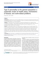
Figure 1 Plasma sCD14 levels in patients w ith BA. Quantitative
analysis of soluble CD14 by ELISA in the plasma of 41 patients with
early-stage biliary atresia (BA), 25 patients followed-up at the
outpatient department (OPD) post-Kasai, 49 patients with late-stage
BA, 9 patients with choledochal cysts (CC), and 7 healthy controls.
Data represent the mean ± SD from duplicate experiments.
Statistical differences were tested by nonparametric Wilcoxon
matched-pairs test. *p<0.05 and **p<0.01 vs. late-stage BA.
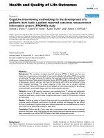
Figure 2 Plasma endotoxin levels in patients with BA. Detection
of plasma endotoxin levels by chromogenic Limulus amebocyte
lysate (LAL) test in 7 healthy controls, 24 patients with early-stage
BA, 18 patients followed-up at the OPD post-Kasai, 18 patients with
late-stage BA, and 9 patients with CC. Data represent the mean ±
SD of duplicate experiments. Statistical differences were tested by
nonparametric Wilcoxon matched-pairs test. *p < 0.05, **p < 0.01.
Chou et al. Journal of Translational Medicine 2010, 8:138
/>Page 4 of 14
BA tissues (Figure 4B and 4E), the CD14 mRNA pre-
sented a constitutive and uniform expression pattern
mainly localized in the hepatocytes and the bile duct
epithelial cells (Figure 4E). The expression of the CD14
mRNA was higher in the early-stage BA tissues (Figure 4E)
than that of control tissues (Figure 4D), but its expres-
sion was significantly decre ased in the late-stage BA
tissues due to loss of hepatocytes (Figure 4F). In addi-
tion, on qRT-PCR analysis, CD14 mRNA levels were
5-fold higher in early-stage BA tissues (n = 9) relative
to the late-stage BA tissues (n = 9) (6.7 ± 1.2 vs.1.4±
0.6, P = 0.002).
The localization of endotoxin in the liver tissues
Immunohistochemical staining using a monoclonal anti-
body against lipid A was performed in liver tissue sections
for detecting the localization of endotoxin. In the normal
liver tissues (Figure 5A), immunoreactivity to lipid A was
weak or absent. However, lipid-A immunoreactivity was
strongly detected around the portal area in hepatocytes,
Kupff er cells, biliary epithelial cells, and some infiltrating
cells in patients with CC (Figure 5B) and in patients with
early-stage BA (Figure 5C). In patients with late-stage BA,
immunoreactivity to lipid A was detected around sites of
fibrous septum forma tion in hepatic parenchymal cells,
Kupffer cells, and biliary epithelial cells (Figure 5D). In the
liver of patients with BA, both hepa tocytes and nonpar-
enchymal liver cells, such as biliary epithelial cells and
Kupff er cells, demonstrated evident uptake of endotoxin,
paralleling the high circulating plasma levels of endotoxin.
Serum enzymes and bilirubin levels in BDL rat model
In the BDL rat model, hepatic injury was associated with
an increase in serum alanine aminotransferase (ALT)
and bilirubin levels. As shown in Figure 6, ALT
increased to 1053 IU/L (BDL vs. sham; P = 0.001) at
Day 1 after ligation, indicating severe liver injury after
BDL. ALT levels decreased afterward and reached a new
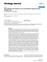
Figure 3 CD14 expression in liver tissues of patients with BA. Comparison of CD14 express ion in paraffi n-embedded liver tissue sections
among the control group (biopsy from neonatal hepatitis and hepatoblastoma) (A), patients with CC (B), patients with early-stage BA (C), and
patients with late-stage BA (D). Liver sections were stained with a monoclonal antibody against CD14 (dark brown) and counterstained with
hematoxylin. Kupffer cells (arrow) and sinusoidal endothelial cells (arrowhead) showed positive immunostain for CD14. Original magnification: × 200.
Chou et al. Journal of Translational Medicine 2010, 8:138
/>Page 5 of 14
Figure 4 CD14 mRNA expression in liver tissues of patients with BA. In situ hybridization of CD14 mRNA in the livers of patients with early-
and late-stage BA. CD14 is stained brown by in situ hybridization with a DIG-labeled CD14 sense (A-C) and antisense RNA probe (D-F). The
paraffin-embedded sections from patients with hepatoblastoma as control (A, D), early-stage BA (B, E) and late-stage BA (C, F) Tissues were
counterstained with hematoxylin. The CD14 mRNA expression pattern mainly localized in the hepatocytes (arrowhead) and the bile duct
epithelial cells (arrow). Original magnification: × 200.
Chou et al. Journal of Translational Medicine 2010, 8:138
/>Page 6 of 14
Figure 5 Endotoxin levels in liver tissues of pa tients with BA. Immunohistochemical staining for endotoxin in the liver tissues of controls
(biopsy from neonatal hepatitis and hepatoplastoma) (A), patients with CC (B), patients with early-stage BA (C), and patients with late-stage BA
(D). Liver sections were stained using a monoclonal antibody against lipid A (HM2046) (left column), mouse IgG1 isotype control antibody
(ab27479) (right column) and counterstained with hematoxylin. Lipid-A immunoreactivity was detected in hepatocytes (arrowhead) and biliary
epithelial cells (arrow), Original magnification: × 200.
Chou et al. Journal of Translational Medicine 2010, 8:138
/>Page 7 of 14
steady-state level of about 180 U/L after Day 7 post-
ligation. However, the total bilirubin continuously
increased after ligation and rea ched its peak at Day 3
(BDL vs. sham; 11.26 ± 1.18 vs. 0.1 ± 0 mg/dL, P < 0.001)
and remained high level throughout the BDL period. The
endotoxin levels in th e plasma and liver tissues were also
significantly increased after Day 1 post-ligation and paral-
leled an increase in plasma bilirubin levels (Figure 7).
CD14 and lipid-A detection in the BLD rat model
Temporal expression of CD14 in hepatocytes was
assessed via immunohistochemical analysis in rats.
CD14 was expressed in the Kupffer cells, sinusoid
endothelial cells and more strongly in hepatocytes
around the portal zones (Figure 8B-F) in rat liver tis-
sues. A significantly higher CD14 expression was dis-
cerned in hepatocytes of BDL rats (Figure 8C-F) as
compared to the sham-operated group. Quantitative
evaluation of CD14 positive cells in live tissues was
performed by an experienced hepatopathologist. If
CD14 positive cells were present in over 10% of the tis-
sue area, CD14 was considered activated. As shown in
Table 2, CD14 activation was a dynamic phenomenon
in BDL group. The expression of CD14 in hepatocytes
was enhanced at 3-6 h post-ligation and returned to
baseline levels by 24 h. Then, CD14 expression was
demonstrated to increase again after 7 days. The BDL
rats also shown a significantly higher CD14 activation
in hepatocytes compared to the sham-operated group
(Figure 8G). Insituhybridization of mRNA of CD14
was performed in rat liver tissues. In additio n to Kupf-
fer cells and sinusoidal endothelial cells, CD14 mRNA
was d emonstrated in hepatocytes and bile duct cells of
the hepatic lobules in control tissues (Figure 9A and
9B).TheexpressionofCD14mRNAinlivertissueof
BDL rats was higher than that of the sham-operated
group at day 14 after BDL, especially in the hepatocytes
(Figure 9D an d 9C).
Hepatic endotoxin levels were higher in the BDL rats
(Figure 10D-F) compared with the sham-operated group
(Figure 10A-C) by immunohistochemical staining. Sig-
nificantly higher endotoxin accumulation was observed
in hepatocytes following BDL. Based on the extent and
intensity of anti-lipid A stain, a semiquantitative method
was used to calculate the ratio with the positive area
over 10% in liver sections. As shown in Table 2 and
Figure 10G, endotoxin was detect ed in liver tissues at 3
h in BDL and sham- operated rats. Like CD14 activation
in the BDL group, endotoxin accumulation was returned
to baseline levels by 24 h and then increased again after
7 days post ligation.
Discussion
Our results demo nstrated for the first time the expres-
sion profile of sCD14 in patients with BA and found sig-
nificantly higher CD14 mRNA and protein levels in
early-stage BA relative to l ate-stage BA and CC. How-
ever, hepatic endotoxin levels remained very high,
despite a signi ficant increase in plasma e ndotoxin levels
in patients with BA compared with control patients.
The liver is thought to be involved in the systemic clear-
ance and detoxication of endotoxin, a nd Kupffer cells
and hepatocytes both contribute to clearing endotoxin
via different recognition systems [24,25]. The production
of sCD14 and LPS binding protein by hepatocytes could
provide a powerful mechanism by which the liver carries
Figure 6 Total bilirubin and ALT levels in rats.Timecourseof
total bilirubin (T-bilirubin; square) and alanine transaminase (ALT;
circle) in rat plasma after bile duct ligation (BDL; closed symbols) or
sham (open symbols) operation. Blood samples were collected at
the time points indicated. T-bilirubin and ALT were assayed using a
biochemistry auto-analyzer (Model 7450; Hitachi, Tokyo, Japan).
Values are mean ± SD (n = 6 in each subgroup). *p < 0.05, **p <
0.005 (sham vs. BDL groups).
Figure 7 Endotoxin levels of plasma and liver tissues in rats.
Time course of endotoxin levels in the liver (circles) and plasma
(squares) after BDL (closed symbols) or sham (open symbols)
operation. Blood samples were collected at the time points
indicated. Endotoxin was assayed using a pyrochrome LAL kit
(Associates of Cape Cod, Falmouth, MA). Values are mean ± SD (n = 6
in each subgroup). *p<0.05, **p < 0.005 (sham vs. BDL groups).
Chou et al. Journal of Translational Medicine 2010, 8:138
/>Page 8 of 14
Figure 8 CD1 4 expre ssion in the liver tissues of rats. CD14 staining in the liver tissues of rats from the sham and BDL groups. Staining of
liver sections using a polyclonal antibody against CD14 shows negligible or no staining in any liver cells in the control (A). Positive staining in
the Kupffer cells (arrow), the sinusoidal endothelial cells (arrowhead) and more strongly in hepatocytes around the portal zones at 3 h after
sham-operation (B), and at 3 h, 1 d, 1w, 3w (C-F) after BDL. Tissues were counterstained with hematoxylin. Original magnification: × 200. The
ratio of CD14 activated (CD14 positive cells were present in over 10% of the tissue area) of the sham (pale bar) and BDL (black bar) groups (G).
Chou et al. Journal of Translational Medicine 2010, 8:138
/>Page 9 of 14
out its function of clearing endotoxin from the blood
stream [26,27].
CD14 expression in the liver increased in many types
of liver disease, including alcohol and cholestatic liver
injuries in rodents [17,28-30]. Immunohistochemical
analysis performed in this study showed higher CD14
expression in Kupffer cells and sinusoidal endothelial
cells in early-stage BA relati ve to late-stage BA. When
the phagocytic function o f Kupffer cells is impaired in
chol estasis, portal derived endotoxin may accumulate in
the liver and spill over into the peripheral circulation
from the intestine [31-33]
.
It is suspected that high
expression of CD14 in Kupffer cells and sinusoidal
endothelial cells may imply a response of these cells to
cholestatic liver injury or to increased endotoxin as a
result of cholestasis. However, the localization of CD14
mRNA was mainly observed in hepatocytes and bile
Table 2 Indexes e of rat liver tissues with positive
reaction
Indexes
CD14 activation Endotoxin
Time Sham Ligation Sham Ligation
N (%)* N (%) N (%) N (%)
3 h (n = 6) 1 (16.7) 4 (66.7) 6 (100) 6 (100)
6 h(n = 6) 1 (16.7) 4 (66.7) 0 4 (66.7)
12 h (n = 6) 2 (33.3) 1 (16.7) 1 (16.7) 3(50)
24 h (n = 6) 1 (16.7) 0 0 1 (16.7)
3 d (n = 6) 0 2 (33.3) 1 (16.7) 1 (16.7)
7 d (n = 6) 0 2 (33.3) 0 5 (83.3)
14 d (n = 6) 1 (16.7) 5 (83.3) 1 (16.7) 6 (100)
21 d (n = 6) 1 (16.7) 6 (100) 0 6 (100)
*Immunohistochemical CD14 and endotoxin staining in the liver tissues of rat
among
sham and common bile duct ligation group. The positive cells were >10% as
positive.
Figure 9 CD14 mRNA expression in the liver tissues of rats. In situ hybridization of CD14 mRNA in the livers from sh am operated and BDL
groups. CD14 is stained brown by in situ hybridization with a DIG-labeled CD14 antisense RNA probe. The paraffin-embedded sections were
hybridized with a sense RNA probe against CD14 in normal tissues as a negative control (A). CD14 is expressed throughout the parenchyma of
the liver tissues of normal controls (B), sham-operated (C) and BDL (D) for 14 days. Tissues were counterstained with hematoxylin. Original
magnification: × 200.
Chou et al. Journal of Translational Medicine 2010, 8:138
/>Page 10 of 14
Figure 10 Endotoxin staining in the liver tissues of rats. Immunohistochemical stain of the liver sections using a monoclonal antibody
against lipid A shows positive staining in the Kupffer cells, hepatocytes, and the sinusoidal endothelial cells at 3 h (A), 1 week r(B) and 3 week
(C) after sham-operated; and at 3 h (D), 1 week (E) and 3 week (F) after BDL. Tissues were counterstained with hematoxylin. Original
magnification: × 200. Statistical analyses of the immunohistochemical score of >10% endotoxin in liver sections of the sham (pale bar) and BDL
groups (black bar) (G).
Chou et al. Journal of Translational Medicine 2010, 8:138
/>Page 11 of 14
duct epithelial cells. Therefore,wecannotruleoutthe
possibility that CD14 production by hepatocytes and
bile duct epithelial cells during cholestasis and its subse-
quent transportation to the Kupffer cells and sinusoidal
endothelial cells produces an unknown downstream
effect. Our results provided evidence that BA d oes not
exempt from endotoxin accumulation in the peripheral
circulation and liver, just like other cholestatic disorders.
sCD14 have been demonstrated direct secretion by
hepatocytes during early-stage of BA to enhance endo-
toxin clearance [15,34]. During cholestasis, the accumu-
lation of endotoxin may induce hepatocyte injury and
impair CD14 synthesis during late-stage BA.
Although studies of liver injury should take into
account the relative contributions of CD14 in Kupffer
cells and hepatocytes, few reports document the propor-
tional change of mCD14 and sCD14 in the liver and the
consequent pathogenetic effects on cholestatic diseases
[16,29]. The balance between activation and inhibition
of endotoxin responses by sCD14 depends on its con-
centration [35,36]. At a higher physiological concentra-
tion, sCD14 can compete with mCD14 and inhibit LPS
activation of CD14-positive cells [35,36] . It is likely that
increa sed plasma levels of sCD14 during early-stage BA
observed in this s tudy implies a protective mechanism
in the liver tha t guards against increased endotoxin due
to cholestasis. Conversely, decreased sCD14 in late-stage
BA, without a concomitant decrease of endotox in in the
liver and blood, may indicate a loss of protection against
CD14-mediated LPS activation in Kupffer cells that pro-
pagates inflammatory reac tions and fibrogenesis, result-
ing in irreversible liver injury a nd end-stage liver
cirrhosis in patients with BA [10,16,35].
In an animal model system injected intraperitoneally
with LPS, the initial and rapid induction of CD14 expres-
sion in myeloid cells is followed by a second, slower
response in epithelial cells, which peak s at 8-16 h [37].
This epithelial cell response appears to require higher
concentrations of LPS induction and is dependent on
TNF-a to promote synthesis of CD14 [38]. Apart from
its apparent role as an LPS receptor that mediates activa-
tion of myeloid cells, CD14 also appears to serve as an
opsonic receptor for engulfment by phagocytes, resulting
in clearance of LPS. Liver is the main clearance organ for
intravenously injected LPS, and this is mediated by Kupf-
fer cells, sinusoidal cells, granulocytes, and hepatocytes in
rats [39] . There is evidence to suggest that LPS may be
cleared from the liver via the bile canalicular system into
the gut. Indeed, 3 h after LPS injection, bile samples
taken from the gall bladder of rabbits contained substan-
tial amounts of LP S, equivalent to that foun d in the
plasma [40]. In our BDL rat model, we confirmed that
endotoxemia and hepatocyte CD14 production occurred
after ligation. CD14 was exp ressed in an LPS-inducible
manner in Kupffer cells, neutrophils, hepatocytes, and
bile duct epithelium, suggesting a possible role for CD14
in hepatocytes during the uptake and clearance of LPS
from the circulation. However the endotoxin levels in
liver tissues were still high due to cholesta sis, and CD14
production was increased again at 7 days after ligation.
Although sCD14 has been observed in normal human
serum and is increased in sera from septic patients
[14,41]. The origin of sCD14 has yet to be determined. It
has been assumed that sCD14 is derived from the mem-
brane bound form present on myeloid cells either by
phospholipase cleavage of the GPI anchor or by protease
digestion [14]. However, patients with paroxysmal noctur-
nal hemoglobinuria have n ormal sCD14 levels in their
sera, although monocytes from these patients do not
express CD14 on their surface [42]. Our observation that
the CD14 mRNA is detected in hepatocytes raises the
possibility that sCD14 may also originate from these cells.
Recent studies have shown that the CD14 antigen is
expressed in man y types of cells and tissues [37,43 ,44].
Some reports suggest that sCD14 behaves like other
acute-phase proteins [14]. Hepatocytes are the major
source of most acute-phase proteins; therefore, hepato-
cytes might be expected to express CD14, which is upre-
gulated during endotoxemia induced by cholestasis
[10,16,39]. These results are in agreement with other pre-
vious reports that demonstrated the synthesis and expres-
sion of CD14 are markedly upregulated by LPS during
endotoxemia induced by cholestasis [3,8,43]. In the liver,
besides hepatocytes, nonparenchymal cells such as Kupf-
fer cells, endothelial cells, neutrophils, and other cells can
also express the CD14 mRNA and synthesize the CD14
protein [21,22,26], but the fact that both isolated hepato-
cytes and hepatic tissues express the CD14 protein and
its mRNA indicate that the nonparenchymal cells could
hardly have any effect on such expression in liver tissues.
Although we do not provide direct evidence here that
sCD14 in the plasma originates from hepatocytes during
endotoxemia, our results showed that there was the pos-
sibility that the liver is an important source of sCD14
during endotoxemia. Pan et al [21]. found that the liver
is one of the major organs involved in the production of
sCD14. Liu et al [26] also reported that CD14 transcrip-
tion rates are significantly increased in hepatocytes from
LPS-treated rats, indicating that the upregulation of
CD14 mRNA levels observed in rat hepatocytes after LPS
treatment is dependent, in part, on incr eased transcrip-
tion. Their observations also support the idea that sCD14
could be an ac ute-p hase protein a nd hepatocytes might
be a source of circulating sCD14 production. Our data
indicated that hepatocytes from BDL rats expressed
higher amounts o f CD14 mRNA and protein and may
have released more sCD14 for promoting endotoxin
clearance.
Chou et al. Journal of Translational Medicine 2010, 8:138
/>Page 12 of 14
Conclusions
In conclusion, our in vivo data indicated the liver as a
main source of sCD14 production during endotoxemia.
However, the significantly decreased sCD14 expression
in late-stage BA without a concomitant decrease in
plasma endotoxin levels suggest ed that the pathogenetic
mechanism underlying CD14-mediated liver injury dur-
ing BA is still unresolved.
Abbreviations
(BA): biliary atresia; (BDL): bile duct ligation; (KP): Kasai procedure; (qRT-PCR):
real-time quantitative reverse-transcription polymerase chain reaction
Acknowledgements
We thank Drs. Chih-Sung Hsieh and Shin-Ye Lee and the liver transplant
team for providing the samples used in this study. This study was supported
by grant #NSC 92-2314-B-182-076 and # NSC 92-2314-B-182-076 from the
National Science Council, Taiwan.
Author details
1
Institute of Basic Medical Sciences, National Chang Kung University, Tainan,
Taiwan.
2
Graduate Institute of Clinical Medical Sciences, Chang Gung
University, Kaohsiung, Taiwan.
3
Department of Surgery, Chang Gung
Memorial Hospital - Kaohsiung Medical Center, Chang Gung University
College of Medicine, Kaohsiung, Taiwan.
4
Department of Pathology, Chang
Gung Memorial Hospital - Kaohsiung Medical Center, Chang Gung University
College of Medicine, Kaohsiung, Taiwan.
5
Department of Laboratory
Medicine, E-DA Hospital/I-SHOU University, Kaohsiung, Taiwan.
6
Department
of Medical Research, E-DA Hospital/I-SHOU University, Kaohsiung, Taiwan.
Authors’ contributions
MHC performed the experiments and drafted the manuscript. JHC designed
the experiment and clinical specimen collection. HLE was the pathologist
and evaluated the histopathology of the cases. CMC participated in
experiment performance and technical support. CHW helped analyze the
data. CLC coordinated the study and drafted the manuscript. TML pro vided
scientific advice, discussions of data and submitted the manuscript. All
authors read and approved the final manuscript.
Competing interests
The authors declare that they have no competing interests.
Received: 13 October 2010 Accepted: 21 December 2010
Published: 21 December 2010
References
1. Bassett MD, Murray KF: Biliary atresia: recent progress. J Clin Gastroenterol
2008, 42:720-729.
2. Hartley JL, Davenport M, Kelly DA: Biliary atresia. Lancet 2009,
374:1704-1713.
3. Wadhwani SI, Turmelle YP, Nagy R, Lowell J, Dillon P, Shepherd RW:
Prolonged neonatal jaundice and the diagnosis of biliary atresia: a
single-center analysis of trends in age at diagnosis and outcomes.
Pediatrics 2008, 121:e1438-1440.
4. Reyes H, Zapata R, Hernandez I, Gotteland M, Sandoval L, Jiron MI, Palma J,
Almuna R, Silva JJ: Is a leaky gut involved in the pathogenesis of
intrahepatic cholestasis of pregnancy? Hepatology 2006, 43:715-722.
5. Marpegan L, Leone MJ, Katz ME, Sobrero PM, Bekinstein TA, Golombek DA:
Diurnal variation in endotoxin-induced mortality in mice: correlation
with proinflammatory factors. Chronobiol Int 2009, 26:1430-1442.
6. Andonegui G, Zhou H, Bullard D, Kelly MM, Mullaly SC, McDonald B,
Long EM, Robbins SM, Kubes P: Mice that exclusively express TLR4 on
endothelial cells can efficiently clear a lethal systemic Gram-negative
bacterial infection. J Clin Invest 2009, 119:1921-1930.
7. Neyrinck AM, Taper HS, Gevers V, Declerck B, Delzenne NM: Inhibition of
Kupffer cell activity induces hepatic triglyceride synthesis in fasted rats,
independent of lipopolysaccharide challenge. J Hepatol 2002, 36:466-473.
8. Klintman D, Li X, Santen S, Schramm R, Jeppsson B, Thorlacius H: p38
mitogen-activated protein kinase-dependent chemokine production,
leukocyte recruitment, and hepatocellular apoptosis in endotoxemic
liver injury. Ann Surg 2005, 242:830-838, discussion 838-839.
9. Ramnath RD, Ng SW, Guglielmotti A, Bhatia M: Role of MCP-1 in
endotoxemia and sepsis. Int Immunopharmacol 2008, 8:810-818.
10. Leicester KL, Olynyk JK, Brunt EM, Britton RS, Bacon BR: Differential findings
for CD14-positive hepatic monocytes/macrophages in primary biliary
cirrhosis, chronic hepatitis C and nonalcoholic steatohepatitis. Liver Int
2006, 26:559-565.
11. Lin YF, Lee HM, Leu SJ, Tsai YH: The essentiality of PKCalpha and
PKCbetaI translocation for CD14+monocyte differentiation towards
macrophages and dendritic cells, respectively. J Cell Biochem 2007,
102:429-441.
12. Nicu EA, van der Velden U, Everts V, Loos BG: Expression of FcgammaRs
and mCD14 on polymorphonuclear neutrophils and monocytes may
determine periodontal infection. Clin Exp Immunol 2008, 154:177-186.
13. Zhao Z, Fleming R, McCloud B, Klempner MS: CD14 mediates cross talk
between mononuclear cells and fibroblasts for upregulation of matrix
metalloproteinase 9 by Borrelia burgdorferi. Infect Immun 2007,
75:3062-3069.
14. Bas S, Gauthier BR, Spenato U, Stingelin S, Gabay C: CD14 is an acute-
phase protein. J Immunol 2004, 172:4470-4479.
15. Ahmed AF, Nio M, Ohtani H, Nagura H, Ohi R:
In situ CD14 expression in
biliary atresia: comparison between early and late stages. J Pediatr Surg
2001, 36:240-243.
16. Shih HH, Lin TM, Chuang JH, Eng HL, Juo SH, Huang FC, Chen CL, Chen HL:
Promoter polymorphism of the CD14 endotoxin receptor gene is
associated with biliary atresia and idiopathic neonatal cholestasis.
Pediatrics 2005, 116:437-441.
17. Isayama F, Hines IN, Kremer M, Milton RJ, Byrd CL, Perry AW, McKim SE,
Parsons C, Rippe RA, Wheeler MD: LPS signaling enhances hepatic
fibrogenesis caused by experimental cholestasis in mice. Am J Physiol
Gastrointest Liver Physiol 2006, 290:G1318-1328.
18. Von Hahn T, Halangk J, Witt H, Neumann K, Muller T, Puhl G, Neuhaus P,
Nickel R, Beuers U, Wiedenmann B, Berg T: Relevance of endotoxin
receptor CD14 and TLR4 gene variants in chronic liver disease. Scand J
Gastroenterol 2008, 43:584-592.
19. Su GL: Lipopolysaccharides in liver injury: molecular mechanisms of Kupffer
cell activation. Am J Physiol Gastrointest Liver Physiol 2002, 283:G256-265.
20. Su GL, Goyert SM, Fan MH, Aminlari A, Gong KQ, Klein RD, Myc A,
Alarcon WH, Steinstraesser L, Remick DG, Wang SC: Activation of human
and mouse Kupffer cells by lipopolysaccharide is mediated by CD14. Am
J Physiol Gastrointest Liver Physiol 2002, 283:G640-645.
21. Pan Z, Zhou L, Hetherington CJ, Zhang DE: Hepatocytes contribute to
soluble CD14 production, and CD14 expression is differentially regulated
in hepatocytes and monocytes. J Biol Chem 2000, 275:36430-36435.
22. Su GL, Dorko K, Strom SC, Nussler AK, Wang SC: CD14 expression and
production by human hepatocytes. J Hepatol 1999, 31:435-442.
23. Wilkinson DG: RNA detection using non-radioactive in situ hybridization.
Curr Opin Biotechnol 1995, 6:20-23.
24. Gegner JA, Ulevitch RJ, Tobias PS: Lipopolysaccharide (LPS) signal
transduction and clearance. Dual roles for LPS binding protein and
membrane CD14. J Biol Chem 1995, 270:5320-5325.
25. Kitchens RL, Munford RS: CD14-dependent internalization of bacterial
lipopolysaccharide (LPS) is strongly influenced by LPS aggregation but
not by cellular responses to LPS. J Immunol 1998, 160:1920-1928.
26. Liu S, Khemlani LS, Shapiro RA, Johnson ML, Liu K, Geller DA, Watkins SC,
Goyert SM, Billiar TR: Expression of CD14 by hepatocytes: upregulation by
cytokines during endotoxemia. Infect Immun 1998, 66:5089-5098.
27. Mimura Y, Sakisaka S, Harada M, Sata M, Tanikawa K: Role of hepatocytes
in direct clearance of lipopolysaccharide in rats. Gastroenterology 1995,
109:1969-1976.
28. Su GL, Rahemtulla A, Thomas P, Klein RD, Wang SC, Nanji AA: CD14 and
lipopolysaccharide binding protein expression in a rat model of
alcoholic liver disease. Am J Pathol 1998, 152
:841-849.
29. Tracy TF Jr, Fox ES: CD14-lipopolysaccharide receptor activity in hepatic
macrophages after cholestatic liver injury. Surgery 1995, 118:371-377.
30. Zuo GQ, Gong JP, Liu CA, Li SW, Wu XC, Yang K, Li Y: Expression of
lipopolysaccharide binding protein and its receptor CD14 in
experimental alcoholic liver disease. World J Gastroenterol 2001, 7:836-840.
Chou et al. Journal of Translational Medicine 2010, 8:138
/>Page 13 of 14
31. Kaser A, Ludwiczek O, Waldenberger P, Jaschke W, Vogel W, Tilg H:
Endotoxin and its binding proteins in chronic liver disease: the effect of
transjugular intrahepatic portosystemic shunting. Liver 2002, 22:380-387.
32. Roughneen PT, Drath DB, Kulkarni AD, Rowlands BJ: Impaired nonspecific
cellular immunity in experimental cholestasis. Ann Surg 1987,
206:578-582.
33. Sasatomi K, Noguchi K, Sakisaka S, Sata M, Tanikawa K: Abnormal
accumulation of endotoxin in biliary epithelial cells in primary biliary
cirrhosis and primary sclerosing cholangitis. J Hepatol 1998, 29:409-416.
34. Lee JW, Paape MJ, Elsasser TH, Zhao X: Recombinant soluble CD14
reduces severity of intramammary infection by Escherichia coli. Infect
Immun 2003, 71:4034-4039.
35. Chuang JH, Chou MH, Wu CL, Du YY: Implication of innate immunity in
the pathogenesis of biliary atresia. Chang Gung Med J 2006, 29:240-250.
36. Kitchens RL, Thompson PA: Modulatory effects of sCD14 and LBP on LPS-
host cell interactions. J Endotoxin Res 2005, 11:225-229.
37. Fearns C, Kravchenko VV, Ulevitch RJ, Loskutoff DJ: Murine CD14 gene
expression in vivo: extramyeloid synthesis and regulation by
lipopolysaccharide. J Exp Med 1995, 181:857-866.
38. Fearns C, Loskutoff DJ: Role of tumor necrosis factor alpha in induction
of murine CD14 gene expression by lipopolysaccharide. Infect Immun
1997, 65:4822-4831.
39. Li SW, Gong JP, Wu CX, Shi YJ, Liu CA: Lipopolysaccharide induced
synthesis of CD14 proteins and its gene expression in hepatocytes
during endotoxemia. World J Gastroenterol 2002, 8:124-127.
40. Schimke J, Mathison J, Morgiewicz J, Ulevitch RJ: Anti-CD14 mAb
treatment provides therapeutic benefit after in vivo exposure to
endotoxin. Proc Natl Acad Sci USA 1998, 95:13875-13880.
41. Landmann R, Reber AM, Sansano S, Zimmerli W: Function of soluble CD14
in serum from patients with septic shock. J Infect Dis 1996, 173:661-668.
42. Schutt C, Schilling T, Grunwald U, Stelter F, Witt S, Kruger C, Jack RS:
Human monocytes lacking the membrane-bound form of the bacterial
lipopolysaccharide (LPS) receptor CD14 can mount an LPS-induced
oxidative burst response mediated by a soluble form of CD14. Res
Immunol 1995, 146:339-350.
43. Kimmings AN, van Deventer SJ, Obertop H, Rauws EA, Huibregtse K,
Gouma DJ: Endotoxin, cytokines, and endotoxin binding proteins in
obstructive jaundice and after preoperative biliary drainage. Gut 2000,
46:725-731.
44. Nishimura M, Naito S: Tissue-specific mRNA expression profiles of human
toll-like receptors and related genes. Biol Pharm Bull 2005, 28:886-892.
doi:10.1186/1479-5876-8-138
Cite this article as: Chou et al.: Endotoxin and CD14 in the progression
of biliary atresia. Journal of Translational Medicine 2010 8:138.
Submit your next manuscript to BioMed Central
and take full advantage of:
• Convenient online submission
• Thorough peer review
• No space constraints or color figure charges
• Immediate publication on acceptance
• Inclusion in PubMed, CAS, Scopus and Google Scholar
• Research which is freely available for redistribution
Submit your manuscript at
www.biomedcentral.com/submit
Chou et al. Journal of Translational Medicine 2010, 8:138
/>Page 14 of 14