Báo cáo hóa học: " Modulation of the major histocompatibility complex by neural stem cell-derived neurotrophic factors used for regenerative therapy in a rat model of stroke" ppt
Bạn đang xem bản rút gọn của tài liệu. Xem và tải ngay bản đầy đủ của tài liệu tại đây (867.22 KB, 10 trang )
RESEARC H Open Access
Modulation of the major histocompatibility
complex by neural stem cell-derived neurotrophic
factors used for regenerative therapy in a rat
model of stroke
Chongran Sun
1,2
, Han Zhang
1
, Jin Li
1
, Hua Huang
1
, Hongbin Cheng
1
, Yajie Wang
3
, Ping Li
4
, Yihua An
1*
Abstract
Background: The relationship between functional improvements in ischemic rats given a neural stem cell (NSC)
transplant and the modulation of the class I major histocompatibility complex (MHC) mediated by NSC-derived
neurotrophins was investigated.
Methods: The levels of gene expression of nerve growth factor (NGF), brain-derived neurotropic factor (BDNF) and
neurotrophin-3 (NT-3) were assayed from cultures of cortical NSC from Sprague-Dawley rat E16 embryos. The levels
of translated NGF in spent culture media from NSC cultures and the cerebral spinal fluid (CSF) of rats with and
without NGF injection or NSC transplant were also measured.
Results: We found a significant increase of NGF, BDNF and NT-3 transcripts and NGF proteins in both the NSC
cultures and the CSF of the rats. The immunochemical staining for MHC in brain sections and the enzyme-linked
immunosorbent assay of CSF were carried out in sham-operated rats and rats with surgically induced focal cerebral
ischemia. These groups were further divided into animals that did and did not receive NGF administration or NSC
transplant into the cisterna magna. Our results sho w an up-regulation of class I MHC in the ischemic rats with NGF
and NSC administration. The extent of caspase-III immunoreactivity was comparable among thre e arms in the
ischemic rats.
Conclusion: Readouts of somatosensory evoked potential and the trap channel test illustrated improvements in
the neurological function of ischemic rats treated with NGF administr ation and NSC transplant.
Introduction
Ischemic stroke is a common neurological disorder and
is one of the leading causes of casualty w orldwide. It is
caused by the occlusion of a cerebral artery with
thrombi and emboli, which leads to an infarction and
the death of neural tissue. Current treatments are pri-
marily palliative and are useful to only a minority of
patients after stroke. Currently, there is no effective
treatment for restoring the neurological functions l ost
during a stroke. Recent studies in pre-clinical and clini-
cal trials have shown that stem cell-based therapy can
lead to symptomatic relief and may offer a novel poten-
tial treatment [1]. Nevertheless, the underlying therapeu-
tic mechanisms f or neural repair and the induction of
functional improvement remains controversial.
The ability of neural stem cells (NSC) to differentiate
into neural cells has been seen in culture [2]. Given the
complexity of both the structure and function of the
central nervous system (CNS), it is critical to understand
the mechanisms by which transplanted neural cells can
replace the damaged cells and interact with healthy host
cells in a well-organized manner. Cell-based therapy
might elicit a chaperone effect i n the at-risk neural tis-
sue surrounding the lesioned area via the up-regulation
of neurotrophic and neurop rotective factors, which help
* Correspondence:
1
Department of Neural Stem Cell, Beijing Neurosurgical Institute, Beijing
Tiantan Hospital, Capital Medical University, China
Full list of author information is available at the end of the article
Sun et al. Journal of Translational Medicine 2010, 8:77
/>© 2010 Sun et al; licensee BioMed Central Ltd. This is an Open Acces s article distributed under the terms of the Creative Commons
Attribution License ( which permits unrestricted use, distribu tion, and reproduction in
any medium, provided the original work is prop erly cited.
to promote the survival, migration and differentiation of
endogenous precursors after stroke [3].
In rats, the administration of nerve growth factor
(NGF) has been shown to enhance the expression of the
class I major histocompatibility complex (MHC) in neu-
rons,butnotinglialcells,anddecreasetheexpression
of the class II MHC in g lias [4]. Immune response and
inflammation are common sources of secondary injury
in neural cells after stroke. In vitro cultures have been
used to demonstrate that NSC, neurons and glias
express b oth class I and II MHCs, which were recently
recognized to be crucial in the activity-dependent refine-
ment and plasticity of neural connections in the devel-
oping and adult CNS [5]. We hypothesized that the
functional improvements in ischemic rats given NSC
transplant might be related to modulation of the class I
MHC, mediated by NSC-derived neurotrophins in the
lesioned micro-environment of the CNS.
Materials and methods
Culture of Neural stem cells
Neural stem cells were harvested from the cortex of E16
Sprague-Dawley rat embryos. The head was decapitated
and the whole brain was removed from the skull.
Meninges, choroid plexus and coherent blood vessels
were carefully stripped off. The tissue was cut into small
pieces, triturated with a glass pipette and allowed to
pass through a 28-mesh copper sieve to remove large
chunks. After three washes with Dulbecco’s modified
Eagle’ s medium (DMEM; Sigma-Aldrich, St. Louis,
), 1.5×10
7
cells were seeded
in 15 mL of high-glucose DMEM/F12 (Sigma-Aldrich,
St. Louis) supplemented with 2 % B27 (Gibco, Carlsbad,
CA, ), 20 μg/L basic fibroblast
growth factor (FGF, PeproTech, NJ, ro-
tech.com) and 20 μg/L epidermal growth factor (EGF,
PeproTech, NJ) onto a 75 cm
2
non-adherent tiss ue cul-
ture flask (Laixin, Shanghai, China, 17.
cebiz.cn) and maintained at 37°C i n a humidified envir-
onment with 5% CO
2
. Cultures were passaged with
0.25% tripsin and titration with a glass pipette once a
week, and half of the spent culture media was replaced.
Enzyme-linked immunosorbent assay (ELISA) of NGF
One-day-old medium from the first seven passages of
NSC cultures and 80 μLofcerebralspinalfluid(CSF)
from 40 Sprague-Dawley rats were collected and centri-
fuged at 400 g for 10 min to remove cellular debris. The
supernatant was stored at -80°C. An ELISA kit (Boster,
Hubei, China, ) was utilized,
following the manufacturer’s protocol, to quantify the
NGF present in the culture supernatants and CSF.
Briefly, 100 μL of sample and standards were added to
plates that were pre-coated with monoclonal anti-NGF
and allowed to react for 1.5 h at 37°C. Samples were
washed thoroughly, and then incubated with 100 μLof
biotin-conjugated a nti-NGF at 37°C for 1 h. Plates w ere
washed to remove the unbound anti-NGF and incubated
again with 100 μL of streptavidin-conjugated horserad-
ish peroxidase at 37°C for 30 min. Signals were
developed by adding 100 μL of chromogenic tetra-
methylbenzidine. The reaction was arrested after 15 min
with 100 μL of stopping solution. The absorbance was
read at 450 nm. The color intensity of this reaction is
proportional to the amount of bound NGF. A standardi-
zation plot was established for NGF standards at 250,
125, 62.5, 31.3, 15.6, 7.8 and 3.9 pg/mL. The diluting
buffer was used as the negative control. All measure-
ments were performed in triplicate.
Molecular Analyses
RNA extraction and cDNA transcription
To test the total RNA, positive and negative controls
were extracted using the RNAqueous®-Midi Kit and the
manufacturer’ s protocol (Applied Biosystems, Foster
City,CA,).Briefly,cellswere
disrupted with the lysis buffer composed of a high con-
centration of guanidinium salt. The lysate was diluted
with a 64% ethanol solution and passed through the
glass fiber filter in the RNAqueous Filter Cartridge.
RNA bound to the filter while othercellularcontents
flowe d through it. The Filter Cartridge was washed with
the wash solutions to remove contaminants, and the
RNA was then eluted using the Elution Solution of very
low ionic strength. RNA integrity was monitored for
two sharp intense bands, 18s and 28s, by running an ali-
quot of the preparation on a denaturing agarose gel and
staining with ethidium bromide. RNA concentration was
determined spectrophotometrically at 260 nm and 280
nm wavelengths. cDNA were transcribed from 1 μgof
RNA with 2.5 μM Oligo dT and 200 U ExScript reverse
transcriptase (TaKaRa, Japan, ara-bio.
com) in a 20 μL reverse-transcription reaction mix con-
taining 500 μM dNTP and 40 U R Nase-Inhibitor
(Sigma-Aldrich, St. Louis). RNA and Oligo dT were
incubated for 5 min at 65°C in a thermal cycler and
quickly chilled on ice. The thermo-profile of the cDNA
generation was 42°C for 15 min and 95°C for 2 min,
ending at 4°C.
Real-time PCR
The level of gene expression for nerve growth factor
( NGF ), brain-derived neurotrophic factor (BDNF)and
neurotrophin-3 (NT-3) from NSC cultures at passages
5-7 were quantified using the ABI 7300 Real -Time PCR
System (Applied Biosystems, Foster City, CA, http://
www.appliedbiosystems.com) and the specific primer-
pairs for BDNF (forwa rd: 5′-ACC CTG AGT TCC ACC
AGG TG-3′ ,reverse:5′-TGG GCG CAG CCT TCA
Sun et al. Journal of Translational Medicine 2010, 8:77
/>Page 2 of 10
T-3′ ), NGF (fo rward:5′-TGG ACC CAA GCT CAC
CTCA-3′, reverse: 5′-GGA TGA GCG CTT GCT CCT-
3′), NT-3 (forward: 5′-GAT CTT ACA GGT GAA CAA
GGT GAT G-3′, reverse: 5′- TTG ATC CAT GTT GTT
GCC TTG-3′) and the house keeping gene b-actin (for-
ward: 5′ -CTA CAA TGA GCT GCG TGT GG-3′ ,
reverse:5′-CAG TCA GGA TCT TCA TGA GG-3′). The
thermo-profile was 50°C for 2 min and 95°C for 10 min,
followed by 40 cycles of 95°C for 15 s, 60°C for 1 min,
and finally 95°C for 15 s, 60°C to 95°C for 30 s, and
95°C for 15 s. Quant ification of the gene of interest was
accomplished by measuring the threshold cycles and
compa ring them to the standard curve to determine the
copy number. The process of calculating threshold
cycles, preparing a standard curve, and determining the
copy number was performed using system software. All
measurements were performed in triplicate in two sepa-
rate experiments.
Preparation of NSC for Transplant
NSC at passage 6 were labeled with 10 μM bromodeoxy-
uridine (Sigma-Aldrich, St. Louis) in the supplemented
culture medium one day prior to transplantation to
ischemic animals for in vivo study. BrdU-labeled cells were
then trypsinized, washed and adjusted to 5 × 10
3
/μlin
Dulbecco’s phosphate-buffered saline (PBS; Boster, Hubei,
China).
Induction of focal cerebral Ischemia in rats
Animal treatments were designed to minimize pain or
discomfort in accordance with the current protocols
approved by the Chinese Medical Ethical Committee for
animal welfare. Forty Sprague-Dawley rats (Weitongli-
hua, Beijing, China, ) at a
mean age of 14 weeks and body weight between 240
and 260 g were maintained on a 12-hour light/dark
schedule and randomly assigned to the following four
groups: (A) normal control with sham-operation (CG),
(B) ischemic group with PBS injection (IG), (C) ischemic
group treated with NGF (NGFG) and (D) ischemic
grouptransplantedwithNSC(NSCG).Animalswere
anesthetized with an intra-peritoneal injection of 400
mg/kg chloral hydrate (Pharmaceutical Plant of Tiantan
Hospital, Beijing, China, ). The rec-
tal temperature was monitored an d maintained at 37.5°
C with a thermal pad throughout the surgical procedure.
A scalp i ncision of 0.5 cm was made at one-third distal
area between the left eye and ear. The temporalis was
separated to expose the zygoma and squamosal bone. A
burr hole of 1.5 × 2 mm was made using a 1 mm
micro-drill rostal to the anterior junction of the zygoma
and the squamosal bone. The dura mater was carefully
pierced with an iris knife. The exposed middle cerebral
artery was isolated and ligated using a 10-0 suture. After
coveringtheburrholewithapieceofgelatinsponge,
the temporalis and overlying skin were sutured. Animals
were then placed in the supine position and a midline
incision was made in the neck. The bilateral common
carotid arteries were isolated. The left artery was ligated
with 4-0 suture, whereas the right artery was occluded
using a micro-aneurysm clip for 1.5 h. The skin was
then sutured. Ope rated animals were kept individually
for a day and then in a cage for six. All 30 lesioned rats
showed signs of consciousness disturbance, including
drowsiness, paucity of movement and coma. Sham-oper-
ated animals did not receive the ligation or occlusion.
Transplantation
Seven days after the induction of focal cerebral ischemia,
animals were anesthetized with an intra-peritoneal injec-
tion of 400 mg/kg chloral hydrate (Pharmaceutical Plant
of Tiantan Hospital, Beijing, China). The rectal tempera-
ture was monitored and maintained at 37.5°C. A scalp
incision was made behind the superior nuchal line at 0.5
cm. The posterior occipital muscle was separated to
expose the atlanto-occipital membrane. 1.5 × 10
5
NSC
at passage six were suspended in 30 μLPBSand
injected into the cisterna magna through the atlanto-
occipital membrane in NSCG. 30 μLof10ngNGFand
PBS were injected into the cisterna magna in NGFG,
and 30 μL PBS were injected into IG.
Behavioral Assessment
Somatosensory response
Evoked potentials are the electrical signals generated by
the nervous system in response to sensory stimuli. The
measurement of somatosensory evoked potentials has
been used in the diagnosis and prognosis of neurologic
disorders[6]. Animals were anesthetized with chloral
hydrate 5 days before and 2 days, 7 days, 14 days and
28 days after NSC transplantation or PBS injection. An
incision of 0.8 cm was made in the midline of the skull.
Bilateral burr holes were made at 1 mm posterior and 3
mm lateral to the bregma by using a micro-drill and 1 ×
3 mm coordinate paper. Titanium alloy electrodes were
placed into th e burr holes and fixed with sterilized bone
wax. The scalp was sutured. Somatosensory evoked
potentials (SEPs) were measured using an Axon electr o-
physiology monitoring system (Axon Instrument, Sun-
nyvale, CA, ). The stimulating
electrode and the reference electrode were placed in the
median nerve of the muscles between the ulner and
radical bone, and the pre-frontal scalp of the midline,
respectively. SEPs were obtained by electrical stimula-
tion of 200 pulses at 2 Hz and 0.8 mA. The measured
latency was the time span from the stimulation to the
beginning of the first w ave, whereas the amplitude was
thevoltagedifferencebetweenthepositiveandthe
negative wave. The rel ative latency and amplitude are
Sun et al. Journal of Translational Medicine 2010, 8:77
/>Page 3 of 10
expressed as r atios of l atencies and amplitudes of the
intact side to the injured side, respectively.
Trap channel test
The trap channel test for the analysis of motor function
in rats has been previously descri bed elsewhere[7].
Briefly, animals were allowed to crawl three times along
a horizontal channel made of Plexiglas that was 75 cm
long, 10 cm wide and 10 cm high, with evenly placed
2-cm long stepping platforms. The motor functions o f
forelimbs and hind-l imbs of rats with and without NSC
transplantat ion or NGF or PBS injection were assessed
onday2,day7,day14andday28bycountingthe
number of foot faults in the left and right forelimbs.
The sum of foot faults (SOFFF) in the forelimbs is
defined as the number of faults of right and left fore-
limb s, whereas the differentiation of forelimb foot faults
(DOFFF) is the difference between the number of faults
for the right and left forelimbs. SOFFF and DOFFF were
used as an index of motor deficit.
CSF aspiration
80 μL of CSF were aspirated from the cis terna magna of
40 chloral hydrate-sedated rats on week 4. The collected
CSF was spun at 400 g for 10 min to remove cell contam-
ination, and the supernatant was then stored at -80°C.
Tissue Processing for histology and immunohistochemical
staining
Upon completion of the in vivo monitoring, rats were
anesthetized with 600 mg/kg chloral hydrate. The thor-
axes were cut open, and the animals were trans-cardia-
cally perfused with physiologic saline and 4%
paraformaldehyde. The brains were then fixed in 30%
sucrose. The freshly isolated brains were cut into 20 μm
thick coronal slices with a cryo-mount and mounted
onto poly-L-lysine-coated slides. To prepare the cells for
immunohistochemical staining, cultures of NSC were
enzymatically segregated wit h trypsin. After thorough
wash ing, 1 × 10
5
separated cells suspended in PBS were
cytospun on slides and kept at -80°C until staining.
Immunohistochemistry staining
Immunohistochemical staining of nestin and BrdU were
performed to assess the property of NSC in cultures and
track the migration and homing of allogeneic BrdU-
labeled NSC in the host brain. In addition to the expres-
sion of th e class I and II MHC, the activation of caspase
III in NSC cultures and brain sections was also assayed.
Primary antibodies, including anti-nestin (1:150; Sigma-
Aldrich, St. Louis), anti-BrdU (1: 400; Sigma-Aldrich, St.
Louis), anti-class I MHC (1:200; AbD Serotec, NC,
), anti-class II MHC (anti-RT-
1B 1:200; AbD Serotec, NC) and anti-caspase III (1:50,
Abcam, Cambridge, UK, ), were
employed. Incubation was conducted at 4°C for
24 hours. After extensive washing, signals were detected
and visualized using Histo stainT M-SP kit and the man-
ufacturer’ s protocol (Zhongshan Beijing, China, http://
gjj.cc/nongye/shengwugongcheng/zsbio.htm).
Statistical Analysis
Results are expressed as mean ± standard deviation
(SD). The non-parametric one-way ANOVA was applied
to analyze continuous variables: the gene expression of
NGF, BDNF and NT-3 derived from different culture
passages, NGF concentration in different NSC culture
passages and CSF aspirated from studied rats, relative
latency and amplitude of SEP and the sum and differen-
tiation of forelimb-foot fault of studied rats at different
points. Data were analyzed using SPSS software version
11.5 (SPSS, IL, ). Differences
between groups were regarded as significant if p ≤ 0.05.
Results
Primary NSC cultures
Primary cells segregated from the neural cortex of E16
Sprague-Dawley rat embryos formed free-floating neuro-
spheres in the serum-free medium supplemented with
FGF and EGF. Cultures were passaged after seven days.
In two separate experiments of two replicate cultures at
passages 1-7, the trypan blue dye exclusion tests
revealed a cell viability of 79 ± 4.4% (range: 72 - 86%).
Immunohistochemical staining demonstrated nestin-
positive neurospheres up to passage 7, suggesting the
successful ex vivo expansion of NSC (Data not shown).
In vitro characterization of NSC
Quantitative real-time PCR was conducted to determine
the gene expression of neurotrophic factors i n in vitro
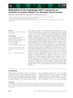
NSC cultures. Figure 1 shows the relative gene e xpres-
sion of NGF, BDNF and NT-3 in NSC culture at
passages 5-7 in three separate experiments. A progres-
sive increase of NGF was noted, and an up-surge of
BDNF and NT-3 was seen in NSC culture at passage 6
(p < 0.0001).
ELISA was performed on one-day-old tissue culture
medium from three replicate cultures at passages 1-7 in
two separate experiments to evaluate the ge ne transla-
tion of NGF (Figure 2) . A stead y increase o f NGF pro-
tein was noted from passages 1-5, followed by a peak of
production at passage 6, which was significantly higher
than those derived from earlier passages (p < 0.0001)
and from subsequent cultures at p assage 7 (p = 0.0006).
Using NGF as a model, our data suggested an in vitro
synthesis of neurotrophic factors in NSC cultures.
In vivo Study of NSC-Derived Cells
Somatosensory function
NSC (1.5 × 10
5
) at passage 6 were transpl anted into the
cisterna magna of 10 ischemic rats seven days after the
Sun et al. Journal of Translational Medicine 2010, 8:77
/>Page 4 of 10
induction of focal cerebral i schemia. Transplanted ani-
mals did not behave abnormally or develop dyskinesia.
Thirty ischemic rats with and without NSC transplant
or NGF injection and 10 sham-operated rats were sub-
jected to the electrophysiological tests before and after
surgery. One ischemic control rat injected with PBS
died after the first electrophysiological test. Compared
to the 10 sham-operated normal control rats, SEP was
not elicited among the 30 ischemic rats on day two after
ischemic induction or day two after the NSC transplant
or NGF administration. On day 7, a very weak SEP was
observed among ischemic rats treated with either NSC
or NGF but not in ischemic control rats (Table 1). A
progressive increase of SEP in terms of the relative
latency and amplitude was seen at week two and four in
the ischemic control rats, suggesting regeneration. At
weeks two and four, transplanted ischemic rats
responded to the somatosensory stimuli more effectively
than ischemic rats with NGF supplement and ischemic
control rats, as shown by the relative latencies (p <
0.0001). The relative amplitudes derived from trans-
planted rats at w eek two were higher than those of the
ischemic rats supplemented with NGF and the ischemic
control rats (p = 0.0086), despite the fact that the rela-
tive amplitude of transplanted rats at week four was sig-
nificantly lower than that of the sham-operated normal
control (p < 0.0001). These data suggest that NSC trans-
plant could improve the somatosensory resp onse after
ischemic stroke.
Motor function
Thirty ischemic rats with (n = 10) and without NSC
transplant (n = 9) or NGF injection (n = 10), and 10
sham-operated rats w ere assessed over four weeks using
a horizontal channel connected to a ladder. Forelimb
faults were summed and differentiated. On examining
the sum of forelimb and foot faults (SOFFF), the three
groups of ischemic rats had higher scores than sham-
operated normal control rats (Figure 3A). On day two,
ischemic rats given the NGF injection showed the least
motor impairment, as measured by the SOFFF, compared
to their ischemic counterparts with or without NSC
transplant (p = 0.025). On week one, the SOFFF was sig-
nificantly lower in ischemic rats transplanted with NSC
than ischemic control rats (p = 0.011), but was compar-
able to that of ischemic rats injected with NGF. The
SOFFF of the three ischemic groups on week two and
four were comparable to (p > 0.05) but higher than that
of sham-operated normal control rats. Figure 3B shows
the relatively s table, but significantly higher, coefficients
of the differentiatio n of forelimb and foot faults (DOFFF)
derived from ischemic control mice over four weeks
compared to the sham-operated normal control rats. The
DOFFF of the three groups of ischemic rats before week
Figure 1 Quantitative real-time PCR showing the relative expression of gene NGF (A), BDNF (B) and NT-3 (C) of triplicate culture
passages five to seven of primary neural stem cells from cortex of E16 Sprague-Dawley rat embryos.
Figure 2 ELISA of nerve growth factor in one-day spent tissue
culture media of culture passages five to seven of primary
neural stem cells from cortices of E16 Sprague-Dawley rat
embryos in three separate experiments. A preponderance of
NGF in pg per day of culture of 1 × 10⁶ cells was evident in culture
passages six and seven.
Sun et al. Journal of Translational Medicine 2010, 8:77
/>Page 5 of 10
two were comparable (p > 0.05). From weeks two to four,
the DOFFF of ischemic rats with NSC transplant was
lower than that of rats injected with NGF, which was in
turn lower than that of ischemic control rats (p < 0 .05).
This suggests that NSC transplant and NGF administra-
tion could enhance symptomatic relief.
NGF synthesis
ELISA of NGF in the CSF aspirated from the IG on day
28 after sham operation displayed a physiological level
of 0.44 ± 0.38 pg/mL. The NGF in CSF aspirated on the
same time line from 9 ischemic control rats and 10
ischemic rats injected with NGF were 14. 25 ± 5.21 pg/
mL (32.4-fold increase ) and 16.22 ± 4.43 pg/mL (36.9-
fold increase), respectively. The NGF in the CSF at week
four of 10 ischemic rats given NSC transplant was 37.86
± 4.12 pg/mL, which was 2 .7-fold and 86-fold higher
than those derived from untreated ischemic rats and
control rats, respectively. These data suggest an ische-
mia-mediated up-regulation of in vivo NGF synthesis
that is augmented by the NSC allograft.
Histology
Animals were sacrificed on week four after CSF aspira-
tion and the completion of behavioral assessments.
Tracking of BrdU
+
NSC revealed that a majority of the
donor cells engrafted to the inf arcted areas of the cor-
tex, hippocampus, striatum and parenchyma near the
third ventricle (Figure 4). Migration of the BrdU
+
cells
along the corpus callosum and the ventricular wall was
noted. Small clusters of BrdU
+
cells and BrdU
+
cells
with glial morphologies of 10-20 μm in size were also
evident.
Immunohistochemical staining of class I MHC
demonstrated high expression levels in the lesioned cor-
tex and brain parenchymas near the ventricular lining in
the three groups of ischemic rats, which was in marked
contrast to the low expression in normal rats (Figure
5A). In addition, the class I MHC was detected in the
hippocampus of ischemic rats wi th either NGF injection
or NSC transplant, but not in control brains or ischemic
brains without therapy. The intensity was more
Table 1 Somatosensory evoked potential at different time points
Relative Latency (Relative Amplitude) of Somatosensory Evoked Potential in Mean ± SD
Period of NSC transplant/NGF injection Day -5 Day 2 Week 1 Week 2 Week 4
Sham-operated normal rats (n = 10) 1.03 ± 0.02
(1.06 ± 0.33)
1.02 ± 0.02
(0.99 ± 0.32)
0.96 ± 0.04
(0.98 ± 0.24)
1.02 ± 0.04
(1.06 ± 0.29)
1.02 ± 0.04
(1.1 ± 0.40)
Ischemic control rats (n = 9) - - - 1.64 ± 0.25
(0.36 ± 0.33)
1.42 ± 0.11
(0.58 ± 0.25)
Ischemic rats with NGF administration (n = 10) - - 1.83 ± 0.06
(0.22 ± 0.11)
1.52 ± 0.10
(0.34 ± 0.16)
1.24 ± 0.07
(0.64 ± 0.15)
Ischemia rats with NSC transplant (n = 10) - - 1.86 ± 0.14
(0.21 ± 0.13)
1.22 ± 0.09
(0.51 ± 0.21)
1.18 ± 0.04
(0.7 ± 0.17)
Figure 3 Analyses of motor function over four weeks of sham-operated normal control rats, ischemic control rats and ischemic rats
with either NGF injection or neural stem cell transplant. A: sum of forelimb foot faults, B: differentiation of forelimb and foot faults.
Sun et al. Journal of Translational Medicine 2010, 8:77
/>Page 6 of 10
profound in the NSC-transplanted group than in the
NGF-injected group. Figure 5B shows that a small
amount of the class II MHC was detected in normal
brain tissue, but was up-regulated under ischemic stress.
The extents of the class II MHC immunoreactivity were
comparable among the three groups of ischemic rats,
irrespective of the treatments given. These data suggest
that ischemia might up-regulate MHC expression, and
that the class I MHC may be further uplifted by NGF
supplement or NSC transplant.
Immunoreactivity of caspase III was almost non-exis-
tent in the control brain parenchyma, except in the
neural tissue adjacent to the third ventricle. Conversel y,
a high l evel of caspase III wa s noted in the cortex, hip-
pocampus, striatum and neural tissue around the third
ventricleofischemicratswithandwithouteitherNGF
administration or NSC transplant (Figure 6). The extent
of caspase III immunoreactivity was comparable among
the three groups of ischemic rats.
Discussion
In this study, we found an up-regulated expression of
the class I and II MHC in rat brains under ischemic
stress. The extent of the class I MHC augmentation was
more remarkable in ischemic rats given an NSC trans-
plant than in rats given an NGF supplement, whereas
class II MHC expression was comparable among
ischemic rats irrespective of NGF or NSC therapy. In
vitro and in vivo analyses of NSC-derived NGF demon-
strated that the NSC-derived, neurotrophin-modulated
MHC expressi on correlated with the degree of transient
symptomatic relief in stroke rats and promoted no sec-
ondary injury, such as apoptotic cell death and inflam-
mation [8,9].
Figure 4 Tracking of BrdU-labeled neural stem cells at passage six in the ischemic brain of rat having undergone cell therapy for four
weeks. A: a representative coronal section of a transplanted rat demonstrated the localization of reddish-brown colored BrdU
+
cells (left panel)
to the cortex, hippocampus, striatum and brain parenchyma near the third ventricle, and of a sham-operated normal control rat without BrdU
postivity (right panel) B: migration of BrdU
+
cells along the corpus callosum. C and D: small clusters of BrdU
+
cells displaying a glial morphology.
Scale bar: 75 μm.
Sun et al. Journal of Translational Medicine 2010, 8:77
/>Page 7 of 10
Figure 5 Immunohistochemical staining of MHC. Reddish-brown immunoreactivity of class I and II MHC were shown in panel A and B,
respectively. A representative coronal section of the hippocampus of an ischemic rat brain without injection of NGF or NSC exhibited no
positivity of class I MHC (A-i). Intense staining of class I MHC was noted in the cytoplasm of pyramidal neurons in the hippocampus of ischemic
rats undergone neural stem cell transplant for four weeks (A-ii). Clusters of cells with class I MHC-positivity were evident in the infarcted brain
parenchyma of transplanted rats (A-iii and A-iv). A comparable extent of class II MHC was noted in ischemic rats irrespective of any therapy but
unremarkable in normal rat (panel B, top row). Reddish-brown staining of Class II MHC was evident in the infarcted brain parenchyma (B-i), along
the meninge (B-ii), areas near the ventricular lining and vascular wall near the hippocampus (B-iv) of transplanted rats. Scale bars: 75 μm
Sun et al. Journal of Translational Medicine 2010, 8:77
/>Page 8 of 10
Data from our p resent and previous studies demon-
strate that a minority of implanted donor stem cells can
migrate along nerve fiber bundles, home to lesioned
brain parenchymas and d ifferentiate into mature cells of
interest [3,10,11]. The low d egree of differentiat ion and
integration of the transplanted cells in the parenchyma
often correlated poorly with the improved functional
benefits [12,13]. As there is little evidence of neuronal
replacement, other mechanisms might account for the
functional recovery. Neurotrophin genes have been
reported to be expressed and transcribed by NSC in
vitro [14]. The administration of neurotrophin-secreti ng
stem cells or neurotrophic factors might be a potential
alternative [15,16].
Neurotrophins, including NGF, BDGF, NT-3, NT-4
and others, are a group of short-lived proteins in the
CNS, which are key regulators of cell fate and cell shape
[17,18]. The growth-enhancing effects of neurotrophins
have also been reported [19]. In this study, we provide
evidence both in vitro and in vivo of neur otrop hin pro-
duction by NSC and confirmed the constitutive secre-
tion that was proposed by Lu et al. [20]. Interestingly,
we noted an increase of NGF in the CSF of rats after
ischemic stress. The extent was further amplified in
ischemic rats that were given a NSC transplant. The
high dose of NGF m ight have a neuroprotective effect
on the injured brain to prevent further secondary inju-
ries, as suggested in this study and that of Chiaretti et
al. [21]. The up-regulation of the class I MHC corre-
lated well with the symptomatic relief in ischemic rats
given the NSC tra nsplant and the upsurge of N GF in
vivo, suggesting an immuno-modulation of the class I
MHC by NSC- derived neurot rophins in the m icro-
environment of the lesioned brain parenchyma.
The MHC is a family of molecules that are responsible
for the immune recognition and are particularly impor-
tant in the context of the adaptive immune response.
The anergy of the regulatory MHC when presenting
inflammatory elements to immuno-competent cells in
the CNS might do more harm than good. Mounting evi-
dence suggests that some forms of immunologic inter-
vention can help protect or restore CNS integrity [22].
The present study shows that an NSC allograft might
boost neural regeneration during focal cerebral i schemia
Figure 6 Immunohist ochemical staining of caspase III. Weak reddish-brown immunore activity was demons trated in neural tissues near the
third ventricle of sham-operated normal control rats, whereas strong reactivities were evident in the cortex, hippocampuses, striata and neural
tissues close to lateral ventricles and third ventricles of ischemic rats with and without NGF administration or neural stem cell transplant. A:
reddish brown coloration of caspase III immunoreactivity along the ventricular lining of cells of ischemic rat having undergone neural stem cell
transplant for four weeks, B: caspase III+ cells took the glial morphology. Scale bar: 75 μm.
Sun et al. Journal of Translational Medicine 2010, 8:77
/>Page 9 of 10
in a rat model via the immuno-modulation of class I
MHC expression by NSC-mediated neurotrophins and
eventually lead to functional recovery without activating
the caspase III inflammatory response. Recently, the
class I MHC was found to be crucial to neural deve lop-
ment, neuronal differentiation, synaptic plasticity and
behavior [23]. Thus, manipulating and targeting MHC
signaling might facilitate NSC-derived neurotrophin-
mediated functional restoration after stroke . This possi-
bility should be elucidated and explored in future
studies.
Conclusions
The findings presented here provide further insights into
the mechanisms of NSC in the regeneration of the CNS.
Should the MHC modulation mediated by NSC-derived
neurotrophins be elucidated, strategic cellular therapy
for neural injuries and neuro-degenerative diseases may
be revolutionized, and novel treatment modaliti es could
be developed.
This paper is not based on a previous communication
to a society or meeting.
Acknowledgements
This study was supported in part by the grant reference 30371452 of the
National Natural Science Foundation of China.
Author details
1
Department of Neural Stem Cell, Beijing Neurosurgical Institute, Beijing
Tiantan Hospital, Capital Medical University, China.
2
Department of
Neurosurgery, 2nd Affiliated Hospital of Zhejiang University Medical College,
Hangzhou, China.
3
Department of Laboratory, Beijing Tiantan Hospital,
Capital Medical University, Beijing, China.
4
Department of Electrophysiology,
Beijing Tiantan Hospital, Capital Medical University, Beijing, China.
Authors’ contributions
CRS conceived of the study, participated in some parts of the research and
wrote the manuscript. HZ carried out the histology test and participated in
ELISA test. JL participated in the in vitro characterization of NSCH. Huang
carried out the culture of NSC and participated in the motor function test.
HBC participated in the creating the animal models. YJW participated in
ELISA test. PL carried out the electrophysiology test. YHA participated in its
design and coordination and helped to draft the manuscript. All authors
read and approved the final manuscript.
Competing interests
The authors declare that they have no competing interests.
Received: 8 November 2009 Accepted: 20 August 2010
Published: 20 August 2010
References
1. Haas S, Weidner N, Winkler J: Adult stem cell therapy in stroke. Curr Opin
Neurol 2005, 18:59-64.
2. Li M, Decherchi P, Raisman G: Transplantation of Olfactory Ensheathing
Cells into Spinal Cord Lesions Restores Breathing and Climbing. J
Neurosci 2003, 23:727-731.
3. Fong SP, Tsang KS, Chan AB, Lu G, Poon WS, Li K, Baum LW, Ng HK:
Trophism of neural progenitor cells to embryonic stem cells: neural
induction and transplantation in a mouse ischemic stroke model. J
Neurosci Res 2007, 85:1851-1862.
4. Stampachiacchiere B, Aloe L: Differential modulatory effect of NGF on
MHC class I and class II expression in spinal cord cells of EAE rats. J
Neuroimmunol 2005, 169:20-30.
5. Modo M, Rezaie P, Heuschling P, Patel S, Male DK, Hodges H:
Transplantation of neural stem cells in a rat model of stroke: assessment
of short-term graft survival and acute host immunological response.
Brain Res 2002, 958:70-82.
6. Sun CR, Wang CC, Tsang KS, Li J, Zhang H, An YH: Modulation and impact
of class I major histocompatibility complex by neural stem cell-derived
neurotrophins on neuroregeneration. Med Hypotheses 2007, 68:176-179.
7. Sun CR, An YH, Liu SL, Xia L, Li J, Zhang H, Huang H, Wang ZH: Trap
channel test: a novel method to evaluate the neurological functions of
cerebral ischemic rats. Chinese Journal Of Neurosurgery 2008, 5:101-104.
8. Modo M, Mellodew K, Rezaie P: In vitro expression of major
histocompatibility class I and class II antigens by conditionally
immortalized murine neural stem cells. Neurosci Lett 2003, 337:85-88.
9. Neumann H, Misgeld T, Matsumuro K, Wekerle H: Neurotrophins inhibit
major histocompatibility class II inducibility of microglia: Involvement of
the p75 neurotrophin receptor. Proc Natl Acad Sci USA 1998, 95:5779-5784.
10. Poon WS, Lu G, Tsang KS, Zhu XL, Chen GG, Ng HK: Migration of bone
marrow stem cells in ischemic brain. Acta Neurochir Suppl 2006,
99:123-124.
11. Li J, Sun CR, Zhang H, Tsang KS, Li JH, Zhang SD, An YH: Co-
transplantation of neural stem cells and Schwann Cells results in
functional recovery in a rat spinal cord contusion injury model. Biomed
Environ Sci 2007, 20:242-249.
12. Lindvall O, Kokaia Z, Martinez-Serrano A: Stem cell therapy for human
neurodegenerative disorders–how to make it work. Nat Med 2004,
10(suppl):S42-50.
13. Shyu WC, Lee YJ, Liu DD, Lin SZ, Li H: Homing genes, cell therapy and
stroke. Front Biosci 2006, 11:899-907.
14. Meltzer C, Kondziolka D, Villemagne VL, Wechsler L, Goldstein S,
Thulborn KR, Gebel J, Elder EM, DeCesare S, Jacobs A:
Serial [18F]
fluorodeoxyglucose positron emission tomography after human
neuronal implantation for stroke. Neurosurgery 2001, 49:586-592.
15. Kondziolka D, Wechsler L, Goldstein S, Meltzer C, Thulborn KR, Gebel J,
Jannetta P, DeCesare S, Elder EM, McGrogan M, Reitman MA, Bynum L:
Transplantation of cultured human neuronal cells for patients with
stroke. Neurology 2000, 55:565-569.
16. Burger H, Foekens JA, Look MP, Meijer-van Gelder ME, Klijn JG, Wiemer EA,
Stoter G, Nooter K: RNA expression of breast cancer resistance protein,
lung resistance-related protein, multidrug resistance-associated proteins
1 and 2, and multidrug resistance gene 1 in breast cancer: correlation
with chemotherapeutic response. Clin Cancer Res 2003, 9:827-836.
17. Barde YA: Trophic factors and neuronal survival. Neuron 1989,
2:1525-1534.
18. Lewin GR, Barde YA: Physiology of the neurotrophins. Annu Rev Neurosci
1996, 19:289-317.
19. Barbara BS, McAtee M, Dai HN, Kuhn PL: Neurotrophic Factors Increase
Axonal Growth after Spinal Cord Injury and Transplantation in the Adult
Rat. Exp Neurology 1997, 148:475-494.
20. Lu P, Jones LL, Snyder EY, Tuszynski MH: Neural stem cells constitutively
secrete neurotrophic factors and promote extensive host axonal growth
after spinal cord injury. Exp Neurol 2003, 181:115-129.
21. Chiaretti A, Piastra M, Polidori G, Di Rocco C, Caresta E, Antonelli A,
Amendola T, Aloe L: Correlation between neurotrophic factor expression
and outcome of children with severe traumatic brain injury. Intensive
Care Med 2003, 29:1329-1338.
22. Schwartz M, Moalem G, Leibowitz-Amit R, Cohen IR: Innate and adaptive
immune responses can be beneficial for CNS repair. Trends Neurosci 1999,
22:295-299.
23. Boulanger LM, Shatz CJ: Immune Signaling in neural development,
synaptic plasticity and disease. Nat Rev Neurosci 2004, 5:521-531.
doi:10.1186/1479-5876-8-77
Cite this article as: Sun et al.: Modulation of the major
histocompatibility complex by neural stem cell-derived neurotrophic
factors used for regenerative therapy in a rat model of stroke. Journal of
Translational Medicine 2010 8:77.
Sun et al. Journal of Translational Medicine 2010, 8:77
/>Page 10 of 10