Điều trị biến chứng của chấn thương gân pdf
Bạn đang xem bản rút gọn của tài liệu. Xem và tải ngay bản đầy đủ của tài liệu tại đây (309.54 KB, 10 trang )
Complications After
Treatment of Flexor
Tendon Injuries
Abstract
The goals of flexor tendon repair are to promote intrinsic tendon
healing and minimize extrinsic scarring in order to optimize
tendon gliding and range of motion. Despite advances in the
materials and methods used in surgical repair and postoperative
rehabilitation, complications following flexor tendon injuries
continue to occur, even in patients treated by experienced surgeons
and therapists. The most common complication is adhesion
formation, which limits active range of motion. Other
complications include joint contracture, tendon rupture, triggering,
and pulley failure with tendon bowstringing. Less common
problems include quadriga, swan-neck deformity, and lumbrical
plus deformity. Meticulous surgical technique and early
postoperative tendon mobilization in a well-supervised therapy
program can minimize the frequency and severity of these
complications. Prompt recognition of problems and treatment with
hand therapy, splinting, and/or surgery may help minimize
recovery time and improve function. In the future, the use of novel
biologic modulators of healing may nearly eliminate complications
associated with flexor tendon injuries.
T
endon lacerations within the
digital sheath are difficult to re-
pair.
1
As a result of poor outcomes
following primary tendon repair
within the digital sheath (zone II),
the area within the sheath contain-
ing the flexor digitorum profundus
(FDP) and flexor digitorum superfi-
cialis (FDS) tendons has been re-
ferred to as “no man’s land.”
2
In the
1960s, the development of stronger
suture materials and improved su-
ture techniques led to a renewed in-
terest in primary repair within the
digital sheath.
3
Primary repair is now
the standard of care. Despite these
advances, outcomes have been rated
fair or poor in 7% to 20% of patients
after flexor tendon repair.
4,5
A thor-
ough knowledge of the basic science
of flexor tendon healing is essential
for improving outcomes and for un-
derstanding, recognizing, and manag-
ing the various complications.
Basic Science of Flexor
Tendon Healing
Anatomy
Tendons are made up of spiraling
bundles of mature tenocytes and pre-
dominantly type I collagen. In the
distal palm and digits, the tendons
are enclosed in a synovial sheath.
The synovial sheath enhances glid-
ing of the tendons and is thickened
Soma I. Lilly, MD
Terry M. Messer, MD
Dr. Lilly is Chief Resident, Department of
Orthopaedics, University of North
Carolina School of Medicine, Chapel
Hill, NC. Dr. Messer is Assistant
Professor, Department of Orthopaedics,
University of North Carolina School of
Medicine, Chapel Hill, NC.
None of the following authors or the
departments with which they are
affiliated has received anything of value
from or owns stock in a commercial
company or institution related directly or
indirectly to the subject of this article:
Dr. Lilly and Dr. Messer.
Reprint requests: Dr. Messer, Wake
Orthopaedics, LLC, 3009 New Bern
Avenue, Raleigh, NC 27610.
J Am Acad Orthop Surg 2006;14:387-
396
Copyright 2006 by the American
Academy of Orthopaedic Surgeons.
Volume 14, Number 7, July 2006 387
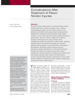
in specific areas between the joints;
these thickened areas are called pul-
leys. The pulleys enhance efficiency
of motion within the digit by pre-
venting tendon bowstringing and
maximizing tendon excursion. Most
critical to this system are the A2 and
A4 pulleys, which are located over
the proximal and middle phalanges,
respectively
6
(Figure 1). The FDP and
FDS tendons are contained within
the digital flexor sheath.
Flexor Tendon Healing
Tendon healing consists of three
phases: inflammatory, proliferative,
and remodeling.
7
The inflammatory
phase occurs during the first week af-
ter injury and involves migration of
fibroblasts and macrophages to the
injured area, with ensuing phagocy-
tosis of the clot and necrotic tissue.
In the proliferative phase, which
lasts from weeks 1 through 3, fibro-
blasts proliferate, and there is imma-
ture collagen deposition and neovas-
cularization. Finally, the remodeling
phase occurs in weeks 3 through 8.
Collagen fibers become organized in
a linear manner parallel to the ten-
don. Adhesion formation between
tendon and sheath is most clinically
evident during this last phase.
Two mechanisms for healing
have been described in the literature:
extrinsic and intrinsic. The extrinsic
mechanism is predominately medi-
ated by an influx of synovial fibro-
blasts and inflammatory cells from
the tendon sheath. Healing also oc-
curs via the intrinsic mechanism, in
which fibroblasts and inflammatory
cells from the tendon and epitenon
invade the injured site. The extrinsic
mechanism is thought to predomi-
nate early in tendon healing and in
cases of digit immobilization; the in-
trinsic mechanism becomes increas-
ingly active after 21 days.
8
The great-
er proliferative and inflammatory
response of the synovial sheath,
along with the greater cytokine reac-
tivity and capacity for matrix degra-
dation of synovial fibroblasts, favor
the extrinsic pathway.
8
Extrinsic
healing produces increased collagen
content at the injury site, but in a
disorganized fashion. Tendon heal-
ing is likely a combination of both
mechanisms, but the predominance
of extrinsic healing leads to scar for-
mation and adhesions between the
tendon and the surrounding sheath.
Requirements for Tendon
Healing
Requirements for tendon healing
include motion and tension at the
repair site, adequate tendon nutri-
tion and vascular perfusion, mini-
mal gap formation at the repair site,
and a strong repair.
9-12
Early-motion
protocols in animal flexor tendons
resulted in a progressively g reater ul-
timate tensile load over time than
was the case in tendons managed
with immobilization protocols.
9
Early-motion protocols also helped
avoid the loss of strength that occurs
in early phases of tendon healing.
10
Additionally, both motion and ten-
sion are needed to stimulate teno-
cyte development and increase col-
lagen amount and organization.
11
Tendon nutrition is provided
through vascular perfusion and sy-
novial fluid diffusion. Flexor tendon
vascular supply originates from ves-
sels in the proximal synovial fold,
segmental branches of digital arter-
ies through the vincular system, and
the osseous insertion of the FDS and
FDP tendons.
13
Diffusion of nutri-
ents through synovial fluid occurs
via imbibition, in which fluid is
forced through interstices on the
surface of the tendon.
14
This process
is facilitated by the pumping mech-
anism created by flexion and exten-
sion of the digit.
Gap formation as a result of cy-
clic loading before tendon failure is
seen routinely after flexor tendon re-
pair.
15
The average gap is 3.2 mm.
16
Gaps have previously been associat-
ed with adhesion formation and poor
gliding.
17
Gelberman et al,
12
howev-
er, demonstrated that gap length has
no relationship to adhesion forma-
tion, but it does have a negative ef-
fect on the acquisition of tendon
tensile proper ties during healing.
In their canine study, repair gaps
>3 mm did not gain stiffness or
strength from 10 to 42 days, but gaps
<3 mm had a 320% increase in stiff-
ness and a 90% increase in strength
over the same period.
12
Techniques for maximizing ten-
don repair strength comprise a large
portion of flexor tendon research. A
strong repair is one that can with-
stand early motion with minimal
gap formation, thereby allowing suc-
cessful tendon healing. Well-
accepted, established principles of
tendon repair include using core su-
tures of 3-0 or 4-0 nonabsorbable
polyfilament material, an increased
Figure 1
Lateral view of the flexor tendon synovial sheath, including the palmar aponeurosis
(PA), five annular (A) pulleys, and three cruciform (C) pulleys. The critical pulleys
are A2 and A4, located over the proximal and middle phalanges, respectively.
(Reproduced with permission from Doyle JR: Anatomy of the finger flexor tendon
sheath and pulley system. J Hand Surg [Am] 1988;13:473-484.)
Complications After Treatment of Flexor Tendon Injuries
388 Journal of the American Academy of Orthopaedic Surgeons
number of sutures crossing the re-
pair, and equal strength across all
strands. In addition, certain locking
suture techniques (ie, transverse
limb of repair passed superficial to
the longitudinal component) have
been shown to increase repair
strength.
18-20
A peripheral locking
epitendinous suture also should be
added to enhance repair strength.
21
Complications
Adhesion Formation
Adhesion formation is the most
common complication following
flexor tendon repair. Prevention of
adhesion formation is facilitated by
optimizing intrinsic healing. Early re-
search reflected the belief that ten-
don healing depended on extrinsic
cellular ingrowth, which required
immobilization. However , the ability
of tendons to heal b y i ntrinsic mech-
anisms alone has since been well
documented.
22
Methods of adhesion
prevention can be divided into me-
chanical and biologic factors de-
signed to promote intrinsic healing.
Mechanical Factors
Mechanical factors for preventing
adhesions include early postopera-
tive motion protocols, preservation
of sheath and pulley components,
partial FDS resection, and atraumat-
ic handling of the tendon and sheath.
Motion, which leads to a predomi-
nance of intrinsic over extrinsic
healing, is critical to preventing ad-
hesions. Three primary motion pro-
tocols are described in the literature:
passive, active, and synergistic. In
1977, Lister et al
23
published the first
results of tendon repair using a con-
trolled passive motion protocol. The
Kleinert splint was used to allow ac-
tive digital extension coupled with
passive digital flexion. Good to ex-
cellent results were reported in 80%
of tendon lacerations in zone II.
23
The splint has since been modified
by adding a midpalmar bar or pulley,
resulting in improved distal tendon
gliding and differential tendon ex-
cursion.
24
The addition of synergistic
wrist motion (wrist flexion–finger
extension combined with wrist ex-
tension–finger flexion) also has been
shown to improve overall tendon
gliding and excursion.
25
Early active motion protocols
subsequently have been developed
to address concerns about variabili-
ty in tendon gliding with passive
protocols. Bainbridge et al
26
reported
on a consecutive series comparing
controlled active motion with active
extension–passive flexion protocols.
Patients treated with controlled ac-
tive motion acquired greater final
motion.
26
Other series using early
active motion have reported good to
excellent results ranging from 57%
to 92%, with rupture rates from 5%
to 46%.
27-29
These findings are com-
parable to rates reported with pas-
sive motion regimens. Improved su-
ture materials and techniques seem
capable of withstanding the higher
forces associated with active motion
protocols.
30-32
However, recent re-
search in repaired canine tendon by
Boyer et al
33
demonstrated no advan-
tage with high-force rehabilitation in
the accrual of either stiffness or
strength compared with low-force
rehabilitation.
The synergistic motion regimen
allows high tendon excursion with
low force on the repair site.
34
This
protocol consists of passive digit flex-
ion combined with active wrist ex-
tension, followed by active wrist flex-
ion combined with passive digit
extension. Zhao et al
35
compared
synergistic motion with passive mo-
tion regimens in the management of
canine flexor tendon repairs. They
noted fewer adhesions with the syn-
ergistic motion group but reported el-
evated gap formation in the motion
group (30%) versus the passive group
(6%).
35
Currently, agreement is uni-
versal that repaired flexor tendons
should be subjected to early mobili-
zation; however , n o single rehabilita-
tion protocol is accepted by all.
Preservation of sheath compo-
nents is controversial. When the vas-
cular source of nutrition is compro-
mised because of trauma, the tendon
sheath can maintain nutrition
through imbibition until the vascu-
lar system is reestablished.
36
Preser-
vation of flexor tendon sheath integ-
rity may reduce adhesions through
its positive effect on intrinsic heal-
ing.
37
However, sheath repair also
may lead to impaired tendon gliding
and increased resistance.
17
Another
study compared sheath repair with
excision and found no difference in
final motion when early mobiliza-
tion was done.
38
Recently, resection of all or part of
the FDS tendon has been suggested
as a method of decreasing gliding re-
sistance of the FDP within the
sheath.
39
Loss of the FDS tendon is
not associated with significant func-
tional compromise. However, this
technique was initially dismissed be-
cause a considerable portion of the
FDP blood supply is provided by cap-
illaries emanating from the FDS ten-
don. In a cadaveric study, FDS resec-
tion was found to be a viable option
for improving the gliding of a bulky
FDP repair . The authors did not dem-
onstrate any advantage of complete
resection versus partial resection.
39
The use of meticulous surgical
technique as a method for decreasing
adhesion formation is well docu-
mented. Adhesion formation is
known to be proportional to the
amount of tissue crushing and to the
number of surface injuries incurred
by the tendon and sheath during re-
pair.
4
Accordingly, stiffness is more
common in digits after crush injuries
as well as in t hose with concomitant
neurovascular and bone injuries.
40
Biologic Factors
Development of novel biologic
factors to provide so-called scarless
healing is an active area of re-
search.
22,41
Advances in this arena
could lead to less reliance on postop-
erative motion for adhesion preven-
tion. Methods currently under inves-
tigation include mechanical barriers
to adhesion formation, as well as
Soma I. Lilly, MD, and Terry M. Messer, MD
Volume 14, Number 7, July 2006 389
chemical and molecular modulation
of scar formation. Many mechanical
barrier methods have been studied,
including silicone, alumina sheaths,
polyethylene, and polytetrafluoro-
ethylene, but none is in widespread
clinical use.
22
ADCON-T/N (Glia-
tech, Cleveland, OH), a gelatin and
carbohydrate polymer, has shown
some potential.
41
In a recent double-
blind randomized study in which
ADCON-T/N was applied to the
tendon after repair, the authors
found no significant effect on final
motion; however, time t o achieve fi-
nal motion was shorter with the use
of ADCON-T/N.
41
Ibuprofen and corticosteroids have
been investigated as possible modu-
lators of adhesion formation.
42,43
Ibu-
profen has been shown to improve
tendon excursion in animal models.
42
Ketchum
43
demonstrated that al-
though corticosteroids decrease the
strength and density of adhesions,
they are associated with smaller,
weaker tendons, diminished wound
healing, and decreased resistance to
infection. These problems have lim-
ited their use in flexor tendon repair.
New Research
Modulation of scar formation on
a molecular level is a new area of
research in tendon healing. This re-
search has been directed toward un-
derstanding the role of cytokines
in tendon metabolism and re-
pair.
22,44,45
Two cytokines, transform-
ing growth factor-β (TGF-β) and ba-
sic fibroblast growth factor (bFGF),
have shown the most potential in
adhesion prevention.
44,45
TGF-β has
been implicated in numerous biolog-
ic activities related to wound heal-
ing, such as fibroblast and macro-
phage recruitment, angiogenesis,
stimulation of collagen production,
downregulation of proteinase activ-
ity, and increased metalloproteinase
inhibitor activity.
44
Chang et al
45
demonstrated that
flexor tendons exposed to transec-
tion and repair exhibit increased
TGF-β in both tenocytes and inflam-
matory cells from the tendon sheath.
These findings are significant be-
cause TGF-β is thought to be in-
volved in the pathogenesis of exces-
sive scar formation. Therefore,
perioperative modulation of this cy-
tokine may lead to decreased adhe-
sion formation. Three isoforms have
been identified; the TGF-β1 isoform
is thought to be primarily responsi-
ble for the proinflammatory and
scarring activities.
22
The TGF-β3 iso-
form demonstrates anti-scarring
properties and acts as an inhibitor of
scarring in injury models.
22
Similar to TGF-β, bFGF has been
implicated in early tendon healing.
45
Basic FGF is a potent stimulator of
angiogenesis and is able to induce
migration and proliferation of endo-
thelial cells in tissue culture. In 1998,
Chang et al
45
found that bFGF was
upregulated in tenocytes, tendon
sheath fibroblasts, and inflammatory
cells from flexor tendons exposed to
a tendon wound environment. With
further research, modification of
bFGF expression may also be useful
in postoperative adhesion reduction.
Research into chemical modula-
tion of cytokines has yielded
5-fluorouracil (5-FU) as a possible can-
didate.
46,47
5-FU is an antimetabolite
that decreases scarring by an un-
known mechanism. Khan et al
46
tested this drug in a rabbit model by
treating the injured synovial sheath
of partially lacerated tendons with a
5-min application of 5-FU before clo-
sure. A significant ( P < 0.001) decrease
in the proliferative and inflammatory
response of synovial fibroblasts was
demonstrated. There was also a sig-
nificant ( P < 0.001) decrease in the ex-
pression of TGF-β in the treated tis-
sue. Others have reported the ability
of 5-FU to reduce postoperative adhe-
sions in a chicken model.
47
These
findings are still experimental, how-
ever, and have not yet been imple-
mented in clinical practice.
When adhesion prevention is un-
successful, early recognition is crit-
ical to ensure a good clinical out-
come and prevent further progression
of stiffness. Adhesion and tendon
rupture present clinically with sim-
ilar physical findings. Both condi-
tions may demonstrate loss of active
flexion, but patients with adhesions
have preservation of some residual
active motion. Imaging studies, such
as magnetic resonance imaging or ul-
trasound, may be indicated to deter-
mine the source of motion loss. Mag-
netic resonance imaging has been
shown to be 100% accurate in distin-
guishing adhesions from rupture.
48
When adhesions are identified, ther-
apy should be directed toward pro-
grams that maximize differential mo-
tion between the FDS and FDP
tendons.
25,26
Splinting also may be a
useful adjunct. When therapy and
splinting fail to produce effective re-
sults, tenolysis may be indicated.
Tenolysis
Flexor tenolysis is indicated when
active range of motion (ROM) mea-
surements do not improve within
several weeks to months, despite
strict patient compliance with splint-
ing and ROM exercises.
49
Tenolysis
should not be considered until the
soft tissues have reached a state of
equilibrium, with supple skin and
subcutaneous tissues. To achieve a
good result, the digit must have min-
imal joint contractures and near-
normal passive ROM.
17
Most sur-
geons recommend waiting for 3 to 6
months after tendon repair or graft-
ing before performing tenolysis.
49,50
When performing flexor tenoly-
sis, a local anesthetic combined with
intravenous sedation is recommend-
ed to allow the patient to perfor m
active flexion in the operating
room.
50
This intraoperative testing is
critical to achieve a successful out-
come. A midlateral or Bruner zigzag
incision is used to expose the length
of the tendon. The neurovascular
bundles are encountered at the ends
of the digital creases, and the sur-
geon must take care t o p revent iatro-
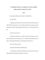
genic injury to these structures. The
scarred tendon and its sheath are vi-
sualized (Figure 2, A),
51
the adhe-
Complications After Treatment of Flexor Tendon Injuries
390 Journal of the American Academy of Orthopaedic Surgeons
sions released, and the tendon bor-
ders identified. A useful technique i s
to pass a small elevator through win-
dows made in less critical parts of
the sheath (Figure 2, C). As much of
the pulley system as possible must
be preserved (Figure 2, B); when this
is not feasible, pulley reconstruction
or a staged tendon implant should be
considered. If pulley reconstruction
requires protected mobilization,
however, the end result may be com-
promised. Additionally, any con-
comitant procedure, such as tendon
lengthening or shortening, skin
grafting, osteotomy, or capsulotomy,
may have an adverse effect on the
outcome of flexor tenolysis.
17
At the
end of the procedure, the patient
should be placed in a splint that per-
mits immediate active ROM. Pa-
tients for whom active ROM im-
proves in the first few weeks after
surgery tend to maintain these gains.
Significant pain and little early im-
provement in motion may be an in-
dication for inserting an indwelling
polyethylene catheter containing lo-
cal anesthetic.
50
One complication of flexor teno-
lysis is tendon or pulley rupture,
which should be managed with a
staged tendon reconstruction. Other
complications include postoperative
edema and pain as well as inadver-
tent neurovascular injury that may
lead to loss of viability in a digit
with marginal preoperative circula-
tion. Flexor tenolysis is a technical-
ly demanding procedure, and the
postoperative rehabilitation is equal-
ly arduous. Not all patients are can-
didates for tenolysis. The surgeon
must evaluate how the loss of active
motion will affect the patient’s
needs and desires as well as the abil-
ity to perform activities of daily liv-
ing and to return to his or her occu-
pation. The surgeon also must
consider the sensory and circulatory
status of the finger, the condition of
the other digits, and the age and gen-
eral health of the patient. Patients
who are noncompliant with therapy
after their initial repair typically are
poor candidates for tenolysis.
Joint Contracture
Even with adherence to early-
motion regimens, the reported rate of
proximal interphalangeal (PIP) and
distal interphalangeal (DIP) joint con-
tracture is 17%.
36
Contractures may
be caused by unrecognized disruption
or scarring of the volar plate, tendon
bowstringing secondary to pulley in-
competence, concomitant fracture or
neurovascular injury, prolonged heal-
ing in a flexed position, collateral lig-
Figure 2
Flexor tenolysis is performed by identifying the scarred
tendon and sheath (A), followed by release of adhesions
and careful preservation of the pulley system (B). C, Re-
lease may be facilitated by passing a small elevator or
dental probe through windows in less critical portions of
the sheath (eg, proximal to A2, or between A2 and A4
pulleys). (Reprinted from Strickland JW: Flexor tenolysis, in
Strickland JW [ed]: Master Techniques in Orthopaedic
Surgery: The Hand. Philadelphia, PA: Lippincott-Raven,
1998, pp 525-538. Illustrations copyright © Gary Schnitz
and the Indiana Hand Center.)
Soma I. Lilly, MD, and Terry M. Messer, MD
Volume 14, Number 7, July 2006 391
ament contracture, skin contracture,
or flexor tendon adhesions. They also
may be secondary to inadequate post-
operative motion regimens and dy-
namic flexion splinting. The latter
may be prevented through correct po-
sitioning of the wrist, hand, and dig-
its in the postoperative splint and
early motion. Most postoperative pro-
tocols involve splinting the metacar-
pophalangeal (MCP) joint in flexion
(approximately 60°) with the inter-
phalangeal (IP) joints fully extended.
Nonsurgical management of joint
contractures consists of early iden-
tification and modification of splint-
ing to allow greater PIP and DIP joint
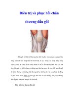
extension. A felt or foam block placed
inside a dorsal splint at the level of
the proximal phalanx, in addition to
increasing MCP joint flexion to relax
the intrinsic mechanism, will help re-
solve PIP joint contracture (Figure 3,
A). This method can be used with
buddy taping and active-assisted ex-
tension exercises. Static nighttime ex-
tension splinting and passive exten-
sion exercises with Velcro bands
applied to the splint to impart an ex-
tension force on the digit also may be
useful. As the tendon continues to
heal and strengthen, finger splints (eg,
Joint Jack, Safety Pin) can be used
(Figure 3, B and C).
When nonsurgical management
of contractures is unsuccessful, sur-
gery should be considered. No abso-
lute guidelines exist regarding the
degree of contracture that requires
surgical release; rather, the decision
for surgery is based on the patient’s
functional limitations and goals.
Preoperatively, the surgeon should
attempt to determine whether the
contracture is caused by extrinsic
factors (eg, skin contracture, proxi-
mal flexor tendon adhesions) or an
intrinsic joint contracture. When ex-
trinsic factors are responsible, PIP
joint extension will improve with
MCP joint flexion. PIP joint release
should be performed only after all
flexor tendon adhesions and skin
contractures have been addressed.
For PIP joint release, exposure is
performed through a Bruner or mid-
lateral incision. The radial and ulnar
neurovascular bundles are identified
and protected. The C1 portion of the
flexor sheath is excised between the
A2 and A3 pulleys, and the FDP and
FDS tendons are exposed
52
(Figure 4,
A). Flexor tenolysis is performed ini-
tially; the checkrein ligaments are
identified by passing a small hemo-
stat or elevator volar to the trans-
verse retinacular vessels as they en-
ter the flexor sheath just proximal to
the collateral ligament origin. The
checkrein ligaments are volar to the
transverse retinacular vessels and
can be divided sharply at this level.
The transverse retinacular vessels
should be preserved whenever possi-
ble because they supply the tendon
vincular system.
When full passive PIP joint exten-
sion cannot be obtained, release of
the collateral ligaments is performed
at their insertion on the head of the
proximal phalanx, beginning with
the accessory collateral ligaments
(Figure 4, B). Release of the collater-
al ligaments should be performed se-
quentially, progressing from palmar
to dorsal, until full extension is
achieved. When full extension can-
not be achieved, release of the volar
plate may be necessary.
Tendon Rupture
Rupture of a tendon repair is not
an uncommon problem. In one
study, a rupture rate of 4% was re-
ported in 728 digital flexor tendon
repairs (440 patients).
53
The authors
were unable to identify the inciting
factor in these failures. Another se-
ries reported a 5.7% rate of rupture
in digital flexor tendon repairs.
19
Factors that predispose tendon re-
pairs to rupture include inadequate
suture material, poor surgical tech-
nique, overly aggressive therapy, or
early termination of postoperative
splinting. Patient noncompliance,
such as removing the splint, lifting
heavy objects, or attempting strong
grasp, is a frequent cause of rup-
ture.
53
Tendon repairs are weakest be-
tween postoperative days 6 and
18.
35
Although rupture is most com-
mon during this period, it may be
Figure 3
Splints used to manage proximal interphalangeal (PIP) flexion contractures. A, Dorsal forearm-based thermoplast splint with a
felt block placed dorsally at the level of the PIP joint. B, Joint Jack Finger Splint (Sammons Preston Rolyan, Bolingbrook, IL).
C, Safety Pin Splint (Sammons Preston Rolyan).
Complications After Treatment of Flexor Tendon Injuries
392 Journal of the American Academy of Orthopaedic Surgeons
seen as late as 6 to 7 weeks after sur-
gery.
36
Timely surgical exploration is
indicated once tendon rupture is
identified. When repair attenuation
is seen without obvious rupture and
<1 cm of scar is present, the scar can
be resected and the primary repair
revised. When the scar is >1 cm, a
tendon grafting procedure should be
considered because excessive distal
advancement of the tendon can lead
to contractures and quadriga.
36
With
complete tendon rupture, the time
from the original repair influences
the course of action. If the rupture
occurs in the early postoperative pe-
riod, the tendon may be primarily re-
paired. When the rupture occurs 4 to
6 weeks after the original repair, ten-
don grafting or a staged reconstruc-
tion is recommended. Staged graft-
ing is preferred when there is
significant scarring within the
sheath. Pediatric urethral or vascular
dilators can be used to expand a con-
stricted but otherwise intact sheath,
thereby eliminating the need for a
two-stage reconstruction.
Triggering
Triggering can occur after tendon
repair and is usually the result of the
repair site’s catching on a pulley or
sheath. Causes of triggering include
a bulbous tendon repair or a tightly
repaired area of the tendon sheath.
The surgeon should intraoperatively
assess tendon gliding to identify ar-
eas that may cause triggering or re-
strict gliding. In the acute setting, a
partial tendon sheath excision or re-
lease may be used. In contrast,
sheath repair may reduce triggering
of a bulky repair by acting as a fun-
nel. Postoperatively, ultrasound or
massage may be helpful. Once the
tendon is healed, a corticosteroid in-
jection may be indicated. Reduction
tenoplasty may be considered when
nonsurgical measures fail; however,
this technique carries a risk of ten-
don rupture.
54
Recent studies have addressed the
feasibility of partial sheath resection
to decrease triggering and gliding re-
sistance. This problem is of particu-
lar concern when it involves the A2
or A4 pulleys. Tang et al
55
found a
decrease in gliding resistance with
partial pulley release. However, a ca-
daveric study by Mitsionis et al
56
demonstrated that, although exci-
sion of up to 25% of both the A2 and
A4 pulleys had no significant effect
on the efficiency of motion, it did
not achieve the goal of decreasing
sheath resistance.
Partial Tendon Injury
Partial tendon lacerations can be
challenging; if not managed proper-
ly, they carry the risk of triggering,
entrapment, or secondary rupture.
57
Repair has been recommended for
lacerations involving >60% of the
tendon substance.
58
In other studies,
the authors reported that trimming
digital flexor tendon lacerations in-
volving >50% of the tendon sub-
stance was not associated with trig-
Figure 4
A, Joint contracture release via excision of the C1 portion of the flexor tendon
sheath between the A2 and A3 pulleys exposes the flexor digitorum superficialis
(FDS) and flexor digitorum profundus (FDP) tendons. B, The checkrein ligaments
are released with subsequent release of the collateral ligaments from palmar to
dorsal. * = transverse retinacular vessels, DIP = distal interphalangeal, PIP =
proximal interphalangeal (Reproduced from Idler RS: Capsulectomies of the
metacarpophalangeal and proximal interphalangeal joints, in Strickland JW [ed]:
Master Techniques in Orthopaedic Surgery: The Hand. Philadelphia, PA: Lippincott-
Raven, 1998, pp 361-379. Illustrations copyright © Gary Schnitz and the Indiana
Hand Center.)
Soma I. Lilly, MD, and Terry M. Messer, MD
Volume 14, Number 7, July 2006 393
gering or rupture.
59
In a study by
Erhard et al
60
that compared trim-
ming with repair of partial lacera-
tions, the lowest gliding resistance
was produced with trimming, with-
out a concomitant decrease in ten-
don strength.
Pulley Failure and
Bowstringing
The A2 and A4 pulleys are re-
sponsible for preserving digital mo-
tion and finger strength (grip and
pinch power). Loss of the integrity
of these pulleys results in bow-
stringing, with loss of the A4 pulley
causing the greatest change in the
efficiency of tendon excursion,
work, and force.
61
Avoidance of bow-
stringing is the best management
strategy and may be facilitated by
performing tendon repair through
cruciate pulley windows, using ex-
ternal pulley rings for compromised
pulleys, and reconstructing pulleys
in a one- or two-stage procedure
when native tissue is unsalvage-
able
36
(Figure 5).
Many techniques for pulley re-
construction have been described,
such as Bunnell, Kleinert, Lister, and
Karev. Nishida et al
62
found that
Lister’s technique of using the exten-
sor retinaculum for pulley recon-
struction had the least resistance to
tendon gliding.
Quadriga
Quadriga is the inability of unin-
jured fingers of the same hand to ob-
tain full flexion. It manifests as a
weak grasp on physical examination.
This complication is caused by func-
tional shortening of the FDP tendon.
Shortening of one FDP tendon af-
fects the function of the FDP ten-
dons of adjacent fingers, causing
overadvancement of the FDP ten-
don, proximal tendon tethering or
adhesions, and insertion of a short
tendon graft. Anatomically, quadriga
occurs because the common FDP
muscle belly to the middle, ring, and
small fingers permits only as much
proximal excursion in each digit as
that of the shortest tendon. Proper
tendon tensioning during repair pre-
vents this problem. When quadriga
occurs, tenolysis of the proximal ad-
hesions or transection of the short-
ened tendon will release the unin-
jured profundi.
7
Swan-neck Deformity
Swan-neck deformity consists of
hyperextension at the PIP joint with
flexion at the DIP joint. In flexor ten-
don repair, common causes include
isolated FDS rupture and volar plate
injury. This complication is infre-
quent, however; loss of the FDS is
usually associated with minimal
functional deficit. Careful attention
to and correction of volar plate inju-
ries at the time of tendon repair pre-
vents this problem. Surgical man-
agement of the hyperextension
deformity may be facilitated through
tenodesis with one slip of the FDS
tendon.
Lumbrical Plus Deformity
Lumbrical plus deformity is the
paradoxical extension at the IP joints
of the injured digit with attempted
forceful flexion. Normally, PIP and
DIP joint flexion occurs in conjunc-
tion with simultaneous relaxation of
the lumbrical muscle (Figure 6, A).
Paradoxical extension arises when
the FDP distal to the lumbrical mus-
cle is functionally too long or is not
present. Flexor tendon force is there-
by transmitted to the lumbrical and
subsequently to the extensor mech-
anism via the lateral bands before
full digital flexion is reached (Figure
6, B). Other causes of lumbrical plus
deformity include avulsion of the
Figure 5
A digit in which pulley reconstruction
necessitated a two-stage revision. The
A2 and A4 pulleys were repaired using
excised flexor tendons sutured to the
retained tendon sheath edge combined
with a silicone rod tendon. (Courtesy
of George S. Edwards, Jr, MD, Raleigh,
NC.)
Figure 6
A, In normal finger mechanics, interphalangeal (IP) flexion occurs with concomitant
lumbrical relaxation. B, In lumbrical plus deformity, extension of the IP joints
paradoxically is through the lateral bands once the limit of lumbrical relaxation is
reached. (Reproduced with permission from Parkes A: The “lumbrical plus” finger. J
Bone Joint Surg Br 1971;53:236-239.)
Complications After Treatment of Flexor Tendon Injuries
394 Journal of the American Academy of Orthopaedic Surgeons
FDP tendon or amputation through
the proximal phalanx.
63
Manage-
ment involves lumbrical muscle re-
lease or placement of a tendon graft
of appropriate length.
Summary
Despite advances in flexor tendon
surgery over the past 50 years, com-
plications continue to occur. The
most common are adhesion forma-
tion and joint contracture. Achiev-
ing optimal outcomes occurs
through meticulous surgical repair
using 3-0 or 4-0 polyfilament core
suture with a minimum of four
strands reinforced with an epitendi-
nous suture, a well-fitting splint,
early controlled mobilization, and
vigilant patient monitoring for com-
pliance with the rehabilitation pro-
gram. Biochemical and molecular
advances in the research into scar-
less healing likely will lead to future
advances.
References
Evidence-based Medicine: Level I/II
prospective studies include referenc-
es 16, 26, 27, 29, 30, 40, and 41. The
remaining references are case-
controlled reports or experimental
observations.
Citation numbers printed in bold
type indicate references published
within the past 5 years.
1. Verdan CE: Half a century of flexor-
tendon surgery: Current status and
changing philosophies. J Bone Joint
Surg Am 1972;54:472-491.
2. Bunnell S: Repair of tendons in the fin-
gers and description of two new in-
struments. Surg Gynecol Obstet
1918;26:103-110.
3. Kleinert HE, Kutz JE, Ashbell TS,
Martinez E: Primary repair of lacerat-
ed flexor tendons in “no man’s land.”
J Bone Joint Surg Am 1967;49:577.
4. Strickland JW: Development of flexor
tendon surgery: Twenty-five years of
progress. J Hand Surg [Am] 2000;25:
214-235.
5. Saldana MJ, Chow JA, Gerbino P,
Westerbeck P, Schacherer TG: Fur-
ther experience in rehabilitation of
zone II flexor tendon repair with dy-
namic traction splinting. Plast
Reconstr Surg 1991;87:543-546.
6. Doyle JR: Anatomy of the fingerflexor
tendon sheath and pulley system.
J Hand Surg [Am] 1988;13:473-484.
7. Strickland JW: Flexor tendons—acute
injuries, in Green DP, Hotchkiss RN,
Pederson WC (eds): Green’s Operative
Hand Surgery,ed4.NewYork,NY:
Churchill Livingstone, 1999, vol 2,
pp 1851-1897.
8. Kakar S, Khan U, McGrouther DA:
Differential cellular response within
the rabbit tendon unit following ten-
don injury. J Hand Surg [Br] 1998;23:
627-632.
9. Gelberman RH, Woo SL, Lothringer
K, Akeson WH, Amiel D: Effects of
early intermittent passive mobiliza-
tion on healing canine flexor tendons.
J Hand Surg [Am] 1982;7:170-175.
10. Aoki M, Kubota H, Pruitt DL, Manske
PR: Biomechanical and histologic
characteristics of canine flexor ten-
don repair using early postoperative
mobilization. J Hand Surg [Am]
1997;22:107-114.
11. Kubota H, Manske PR, Aoki M, Pruitt
DL, Larson BL: Effect of motion and
tension on injured flexor tendons in
chickens. J Hand Surg [Am] 1996;21:
456-463.
12. Gelberman RH, Boyer MI, Brodt MD,
Winters SC, Silva MJ: The effect of gap
formation at the repair site on the
strength and excursion of intrasy-
novial flexor tendons: An experimen-
tal study on the early stages of tendon-
healing in dogs. J Bone Joint Surg Am
1999;81:975-982.
13. Ochiai N, Matsui T, Miyaji N, Merk-
lin RJ, Hunter JM: Vascular anatomy
of flexor tendons: I. Vincular system
and blood supply of the profundus ten-
don in the digital sheath. J Hand Surg
[Am] 1979;4:321-330.
14. Weber ER, Hardin G, Haynes DW:
Synovial fluid nutrition of flexor ten-
dons. Presented at the 25th Annual
Meeting of the Orthopaedic Research
Society, San Francisco, CA, February
20-22, 1979.
15. Pruitt DL, Manske PR, Fink B: Cyclic
stress analysis of flexor tendon repair.
J Hand Surg [Am] 1991;16:701-707.
16. Silfverskiöld KL, May EJ, Törnvall
AH: Gap formation during controlled
motion after flexor tendon repair in
zone II: A prospective clinical study.
J Hand Surg [Am] 1992;17:539-546.
17. Boyer MI, Strickland JW, Engles D,
Sachar K, Leversedge FJ: Flexor ten-
don repair and rehabilitation: State of
the art in 2002. Instr Course Lect
2003;52:137-161.
18. Hatanaka H, Zhang J, Maske PR: An
in vivo study of locking and grasping
techniques using a passive mobiliza-
tion protocol in experimental ani-
mals. J Hand Surg [Am] 2000;25:260-
269.
19. Tanaka T, Amadio PC, Zhao C, Zobitz
ME, Yang C, An KN: Gliding charac-
teristics and gap formation for locking
and grasping tendon repairs: A biome-
chanical study in a human cadaver
model. J Hand Surg [Am] 2004;29:
6-15.
20. Barrie KA, Tomak SL, Cholewicki J,
Merrell GA, Wolfe SW: Effect of su-
ture locking and suture caliber on fa-
tigue strength of flexor tendon repairs.
J Hand Surg [Am] 2001;26:340-346.
21. Lin GT, An KN, Amadio PC, Cooney
WP III: Biomechanical studies of run-
ning suture for flexor tendon repair in
dogs. J Hand Surg [Am] 1988;13:553-
558.
22. Beredjiklian PK: Biologic aspects of
flexor tendon laceration and repair.
J Bone Joint Surg Am 2003;85:539-
550.
23. Lister GD, Kleinert HE, Kutz JE,
Atasoy E: Primary flexor tendon re-
pair followed by immediate con-
trolled mobilization. J Hand Surg
[Am] 1977;2:441-451.
24. Chow JA, Thomes LJ, Dovelle S,
Milnor WH, Seyfer AE, Smith AC: A
combined regimen of controlled mo-
tion following flexor tendon repair in
“no man’s land.” Plast Reconstr Surg
1987;79:447-453.
25. Horii E, Lin GT, Cooney WP, Lin-
scheid RL, An KN: Comparative flex-
or tendon excursion after passive mo-
bilization: An in vitro study. J Hand
Surg [Am] 1992;17:559-566.
26. Bainbridge LC, Robertson C, Gillies
D, Elliot D: A comparison of post-
operative mobilization of flexor ten-
don repairs with “passive flexion-
active extension” and “controlled
active motion” techniques. J Hand
Surg [Br] 1994;19:517-521.
27. Peck FH, Bücher CA, Watson JS, Roe
A: A comparative study of two meth-
ods of controlled mobilization of flex-
or tendon repairs in zone 2. J Hand
Surg [Br] 1998;23:41-45.
28. Riaz M, Hill C, Khan K, Small JO:
Long ter m outcome of early active
mobilization following flexor tendon
repair in zone 2. J Hand Surg [Br]
1999;24:157-160.
29. Kitsis CK, Wade PJ, Krikler SJ, Parsons
NK, Nicholls LK: Controlled active
motion following primary flexor ten-
don repair: A prospective study over
Soma I. Lilly, MD, and Terry M. Messer, MD
Volume 14, Number 7, July 2006 395
9 years. J Hand Surg [Br] 1998;23:
344-349.
30. Wada A, Kubota H, Miyanishi K,
Hatanaka H, Miura H, Iwamoto Y:
Comparison of postoperative early ac-
tive mobilization and immobilization
in vivo utilising a four-strand flexor
tendon repair. J Hand Surg [Br] 2001;
26:301-306.
31. Tang JB, Wang B, Chen F, Pan CZ, Xie
RG: Biomechanical evaluation of flex-
or tendon repair techniques. Clin
Orthop Relat Res 2001;386:252-259.
32. Labana N, Messer T, Lautenschlager
E, Nagda S, Nagle D: A biomechanical
analysis of the modified Tsuge suture
technique for repair of flexor tendon
lacerations. J Hand Surg [Br] 2001;
26:297-300.
33. Boyer MI, Gelberman RH, Burns ME,
Dinopoulos H, Hofem R, Silva MJ: In-
trasynovial flexor tendon repair: An
experimental study comparing low
and high levels of in vivo force during
rehabilitation in canines. J Bone
Joint Surg Am 2001;83:891-899.
34. Lieber RL, Silva MJ, Amiel D, Gelber-
man RH: Wrist and digital joint mo-
tion produce unique flexor tendon
force and excursion in the canine fore-
limb. J Biomech 1999;32:175-181.
35. Zhao C, Amadio PC, Momose T, Cou-
vreur P, Zobitz ME, An KN: Effect of
synergistic wrist motion on adhesion
formation after repair of partial flexor
digitorum profundus tendon lacera-
tions in a canine model in vivo.
J Bone Joint Surg Am 2002;84:78-84.
36. Taras JS, Gray RM, Culp RW: Compli-
cations of flexor tendon injuries.
Hand Clin 1994;10:93-109.
37. Peterson WW, Manske PR, Dunlap J,
Horwitz DS, Kahn B: Effect of various
methods of restoring flexor sheath in-
tegrity on the formation of adhesions
after tendon injury. J Hand Surg
[Am] 1990;15:48-56.
38. Gelberman RH, Woo SL, Amiel D,
Horibe S, Lee D: Influences of flexor
sheath continuity and early motion
on tendon healing in dogs. J Hand
Surg [Am] 1990;15:69-77.
39. Zhao C, Amadio PC, Zobitz ME, An
KN: Resection of the flexor digitorum
superficialis reduces gliding resis-
tance after zone II flexor digitorum
profundus repair in vitro. J Hand
Surg [Am] 2002;27:316-321.
40. Chow SP, Pun WK, So YC, et al: A pro-
spective study of 245 open digital frac-
tures of the hand. J Hand Surg [Br]
1991;16:137-140.
41. Golash A, Kay A, Warner JG, Peck F,
Watson JS, Lees VC: Efficacy of
ADCON-T/N after primary flexor
tendon repair in zone II: A controlled
clinical trial. J Hand Surg [Br] 2003;
28:113-115.
42. Kulick MI, Smith S, Hadler K: Oral
ibuprofen: Evaluation of its effect on
peritendinous adhesions and the
breaking strength of a tenorrhaphy.
J Hand Surg [Am] 1986;11:110-120.
43. Ketchum LD: Effects of triamcino-
lone on tendon healing and function:
A laboratory study. Plast Reconstr
Surg 1971;47:471-482.
44. Chang J, Most D, Stelnicki E, et al:
Gene expression of transforming
growth factor beta-1 in rabbit zone II
flexor tendon wound healing: Evi-
dence for dual mechanisms of repair.
Plast Reconstr Surg 1997;100:937-
944.
45. Chang J, Most D, Thunder R, Mehrara
B, Longaker MT, Lineaweaver WC:
Molecular studies in flexor tendon
wound healing: The role of basic fibro-
blast growth factor gene expression.
J Hand Surg [Am] 1998;23:1052-
1058.
46. Khan U, Kakar S, Akali A, Bentley G,
McGrouther DA: Modulation of the
formation of adhesions during the
healing of injured tendons. J Bone
Joint Surg Br 2000;82:1054-1058.
47. Moran SL, Ryan CK, Orlando GS,
Pratt CE, Michalko KB: Effects of
5-fluorouracil on flexor tendon repair.
J Hand Surg [Am] 2000;25:242-251.
48. Matloub HS, Dzwierzynski WW,
Erickson S, Sanger JR, Yousif NJ,
Muoneke V: Magnetic resonance im-
aging scanning in the diagnosis of
zone II flexor tendon rupture. J Hand
Surg [Am] 1996;21:451-455.
49. Strickland JW: Flexor tenolysis.
Hand Clin 1985;1:121-132.
50. Feldscher SB, Schneider LH: Flexor
tenolysis. Hand Surg 2002;7:61-74.
51. Strickland JW: Flexor tenolysis, in
Strickland JW (ed): Master Tech-
niques in Orthopaedic Surgery: The
Hand. Philadelphia, PA: Lippincott-
Raven, 1998, pp 525-538.
52. Idler RS: Capsulectomies of the
metacarpophalangeal and proximal
interphalangeal joints, in Strickland
JW (ed): Master Techniques in Ortho-
paedic Surgery: The Hand. Philadel-
phia, PA: Lippincott-Raven, 1998,
pp 361-379.
53. Harris SB, Harris D, Foster AJ, Elliot
D: The aetiology of acute rupture of
flexor tendon repairs in zones 1 and 2
of the fingers during early mobiliza-
tion. JHandSurg[Br]1999;24:275-
280.
54. Seradge H, Kleinert HE: Reduction
flexor tenoplasty: Treatment of
stenosing flexor tenosynovitis distal
to the first pulley. J Hand Surg [Am]
1981;6:543-544.
55. Tang JB, Wang YH, Gu YT, Chen F: Ef-
fect of pulley integrity on excursions
and work of flexion in healing flexor
tendons. J Hand Surg [Am] 2001;26:
347-353.
56. Mitsionis G, Bastidas JA, Grewal R,
Pfaeffle HJ, Fischer KJ, Tomaino MM:
Feasibility of partial A2 and A4 pulley
excision: Effect on finger flexor ten-
don biomechanics. J Hand Surg [Am]
1999;24:310-314.
57. Schlenker JD, Lister GD, Kleinert HE:
Three complications of untreated par-
tial laceration of the flexor tendon-
entrapment, rupture, and triggering.
J Hand Surg [Am] 1981;6:392-398.
58. Bishop AT, Cooney WP III, Wood MB:
Treatment of partial flexor tendonlac-
erations: The effect of tenorrhaphy
and early protected mobilization.
J Trauma 1986;26:301-312.
59. al-Qattan MM: Conservative man-
agement of zone II partial flexor ten-
don lacerations greater than half the
width of the tendon. J Hand Surg [Am]
2000;25:1118-1121.
60. Erhard L, Zobitz ME, Zhao C, Amadio
PC, An KN: Treatment of partial lac-
erations in flexor tendons by trim-
ming: A biomechanical in vitro study.
J Bone Joint Surg Am 2002;84:1006-
1012.
61. Rispler D, Greenwald D, Shumway S,
Allan C, Mass D: Efficiency of the
flexor tendon pulley system in human
cadaver hands. J Hand Surg [Am]
1996;21:444-450.
62. Nishida J, Amadio PC, Bettinger PC,
An KN: Flexor tendon-pulley interac-
tion after pulley reconstruction: A
biomechanical study in a human
model in vitro. J Hand Surg [Am]
1998;23:665-672.
63. Parkes A: The “lumbrical plus” fin-
ger. J Bone Joint Surg Br 1971;53:236-
239.
Complications After Treatment of Flexor Tendon Injuries
396 Journal of the American Academy of Orthopaedic Surgeons