Phẫu thuật chỉnh hình doc
Bạn đang xem bản rút gọn của tài liệu. Xem và tải ngay bản đầy đủ của tài liệu tại đây (308 KB, 12 trang )
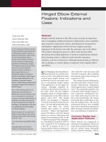
Hinged Elbow External
Fixators: Indications and
Uses
Abstract
Hinged external fixation of the elbow joint can play an important
role in managing complicated fracture-dislocations, joint instability
after extensive contracture release, and distraction interposition
arthroplasty. Application of these devices requires accurate
alignment of the fixator axis with the anatomic axis of the elbow.
The primary therapeutic goal is to allow joint motion while
protecting the healing ligaments. Common complications include
pin loosening, injury to adjacent neurovascular structures,
cellulitis, and loss of reduction. Although reported data are limited,
this technique is a useful adjunct in patients with complex elbow
instability.
S
ince Malgaigne first described ex-
ternal fixation for patellar frac-
tures in 1843, external fixators have
evolved from simple devices used
exclusively in lower extremity trau-
ma to articulating hinged frames
with applications in the upper ex-
tremity. Although originally de-
scribed in the Russian literature in
the early 1970s, the first English-
language report of a hinged distrac-
tion apparatus for the elbow did not
appear until 1975.
1
This hinged
device was designed to eliminate
“excessive friction between the sur-
faces,” prevent abnormal joint kine-
matics, and allow the “newly
formed joint surfaces to develop
correctly.”
1
Volkov and Oganesian
1
treated 28 elbows with the hinged
fixator for various indications and
reported encouraging results. In the
13 elbows managed with arthroplas-
ty, 7 had final arc of motion of 80° to
120°, 5 had 50° to 70° of motion, and
1 had 40° of motion. In the 11 el-
bows treated for flexion contracture,
6 had final arc of motion of 70° to
120°, 4 had 50° to 60° of motion, and
1 had 40° of motion. The remaining
four patients had a final average arc
of motion of 102°.
The concept of an articulating fix-
ator about the elbow is based on the
normal ulnohumeral kinematics,
which approximate a simple hinged
joint.
2-4
Recreating the anatomic axis
of rotation with a hinged fixator al-
lows concentric ulnohumeral mo-
tion while protecting the joint sur-
faces and periarticular soft tissues
from loads that would injure or dis-
rupt the healing tissue. The several
commercial devices now available
that seek to satisfy this requirement
differ in design, method of mobiliza-
tion, and technique of application.
Common Design and
Application Features
Elbow fixators are categorized as
unilateral ormultiplanar. These two
types share common design features
and are affixed using essentially
similar surgical techniques. Advan-
Virak Tan, MD,
Aaron Daluiski, MD,
John Capo, MD, and
Robert Hotchkiss, MD
Dr. Tan is Associate Professor, Division
of Hand and Microsurgery, Department
of Orthopaedic Surgery, University of
Medicine and Dentistry of New Jersey–
The New Jersey Medical School,
Newark, NJ. Dr. Daluiski is Assistant
Professor, Division of Hand Surgery,
Department of Orthopaedic Surgery,
The Hospital for Special Surgery, New
York, NY. Dr. Capo is Assistant
Professor, Division of Hand and
Microsurgery, Department of
Orthopaedic Surgery, University of
Medicine and Dentistry of New Jersey–
The New Jersey Medical School. Dr.
Hotchkiss is Associate Professor,
Division of Hand Surgery, Department of
Orthopaedic Surgery, The Hospital for
Special Surgery.
None of the following authors or the
departments with which they are
affiliated has received anything of value
from or owns stock in a commercial
company or institution related directly or
indirectly to the subject of this article:
Dr. Tan, Dr. Daluiski, and Dr. Capo. Dr.
Hotchkiss or the department with which
he is affiliated has received royalties
from Smith & Nephew.
Reprint requests: Dr. Tan, University of
Medicine and Dentistry of New Jersey,
90 Bergen Street, DOC 1200, Newark,
NJ 07101-1709.
J Am Acad Orthop Surg 2005;13:503-
514
Copyright 2005 by the American
Academy of Orthopaedic Surgeons.
Volume 13, Number 8, December 2005 503
tages of the unilateral frames in-
clude (presumably) less ulnar nerve
irritation; a lower profile, which is
more tolerable to the patient; and
ease of application. Advantages of
the multiplanar fixators include
more rigid skeletal fixation as well
as better control of varus/valgus
alignment and joint distraction.
Application of a hinged elbow
external fixator can be demanding.
The most critical step is correct
placement of the axis pin. To mini-
mize resistance to motion and half-
pin loosening, this pin must be
colinear with the center of rotation
of the elbow joint. Madey et al
5
re-
ported that misalignment of 5°
caused a 3.7-fold increase in motion
energy; a 10° mismatch yielded a
7.1-fold increase. The anatomic axis
of rotation lies at the center of the
capitellum and trochlear spool and is
usually determined from anatomic
landmarks. Medially, this point lies
just distal and anterior to the medi-
al epicondyle; laterally, it lies just
slightly distal to the lateral epi-
condyle.
The axis pin starting point should
be verified with fluoroscopy before
advancing into bone. The pin should
be slowly advanced and the position
confirmed on lateral and anteropos-
terior radiographic views. The true
lateral view should show the pin as
a dot within the center of the troch-
lear spool, while the anteroposterior
view should show it traversing paral-
lel to the joint, along the normal val-
gus angulation of the distal humerus
(Figure 1). With a unilateral frame,
the axis pin is inserted only far
enough to ensure proper orientation
and stable bone purchase. With a
multiplanar frame, a single axis pin
is advanced across the distal humer-
us, or two pins may be placed from
both the medial and lateral sides.
Care must be taken on the medial
side to protect the ulnar nerve dur-
ing advancement of the axis pin.
The external fixator frame is as-
sembled around the axis pin and at-
tached to the skeleton with half
pins. These half pins should be
placed without impaling any major
muscle-tendon units or jeopardizing
neurovascular structures. The hu-
meral pins are usually placed first.
Lateral pins are more easily placed
because of patient positioning. The
most proximal lateral pin may lie
near the course of the radial nerve,
which should be avoided by careful
pin placement. Pins placed medially
should be inserted through an open
incision to protect the ulnar nerve.
All half pins should have bicortical
purchase.
With the elbow concentrically re-
duced, the fixator frame is attached
to the ulna. It is useful to hold the el-
bow in flexion with the arm in the
overhead position to take advantage
of gravity to assist in concentric re-
duction and placement of the ulnar
half pins. Depending on the fixator
used, the ulnar pins are inserted ei-
ther in a dorsal-to-volar or in a
lateral-to-medial direction. After the
pins are inserted into the ulna, the
frame is secured to the ulnar pins.
With a highly unstable reduction,
the joint can be temporarily pinned
with a stout Kirschner wire before
ulnar pin insertion.
After ensuring that all the connec-
tions are secure, the axis pin is re-
moved and the elbow is taken
through a range of motion (ROM) un-
der fluoroscopy to evaluate for con-
centric reduction and stability. The
half pins should be checked for
proper skin clearance, and the ulnar
nerve should be sensitive during flex-
ion and extension of the elbow. Fine-
tuning of the frame to achieve the de-
sired amount of distraction and
varus/valgus angulation is done at
the conclusion of frame application.
Figure 1
Intraoperative fluoroscopic anteroposterior (A) and lateral (B) views of the axis pin position.
Hinged Elbow External Fixators
504 Journal of the American Academy of Orthopaedic Surgeons
Specific Hinged
Fixators
Currently, there are four commer-
cially available hinged external fix-
ators for the elbow: Compass Uni-
versal Hinge (Smith & Nephew,
Memphis, TN), OptiROM Elbow
Fixator (EBI, Parsippany, NJ), Ortho-
fix Elbow Fixator (Intavent Orthofix
Ltd, Berkshire, United Kingdom),
and Dynamic Joint Distractor II
(Stryker Howmedica Osteonics,
Mahwah, NJ) (Table 1). Each specif-
ic fixator has unique features, and
the choice of fixator is usually based
on the surgeon’s familiarity and
comfort with the system.
The Compass Universal Hinge
(Figure 2, A) is a multiplanar fixator
that allows incremental passive
joint ROM. The frame, which is
composed of radiolucent 1/2-in and
5/8-in rings, is assembled before ap-
plication. The humeral half pins are
placed in both medial and lateral
multiplanar positions. The ulno-
humeral articulation must be con-
centrically reduced before placing
the ulnar pins and attaching the
frame. The ulnar pins are inserted
from the dorsal surface of the ulna in
a dorsal-to-volar direction. The
frame has a self-telescoping mecha-
nism to allow a 20° arc of varus/
valgus adjustment. Distraction
screws allow joint displacement/
distraction that is independent of
the varus/valgus alignment. Addi-
tionally, a precision worm gear per-
mits motion within a specified range
and can be “ungeared” for active and
passive motion or kept locked for in-
cremental gear-driven passive mo-
tion. The patient or occupational
therapist can easily operate the gear.
One disadvantage of the Compass
Universal Hinge is that its applica-
tion can be technically demanding.
Additionally, there is less room for
adjustment when the frame is placed
with the elbow subluxated. Because
the frame impinges against the chest
wall, patient comfort may be an is-
sue.
The OptiROM Elbow Fixator
(Figure 2, B) is a unilateral frame
based on multiple stout adjustable
linkages that allow many degrees of
freedom. The axis pin is placed from
lateral to medial, after which the hu-
meral por tion of the frame may be
applied with appropriate half pins.
Alternatively , the humeral pins may
be placed and secured to the frame
before the axis pin is inserted. The
Table 1
Commercially Available Hinged Elbow External Fixators
Fixator Type Features Advantages Disadvantages
Compass
Universal
Hinge
Multiplanar Ilizarov concept
Radiolucent arcs with
bilateral hinges
Varus/valgus control
Distraction control
Worm gear
Frame stability
Independent angulation
and distraction control
Passive gear-driven
motion allows for
soft-tissue
stress/relaxation
Steep learning curve
Concentric reduction
required before fixation to
ulna
Less patient comfort
More exposure needed for
medial half pins
OptiROM Elbow
Fixator
Unilateral Multiple adjustable
linkages
More patient comfort due
to unilateral design
Flexibility to allow
adjustment of frame to
elbow axis of rotation
Noninvasive technique for
axis placement
Less frame stability
No independent angulation
and distraction control
Conical half-pin design
limits depth adjustment
Orthofix Elbow
Fixator
Unilateral Linked components
with central
connecting units
Low profile
Compression-distraction
unit can be applied for
static progressive ROM
Concentric reduction
required before fixation to
ulna
Frame allows less flexibility
Extensor mass may be
impaled with pin
placement
Dynamic Joint
Distractor II
Unilateral/
bilateral
Simplified frame
construction with
integrated hinge
Compatible with
Hoffman II
Compact couplings
Ease of application
Lowest profile for
increased patient
comfort
Independent half-pin
placement
Frame stability increased
with bilateral application
Varus/valgus controlled at
pin-to-rod coupling
No passive-motion
mechanism in the frame
Concentric reduction
required before fixation to
ulna
Ulnar pin placement
impales the common
extensor muscles
Virak Tan, MD, et al
Volume 13, Number 8, December 2005 505
axis guide ring can be adjusted to lie
over the axis pin. The axis ring
should slide easily over the guide pin
for several centimeters to ensure
proper alignment of the frame with
the elbow axis of rotation. An addi-
tional benefit is that the ulnar half
pins can be inserted either from dor-
sal to volar (preferred by the authors)
or lateral to medial and locked to the
frame with the elbow in an unre-
duced position. Ulnohumeral joint
reduction can be done with the
frame in place and the universal
joints tightened to maintain reduc-
tion, thus avoiding the sometimes
difficult task of placing the ulnar
pins while maintaining perfect re-
duction of the elbow. A new addition
to this frame allows static progres-
sive ROM with application of torque
through an adjustment screw.
The Orthofix Elbow Fixator (Fig-
ure 2, C) is similar to the OptiROM.
This unilateral frame consists of
two linked components (ulnar and
humeral) with a central connecting
unit. The frame is placed over the
axis pin, the humeral pins are placed
unicortically from lateral to medial,
and the ulnar pins are placed from
lateral to medial. A distraction unit
can be applied to either the ulnar or
humeral link. Additionally, a com-
pression-distraction bar can be at-
tached to the cams of the fixator for
static progressive ROM, which can
be done by either the patient or the
therapist. One disadvantage of this
device is less flexibility of the frame;
there are only two adjustable link-
ages, which limits the degrees of
freedom. Another disadvantage is
impalement of the common exten-
sor muscles resulting from the later-
al-to-medial placement of the ulnar
pins.
The Dynamic Joint Distractor II
(Figure 2, D) is based on the same
concept as its predecessor, the
Mayo Dynamic Joint Distractor. The
Figure 2
A, Compass Universal Hinge. (Courtesy of Smith & Nephew, Memphis, TN.) B, OptiROM Elbow Fixator. (Courtesy of EBI,
Parisippany, NJ.) C, Orthofix Elbow Fixator. (Courtesy of Intavent Orthofix, Berkshire, UK.) D, Dynamic Joint Distractor II.
(Courtesy of Stryker Howmedica Osteonics, Mahwah, NJ.)
Hinged Elbow External Fixators
506 Journal of the American Academy of Orthopaedic Surgeons
frame can be applied in a unilateral
or bilateral configuration. Using a
humeral axis guide, the axis pin is
placed via open technique on the
medial side and percutaneously on
the lateral side. The guide clamps,
the center points on the medial
and lateral sides, and the axis pin
are placed through the cannulated
guide. The frame is applied over
the axis pin, and pin guides are
used to place the humeral pins.
The half pins are connected to the
frame with standard Hoffman II
Compact clamps (Stryker Howmed-
ica Osteonics). The ulnar pins are
placed percutaneously from lateral
to medial, which has the disad-
vantage of impaling the common
extensor muscles. The built-in
distraction-compression device can
be progressively adjusted. Dual me-
dial and lateral frames can be applied
for more stability. The advantages of
this frame are its low profile and rel-
ative ease of application.
Complications
Although the true incidence of com-
plications is difficult to determine,
they are relatively common and
should be anticipated. Infection can
range from cellulitis around the pin
tract to deep-seated sepsis.
1,5-7
In the
early phase, when there is only
erythema and tenderness around the
pin site and the pin is not loose, cel-
lulitis may be treated with oral anti-
biotics for 10 to 14 days. When there
is drainage around the pin despite
antibiotic treatment or when the pin
is loose, removal with insertion of a
new pin or pins in healthy tissue
may be necessary. Rarely, osteomy-
elitis can develop, in which case the
entire fixator must be removed
1,8
and intravenous antibiotics given for
6 weeks. Vigilance and local pin care
are the keys to minimizing this
complication.
Loss of reduction can occur from
improper placement of the fixator
axis or from hardware failure.
8-11
Pe-
riodic radiographic evaluation is
mandatory to confirm that the joint
remains reduced and the frame is se-
cure. With more vigorous rehabilita-
tion and motion, more stress is
placed on the components, which
can lead to pin loosening or break-
age. Pin replacement may be neces-
sary to maintain alignment between
the fixator axis and that of the el-
bow.
Despite awareness that the ulnar
nerve is at risk during surgical proce-
dures around the elbow, injury still
may occur.
7,9,12
Causes include inju-
dicious placement of the axis or me-
dial humeral half pins, over-
penetration of lateral humeral half
pins, and increased elbow flexion af-
ter a contracture release. Ulnar
nerve injury can be avoided with
precise pin insertion and protection
and/or transposition of the ner ve.
For laterally based unilateral frames,
care must be taken to protect the ra-
dial nerve during application of the
most proximal pin.
13
With the Com-
pass Universal Hinge, the low later-
al humeral pin should be placed
from posterolateral to anteromedial,
staying posterior to the course of the
radial nerve. Injury to the posterior
interosseous nerve also has been re-
ported.
7,14
In addition to avoiding
neurovascular structures, the half
pins should be placed without im-
paling any major muscle-tendon
units. Such injury may impede mo-
tion or cause pain.
15
Fracture of the ulna during vigor-
ous therapy or as a result of a fall are
less commonly reported complica-
tions.
11
Using smaller diameter pins
for the ulna can help reduce the
stress riser effect. When fracture
does occur, internal fixation with
plating may be necessary. Reflex
sympathetic dystrophy also has been
reported after hinged fixation
10
and
should be treated expeditiously.
Indications
Instability
The elbow is a relatively stable
joint because of its bony anatomy
and capsuloligamentous complex.
However, disruption of these struc-
tures can render the joint unstable.
Causes of instability include disloca-
tion with medial collateral and liga-
ment tear, coronoid and radial head
fractures (the so-called terrible triad),
medial collateral ligament injury
with concomitant radial head frac-
ture, comminuted olecranon and/or
distal humerus fractures, and post-
contracture release of a stiff joint.
Ulnohumeral instability can be cat-
egorized as acute, recurrent, or
chronic. Acute instability is present
at the time of the initial surgical
treatment; recurrent instability is re-
ducible, but with persistent instabil-
ity (after the initial stabilization) in
the postoperative period; and chron-
ic instability is late, unreduced dislo-
cation that has become irreducible
by closed manipulation.
In the setting of unstable elbows,
hinged fixators are indicated for per-
sistent acute or recurrent instability
despite attempted fracture stabiliza-
tion and ligament repair; for protect-
ing nonrigid fracture fixation and/or
non-secure ligamentous repair dur-
ing postoperative rehabilitation; and
for chronically unreduced disloca-
tion; and acute gross instability that
cannot be splinted in concentric re-
duction in a patient who is unable to
tolerate a prolonged surgical proce-
dure (Figure 3).
Acute and Recurrent
Instability
Acute elbow instability encom-
passes a spectrum of conditions,
ranging from subtle ulnohumeral
subluxation to simple dislocation to
the terrible triad. Acute subluxation
and simple dislocation respond well
to cloned reduction followed by non-
surgical management with super-
vised rehabilitation. At the other end
of the spectrum, complex elbow dis-
location with associated radial head
and coronoid fractures and/or collat-
eral ligament disruption may render
the joint very unstable. Under these
circumstances, the radial head and
Virak Tan, MD, et al
Volume 13, Number 8, December 2005 507
coronoid should be repaired, recon-
structed, or replaced, and the collat-
eral ligament or ligaments repaired or
reconstructed. In some patients, even
with osteosynthesis and repair of the
ligaments, the elbow may still be un-
stable because of severe bony and
soft-tissue defects. With such injury
patterns, ulnohumeral instability
may not be immediately evident be-
cause the surgeon does not want to
test or stress the repair; thus, an im-
perfect or unstable reduction may
not be detected until the postopera-
tive period.
Regardless of the timing, persis-
tent instability that is present either
acutely or in the early postoperative
period is an indication for a hinged
external fixator. The function of the
fixator is to maintain concentric re-
duction of the ulnohumeral joint,
protect the bony and/or ligament re-
pair or reconstruction, and allow ear-
ly postoperative motion.
11,15
For a
complex fracture-dislocation of the
elbow, a hinged device should be
available in the operating room at
the time of initial surgery. Careful
assessment of the quality of fracture
fixation and ligament repair must be
done intraoperatively to determine
whether the ulnohumeral joint is
stable enough to tolerate early post-
operative motion. In some settings,
the position of maximal instability
Figure 3
Closed
reduction
Acute
instability
Recurrent
instability
Acute gross instability;
medically unstable*
Chronic unreduced
dislocation
Repair or
reconstruct
ligament(s)
Total elbow
arthroplasty
Open reduction
and/or interposition
and/or ligament repair(s)
Hinged
external
fixator
Rehabilitation
program
Associated
fractures
Salvageable
No
Yes
No
Yes
YesYes
Stable range
of motion
No
No (ie, nonrigid
fracture fixation)
Stable fixation/
reconstruction
achieved
Stable
range of
motion
Yes
No (ie,
nonsecure
ligamentous
repair)
No
Yes
Stable range
of motion
Treatment algorithm for complex elbow instability and the potential use of a hinged fixator.
* A static external fixator across the elbow also may be appropriate in this situation.
Hinged Elbow External Fixators
508 Journal of the American Academy of Orthopaedic Surgeons
can be avoided, and the elbow reha-
bilitation program may proceed with
a hinged brace or splinting. When
persistent instability through ROM
precludes rehabilitation, the hinged
fixator should be applied. For recur-
rent subluxation in the postopera-
tive period, the device can be applied
by percutaneous techniques provid-
ed that the ulnar nerve is protected.
Although no large studies have
been done, several authors have re-
ported cautious optimism with use
of a hinge for acute and/or recurrent
complex instability. McKee et al
10
used hinged fixation to treat com-
plex elbow instability in 16 patients.
In two patients, the fixator was ap-
plied at the time of the original treat-
ment because the elbow remained
unstable after open reduction and
internal fixation and soft-tissue re-
construction. The other 14 patients
failed conventional treatment; 11
had recurrent dislocation and 3 had
recurrent subluxation.
10
In these 14
patients, hinge application was done
at a mean of 4.8 weeks (range, 2 to
9 weeks) after the primary treat-
ment. The fixators remained in place
for a mean of 8.5 weeks (range, 6 to
11 weeks). On final follow-up 23
months after surgery (range, 14 to
40 months), 15 of 16 elbows had
achieved concentric reduction. The
mean arc of flexion-extension was
105° (range, 65° to 150°) with prona-
tion of 76° (range, 20° to 90°) and su-
pination of 75° (range, 15° to 90°). Six
patients experienced complications,
including recurrent subluxation, re-
flex sympathetic dystrophy, pin tract
infection, wound infection, and tran-
sient radial nerve palsy. The one re-
current instability occurred in a non-
compliant patient who had incorrect
placement of the center axis of rota-
tion and early loosening of the hu-
meral pins.
Cobb and Morrey
11
reviewed
seven patients who had unstable el-
bow dislocations associated with
coronoid fractures. Hinged external
fixation was applied acutely in one
patient and postoperatively in three
patients. (The other three patients
underwent “resurfacing” distraction
arthroplasty.) At final follow-up (44
months), three of the four patients
had a stable elbow, with a flexion-
extension arc of 95° and a pronation-
supination arc of 115°. One patient
had persistent instability and went
on to have a total elbow arthroplasty.
Ruch and Triepel
12
evaluated a
unilateral hinged elbow frame for
recurrent instability following frac-
ture-dislocation. Three of eight pa-
tients had acute instability and were
treated with hinge stabilization for a
mean of 43 days (range, 40 to 47
days) because of inability to achieve
complete osseous and ligamentous
repair. The average postoperative
arc of motion was 120° (range, 105°
to 130°), with average pronation of
90° and average supination of 67°.
The other five patients were treated
with an articulated fixator as an “al-
ternative to complete osseous and
ligamentous reconstruction” for re-
current instability at 6 weeks to 9
months after the initial injury.
Mean duration of external fixation
was 62 days (range, 54 to 80 days).
Their average postoperative arc of
motion was 84° (range, 75° to 95°),
with average pronation of 68° and
average supination of 43°.
In another study of a unilateral ar-
ticulating fixator, von Knoch et al
6
reported on 13 patients, 9 with acute
elbow trauma. The average duration
of external fixation was 7.6 weeks
(range, 3 to 18 weeks). These authors
did not stratify the results of the
acute traumatic injury from the
posttraumatic reconstruction (eg,
joint stiffness/contracture, hetero-
topic ossification, distal humerus
nonunion). The 11 patients who
were followed had an average arc of
motion of 81° (range, 50° to 125°).
Complications were confined to five
patients who developed pin tract in-
fection, which resolved with oral an-
tibiotics.
The hinged fixator is not a pana-
cea for complex elbow instability.
These injuries are difficult to man-
age, and patients usually have a less
than satisfactory result. Initially , the
principles of osteosynthesis with or
without collateral ligament repair
should be followed. The hinged el-
bow devices should be used as an ad-
junct to, not in lieu of, convention-
al stabilization. The only exception
is in the patient with gross elbow in-
stability who cannot medically tol-
erate a prolonged surgical interven-
tion.
15
Because of the instability,
closed reduction cannot be obtained
and maintained by external splint-
ing. In such patients, an external fix-
ator (either static or hinged) can be
used as a primary temporizing de-
vice until definitive stabilization can
be performed.
Chronic Dislocation
Morrey
16
described two major
types of chronic elbow instability,
based on the degree of displacement:
subluxation and dislocation. Chron-
ic subluxation (ie, posterolateral ro-
tatory instability) is more common;
of these, the chronic dislocation can
be best treated with a hinged fixator.
Patients with chronic complete dis-
location of the elbow often have had
a neglected or irreducible elbow
dislocation.
17-19
Neglected disloca-
tion is more commonly seen in pa-
tients in underdeveloped countries.
However, such instances occur in
North America when the disloca-
tion is unrecognized in an unrespon-
sive multiply traumatized patient.
They also occur when the elbow re-
dislocates in a patient who fails to
follow up, or in a patient who did not
initially seek medical attention.
Marked deformity of the elbow can
result, with severely limited func-
tion caused by pain and restricted
motion.
18
Often, there are associated
fractures along with the chronic dis-
location.
19
Various treatment strategies have
been reported for chronic elbow dis-
locations; however, only open reduc-
tion (with or without hinged ex-
ternal fixation) and total elbow
replacement are reasonable options.
Virak Tan, MD, et al
Volume 13, Number 8, December 2005 509
Several investigators have reported
satisfactory results with open reduc-
tion without distraction of the joint.
Billett
18
reported on six unreduced
posterior dislocations in which re-
duction was achieved by open exci-
sion of all fibrous tissue and by divi-
sion of the medial and lateral
collateral ligaments before joint re-
duction. Because of the induced in-
stability required for the reduction, a
Kirschner wire was used to tempo-
rarily transfix the joint for 2 weeks.
Range of motion (flexion and exten-
sion) improved postoperatively. One
patient who did not have the joint
pinned redislocated. Naidoo
19
re-
ported on 23 unreduced posterior dis-
locations treated with release of the
anterior capsule and collateral liga-
ments as well as temporary trans-
fixion of the ulnohumeral joint.
Although follow-up was limited be-
cause of socioeconomic factors, use-
ful ROM was obtainable even in dis-
locations older than 3 months and in
patients older than age 40 years. Ara-
files
17
described open reduction and
an intra-articular “cruciate” liga-
ment reconstruction with a free
tendon graft in 11 patients with ne-
glected elbow dislocation. This pro-
cedure was devised to allow early el-
bow motion in the flexion-extension
plane after the open reduction. At
32-month follow-up, the flexion-
extension arc of motion averaged
105°, with a mean valgus-varus lax-
ity of 13°.
Because open reduction of a chron-
ically unreduced elbow requires ex-
tensile release of the contracted soft
tissues, including the capsule, collat-
eral ligaments, and possibly the tri-
ceps, Morrey
16
advocated reconstruc-
tion of the collateral ligaments
through bone tunnels and application
of a hinged fixator, both to allow im-
mediate motion and to protect the
repaired collateral ligaments. In three
of four patients, a stable arc of elbow
motion >90° was achieved; these pa-
tients had mild or no pain.
16
Jupiter and Ring
20
treated un-
reduced elbow dislocations with
hinged external fixation in five pa-
tients. Surgery was performed at an
average of 11 weeks (range, 6 to 30
weeks) after the original dislocation.
The elbow joint was exposed both
medially and laterally, but the origin
of the flexor-pronator mass was left
attached to the medial epicondyle.
Adhesions and the entire lateral cap-
sule were resected, after which the
ulnohumeral joint was reduced and
the hinged fixator applied. At an av-
erage of 38 months (range, 12 to 98
months), all patients had stable con-
centric reduction and a satisfactory
Mayo Elbow Performance Index (av-
erage score, 89 points). The average
arc of flexion was 123°, and all pa-
tients had full forearm rotation.
In cases of chronic unreduced el-
bow dislocation, a hinge-distraction
device can be useful to maintain ul-
nohumeral joint reduction without
transfixing the joint and to start im-
mediate concentric motion. A hinge-
distraction device also allows the
joint to unload by distraction and
the soft-tissue sleeves to heal in the
optimal position for motion.
Distraction Interposition
Arthroplasty
Distraction interposition arthro-
plasty has been developed from two
procedures—distraction and biolog-
ic resurfacing of the joint—used to
treat incapacitating elbow pain and
loss of motion. Volkov and Ogane-
sian
1
advocated joint separation
while gradually restoring motion
through a hinged-distraction device.
In their arthroplasty group of pa-
tients, in whom preoperative motion
was severely limited (arc of motion
ranged from 0° to 50°), postoperative
motion increased by 70° to 120° in
six patients, by 50° to 60° in four pa-
tients, and by only 40° in one pa-
tient. Nine patients returned to their
previous occupations, although
three of these remained in pain. Sim-
ilarly, Morrey
8
reported on 14 pa-
tients who underwent distraction
without joint resurfacing for elbow
contracture. The average arc of mo-
tion increased from 32° (range, 0° to
75°) preoperatively to 99° (range, 70°
to 125°) postoperatively.
Interposition arthroplasty, in
which a biologic material is used to
resurface the joint, has had mixed
success in the elbow.
21-24
This proce-
dure has been used for posttraumatic
or postinfectious ankylosis, hemo-
philic arthropathy, and rheumatoid
arthritis.
21-24
In 1952, Knight and Van
Zandt
24
reported the results of fascia
lata interposition arthroplasty in 45
patients with partial or complete
elbow ankylosis. At an average
follow-up of 14 years, there were 25
good, 10 fair, and 10 poor or failed el-
bows. In 1976, Froimson et al
23
re-
ported satisfactory results using deep
dermal skin interposition arthro-
plasty in five patients. However, two
patients had varus-valgus instability
of 20° and 30°, respectively. Ljung et
al
22
found that the results of interpo-
sition arthroplasty in 35 rheumatoid
elbows were good in terms of pain re-
lief but only fair in terms of joint mo-
bility and stability. The inconsistent
outcome of interposition arthro-
plasty alone raised questions about
the effectiveness of this procedure in
patients with arthritic elbows.
22
The addition of joint distraction
to interposition arthroplasty (ie, dis-
traction interposition arthroplasty)
was done to address concerns of
postoperative instability and de-
gradation of the interposed tissue
when early motion is started.
4,25
Us-
ing a hinged external fixator allows
for distraction across the joint to
minimize shear forces across the in-
terposed tissue. A hinged external
fixator also permits immediate post-
operative motion. The medial and
lateral ligaments remain protected
through the postoperative healing
period.
The surgical procedure, which
has been well described,
4,8,25
begins
with contracture release using the
lateral column and/or medial over-
the-top approach.
26
After the release,
if visual inspection of the joint sur-
face reveals loss of articular cartilage
Hinged Elbow External Fixators
510 Journal of the American Academy of Orthopaedic Surgeons
≥50%, significant intra-ar ticular ad-
hesions causing avulsion of cartilage
during motion, or an intra-articular
malunion requiring recontouring,
then interposition arthroplasty
should be performed.
8
The lateral
ligament complex is sharply divided
from the humerus. If necessary for
exposure, the triceps is mobilized as
a continuous sleeve from the ulna.
Manual distraction applied across
the ulnohumeral articulation often
provides sufficient exposure to per-
form the operation without remov-
ing the triceps insertion. The ulno-
humeral joint surfaces are then
prepared by contouring them into
matching surfaces. Bone resection
should be sufficient to allow a gap of
at least 3 mm. The radial head is re-
tained when there is painless fore-
arm rotation. In some cases, the ul-
nar articular margin for the radial
head is removed (ie, “radialization”
procedure) to increase forearm mo-
tion.
25
Although a variety of interposi-
tion materials has been used, autol-
ogous fascia lata is usually the graft
of choice. A sheet of fascia 5 cm × 12
cm is harvested from the thigh. The
fascia is then sutured to the distal
end of the humerus with suture an-
chors or drill holes through the hu-
merus. Once the interposition graft
is in place, the ulnohumeral joint is
located, and fluoroscopic views of
the joint are taken to verify reduc-
tion. Care must be taken not to
translate the ulna too far radially.
Proper repair or reconstruction with
tendon grafts of the lateral ligament
complex is then performed. A
hinged fixator is applied, and distrac-
tion of at least 3 mm is maintained
through a full arc of motion.
Few reports of the outcome of dis-
traction interposition arthroplasty
have been published (Table 2). Many
of these results are embedded with-
in larger study cohorts. Although the
results are not as reproducible as for
patients undergoing total elbow ar-
throplasty, most patients have im-
provement in ROM and moderate to
significant pain relief.
Distraction interposition arthro-
plasty likely will become more com-
mon as the number of patients with
posttraumatic elbow arthropathy in-
creases. This procedure is indicated
for patients with intra-articular pa-
thology who are too young for a total
elbow prosthesis. It also is indicated
in certain patients with inflammatory
arthropathy , and in patients who have
experienced trauma or infection.
Postoperative Control of
Motion
Elbow contracture releases, espe-
cially revision cases, often require
extensive excision of soft tissues and
bone structures to regain motion.
These structures include the collat-
eral ligaments, heterotopic bone, os-
teophytes, and the hypertrophic cap-
sule. Because the goal of surgery is to
improve elbow motion, the surgeon
should not abandon this goal until
full or nearly full motion is obtained
on the operating table. In the course
of these excisions, the joint may be
rendered unstable,
4
requiring the ap-
plication of hinged fixation to allow
controlled motion until the soft tis-
sues heal adequately to provide sta-
bility. Posttraumatic contracture,
which involves an intrinsic cause,
may benefit from distraction inter-
position arthroplasty.
Within the spectrum of contrac-
ture is complete ankylosis of the
joint (Figures 4 and 5). In a report of
20 elbows (15 patients) that under-
went surgical release for this condi-
tion, Ring and Jupiter
27
used a
hinged device in three patients to
treat elbow subluxation or disloca-
tion. The true outcome of these
three patients is unknown because
the results were not stratified ac-
Table 2
Results of Distraction Interposition Arthroplasty
Study
No. of
Patients
Mean
Follow-up
(months)
Mean Preoperative
Flexion-Extension Arc
(range)
Mean Postoperative
Flexion-Extension Arc
(range) Results
Morrey
8
6 33 27° (0°-60°) 107° (70°-150°) —
Morrey
4
20 — — — 80% satisfactory
Cobb and
Morrey
11
2 30 38° (35°-40°) 84° (57°-110°) Both satisfactory
Cheng and
Morrey
25
13* 63 60° (24°-100°) 84° (40°-135°) 69% satisfactory
pain relief, 62%
excellent or good
result
Pignatti et al
9
12 Range, 8-33 35° (0°-90°) 91° (—) 92% satisfied with
outcome
*Does not include four patients converted to total elbow arthroplasty
— = data could not be determined from the published material
Virak Tan, MD, et al
Volume 13, Number 8, December 2005 511
cording to hinge use. However, 7 of
the 20 elbows had recurrence of con-
tracture, and 6 underwent a subse-
quent contracture release. One oth-
er elbow had ulnar plate fracture
fixation. Even so, using a hinged de-
vice to manage complete ankylosis
may be beneficial because of the dis-
traction component and greater abil-
ity to control postoperative motion.
Another uncommon use of
hinged fixation is protecting elbow
motion and/or repair in the obese
patient. Because of large body habi-
tus, it may difficult or impossible to
adequately control elbow motion
with postoperative splinting or brac-
ing. In such patients, skeletal fixa-
tion with a hinged fixator allows
protected and controlled motion.
Summary
Hinged external fixation about the
elbow is generally viewed as chal-
lenging. Similar to other surgical de-
vices, success is dependent on when
and how to apply the fixator. An un-
derstanding of the anatomy, specific
technique, and indications for each
problem is crucial for restoring el-
bow function. Current indications
include acute and recurrent instabil-
ity after osteosynthesis and ligament
repair, chronic dislocation, distrac-
tion interposition arthroplasty, and
postoperative control of motion.
Hinged external fixation is also indi-
cated in the uncommon case of
acute elbow instability when con-
centric reduction cannot be achieved
by splinting or in a patient who can-
not tolerate a prolonged intraopera-
tive surgical procedure. The two
main types of hinged elbow fixators
are unilateral and multiplanar. Uni-
lateral frames inflict less ulnar nerve
irritation, have a lower profile, and
Figure 4
A 15-year-old girl sustained a fracture-dislocation of the elbow in a fall from a horse. A, Lateral radiograph demonstrating
complete ankylosis of the ulnohumeral joint, fixed at 75°. B, Intraoperative photograph demonstrating takedown of the bony
ankylosis and distraction interposition arthroplasty 4 years after injury. Postoperative anteroposterior (C) and lateral (D)
radiographs demonstrating the new elbow articulations.
Hinged Elbow External Fixators
512 Journal of the American Academy of Orthopaedic Surgeons
are relatively easy to apply. Multi-
planar fixators provide more rigid
fixation as well as better control of
alignment and distraction across the
joint. Despite the complexity and
complications, a hinged external fix-
ator can be very helpful in treating
elbow instability in the absence of
other reasonable alternatives.
References
No randomized Level I studies cited.
1. Volkov MV, Oganesian OV: Restora-
tion o f function in the knee and elbow
with a hinge-distractor apparatus.
J Bone Joint Surg Am 1975;57:591-
600.
2. Deland JT, Garg A, Walker PS: Biome-
chanical basis for elbow hinge-
distractor design. Clin Orthop Relat
Res 1987;215:303-312.
3. London JT: Kinematics of the elbow.
J Bone Joint Surg Am 1981;63:529-
535.
4. Morrey BF: Distraction arthroplasty:
Clinical applications. Clin Orthop
Relat Res 1993;293:46-54.
5. Madey SM, Bottlang M, Steyers CM,
Marsh JL, Brown TD: Hinged external
fixation of the elbow: Optimal axis
alignment to minimize motion resis-
tance. J Orthop Trauma 2000;14:41-
47.
6. von Knoch F, Marsh JL, Steyers C,
McKinley T, O’Rourke M, Bottlang
M: A new articulated elbow external
fixation technique for difficult elbow
trauma. Iowa Orthop J 2001;21:13-
19.
7. Fox RJ, Varitimidis SE, Plakseychuk
A, Vardakas DG, Tomaino MM, So-
tereanos DG: The Compass Elbow
Hinge: Indications and initial results.
J Hand Surg [Br] 2000;25:568-572.
8. Morrey BF: Post-traumatic contrac-
ture of the elbow: Operative treat-
ment, including distraction arthro-
plasty. J Bone Joint Surg Am 1990;72:
601-618.
9. Pignatti G, Ferrari D, Tigani D, et al:
The treatment of post-traumatic stiff-
ness of the elbow. Chir Organi Mov
2000;85:381-387.
10. McKee MD, Bowden SH, King GJ, et
al: Management of recurrent, com-
plex instability of the elbow with a
hinged external fixator. J Bone Joint
Surg Br 1998;80:1031-1036.
11. Cobb TK, Morrey BF: Use of distrac-
tion arthroplasty in unstable fracture
dislocations of the elbow. Clin
Orthop Relat Res 1995;312:201-210.
12. Ruch DS, Triepel CR: Hinged elbow
fixation for recur rent instability fol-
lowing fracture dislocation. Injury
2001;32(suppl 4):SD70-SD78.
13. Pennig D, Gausepohl T, Mader K:
Transarticular fixation with the ca-
pacity for motion in fracture disloca-
tions of the elbow. Injury
2000;31(suppl 1):35-44.
14. Tomaino MM, Sotereanos DG, West-
kaemper J, Plakseychuk A: Posterior
interosseous nerve palsy following
placement of the compass elbow
hinge for acute instability: A case
report.J Hand Surg [Am] 1999;24:
554-560.
15. O’Driscoll SW, Jupiter JB, King GJ,
Hotchkiss RN, Morrey BF: The unsta-
ble elbow. Instr Course Lect 2001;50:
89-102.
16. Morrey BF: Chronic unreduced elbow
dislocation, in Morrey BF (ed): The El-
bow and its Disorders. Philadelphia,
PA: WB Saunders, 2000, pp 431-436.
17. Arafiles RP: Neglected posterior dis-
location of the elbow: A reconstruc-
tion operation. J Bone Joint Surg Br
1987;69:199-202.
18. Billett DM: Unreduced posterior dis-
location of the elbow. J Trauma 1979;
19:186-188.
19. Naidoo KS: Unreduced posterior dis-
locations of the elbow. J Bone Joint
Surg Br 1982;64:603-606.
Figure 5
Same patient as in Figure 4. At 1 year postoperatively, the patient had a stable
elbow, with active elbow motion from 35° (A) to 125° (B), forearm supination of
45° (C), and pronation of 65° (D). Although there was mild crepitus with motion, the
patient experienced only occasional discomfort.
Virak Tan, MD, et al
Volume 13, Number 8, December 2005 513
20. Jupiter JB, Ring D: Treatment of unre-
duced elbow dislocations with hinged
external fixation. J Bone Joint Surg
Am 2002;84:1630-1635.
21. Dickson RA, Stein H, Bentley G: Exci-
sion ar throplasty of the elbow in rheu-
matoid disease. J Bone Joint Surg Br
1976;58:227-229.
22. Ljung P, Jonsson K, Larsson K, Ryd-
holm U: Interposition arthroplasty of
the elbow with rheumatoid arthritis.
J Shoulder Elbow Surg 1996;5(2 pt 1):
81-85.
23. Froimson AI, Silva JE, Richey D: Cu-
tis arthroplasty of the elbow joint.
J Bone Joint Surg Am 1976;58:863-
865.
24. Knight RA, Van Zandt IL: Ar throplas-
ty of the elbow: An end-result study.
J Bone Joint Surg Am 1952;34:610-
618.
25. Cheng SL, Morrey BF: Treatment of
the mobile, painful arthritic elbow by
distraction interposition arthro-
plasty. J Bone Joint Surg Br 2000;82:
233-238.
26. Hotchkiss RN: Elbow contracture, in
Green DP, Hotchkiss RN, Pederson
WC (eds): Green’s Operative Hand
Surgery, ed 4. Philadelphia, PA:
Churchill Livingstone, 1999, vol 1, pp
667-682.
27. Ring D, Jupiter JB: Operative release
of complete ankylosis of the elbow
due to heterotopic bone in patients
without severe injury of the central
nervous system. J Bone Joint Surg
Am 2003;85:849-857. Therapeutic
study, level IV.
Hinged Elbow External Fixators
514 Journal of the American Academy of Orthopaedic Surgeons