Phẫu thuật chỉnh hình đầu gối ppt
Bạn đang xem bản rút gọn của tài liệu. Xem và tải ngay bản đầy đủ của tài liệu tại đây (252.04 KB, 11 trang )
Osteochondritis Dissecans
of the Knee
Abstract
Osteochondritis dissecans is a condition of the joints that appears
to affect subchondral bone primarily, with secondary effects on
articular cartilage. With progression, this pathology may present
clinically with symptoms related to the integrity of the articular
cartilage. Early signs, associated with intact cartilage, may be
related to a softening phenomenon and alteration in the
mechanical properties of cartilage. Later stages, because of the lack
of underlying support of the cartilage, can present with signs of
articular cartilage separation, cartilage flaps, loose bodies,
inflammatory synovitis, persistent or intermittent joint effusion,
and, in severe cases, secondary joint degeneration. Selecting and
recommending a surgical intervention require balancing
application of nonsurgical interventions with assessment of the
degree of articular cartilage stability and the potential for
spontaneous recovery.
T
he etiology of osteochondritis
dissecans (OCD), in contrast to
its etymology, remains unclear. Al-
though also described by Pare and
Paget, the disease was named by
Konig
1
in 1888, who by his nomen-
clature indicated an inflammatory
basis to explain a phenomenon of
loose bodies in the joint.
1,2
No theory
regarding the cause of OCD is uni-
versally accepted, even though, as
Konig later recognized, an inflamma-
tory origin is unlikely. Repetitive mi-
crotrauma, secondary effects associ-
ated with vascular insufficiency, and
potentially inherited factors remain
important areas for investigation and
clarification. Classification of OCD
in the knee involves identification of
a specific location, potential frag-
mentation and/or displacement, and
the status of the growth plate (Table
1). Skeletal age at onset of symptoms
appears to be the most important de-
terminant of prognosis and remains
an essential factor , directing the tim-
ing and nature of treatment deci-
sions. Confusion regarding the etiol-
ogy, treatment, and natural history o f
these lesions is compounded by the
common practice of referring to both
osteochondritis dissecans and osteo-
chondral defects (which can be sec-
ondary to osteochondritis dissecans
or to traumatic osteochondral frac-
ture) as OCD.
Incidence of OCD has been esti-
mated at between 0.02% and 0.03%,
based on a survey of knee radio-
graphs, and at 1.2%, based on knee
arthroscopy.
3,4
The highest rates ap-
pear among patients aged between
10 and 15 years. Male-to-female ra-
tio historically is approximately 2:1.
Bilateral lesions, typically in differ-
ent phases of development, are re-
ported in 15% to 30% of cases, man-
dating assessment of both knees in
Dennis C. Crawford, MD, PhD
Marc R. Safran, MD
Dr. Crawford is Assistant Professor,
Sports Orthopaedic and Arthroscopic
Surgery, Department of Orthopaedics
and Rehabilitation, Oregon Health &
Science University, Portland, OR.
Dr. Safran is Associate Professor and
Director, Sports Medicine, Department
of Orthopaedic Surgery, University of
California, San Francisco, San
Francisco, CA.
None of the following authors or the
departments with which they are
affiliated has received anything of value
from or owns stock in a commercial
company or institution related directly or
indirectly to the subject of this article:
Dr. Crawford and Dr. Safran.
Reprint requests: Dr. Safran,
Department of Orthopaedic Surgery,
University of California, 500 Parnassus
Avenue, MU 320W, San Francisco, CA
94143-0728.
J Am Acad Orthop Surg 2006;14:90-
100
Copyright 2006 by the American
Academy of Orthopaedic Surgeons.
90 Journal of the American Academy of Orthopaedic Surgeons
all patients presenting with this di-
agnosis.
5
Etiology
Lesions described as OCD can be
caused by several factors, the signif-
icance of which may vary depending
on the area of the knee affected.
1,6
The essential mechanisms responsi-
ble are divided into constitutional or
hereditary, vascular, and traumatic.
Constitutional Factors
Ribbing
7
suggested that OCD
may represent a variation or sub-
group of epiphyseal dysplasia and
thus may display a similar inheri-
tance pattern. One report of familial
predisposition to OCD-type lesions
supports this idea;
8
however, Petrie,
9
in a study showing minimal trans-
mittance to first-degree relatives,
found limited evidence for a genetic
pattern and suggested that the usual
presentation is not familial. Despite
this determination, an association of
OCD has been found with a variety
of inherited conditions, including
dwarfism (described only as “short
stature”), tibia vara, Legg-Calvé-
Perthes disease, and Stickler’s syn-
drome.
5
The relationship between the na-
ture of developing OCD lesions, pos-
sible hereditary factors, and the po-
tential for abnormal ossification of
the growth plate remains uncertain.
Abnormalities of epiphyseal matura-
tion are common and typically re-
solve without long-term sequelae.
Distinguishing normal ossification
centers of the distal femur is critical
in evaluating the young patient with
knee pain. Caffey et al
10
describe the
presence of irregularities of ossifica-
tion in the distal femoral growth
plates as the rule and explain them
as an imbalance between rapid carti-
lage proliferation and ossification.
These areas are typically benign, re-
solve without sequelae, and should
not be confused with OCD lesions.
Some investigators, however, have
proposed that accessory nuclei
might separate from these epiphys-
eal areas and subsequently act as the
precursors for OCD lesions.
9,11
More
recent data from equine OCD stud-
ies suggest a role for elevated matrix-
metalloproteinase activity in sub-
chondral bone.
12,13
Whether a
predisposition to this condition is
manifest in genetic inheritance is
likely to prove to be multifactorial.
Vascular Factors
Analogous pathophysiology be-
tween osteonecrosis and OCD con-
stituted a popular theory for etiolo-
gy among many early investigators
(eg, Paget, Ficat, Enneking).
2
En-
neking specifically championed a
theory centered on poor end-arterial
cascades in the distal femur and a
predisposition for this bone to devel-
op and behave in a manner analo-
gous to a sequestrum. Often cited as
evidence against this hypothesis is
an anatomic study of 200 adult, 16
newborn, and 4 “juvenile” femurs
that indicated extensive vascular
anastomosis.
14
Further proof is
found in another study that exam-
ined six detached lesions from pa-
tients with diagnosed osteochondri-
tis with no histopathologic evidence
of osteonecrosis.
11
Despite these in-
vestigations, several recent reports
have suggested that the cause is a
paucity of vascular supply to the me-
dial femoral condyle subjacent to the
posterior cruciate ligament inser-
tion, an area most commonly associ-
ated with “classic” lesions of
OCD.
15,16
Similarly, Linden and Tel-
hag
17
demonstrated limited uptake
of tetracycline and radionucleotide
in 14 adults with OCD lesions and
so concluded that the reparative pro-
cess of subchondral bone was arrest-
ed at a fibrocartilage stage, possibly
the result of poor blood supply.
Traumatic Factors
A history of injury is reported in
as many as 40% of patients with
OCD, although some studies suggest
a far more limited role for direct in-
jury.
18
Cahill and colleagues
19,20
found no specific history of direct
trauma among 204 patients in
whom they attributed the pathology
to a stress fracture. This theory is
based on the unproven hypothesis
that a series of pathologic reactions
within articular cartilage and sub-
chondral bone occur secondary to re-
petitive microtrauma and yield a
chronic osteochondral injury that
manifests as an OCD lesion.
Shear forces particular to the lat-
eral aspect of the medial femoral
condyle may be a contributing fac-
tor. Fairbank
21
described repetitive
impingement from the tibial spine
as causal for OCD of the lateral as-
pect of the medial femoral condyle.
In this hypothesis, supported by bio-
mechanical studies later performed
by Nambu et al,
22
shear forces
caused by impingement are generat-
ed as the knee rotates medially with
Table 1
Imaging and Arthroscopic Criteria for Classifying Osteochondritis
Dissecans Lesions in the Knee
Radiographic
Magnetic Resonance
Imaging* Arthroscopic
Open vs closed
physis
Location of lesion
Size of lesion
Presence of loose
bodies
Low signal between the
osseous fragments
Low signal breaching
the cartilage
Focal defect ≥5mm
Stable: Cartilage
softening, cartilage
breach
Unstable: Cartilage flag
tears, osteochondral
loose body
Osteochondral defects
* T2-weighted (fluid-weighted) sequence
Dennis C. Crawford, MD, PhD, and Marc R. Safran, MD
Volume 14, Number 2, February 2006 91
loading in flexion. Smillie
23
also fa-
vored this hypothesis, citing factors
that could increase contact forces,
including meniscectomy, instability,
genu recurvatum, and condylar flat-
tening. Several investigators subse-
quently have shown an association
between discoid lateral meniscus
and the less common lateral femoral
condylar lesion.
24-26
Specifically, the
amount of direct microtrauma or
macrotrauma necessary to produce
an osteochondritis cannot be ascer-
tained from the literature. What is
clear is that distinguishing osteo-
chondral fractures that fail to unite
from lesions of OCD based on static
radiographic and histologic evidence
has proved to be difficult and re-
mains controversial.
Smillie
23
distinguished two essen-
tial forms of OCD, juvenile and
adult, and suggested unique etiolo-
gies. In the variety manifested in the
skeletally immature individual, there
may be a fundamental disturbance of
the epiphyseal development, with re-
sultant formation of small accessory
areas of subchondral bone that sepa-
rate from the principal ossification
center of the epiphyseal plate. Min-
imal trauma, whether repetitive mi-
crotrauma or direct macrotrauma,
then may cause osteonecrosis within
this region, as separation of the frag-
ments disturbs the balance of oxygen
tension necessary for ossification. In
contrast to this type of developmen-
tal etiology, Smillie postulated a
more direct traumatic causation for
the adult form.
Clinical Presentation
Cahill
19
and Mubarak and Carroll
8
emphasized a distinction between
the juvenile and adult types of OCD,
based on the osseous age of the pa-
tient at the time of symptom onset.
Those with open physes are consid-
ered to have juvenile-onset OCD,
whereas those with skeletal maturity
are considered to have the adult
form. Cahill
19
reported cases of adult-
onset OCD in which radiographs
taken during childhood did not reveal
OCD. Later, during adulthood, the
patients did have radiographically ap-
parent OCD. However, the patients
presented by Cahill likely had child-
hood OCD that may not have been
apparent on the plain radiographs,
possibly because of the position of
the lesion relative to the angle of the
knee during radiography. It has
clearly been shown that OCD (in the
“classic” location, the lateral aspect
of the medial femoral condyle) may
be missed on posteroanterior radio-
graphs with the knee in full exten-
sion, yet may be visualized on flexed-
knee views.
26
Adult-onset OCD may
simply be a delayed onset of previ-
ously asymptomatic juvenile OCD
that failed to heal and presents later
with loosening and joint degenera-
tion.
Early presentation often encom-
passes poorly defined complaints.
Pain is generalized to the anterior
knee, with variable amounts of
swelling that is typically intermit-
tent. Anecdotal but consistent re-
ports suggest an association between
periods of increased activity and ep-
isodes of swelling and effusion. An
effusion may be found in association
with joint synovitis and does not
necessarily reflect a loose osteocarti-
laginous fragment. The true source
of this synovitis and/or effusion is
elusive. In patients with more ad-
vanced OCD, persistent swelling or
effusion may be accompanied by
catching, locking, or giving way. In
late-stage disease, the sensation of a
loose body is often described.
Physical findings may be correlat-
ed with the area of the lesion. Wil-
son
27
describes an external rotation
of the tibia during gait as signifying
compensation for impingement of
the tibial eminence on an OCD le-
sion of the medial femoral condyle.
Wilson’s test involves reproduction
of pain on examination by internal-
ly rotating the tibia during extension
of the knee between 90° and 30°,
then relieving the pain with tibial
external rotation. The presumption
is that, in internal rotation and ex-
tension, the tibial eminence imping-
es on the OCD lesion, causing pain,
and that external rotation moves the
eminence away from the lesion, re-
lieving the pain. A recent case series
has shown a poor predictive value of
this maneuver with radiographically
proven OCD.
28
However, the same
authors suggest use of this maneu-
ver, when it is initially positive, as a
tool for following disease resolution.
Standard techniques for testing sta-
bility and joint palpation are neces-
sary to identify concurrent patholo-
gy, including loose bodies, associated
meniscal tears, malalignment, and
ligamentous injury.
Imaging Studies
Characterizing the lesion type
and assessing growth plate status
typically begins by making standard
weight-bearing anteroposterior and
lateral radiographs of both knees.
Lateral radiographs allow recogni-
tion of a relatively anteroposterior
lesion location and identification of
normal, benign accessory ossifica-
tion centers in the skeletally imma-
ture knee, as described by Caffey et
al.
10
An axial view can be added
when lesions of the patella or troch-
lea are suspected. In addition, the ra-
diographic “notch view,” taken with
the knees bent 30° to 50°, may help
identify the lesions in the posterior
condyles.
Plain radiographs provide initial
data to determine lesion size, pres-
ence or absence of sclerosis, poten-
tial dissection, and assignment to
one of several classification systems.
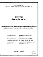
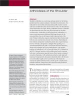
Cahill and Berg
29
describe a method
of localizing lesions by dividing the
knee into 15 distinct alphanumeric
zones (Figure 1). From medial to lat-
eral, five zones numbered 1 through
5 are divided centrally by the notch;
each compartment is then divided in
half. The lateral radiograph uses Blu-
mensaat’s line anteriorly and the
posterior cortical line to divide zone
A (anterior) from B (central) and C
(posterior). This alphanumeric sys-
Osteochondritis Dissecans of the Knee
92 Journal of the American Academy of Orthopaedic Surgeons
tem provides standardization for re-
search purposes, although it has
found limited application to
date.
6,26
Cahill and Berg
29
also describe a
classification system for juvenile
OCD based on technetium Tc 99m
phosphate scintigraphy findings.
Grading is based on the relative de-
gree of scintigraphic activity in rela-
tion to plain radiographs. Stage 0 is
normal in both. Stage 1 demon-
strates a defect on plain radiographs
but no increased activity on the bone
scan. Stage 2 shows increased uptake
in the lesion but not in the adjacent
femoral condyle. Stage 3 indicates
isotope uptake in both the lesion and
the adjacent condyle. Finally, stage 4
demonstrates increased isotope up-
take in both the lesion and adjacent
tibial surface. Patients with stage 3
or 4 disease were described as having
symptomatic OCD. Cahill et al
20
lat-
er reported limited correlation be-
tween this staging system and pre-
diction of lesion stability or the need
for subsequent surgery. However,
Paletta et al
30
suggested a role for
this imaging technique that distin-
guishes between results in juveniles
and those in adults. They reported
that four of four patients with open
physeal plates and increased activity
on bone scan healed with nonsurgi-
cal treatment, whereas the two pa-
tients without increased activity did
not heal. In contrast, among patients
with closed growth plates, only 33%
(2/6) healed despite having similar
increased activity within the le-
sion.
Magnetic resonance imaging
(MRI) has proved to be particularly
valuable in assessing osteochondral
lesions. Several investigators have
attempted to characterize the stabil-
ity of the OCD lesion with findings
on MRI. Dipaola et al
31
classified le-
sions according to appearance on
MRI and associated specific findings
with the potential for fragment de-
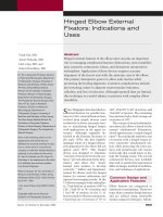
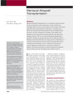
tachment. They described lesions
containing fluid behind the joint as
partially or completely detached, as
evidenced by high signal intensity
on T2-weighted images when a
breach of the cartilage surface was
detected. They distinguished carti-
lage breach with an attached frag-
ment by interpreting interposed low
signal intensity on the rim as fibrous
tissue (Figure 2).
Others have added criteria for
fragment instability to include the
following: an area of increased ho-
mogenous signal ≥5 mm in diameter
beneath the lesion; a focal defect ≥5
mm in the articular surface; and a
high signal line traversing the sub-
chondral plate into the lesion.
32
In
cases of limited joint effusion,
Kramer et al
33
expressed a high level
of confidence for predicting lesion
stability using intra-articular gado-
linium Gd 153 contrast material.
More recent advances in MRI tech-
nology (eg, cartilage-specific se-
quences) may eliminate the necessi-
ty of intra-articular injections and
allow distinction between areas of
interposed synovial fluid, fibrocarti-
lage, and degenerated or lytic sub-
chondral bone.
Classification and
Characterization
Distribution of OCD lesions in
the knee are most commonly associ-
ated with the lateral aspect of the
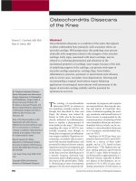
medial femoral condyle. Aichroth
26
described this as the classic location
and confirmed it in 69% (72) of 105
knees (Figure 3). The patella was in-
volved in five patients (5%); the re-
mainder of the lesions involved the
lateral femoral condyle (15% [16])
and the medial femoral condyle
(69% [72]). In a large multicenter ret-
Figure 1
Anteroposterior (A) and lateral (B) views of the knee, demonstrating the 15
alphanumeric radiographic regions described by Cahill and Berg.
29
The five
numbered zones on the anteroposterior view are divided centrally by the notch
(zone 3). The lettered zones on the lateral view are divided by Blumensaat’s line
anteriorly and the posterior cortical line. The half-moon–shaped shaded area in each
view of the distal femur represents an old lesion. (Adapted with permission from
Cahill BR, Berg BC: 99m-Technetium phosphate compound joint scintigraphy in the
management of juvenile osteochondritis dissecans of the femoral condyles.
Am J Sports Med 1988;11:329-335.)
Dennis C. Crawford, MD, PhD, and Marc R. Safran, MD
Volume 14, Number 2, February 2006 93
rospective study of 713 patients and
798 knees, Hefti et al
5
described a
slightly different distribution. The
medial femoral condyle was typical-
ly affected, with the majority of le-
sions involving the lateral aspect
(51%), 19% the central, and 7% the
medial aspect. Involvement of the
lateral condyle in all areas encom-
passed 17% of lesions; those of the
patella, 7%; and 0.2% (one lesion),
the tibial plateau. Lateral condylar
lesions are more commonly associ-
ated with discoid meniscus or with
occurrence after meniscal sur-
gery.
26,34
Knee radiographs provide not only
the initial basis for distinction of
growth plate maturation but also as-
sessment of lesion location and sta-
bility (ie, free or loose bodies). Berndt
and Harty
35
described four stages of
chondral lesions based on plain ra-
diographs of the talus; this system
has been widely applied to lesions
about the knee: stage I, involvement
of a small area of compression of the
subchondral bone; stage II, partially
detached osteochondral fragment;
stage III, completely detached frag-
ment that remains in the underlying
crater; and stage IV, complete
detachment/loose body. Other crite-
ria, such as lesion size, have been
used to assess the potential for heal-
ing with nonsurgical intervention.
Several authors
20,32,36
have thought
that patients could be successfully
treated nonsurgically when the
mean area was smaller than between
194 and 424 mm
2
. In contrast, le-
sions larger than between 436 to 815
mm
2
were associated with poor out-
comes. Others have suggested the
presence of “marked sclerosis” as a
poor predictor of successful nonsur-
gical management.
37-39
Understanding and characterizing
the spectrum of OCD lesions as sta-
ble or unstable is often considered
central to the treatment plan. How-
ever, this characterization has
proved to be difficult to determine
prior to surgical intervention and of-
ten remains a clinical judgment.
MRI criteria have proved to be rea-
sonably accurate compared with the
gold standard of arthroscopic find-
ings in predicting lesion integrity.
Strict adherence to the MRI criteria
of Dipaola was shown in one
study
40
to have an 85% correlation
with arthroscopic findings when ap-
plying Guhl’s arthroscopic staging
system. Guhl’s intraoperative classi-
fication is defined by cartilage integ-
rity and fragment stability.
37
Type I
Figure 2
Sagittal MRI scans of unstable osteochondritis lesions of the distal femur. T2-
weighted (fluid-weighted) images of osteochondral separation are indicated by high
signal line between the osseous components (A) and extending from the
intraosseous portion to the joint surface, “breaching” the cartilage (B).
Figure 3
Anteroposterior radiographs demonstrating an OCD lesion on the lateral aspect of
the medial femoral condyle before (A) and after (B) displacement.
Osteochondritis Dissecans of the Knee
94 Journal of the American Academy of Orthopaedic Surgeons
represents softening of cartilage but
no breach; type II, breached cartilage
that is stable; type III, a definable
fragment that remains partially at-
tached (flap lesion); and type IV, a
loose body and osteochondral defect
at the donor site.
Further surgical characterization
of OCD lesions, however, should not
be limited to this system. Assess-
ment of the size and number of loose
fragments, the presence of bone as-
sociated with each chondral frag-
ment and its potential reparability,
and the quality and character of the
underlying subchondral bone (pres-
ence of fibrocartilage or cystic de-
generative material) are important
factors in characterizing and surgi-
cally treating this heterogenous pa-
thology.
Natural History and
Prognosis
No randomized, controlled clinical
trials exist for either surgical or non-
surgical interventions for OCD of
the knee. In general, physeal maturi-
ty, dissection of the lesion from the
adjacent subchondral bone (stabili-
ty), size and location of lesions, and
integrity of the cartilage surface
have been used as predictive criteria
for surgical intervention. Historical-
ly, data from case reports, case se-
ries, and retrospective reviews have
directed care of patients with OCD
of the knee. Thus, caution should be
exercised in proceeding with recom-
mendations from these studies be-
cause they provide only a limited
ability to predict the natural history
of OCD; that is, they are level IV and
V evidence-based studies.
A large, recent multicenter re-
view of the European Paediatric Or-
thopedic Society study (509 knees
[318 juvenile, 191 adult] in 452 pa-
tients) has provided notable data.
5
The authors made several important
distinctions and reached a number of
conclusions. (1) When there are no
signs of dissection (defined as a sta-
ble fragment), the prognosis is mark-
edly better than it is with signs of
dissection. (2) Pain and swelling are
not good indicators of dissection. (3)
Plain radiography and computed to-
mography are not useful in predict-
ing dissection. (4) Sclerosis on plain
radiography predicts poor response
to drilling. (5) Lesions >2 cm in di-
ameter have a worse prognosis than
smaller lesions. (6) When there is ev-
idence of dissection, surgical results
are better than those of nonsurgical
treatment. (7) Lesions in the classic
location had a better prognosis. (8)
Although patients with adult-onset
symptoms had a higher proportion
of abnormal findings on radiographs
after the treatment period (42%),
more than one in five of those with
open epiphyseal plates (22%) had ab-
normal knee radiographs an average
of 3 years after starting treatment.
Pill et al
6
recently compared the
value of MRI and clinical criteria for
predicting the success of nonsurgical
treatment of OCD. Their retrospec-
tive review correlated outcomes to
radiographic measures using the
MRI criteria of DeSmet et al
32
and
the radiographic criteria of Cahill
and Berg.
29
Although they found no
single factor to be uniformly predic-
tive of successful nonsurgical treat-
ment, several important associa-
tions were found. Older patients
with one or more signs of chondral
disruption by MRI failed nonsurgical
treatment most often. Similarly,
larger lesions and lesions deemed to
be within the weight-bearing area, as
indicated by the lateral radiograph,
also were most likely to fail nonsur-
gical treatment. Younger patients
with no MRI criteria for instability
were most likely to recover with
nonsurgical treatment.
Management and
Outcomes
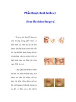
An algorithm for management deci-
sions is given in Figure 4. The goal of
nonsurgical treatment is to promote
healing of lesions in situ and prevent
lesion displacement. The preferred
surgical goals are salvage of native
cartilage, when possible, followed by
restoration procedures.
Initial discussion in early OCD
involves patient, family, and physi-
cian education. Understanding the
nature of this disease, the potential
long-term implications, and the
timeline for management are crucial
early strategies to help both the pa-
tient and surgeon avoid frustration.
Symptoms that are exacerbated by
activity, particularly episodes of
trauma and athletic and weight-
bearing activities, should be identi-
fied. Significantly limiting sports
and high-impact activities is univer-
sally recommended.
Nonsurgical treatment is primari-
ly mitigated through activity modi-
fication. It may include a wide spec-
trum of approaches that historically
have included crutches (for limited
weight bearing) as well as braces or
even casts for noncompliant pa-
tients. The efficacy of limiting activ-
ity compared with limited or non–
weight-bearing activity has not been
studied in a controlled trial. Sales de
Gauzy et al
38
followed a group of 30
children (mean age, 11 years 4
months) to complete resolution of
symptoms by discontinuing sports
activities; the authors recommended
no surgical intervention because
symptoms resolved with discontinu-
ation of sports activities. In patients
without marked sports participa-
tion, prescribing a non–weight-
bearing status and range-of-motion
knee exercises may be beneficial to
cartilage and may help avoid the po-
tential disaster of “cast disease” (eg,
joint stiffness, muscle atrophy, os-
teoporosis) and arthrofibrosis associ-
ated with cast immobilization.
Symptom relief may be gained
with nonsteroidal anti-inflammatory
drugs (NSAIDs), but doing so may in-
terfere with monitoring disease pro-
gression. In young patients with ra-
diographically and clinically stable
lesions, we prefer to control pain
with a non–anti-inflammatory med-
ication (eg, acetaminophen). This
Dennis C. Crawford, MD, PhD, and Marc R. Safran, MD
Volume 14, Number 2, February 2006 95
avoids the theoretical negative influ-
ence of NSAIDs on bone healing. In
this strategy, we record days with
swelling and those without and plot
trends versus compliance with peri-
ods of inactivity. This semiobjective,
patient-reliant practice helps support
recommendations for reduced activ-
ity over a potentially long period of
limited intervention. For patients
with persistent pain or continued ep-
isodes of swelling/effusions, deci-
sions about surgical intervention
may be supported by the data. In
adults without evidence of loose
fragments or unstable lesions, we
employ a strategy similar to that
used for patients with early, focal os-
teoarthritis. Pain medication, activ-
ity modification, strengthening, and
weight control are the central tenets.
Concurrent pathology (ie, malalign-
ment, instability, osteoarthritis) is
more likely in older patients and is
an important consideration for both
surgical and nonsurgical manage-
ment of OCD.
Choosing surgical intervention
for OCD, and selecting a strategy for
repair versus reconstruction or re-
moval of osteochondral lesions, es-
sentially depend on the stability of
the inciting lesion and the integrity
of the overlying cartilage. With fail-
ure of the nonsurgical approach, sur-
gical management often begins with
arthroscopy. Classification using ar-
throscopic findings is based on a de-
scription of the lesion using two es-
sential criteria: the integrity of the
overlying articular cartilage and the
stability of the lesion to distinguish
three categories (intact, not intact
but stable, not intact and unsta-
ble).
19,37
Lesions with intact cartilage
are considered either stable or unsta-
ble. Lesions with damaged cartilage
surfaces comprise a mixture of ad-
vanced lesions and may be either
disrupted or macerated, and by Ca-
hill’s definition are unstable. Ca-
hill
19
further subdivided the
unstable-cartilage, injured lesion as
predetached, hinged, or loose, attrib-
uting characteristics of temporal
symptoms and reduction congruity
to each. Using this system, he rec-
ommended a treatment algorithm,
based on arthroscopic findings and
scintigraphic data. We employ a sim-
ilar approach using symptoms, ra-
diographic and MRI findings, and ar-
throscopic observations to apply
treatments based on skeletal maturi-
ty, osteochondral stability and re-
ducibility, and articular cartilage in-
tegrity (Figure 4).
Surgical treatment for stable le-
sions with normal articular cartilage
involves drilling the subchondral
bone with the intention of stimulat-
Figure 4
Juvenile
(open physis)
Radiographs
Adult
(closed physis)
Stable
Physical examination
Stable
MRI
Stable
Bone scan
Activity restriction (3 mos)
• Impending physeal closure
• Clinical signs of instability
• Expanding lesion on plain films
Arthroscopy
Stable
Physical examination
MRI
Stable
Malalignment
Yes
Treat symptomatically
Stable
Unstable
Osteotomy
Stable
Unstable
reducible
Fixation
graft
Fixation and
graft
chondrocyte transplant, or
osteochondral graft
Not
positive
No
Positive
Unstable with
fragmentation
or osteolysis
Unstable
and chondral
damage
Fixation and grafts,
Transchondral
drilling
Treatment algorithm for knee osteochondritis dissecans.
Osteochondritis Dissecans of the Knee
96 Journal of the American Academy of Orthopaedic Surgeons
ing vascular ingrowth and subchon-
dral bone healing. Retrograde tech-
niques (defined as methods that
avoid articular cartilage disruption
using a transosseous approach) have
given way to arthroscopically assist-
ed methods that have proved to be
highly efficacious in skeletally im-
mature patients. Anderson and col-
leagues
39,41
described success with
this technique in 24 patients fol-
lowed for an average of 5 years. Av-
erage time to healing was 4 months.
Twenty-two patients had good or ex-
cellent results based on the Hugh-
ston rating scale; in two of four skel-
etally mature patients, the lesion did
not heal.
Kocher et al
42
treated 30 knees in
23 skeletally immature patients who
had failed 6 months of nonsurgical
therapy (average age, 12.3 years). Us-
ing arthroscopically directed ante-
grade (transchondral) drilling of sta-
ble lesions, the authors reported
radiographic healing in all 30 knees
at an average of 4.4 months. They
advocated this treatment in patients
with intact articular surfaces who
had failed nonsurgical treatment.
Our experience has been the same,
that drilling works better in skeletal-
ly immature patients than in older
patients, although it is still worth at-
tempting in all patients with a per-
sistently symptomatic lesion and in-
tact articular cartilage.
Surgical treatment for unstable
lesions typically is attributed to
Smillie
23
because he developed a nail
for open reduction and internal fixa-
tion of displaced and unstable le-
sions. Surgical intervention to en-
hance union has included Kirschner
wires, cannulated screws, Herbert
screws, and bone pegs. These typi-
cally require a second surgery to re-
move the device and have been asso-
ciated with several complications,
including wire migration, adjacent
cartilage damage, and implant frac-
ture. Biodegradable implants (ie, pins
and screws) have the potential ad-
vantage of not requiring removal;
however, some of these devices have
been associated with sterile abscess
formation, synovitis, and loss of fix-
ation.
43
Compressive devices provide
the possibility of loading the osseous
components, a technical advantage
that may facilitate healing (Figure 5,
E). When indicated, hardware re-
moval often can be done arthroscop-
ically, with low morbidity, and can
provide an opportunity to directly
assess healing and cartilage integrity.
Simple removal of a loose or de-
tached fragment is rarely considered
to be an effective treatment, aside
from cases in which the fragment is
macerated and irreparable. Wright et
al
44
reported only 35% good and ex-
cellent results with fragment exci-
sion at an average of 9 years after
surgery.
In cases in which simple trans-
chondral drilling is unsuccessful, or
when the lesion is hinged, loose, or
displaced, the objective is to restore
articular congruency by stimulating
subchondral bone repair via com-
pression and bone grafting, when
necessary. In this manner, the os-
seous portion of the fragment may
heal and allow stabilization of the
overlying articular surface. This
strategy also would provide protec-
tion to the adjacent uninjured car ti-
lage that, after fragment excision,
could be subject to increased contact
stress and shear forces secondary to
surface irregularity, step-off, and re-
sultant edge loading. Green
18
used a
similar argument to suggest that the
technical difficulty of repairing loose
fragments and subsequent malreduc-
tion could have similar consequenc-
es. His advice, which remains a te-
net of joint surgery, was to replace
larger fragments and remove those
that were essentially too small to be
fixed anatomically. Cahill
19
similar-
ly advocated fixation whenever pos-
sible because the results of excision
usually are ineffective.
Discrepancy in the size of a lesion
as a result of overgrowth of displaced
fragments, loss of fragment substance
because of mechanical damage, or
craterization of the donor site all have
been described and provide technical
challenges. Several strategies have
been described to address these poten-
tial issues. Johnson et al
45
treated 35
knees via an arthroscopically assisted
technique that involved fragment fix-
ation using cannulated AO-type
screws. Results, comparable with
other in situ methods, were good or
excellent in 90% of cases.
When poor congruency of the
fragment-donor interface exists, a
technique similar to that described
by Anderson et al
41
may be used. In
this method, the lesion is evaluated
arthroscopically, followed by ante-
grade open curettage, grafting, reduc-
tion, and fixation (Figure 5, C and D).
This method is done by reflecting a
partially attached fragment, or re-
moving it temporarily, to allow in-
spection of the osseous surfaces and
removal of fibrocartilage from the op-
posing subchondral inter face. The
ensuing fragment-crater mismatch
then can be grafted with autogenous
bone (tibial metaphysis) before com-
pression screw fixation. We have
used cannulated Acutrak (Acumed,
Hillsboro, OR) headless screws or a
4.0-mm headed screw countersunk 1
to 2 mm below the cartilage surface
to avoid causing articular car tilage le-
sions on the opposing surface. Fol-
lowing a period of strict non–weight-
bearing and range-of-motion exercise,
hardware is removed arthroscopically
after a minimum of 6 weeks to as
long as 12 weeks.
Several techniques for salvage of
full-thickness defects of articular
cartilage, including autologous
chondrocyte implantation, mosaic-
plasty, and osteochondral allograft,
have been advocated. Browne and
Branch
46
have presented an algo-
rithm for approaching these types of
injuries. The efficacy of their tech-
niques for addressing the symptoms
of advanced OCD has been mixed.
Some have advocated fixing loose
osteochondral fragments with autol-
ogous osteochondral autografts or by
using autologous osteochondral au-
tografts for filling empty craters to
Dennis C. Crawford, MD, PhD, and Marc R. Safran, MD
Volume 14, Number 2, February 2006 97
decrease edge loading. Case reports
have indicated generally favorable
results for these procedures but have
limited follow-ups. Yoshizumi et
al
47
describe successful union by 6
months in three cases of adult OCD
using a modification of a method de-
scribed by Berlet et al.
48
In the Berlet
technique, the OCD lesion is essen-
tially fixed in situ by applying pe-
ripheral autologous osteochondral
plugs. However, Yoshizumi et al
47
describe a technique using one cen-
tral plug to fix the lesion. Others
have advocated using either tech-
nique to reduce edge loading. Anoth-
er method for unloading cartilage for
adult OCD patients was described
by Slawski.
49
He performed seven
valgus osteotomies for medial femo-
ral condyle OCD and, at 2 years, de-
scribed an average improvement in
the Lysholm score from 39 to 89,
with an average postoperative knee
angle of 9º valgus.
Use of autologous chondrocyte
transplantation has been discussed
and advocated by several authors.
Peterson et al
50
reviewed their expe-
rience with autologous chondrocyte
Figure 5
Surgical reduction and fixation of an
unstable osteochondritis dissecans
injury. A, Preoperative anteroposterior
radiograph demonstrating a loose
and fragmenting chondritis dissecans
of the medial femoral condyle. B,
Intraoperative photograph indicating
the margin (dotted area) of the extent of
the loose/unattached articular
cartilage. C, The osteochondral
fragment is elevated. The fibrocartilage
has been débrided from the interval
between the osseous component prior
to bone grafting. D, Fixation with two
compression screws and absorbable
pins. E, Postoperative anteroposterior
radiograph.
Osteochondritis Dissecans of the Knee
98 Journal of the American Academy of Orthopaedic Surgeons
implantation in 94 patients at a min-
imum of 2 years; 18 patients (19%)
had chondral defects secondary to
OCD. Defects in this group were
characterized as particularly recalci-
trant to previous surgery and often
required a longer period to mature,
compared with the other articular le-
sions studied. Interestingly, 89% of
patients (16/18) improved, with a
similar distribution of excellent,
good, and fair results compared with
posttraumatic isolated femoral
condyle lesions.
Subsequently, these authors
51
re-
ported results of 58 patients with a
variety of OCD lesions. Results at a
mean of 5.6 years were similar to
those at a minimum of 2 years in the
previous study.
50
Interestingly, in
this larger study, a small group of pa-
tients received bone graft for sub-
chondral defects to provide a chon-
drocyte bed prior to transplantation.
Results of autologous osteochondral
and allograft transplantation are gen-
erally difficult to interpret; indica-
tions in most studies for such proce-
dures involve a variety of conditions,
including osteonecrosis, osteoarthri-
tis, trauma, osteochondral fracture,
and OCD.
Summary
Understanding of the origins and
natural history of OCD of the knee
continues to progress. Tw o principle
factors, skeletal maturity at symp-
tom onset and contiguity of the sub-
chondral and bone-cartilage surface,
remain the most important determi-
nants in choosing treatment. The
challenge is identifying those deter-
minants within the spectrum of dis-
ease (eg, who may benefit from
longer periods of nonsurgical man-
agement versus earlier surgical treat-
ment). OCD is not a benign condi-
tion, even in the skeletally
immature knee. Despite the fact that
many patients are asymptomatic,
the potential for arthrosis and degen-
eration, demonstrated radiographi-
cally, remains a problem, the exact
consequences of which remain un-
certain. Additional significant chal-
lenges include dissecting and defin-
ing the different subtypes of OCD,
determining the potential of each for
spontaneous healing or progression,
and improving opportunities and
techniques for intervention to main-
tain and restore joint integrity.
References
Evidence-based Medicine: The au-
thors note that there are no Level I or
Level II evidence-based studies.
Citation numbers printed in bold
type indicate references published
within the past 5 years.
1. Konig F: Ueber freie Korper in den
Glenken. Zeiteschr Chir 1888;27:90-
109.
2. Schenck RC Jr, Goodnight JM: Osteo-
chondritis dissecans. J Bone Joint
Surg Am 1996;78:439-453.
3. Lindén B: Theincidence ofosteochon-
dritis dissecans in the condyles of the
femur. Acta Orthop Scand 1976;47:
664-667.
4. Bradley J, Dandy DJ: Osteochondritis
dissecans and other lesions ofthe fem-
oral condyles. J Bone Joint Surg Br
1989;71:518-522.
5. Hefti F, Beguiristain J, Krauspe R, et
al: Osteochondritis dissecans: A mul-
ticenter study of the European Pediat-
ric Orthopedic Society. J Pediatric
Orthop B 1999;8:231-245.
6. Pill SG, Ganley TJ, Milam RA, et al:
Role of magnetic resonance imaging
and clinical criteriain prediciting suc-
cessful nonoperative treatment of os-
teochondritis dissecans in children.
J Pediatr Orthop 2003;23:102-108.
7. Ribbing S: The hereditary multiple
epiphyseal disturbance and its conse-
quences for the etiologies of local
malacia-particularly the osteochon-
dritis dissecans. Acta Orthop Scand
1955;24:286-998.
8. Mubarak SJ, Carroll NC: Familial os-
teochondritis dissecans of the knee.
Clin Orthop Relat Res 1979;140:131-
136.
9. Petrie PW: Etiology of osteochondritis
dissecans: Failure to establish a famil-
ial background. J Bone Joint Surg Br
1977;59:366-367.
10. Caffey J, Madell SH, Royer C, Morales
P: Ossification of the distal femoral
epiphysis. J Bone Joint Surg Am
1958;40:647-654.
11. Chiroff RT, Cooke CP III: Osteochon-
dritis dissecans: A histologic and mi-
croscopic analysis of surgically ex-
cised lesions. J Trauma 1975;15:689-
696.
12. Laverty S, Okouneff S, Ionescu M, et
al: Excessive degradation of type II
collagen in articular cartilage in
equine osteochondrosis. J Orthop
Res 2002;20:1282-1289.
13. Al-Hizab F, Clegg PD, Thompson CC,
Carter SD: Microscopic localization
of active gelatinases in equine osteo-
chondritis dissecans (OCD) cartilage.
Osteoarthritis Cartilage 2002;10:
653-661.
14. Rogers WM, Gladstone H: Vascular
foramina and arterial supply of the
distal end of the femur. J Bone Joint
Surg Am 1950;32:867-875.
15. Reddy AS, Frederick RW: Evaluation
of the intraosseous and extraosseous
blood supply to the distal femoral
condyles. Am J Sports Med 1998;26:
415-419.
16. Lankes M, Petersen W, Hassenpflug J:
Arterial supplies of the femoral
condyle [German]. Z Orthop Ihre
Grenzgeb 2000;138:174-180.
17. Linden B, Telhag H: Osteochondritis
dissecans: A histologic and autoradio-
graphic study in man. Acta Orthop
Scand 1977;48:682-694.
18. Green JP: Osteochondritis dissecans
of the knee. J Bone Joint Surg Br
1966;48:82-91.
19. Cahill BR: Osteochondritis dissecans
of the knee: Treatment of juvenile and
adult forms. J Am Acad Orthop Surg
1995;3:237-247.
20. Cahill BR, Phillips MR, Navarro R:
The results of conservative manage-
ment of juvenile osteochondritis dis-
secans using jointscintigraphy: A pro-
spective study. Am J Sports Med
1989;17:601-605.
21. Fairbank HAT: Osteochondritis disse-
cans. J Bone Joint Surg Br 1933;21:67-
73.
22. Nambu T, Gasser B, Schneider E, Ban-
di W, Perren SM: Deformation of the
distal femur: A contribution towards
the pathogenesis of osteochondritis
dissecans in the knee joint.
J Biomech 1991;24:421-433.
23. Smillie IS: Osteochondritis Disse-
cans: Loose Body in Joints. Edin-
burgh, UK: Churchill Livingstone,
1960.
24. Aichroth PM, Patel DV, Marx CL:
Congenital discoid lateral meniscus
in children: A follow-up study and
evolution of management. J Bone
Joint Surg Br 1991;73:932-936.
25. Stanitski CL, Bee J: Juvenile osteo-
Dennis C. Crawford, MD, PhD, and Marc R. Safran, MD
Volume 14, Number 2, February 2006 99
chondritis dissecans of the lateral
femoral condyle after lateral discoid
meniscal surgery. Am J Sports Med
2004;32:797-801.
26. Aichroth P: Osteochondritis disse-
cans of the knee: A clinical survey.
J Bone Joint Surg Br 1971;53:440-447.
27. Wilson JN: A diagnostic sign in osteo-
chondritis dissecans of the knee.
J Bone Joint Surg Am 1967;49:477-
480.
28. Conrad JM, Stanitski CL: Osteochon-
dritis dissecans: Wilson’s sign revisit-
ed. Am J Sports Med 2003;31:777-
778.
29. Cahill BR, Berg BC: 99m-Technetium
phosphate compound scintigraphy in
the management of juvenile osteo-
chondritis dissecans of the femoral
condyles. Am J Sports Med 1983;11:
329-335.
30. Paletta GA Jr, Bednarz PA, Stanitski
CL, Sandman GA, Stanitski DF, Kot-
tamasu S: The prognostic value of
quantitative bone scan in knee osteo-
chondritis dissecans: A preliminary
report. Am J Sports Med 1998;26:7-
14.
31. Dipaola JD, Nelson DW, Colville MR:
Characterizing osteochondritis disse-
cans lesion by magnetic resonance im-
aging. Arthroscopy 1991;7:101-104.
32. De Smet AA, Ilahi OA, Graf BK: Un-
treated osteochondritis dissecans of
the femoral condyles: Prediction of
patient outcome using radiographic
and MR findings. Skeletal Radiol
1997;26:463-467.
33. Kramer J, Stiglbauer R, Engel A,
Prayer L, Imof H: MR contrast ar-
thrography (MRA) in osteochondritis
dissecans. J Comput Assist Tomogr
1992;16:254-260.
34. Yoshida S, Ikata T, Takai H, Kashi-
waguchi S, Katoh S, Takeda Y: Osteo-
chondritis dissecans of the femoral
condyle in the growth stage. Clin
Orthop Relat Res 1998;346:162-170.
35. Berndt AL, Harty M: Transcondylar
fractures (osteochondritis dissecans)
of the talus. J Bone Joint Surg Br
1959;41:988-1020.
36. Hughston JC, Hergenroeder PT,
Courtenay BG: Osteochondritis dis-
secans of the femoral condyles.
J Bone Joint Surg Am 1984;66:1340-
1348.
37. Guhl JF: Arthroscopic treatment of
osteochondritis dissecans. Clin
Orthop 1982;167:65-74.
38. Sales de Gauzy J, Mansat C, Darodes
PH, Cahuzac JP: Natural course of os-
teochondritis dissecans in children.
J Pediatr Orthop B 1999;8:26-28.
39. Anderson AF, Richards DB, Pagnani
MJ, Hovis WD: Antegrade drilling of
osteochondritis dissecans of the knee.
Arthroscopy 1997;13:319-324.
40. O’Connor MA, Palaniappan M, Kahn
N, Bruce CE: Osteochondritis disse-
cans of the knee in children: A com-
parison of MRI and arthroscopic find-
ings. J Bone Joint Surg Br 2002;84:
258-262.
41. Anderson AF, Lipscomb AB, Coulam
C: Antegrade curettement,bone graft-
ing and pinning of osteochondritis
dissecans in the skeletally mature
knee. Am J Sports Med 1990;18:254-
261.
42. Kocher MS, Micheli LJ, Yaniv M,
Zurakowski D, Ames A, Adrignolo
AA: Functional and radiographic out-
comes of juvenile osteochondritis dis-
secans of the knee treated with trans-
articular arthroscopic drilling. Am J
Sports Med 2001;29:562-572.
43. Dervin GF, Keene GC, Chissel HR:
Biodegradable rods in adult osteo-
chondritis dissecans of the knee.
Clin Orthop 1998;356:213-221.
44. Wright RW, McLean M, Matava MJ,
Shively RA: Osteochondritis disse-
cans of the knee: Long-term results of
excision of the fragment. Clin
Orthop 2004;424:239-245.
45. Johnson LL, Uitvlugt G, Austin MD,
Detrisac DA, Johnson C: Osteochon-
dritis dissecans of the knee: Arthro-
scopic compression screw fixation.
Arthroscopy 1990;6:179-189.
46. Browne JE, Branch TP: Surgical alter-
natives for treatment of articular car-
tilage lesions. J Am Acad Orthop
Surg 2000;8:180-189.
47. Yoshizumi Y, Sugita T, Kawamata T,
Ohnuma N, Maeda S: Cylindrical os-
teochondral graft for osteochondritis
dissecans of the knee: A report of
three cases. Am J Sports Med 2002;
30:441-445.
48. Berlet GC, Mascia A, Miniaci A:
Treatment of unstable osteochondri-
tis dissecans lesions of the knee using
autogenous osteochondral grafts (mo-
saicplasty). Arthroscopy 1999;15:
312-316.
49. Slawski DP: High tibial osteotomy in
the treatment of adult osteochondri-
tis dissecans. Clin Orthop Relat Res
1997;341:155-161.
50. Peterson L, Minas T, Brittberg M,
Nilsson A, Sjögen-Jansson E, Lindahl
A: Two- to 9-year outcome after autol-
ogous chondrocyte transplantation of
the knee. Clin Orthop 2000;374:212-
234.
51. Peterson L, Minas T, Brittberg M,
Lindahl A: Treatment of osteochon-
dritis dissecans of the knee with au-
tologous chondrocyte transplanta-
tion. J Bone Joint Surg Am
2003;85(suppl 1):17-24.
Osteochondritis Dissecans of the Knee
100 Journal of the American Academy of Orthopaedic Surgeons