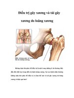
Loạn dưỡng xương do thận pdf
Bạn đang xem bản rút gọn của tài liệu. Xem và tải ngay bản đầy đủ của tài liệu tại đây (243.13 KB, 9 trang )
Renal Osteodystrophy
Abstract
The incidence of chronic renal disease is increasing, and the
pattern of renal osteodystrophy seems to be shifting from the
classic hyperparathyroid presentation to one of low bone turnover.
Patients with persistent disease also live longer than previously
and are more physically active. Thus, patients may experience
trauma as a direct result of increased physical activity in a setting
of weakened pathologic bone. Patient quality of life is primarily
limited by musculoskeletal problems, such as bone pain, muscle
weakness, growth retardation, and skeletal deformity. Chronic
renal disease also increases the risk of comorbidity, such as
infection, bleeding, and anesthesia-related problems. Current
treatment strategies include dietary changes, plate-and-screw
fixation, and open reduction and internal fixation.
R
enal osteodystrophy refers to
pathologic bone conditions in
patients with known kidney disease.
The kidneys monitor the physiolog-
ic homeostasis of mineral metabo-
lism; thus, any deficiency in their
operation directly affects bone min-
eralization because of the conse-
quent negative effect on calcium and
phosphate regulation. This is note-
worthy because the rising incidence
of chronic renal disease translates
into more patients with bone pathol-
ogy presenting to orthopaedic sur-
geons for elective surgery and to
emergency trauma units because of
pathologic fractures.
Musculoskeletal problems signif-
icantly limit quality of life in pa-
tients with renal failure.
1
According
to the Health Care Financing Ad-
ministration, each year 325,000
Americans are treated for end-stage
renal disease, and more than 1.2 mil-
lion patients worldwide receive dial-
ysis.
2
These figures were growing by
about 8% annually, although the in-
cidence seems to be leveling out.
The United States Renal Data Sys-
tem reports an incidence of 338 per
million of population in 2003, with
the largest proportion in patients
aged 45 to 64 years.
3
Patient Demographics
According to the US Renal Data Sys-
tem 2003 Annual Report, in 2001
the median age of patients with end-
stage renal disease was 64.5 years.
2
Caucasians had the highest median
age (67.1 years) and Hispanics, the
lowest (60.6 years). Overall inci-
dence in the US population is 334
cases per million. In 2001, the inci-
dence of end-stage renal disease was
highest in African-Americans (988
cases per million) and lowest in Cau-
casians (254 cases per million), ad-
justed for age and sex. Patients aged
45 to 64 years represented the largest
proportion of new cases in 2001
(36%), with an incidence of 625 per
million, adjusted for sex and race.
However, the incidence was much
higher in patients aged 65 to 74 years
(1,402 per million) and in those age
75 years and older (1,542 per mil-
Nirmal C. Tejwani, MD
Aaron K. Schachter, MD
Igor Immerman, BS
Pramod Achan, MBBS, FRCS
(Orth)
Dr. Tejwani is Associate Professor,
Department of Orthopaedics, Bellevue
Hospital, NYU–Hospital for Joint
Diseases, New York, NY. Dr. Schachter
is Resident, NYU—Hospital for Joint
Diseases. Mr. Immerman is a Medical
Student, NYU—Hospital for Joint
Diseases. Dr. Achan is Fellow,
Department of Orthopaedics,
NYU—Hospital for Joint Diseases.
None of the following authors or the
departments with which they are
affiliated has received anything of value
from or owns stock in a commercial
company or institution related directly or
indirectly to the subject of this article:
Dr. Tejwani, Dr. Schachter, Mr.
Immerman, and Dr. Achan.
Reprint requests: Dr. Tejwani, Bellevue
Hospital, 550 First Avenue, NBV
21W37, New York, NY 10016.
J Am Acad Orthop Surg 2006;14:303-
311
Copyright 2006 by the American
Academy of Orthopaedic Surgeons.
Volume 14, Number 5, May 2006 303
lion). Males are more likely than fe-
males to be diagnosed with end-
stage renal disease. In 2001, the
incidence rate adjusted by age and
race was 404 per million in males
compared with 280 per million in fe-
males.
Disease
Pathophysiology
The kidneys are responsible for
monitoring and regulating calcium
homeostasis as well as for control-
ling levels of phosphate, magne-
sium, and other minerals (Figure 1).
The kidneys act both as target or-
gans for parathyroid hormone (PTH)
and for excreting it. The proximal
convoluted tubules of the kidneys
are the site of production of 1,25-
dihydroxycholecalciferol (the active
form of vitamin D following hydrox-
ylation of 25-hydroxycholecalciferol
catalyzed by 1α-hydroxylase), the
foremost regulator of intestinal cal-
cium absorption. This hormone also
promotes osteoclastic resorption of
bone and the feedback inhibition of
PTH synthesis. The kidneys serve as
the primary route for the excretion
of metals, such as aluminum. Mod-
est changes in the efficacy of renal
excretion dramatically alter the
body’s ability to maintain mineral
homeostasis.
The bony manifestations of renal
compromise are subdivided into
high turnover, caused by persistent-
ly elevated levels of PTH (secondary
hyperparathyroidism), and low turn-
over, seen with either excess alumi-
num deposition in bone or normal or
reduced PTH levels. Cannata Andia
4
described the increasing prevalence
of low-turnover renal osteodystro-
phy. Sherrard et al
5
distinguished be-
tween peritoneal dialysis patients
with the low-turnover form, com-
pared with hemodialysis patients
with high-turnover lesions. Bushin-
sky
6
emphasized the presence of two
distinct histologic entities present-
ing with a common clinical picture.
High-dose corticosteroids used
therapeutically for chronic renal dis-
ease play a role in osteopenia and—
more significantly—in osteonecro-
sis. Approximately 15% of patients
with renal transplantation develop
osteonecrosis within 3 years of sur-
gery.
7,8
High-Turnover Renal
Osteodystrophy
High-turnover renal osteodystro-
phy is the classic form of this dis-
ease. PTH secretion is increased and,
in the absence of medical interven-
tion, leads to parathyroid gland hy-
perplasia. This hyperplasia is associ-
ated with loss of feedback inhibition
in normal regulation of PTH secre-
tion; consequently, even after
correction of the renal disease, the
kidneys continue to secrete exces-
sive levels of PTH. This condition is
called secondary hyperparathyroid-
ism. The sustained increase in PTH
secretion may be caused by
hypocalcemia, hyperphosphatemia,
impaired renal production of 1,25-
dihydroxycholecalciferol, alteration
in the skeletal response to PTH, or
alteration in the control of PTH gene
transcription. Serum levels of PTH
may be 5 to 10 times above the up-
per level of normal in patients with
secondary hyperparathyroidism; in
patients with severe end-stage renal
disease, the upper level may be ex-
ceeded by 20 to 40 times. In the pres-
ence of excessive PTH levels, bone
turnover remains high because of in-
creased activity of both osteoblasts
and osteoclasts. If unchecked, this
process can lead to the development
of osteitis fibrosa cystica (Figure 2).
Low-Turnover Renal
Osteodystrophy (Adynamic
Lesions)
Before the advent of modern med-
ical treatment of renal disease, sec-
ondary hyperparathyroidism was an
almost inevitable consequence of
chronic renal failure. With the effi-
cient management of this condition,
including early diagnosis and insti-
gation of appropriate dialysis,
patients with renal disease and sec-
ondary bone pathology without ab-
normal levels of PTH are presenting
with low-turnover (adynamic) bone.
According to Sherrard et al,
9
the
aplastic lesion has low bone forma-
tion without an increase in unmin-
eralized osteoid. With continued ear-
ly detection and management of
renal disease, more adynamic bone
lesions will be encountered. Unlike
osteomalacia, the bone does not
have defective osteoid (unmineral-
ized collagen). It was believed that
the failure of the kidney to excrete
aluminum led to overload and sec-
ondary bone deposition. In bone, alu-
minum impairs both proliferation of
osteoblasts as well as differentiation
from precursors to mature osteo-
Figure 1
Normal calcium homeostasis. In
response to low serum calcium, the
parathyroid gland secretes parathyroid
hormone (PTH). This hormone acts
indirectly at the gut (A) with vitamin D
to increase dietary calcium absorption,
at the kidney (C) within the distal renal
tubules by increasing calcium
reabsorption, and at the bone by
increasing osteoclastic resorption (B).
All of these mechanisms result in net
increase in serum calcium.
Renal Osteodystrophy
304 Journal of the American Academy of Orthopaedic Surgeons
blasts. However, these lesions are
noted even after managing the alu-
minum overload.
Histologic Features of
Bone in Renal Disease
Bone biopsies provide information
on the quality of osteoid, number of
osteoblasts and osteoclasts, the ex-
tent of areas of resorption, and evi-
dence of fibrosis within the marrow.
Ho and Sprague
10
stated that bone bi-
opsy is “an essential tool in the un-
derstanding of underlying bone pa-
thology and in directing therapeutic
intervention.” Bone formation rate
can be assessed via tetracycline la-
beling. After a preload of tetracy-
cline, bone turnover is assessed un-
der fluorescent microscopy after a
defined period of time.
11-13
Osteitis fibrosa lesion, which is
the response to prolonged elevation
of PTH levels, is seen in high-
turnover disease. Osteoclasts are
numerous and enlarged, with an in-
creased number of Howship’s lacu-
nae. Fibrous tissue is seen adjacent
to trabecular bone or within the
marrow. The increased number of
osteoblasts is caused by the action of
PTH on cell receptor osteoblasts,
which causes increased osteoclastic
activity via PTH receptor 1 (PTRH1),
resulting in newly formed osteoid
with disordered collagen. Hoyland
and Picton
14
showed downregulation
of PTHR1 mRNA by osteoblasts in
renal bone compared with normal,
fractured, or pagetoid bone.
In low-turnover disease, the his-
tologic appearance is that of osteo-
malacia. Excess osteoid accumulates
in bone because of abnormal miner-
alization, and wide osteoid seams
with reduced osteoblastic activity
secondary to poor bone turnover are
seen on tetracycline labeling studies.
The histologic finding of increased
aluminum deposition is no longer as
consistent because this condition is
now identified and managed earlier.
Clinical Manifestations
In renal osteodystrophy, bone pain is
diffuse and nonspecific and may be
associated with weight bearing.
Whether this pain is a consequence
of microfractures within the str uc-
turally weaker bone remains uncon-
firmed. Occasionally, the initial
manifestation of pain is periarticu-
lar, akin to an exacerbation of an ar-
thritic condition. The pain is more
severe in aluminum-related bone
disease.
15
Muscle weakness is commonly as-
sociated with renal disease, usually
with a proximal myopathic distribu-
tion. The physiologic basis for this
weakness is not clear.
16,17
Such weak-
ness may have an adverse impact on
the patient’s ability to rehabilitate ad-
equately after surgery. In some pa-
tients, clinical weakness resolves
with treatment of the renal disease.
Growth retardation, seen in chil-
dren with chronic renal failure, is a
result of renal bone disease, malnu-
trition, and chronic acidosis.
18
The
pediatric orthopaedic surgeon may
encounter a child with both growth
retardation and progressive skeletal
deformity. Treatment requires cor-
recting the angular deformity as well
as selective limb lengthening.
Skeletal deformity is the most sig-
nificant clinical manifestation of re-
nal osteodystrophy. It may affect the
appendicular as well as the axial skel-
eton and is often more pronounced in
children. Radiographically, the defor-
mity resembles that seen in vitamin
D–deficient rickets, with rachitic ro-
sary, enlargement of the metaphyses
(eg, thickened wrists and ankles),
bowing of long bones (most classi-
cally, genu varum), frontal bossing,
and ulnar deviation at the wrists.
Slipped capital femoral epiphysis is
seen in adolescents with renal dis-
ease;
19,20
although the physis has been
shown to be more nearly vertical in
these children, it has not been shown
to be weaker.
21
Adults tend to have
less appendicular involvement.
16
The clinical manifestations of re-
nal osteodystrophy are diverse and
show poor specificity. They also
show a poor correlation with the se-
verity of the disease, biochemical
markers, or radiologic appearance.
Bone density is reduced in patients
with renal osteodystrophy, but Lima
et al
22
showed the value of peripheral
quantitative computed tomography
in distinguishing between cortical
bone density (CBD) and trabecular
bone density (TBD). In patients with
renal osteodystrophy, TBD values
Figure 2
A, Histopathologic hematoxylin-eosin stain demonstrating extensive osteoclast
proliferation, bone resorption, and hypervascularity caused by high levels of
parathyroid hormone. Note the increased presence of multinucleated giant cells
and marrow stroma. B, High-power view of the multinucleated osteoclasts found in
high-turnover renal osteodystrophy. Note the paucity of osteoid with numerous,
interspersed Howship’s lacunae.
Nirmal C. Tejwani, MD, et al
Volume 14, Number 5, May 2006 305
were higher than in control subjects,
but the CBD values were lower. The
authors also reported that TBD was
lower in low-turnover disease than in
high-turnover lesions; conversely, the
CBD was lower in high-turnover
than in low-turnover lesions. The
most striking manifestation in chil-
dren is growth retardation. In adults,
renal osteodystrophy manifests pri-
marily as pain, weakness, skeletal de-
formity, and heterotopic calcifica-
tion.
The extraskeletal manifestations
of renal osteodystrophy include peri-
articular calcification that simulates
inflammatory arthritides; vascular
calcification of medium and small
arteries (Mönckeberg’s sclerosis),
making peripheral vascular status
difficult to interpret; and visceral
calcification affecting the lungs,
heart, kidneys, and skeletal muscle.
The patient may develop restrictive
lung disease, which has associated
anesthetic implications. The patient
with extremely severe renal osteo-
dystrophy may present with calci-
phylaxis, a rare clinical condition in
which the patient suffers ischemic
necrosis of the skin, subcutaneous
tissues, and skeletal muscle with
catastrophic consequences. The con-
sequences are especially dire with
surgical wounds.
23
Radiologic
Manifestations
Radiologically, renal osteodystrophy
may present as osteomalacia, osteo-
sclerosis, fracture, amyloid deposi-
tion, and soft-tissue calcification and
bone resorption. Osteomalacia may
be evident as osteopenia only when
significant amounts of bone loss
have occurred; in extreme circum-
stances, however, its presentation is
dramatic (Figure 3). Osteopenia is
particularly common following re-
nal transplantation; evidence of de-
creased bone mass is present in near-
ly all patients within 5 years of
surgery.
24
Large immunosuppressive
doses of corticosteroids also may sig-
nificantly contribute to osteopenia.
Sclerosis may appear as patchy and
nonspecific or, as in the spine, show
concentrated end plate involvement.
Chondrocalcinosis may be seen in
the hyaline or fibrocartilage around
the knee, at the pubic symphysis, or
in the triangular fibrocartilaginous
complex at the wrist.
25-29
Looser’s
zones—microfracture lines or com-
plete fractures following an os-
teoporotic insufficiency pattern—
may be noted.
Bone resorption may be subchon-
dral, endosteal, subperiosteal, or sub-
ligamentous. The classic sites of sub-
chondral resorption are the distal
clavicle, sacroiliac joints, and pubic
symphysis.
25-29
Endosteal resorption
is evident in the long bone diaphysis.
Subperiosteal resorption occurs at
the joint margins, giving the appear-
ance of rheumatoid marginal ero-
sions; the hands and feet demon-
strate subperiosteal erosion along the
radial border of the middle phalanges
and at the tufts of the distal phalan-
ges. Subligamentous or subtendinous
erosions can be seen at the calcaneal
insertion of the plantar fascia, the tri-
ceps insertion on the olecranon, and
the hamstring attachment at the is-
chial tuberosities.
30
In children with renal osteodys-
trophy, the radiographic appearance
is that of osteomalacia with rachitic
changes, including widening and
elongation of the growth plates and
cupping of the metaphyses.
31
Management
The orthopaedic surgeon will en-
counter patients with bone patholo-
gy secondary to chronic renal disease
and the consequences of associated
medical therapy. These consequenc-
es include corticosteroid-induced os-
teonecrosis as well as immunologic
compromise leading to increased
risk of infection and significant an-
esthetic risk.
Figure 3
A, Anteroposterior radiograph of the knee demonstrating severe osteopenia,
advanced cystic resorption, joint deformity, and arthritic changes in a patient with
advanced renal failure. Note the presence of severe atherosclerosis and large
vessel calcification. B, Lateral radiograph of the femur demonstrating marked
osteopenia, femoral bowing, and calcification of the femoral artery.
Renal Osteodystrophy
306 Journal of the American Academy of Orthopaedic Surgeons
Nonsurgical Treatment
The main objectives of medical
management in patients with renal
osteodystrophy are maintaining min-
eral homeostasis (especially calcium
and phosphorus), avoiding aluminum
and iron toxicity, and preventing ex-
traskeletal calcification. Dietary re-
striction of phosphorus can help reg-
ulate serum phosphate levels.
32,33
This is important in preventing
soft-tissue calcification and control-
ling secondary hyperparathyroidism.
Such diets are often unpalatable,
34
however, and patients may prefer reg-
ular ingestion of phosphate-binding
antacids, which reduce intestinal
phosphate absorption by forming
complexes with dietary phosphorus.
Even with dietary phosphate re-
striction, adequate calcium intake,
and use of phosphate-binding agents,
a substantial number of patients will
develop secondary hyperparathyroid-
ism. These patients are treated with
active vitamin D sterols, most com-
monly calcitriol (in the United
States) or 1α-hydroxylase (in Europe
and Japan).
35
These sterols have been
shown to be effective in reducing
bone pain as well as improving mus-
cle strength and efficiency of gait.
36-38
Aluminum intoxication can be
effectively treated with deferox-
amine (a chelating agent) during he-
modialysis or peritoneal dialysis.
There is an associated risk of serious
and potentially lethal infection,
however, particularly with Yersinia
species.
39,40
Surgical Treatment
The patient with renal osteodys-
trophy generally presents in one of
four distinct settings: (1) the pediat-
ric patient with growth disturbance
and skeletal deformity; (2) the adult
patient presenting for elective sur-
gery; (3) the adult patient with
pathologic fracture; and (4) the in-
fected adult patient with osteomy-
elitis, either in isolation or around a
joint or fracture implant.
Pediatric Osteodystrophy
In the pediatric patient with
growth disturbance and skeletal de-
formity, the principles of deformity
correction are similar to strategies
for managing rickets; the surgeon
makes careful use of the child’s re-
modeling potential as well as re-
maining growth to allow correction.
Presurgical planning is crucial in or-
der to assess the exact extent of de-
formity in all three planes. Depend-
ing on the extent of deformity,
angular and rotational deformity
may be managed with corrective os-
teotomies or with gradual correction
through a corticotomy site. Osteot-
omies and fractures tend to heal fast-
er in children than in adults; howev-
er, there is no evidence that they
heal at a different rate in children
with skeletal deformity than in chil-
dren with normal bone. Commonly
used implants include plates and
screws, intramedullary devices
(avoiding the growth plates in the
younger patient), and external fix-
ators (monoaxial or Ilizarov). Nu-
merous authors have examined the
characteristics of implant failure in
adult osteoporotic bone,
41-46
but
there are no reports in the literature
on the rates of implant cutout in
children with osteopenic bone. Par-
ents and older children need to be
warned that, despite the success of
initial realignment procedures, fu-
ture corrective procedures may be
required.
In the patient with a slipped cap-
ital femoral epiphysis, prophylactic
pinning on the contralateral side is
advocated.
47,48
Standard screw fixa-
tion seems to be adequate, although
the literature regarding outcomes is
limited.
Adult Osteodystrophy
Hip Arthroplasty The adult pa-
tient presenting for elective surgery
likely requires joint arthroplasty to
address cor ticosteroid-induced hip
osteonecrosis or osteoarthritis (Fig-
ure 4). Osteoarthritis may be a pri-
mary deformity or may be secondary
to periarticular erosion and osteope-
nia. Renal transplant recipients have
a cumulative incidence of total hip
arthroplasty (THA) of 5.1 episodes
per 1,000 person-years—five to eight
times higher than in the general pop-
ulation.
49
Osteonecrosis of the hip
was the most frequent primary diag-
nosis requiring THA in this popula-
tion (72% of cases).
49
When aseptic
necrosis occurs in transplant pa-
tients, it usually does so within the
first 7 to 15 months after surgery.
50-52
Although clinical symptoms of pain
and disability fulfill the criteria for
surgery, the radiographic appearance
of sparse bone make it a daunting
prospect. Careful preoperative plan-
ning is crucial to account for angular
deformity affecting mechanical axes
of the involved limb. Long bone ra-
diographs may reveal the need for
Figure 4
Anteroposterior radiograph taken 2
years after total hip arthroplasty using
cemented acetabular and femoral
components. Note the heterotopic
bone at the calcar and greater
trochanter.
Nirmal C. Tejwani, MD, et al
Volume 14, Number 5, May 2006 307
custom-built implants. The absence
of significant bone stock may predis-
pose the surgeon to using cemented
implants for both the femoral and
acetabular components. Immune-
compromised patients require the
usual antibiotic prophylaxis but also
may need careful screening for infec-
tive foci before surgery is considered.
Cheng et al
53
examined the long-
term results of THA using bone ce-
ment after renal transplantation and
concluded that the results were sat-
isfactory and comparable with those
of age-matched patients without a
renal transplant. They reported a
low infection rate (early [3 weeks],
1.3%) but a high dislocation rate
(16%).
53
In an earlier study, Murzic
and McCollum
54
reported a 46% rate
of loosening in 32 cemented hips at
a mean of 8 years after THA. In their
retrospective study of 15 patients (24
hips), Toomey and Toomey
55
report-
ed a 58% failure rate, requiring revi-
sion at a mean of 8 years.
Pathologic Fracture In the patient
with a pathologic fracture (Figure 5),
the weakened bone is prone to failure
under physiologic loads. Injury pat-
terns are similar to osteoporotic frac-
tures in the elderly.
56-60
Within the
first 3 years after renal transplant, re-
cipients have a greater incidence of
fracture than the general population
and a decreased rate of patient surviv-
al.
60
These fractures are often commi-
nuted and, as with most insuffi-
ciency fractures, occur at the distal
radius, proximal femur, vertebrae,
and ankles (Figure 6, A). The surgeon
will encounter problems associated
Figure 5
Anteroposterior radiograph of the
humerus in a patient with renal
osteodystrophy with pathologic
fracture of the humeral shaft. Note the
cystic changes and profound cortical
thinning.
Figure 6
A 42-year-old woman with end-stage renal disease sustained bilateral femur
fractures after a fall from standing height (right femur) and, 2 days after the first
fracture was fixed, by turning in bed (left femur). A, Posteroanterior view of the right
femur demonstrating significant comminution and displacement. B, Posteroanterior
view of the left femur demonstrating a long spiral fracture. C, The right femur was
fixed with an antegrade femoral nail 2 days after the fracture. D, The left femur was
also treated 2 days after fracture with an antegrade intramedullary nail.
Renal Osteodystrophy
308 Journal of the American Academy of Orthopaedic Surgeons
with both internal fixation of frac-
tures in weak and fragmented bone
as well as the extent of preinjury de-
formity. Any modification to these
contours compromises the strength
of the implant and, with the locking
plate, distorts the shape of the hole,
thereby preventing the screw from
locking into the plate.
Careful presurgical planning and
consideration is vital to a successful
outcome; structural augmentation
with implants, bone cement, or bone
graft may be required. Postoperative
rehabilitation should be less aggres-
sive in terms of load bearing. Early
mobilization of the joints is crucial,
however, because of the risk of
periprosthetic fracture at the im-
plant tip when mobilization is begun
in a stiff joint. In the advanced stag-
es of the disease, in the presence of
marked bony deformity, loss of bony
cortices, and limited ambulation,
nonsurgical management may be
the best option.
Sepsis The infected patient may
present with osteomyelitis either in
isolation or around a joint or fracture
implant. The patient with chronic
renal disease is immunologically
compromised because of disease as
well as corticosteroid therapy. This
compromised immunologic state,
along with regular renal dialysis ses-
sions (hemodialysis or peritoneal di-
alysis), leaves the patient with a
constantly high circulatory microbi-
ologic load.
39,61-65
Hematogenous in-
fection is a common consequence.
Managing chronic bone infection re-
mains difficult; the acute infective
episode requires incision, bone and
soft-tissue treatment, and packing of
the resulting bone defect with anti-
biotic beads. Secondary wound clo-
sure is performed later. Complete
eradication of the infection may not
be possible.
The situation becomes more com-
plex with the total joint implant left
in situ. Two-stage revision is ideal for
managing infected joint arthroplasty.
However, with weak fragile bone and
lack of bone stock, the surgeon may
prefer débridement with washout,
liner exchange, and retention of the
total joint despite the presence of
deep-seated infection. Resection pro-
cedures (eg, Girdlestone excision ar-
throplasty) may have to be consid-
ered despite the associated morbidity .
Achieving anatomic reduction and
stable fixation will eventually lead to
fracture union, even in the presence
of infection. In these instances, the
infection is managed with antimicro-
bial drugs until union is achieved, af-
ter which the hardware is removed in
an attempt to eradicate the infection.
The literature contains sparse infor-
mation regarding the best course of
treatment in this subset of patients.
Summary
Chronic renal disease is marked by
potentially life-altering manifesta-
tions of musculoskeletal disease.
Mild forms of musculoskeletal dis-
ease should improve with manage-
ment of the underlying renal disease.
In children and adolescents, the ad-
vanced sequelae may be categorized
as deformity. In the adult, advanced
sequelae include secondary osteoar-
thritis, pathologic fracture, and
chronic infection in the presence of
immunosuppression. All of these en-
tities require orthopaedic interven-
tion.
Management of pediatric defor-
mity involves extensive preopera-
tive planning and the application of
orthopaedic devices that enable de-
formity correction in three planes.
Adequate planning and correct appli-
cation of devices are required to re-
store proper mechanical alignment.
Counseling for the child and parents
is vital, particularly when further
surgery may be required to correct
secondary deformity. Pinning of
slipped epiphyses as well as prophy-
lactic pinning of the contralateral
side are recommended.
In adults, degenerative joint dis-
ease is often present, the result of os-
teonecrosis. Additionally, the patient
is often younger than the typical ar-
throplasty patient. Thus, specific at-
tention should be paid to variables
such as the existence of deformity , ab-
sence of bone stock, and chronologic
age. A modular joint arthroplasty sys-
tem that allows offsetting of correc-
tion may be useful. Open reduction
and internal fixation of pathologic
fracture allows early mobilization of
joints after surgery and may help re-
duce associated morbidity. Infection
remains a difficult problem in the pa-
tient with renal osteodystrophy. The
principles governing care of the im-
munologically compromised patient
are the same as those for the manage-
ment of all patients with osteomyeli-
tis. The orthopaedic surgeon should
work with the involved endocrinol-
ogist and/or nephrologist to provide
optimal care for the patient with re-
nal osteodystrophy.
References
Citation numbers printed in bold
type indicate references published
within the past 5 years.
1. Bardin T: Musculoskeletal manifesta-
tions of chronic renal failure. Curr
Opin Rheumatol 2003;15:48-54.
2. 2003 USRDS Annual Report Atlas.
Minneapolis, MN: United States Re-
nal Data System, 2003. Available at
/>Accessed March 20, 2006.
3. 2005 USRDS Annual Data Report At-
las. Minneapolis, MN: United States
Renal Data System, 2005. Available at
/>Accessed March 20, 2006.
4. Cannata Andia JB: Adynamic bone
and chronic renal failure: An over-
view. Am J Med Sci 2000;320:81-84.
5. Sherrard DJ, Hercz G, Pei Y, Segre G:
The aplastic form of renal osteodys-
trophy. Nephrol Dial Transplant
1996;11(suppl 3):29-31.
6. Bushinsky DA: Bone disease in mod-
erate renal failure: Cause, nature and
prevention. Annu Rev Med 1997;48:
167-176.
7. Murray WR: Hip problems associated
with organ transplants. Clin Orthop
Relat Res 1973;90:57-69.
8. Bewick M, Stewart PH, Rudge C, Far-
rand C, McColl I: Avascular necrosis
of bone in patients undergoing renal
allotransplantation. Clin Nephrol
1976;5:66-72.
Nirmal C. Tejwani, MD, et al
Volume 14, Number 5, May 2006 309
9. Sherrard DJ, Hercz G, Pei Y, et al: The
spectrum of bone disease in end-stage
renal failure: An evolving disorder.
Kidney Int 1993;43:436-442.
10. Ho LT, Sprague SM: Percutaneous
bone biopsy in the diagnosis of renal
osteodystrophy. Semin Nephrol
2002;22:268-275.
11. Hulth A, Olerud S: Tetracycline label-
ling of growing bone. Acta Soc Med
Ups 1962;67:219-231.
12. Frst HM, Villanueva AR, Ramser JR,
Ilnicki L: Bone biodynamics in 39 os-
teoporotic cases measured by tetracy-
cline labelling [German]. Internist
(Berl) 1966;7:572-578.
13. Deeb S, Herrmann HJ: Tetracycline-
labelling as a method for detec-
ting the bone demineralization of
parathormone-treated rats. Acta
Histochem 1974;50:35-42.
14. Hoyland JA, Picton ML: Cellular
mechanisms of renal osteodystrophy.
Kidney Int Suppl 1999;73:S8-13.
15. Llach F, Felsenfeld AJ, Coleman MD,
Keveney JJ Jr, Pederson JA, Medlock
TR: The natural course of dialysis os-
teomalacia. Kidney Int Suppl 1986;
18:S74-S79.
16. Goodman WC, Coburn JW, Slatopol-
sky E, Salusky IB, Quarles LD: Renal
osteodystrophy in adults and chil-
dren, in Favus M (ed): Primer on the
Metabolic Bone Diseases and Disor-
ders of Mineral Metabolism,ed3.
Washington, DC: American Society
for Bone and Mineral Research, 2003,
pp 341-360.
17. Coburn JWS, Slatopolsky E: Vitamin
D, parathyroid hormone, and the re-
nal osteodystrophies, in Brenner BR
(ed): The Kidney, ed 7. Philadelphia,
PA: WB Saunders, 1990, p 2076.
18. Stickler GB, Bergen BJ: A review:
Short stature in renal disease.
Pediatr Res 1973;7:978-982.
19. Loder RT, Hensinger RN: Slipped cap-
ital femoral epiphysis associated
with renal failure osteodystrophy.
J Pediatr Orthop 1997;17:205-211.
20. Oppenheim WL, Bowen RE, McDon-
ough PW, Funahashi TT, Salusky IB:
Outcome of slipped capital femoral
epiphysis in renal osteodystrophy.
J Pediatr Orthop 2003;23:169-174.
21. Mehls O: Renal osteodystrophy in
children: Etiology and clinical as-
pects, in Fine RG (ed): Endstage Renal
Disease in Children. Philadelphia,
PA: WB Saunders, 1984, pp 227-250.
22. Lima EM, Goodman WG, Kuizon BD,
et al: Bone density measurements in
pediatric patients with renal osteo-
dystrophy. Pediatr Nephrol 2003;18:
554-559.
23. Gipstein RM, Coburn JW, Adams DA,
et al: Calciphylaxis in man: A syn-
drome of tissue necrosis and vascular
calcification in 11 patients with
chronic renal failure. Arch Intern
Med 1976;136:1273-1280.
24. Rodino MA, Shane E: Osteoporosis af-
ter organ transplantation. Am J Med
1998;104:459-469.
25. Shapiro R: Radiologic aspects of renal
osteodystrophy. Radiol Clin North
Am 1972;10:557-568.
26. Reginato AJ, Falasca GF, Pappu R,
McKnight B, Agha A: Musculo-
skeletal manifestations of osteomala-
cia: Report of 26 cases and literature
review. Semin Arthritis Rheum
1999;28:287-304.
27. Murphey MD, Sartoris DJ, Quale JL,
Pathria MN, Martin NL: Musculo-
skeletal manifestations of chronic re-
nal insufficiency. Radiographics
1993;13:357-379.
28. Mankin HJ: Rickets, osteomalacia,
and renal osteodystrophy: An update.
Orthop Clin North Am 1990;21:81-
96.
29. Mankin HJ: Rickets, osteomalacia,
and renal osteodystrophy: Part II.
J Bone Joint Surg Am 1974;56:352-
386.
30. Pugh DG: Subperiosteal resorption of
bone: A roentgenologic manifestation
of primary hyperparathyroidism
and renal osteodystrophy. Am J
Roentgenol Radium Ther Nucl Med
1951;66:577-586.
31. Steinbach HL, Noetzli M: Roentgen
appearance of the skeleton in osteo-
malacia and rickets. Am J
Roentgenol Radium Ther Nucl Med
1964;91:955-972.
32. Portale AA, Halloran BP, Morris RC
Jr: Dietary intake of phosphorus mod-
ulates the circadian rhythm in serum
concentration of phosphor us: Impli-
cations for the renal production of
1,25-dihydroxyvitamin D. J Clin
Invest 1987;80:1147-1154.
33. Portale AA, Halloran BP, Morris RC
Jr: Physiologic regulation of the se-
rum concentration of 1,25-dihydroxy-
vitamin D by phosphorus in normal
men. J Clin Invest 1989;83:1494-
1499.
34. Barsotti G, Guiducci A, Ciardella F,
Giovannetti S: Effects on renal func-
tion of a low-nitrogen diet supple-
mented with essential amino acids
and ketoanalogues and of hemodialy-
sis and free protein supply in patients
with chronic renal failure. Nephron
1981;27:113-117.
35. Coburn JW, Henry DA: Renal osteo-
dystrophy. Adv Intern Med 1984;30:
387-424.
36. Baker LR, Abrams L, Roe CJ, et al:
1,25(OH)2D3 administration in mod-
erate renal failure: A prospective
double-blind trial. Kidney Int 1989;
35:661-669.
37. Baker LR, Abrams SM, Roe CJ, et al:
Early therapy of renal bone disease
with calcitriol: A prospective double-
blind study. Kidney Int Suppl 1989;
27:S140-S142.
38. Baker LR: Prevention of renal osteo-
dystrophy. Miner Electrolyte Metab
1991;17:240-249.
39. Hoen B, Renoult E, Jonon B, Kessler
M: Septicemia due to Yersinia entero-
colitica in a long-term hemodialysis
patient after a single desferrioxamine
administration. Nephron 1988;50:
378-379.
40. Gallant T, Freedman MH, Vellend H,
Francombe WH: Yersinia sepsis in pa-
tients with iron overload treated with
deferoxamine. N Engl J Med
1986;314:1643.
41. Augat P, Rapp S, Claes L: A modified
hip screw incorporating injected ce-
ment for the fixation of osteoporotic
trochanteric fractures. J Orthop
Trauma 2002;16:311-316.
42. Moroni A, Orienti L, Stea S, Visentin
M: Improvement of the bone-pin in-
terface with hydroxyapatite coating:
An in vivo long-term experimental
study. J Orthop Trauma 1996;10:236-
242.
43. Moroni A, Faldini C, Marchetti S,
Manca M, Consoli V, Giannini S: Im-
provement of the bone-pin interface
strength in osteoporotic bone with
use of hydroxyapatite-coated tapered
external-fixation pins: A prospective,
randomized clinical study of wrist
fractures. J Bone Joint Surg Am 2001;
83:717-721.
44. Moroni A, Faldini C, Rocca M, Stea S,
Giannini S: Improvement of the bone-
screw interface strength with
hydroxyapatite-coated and titanium-
coated AO/ASIF cortical screws.
J Orthop Trauma 2002;16:257-263.
45. Kwon BK, Goertzen DJ, O’Brien PJ,
Broekhuyse HM, Oxland TR: Biome-
chanical evaluation of proximal hu-
meral fracture fixation supplemented
with calcium phosphate cement.
J Bone Joint Surg Am 2002;84:951-
961.
46. Ferguson SJ, Winkler F, Nolte LP: An-
terior fixation in the osteoporotic
spine: Cut-out and pullout character-
istics of implants. Eur Spine J 2002;
11:527-534.
47. Hagglund G: The contralateral hip in
Renal Osteodystrophy
310 Journal of the American Academy of Orthopaedic Surgeons
slipped capital femoral epiphysis.
J Pediatr Orthop B 1996;5:158-161.
48. Wells D, King JD, Roe TF, Kaufman
FR: Review of slipped capital femoral
epiphysis associated with endocrine
disease. J Pediatr Orthop 1993;13:
610-614.
49. Bucci JR, Oglesby RJ, Agodoa LY, Ab-
bott KC: Hospitalizations for total hip
arthroplasty after renal transplanta-
tion in the United States. Am J
Transplant 2002;2:999-1004.
50. Spencer JD, Brookes M: Avascular ne-
crosis and the blood supply of the fem-
oral head. Clin Orthop Relat Res
1988;235:127-140.
51. Spencer JD, Maisey M: A prospective
scintigraphic study of avascular ne-
crosis of bone in renal transplant pa-
tients. Clin Orthop Relat Res 1985;
194:125-135.
52. Parfrey PS, Farge D, Parfrey NA, Han-
ley JA, Guttman RD: The decreased
incidence of aseptic necrosis in renal
transplant recipients: A case control
study. Transplantation 1986;41:182-
187.
53. Cheng EY, Klibanoff JE, Robinson HJ,
Bradford DS: Total hip arthroplasty
with cement after renal transplanta-
tion: Long-term results. J Bone Joint
Surg Am 1995;77:1535-1542.
54. Murzic WJ, McCollum DE: Hip ar-
throplasty for osteonecrosis after re-
nal transplantation. Clin Orthop
Relat Res 1994;299:212-219.
55. Toomey HE, Toomey SD: Hip arthro-
plasty in chronic dialysis patients.
J Arthroplasty 1998;13:647-652.
56. Vautour LM, Melton LJ III, Clarke BL,
Achenbach SJ, Oberg AL, McCarthy
JT: Long-term fracture risk following
renal transplantation: A population-
based study. Osteoporos Int 2004;15:
160-167.
57. Atsumi K, Kushida K, Yamazaki K,
Shimizu S, Ohmura A, Inoue T: Risk
factors for vertebral fractures in renal
osteodystrophy. Am J Kidney Dis
1999;33:287-293.
58. Ball AM, Gillen DL, Sherrard D, et al:
Risk of hip fracture among dialysis
and renal transplant recipients.
JAMA 2002;288:3014-3018.
59. Alem AM, Sherrard DJ, Gillen DL, et
al: Increased risk of hip fracture
among patients with end-stage renal
disease. Kidney Int 2000;58:396-399.
60. Abbott KC, Oglesby RJ, Hypolite IO,
et al: Hospitalizations for fractures af-
ter renal transplantation in the Unit-
ed States. Ann Epidemiol 2001;11:
450-457.
61. Abbott KC, Duran M, Hypolite I, Ko
CW, Jones CA, Agodoa LY: Hospital-
izations for bacterial endocarditis af-
ter renal transplantation in the Unit-
ed States. J Nephrol 2001;14:353-360.
62. Abbott KC, Oliver JD III, Hypolite I, et
al: Hospitalizations for bacterial sep-
ticemia after renal transplantation in
the United States. Am J Nephrol
2001;21:120-127.
63. Abbott KC, Napier MG, Agodoa LY:
Hospitalizations for bacterial septice-
mia in patients with end stage renal
disease due to diabetes on the renal
transplant waiting list. J Nephrol
2002;15:248-254.
64. Teehan GS, Bahdouch D, Ruthazer R,
Balakrishnan VS, Snydman DR, Jaber
BL: Iron storage indices: Novel predic-
tors of bacteremia in hemodialysis pa-
tients initiating intravenous iron
therapy. Clin Infect Dis 2004;38:
1090-1094.
65. Tveit DJ, Hypolite IO, Poropatich RK,
et al: Hospitalizations for bacterial
pneumonia after renal transplanta-
tion in the United States. J Nephrol
2002;15:255-262.
Nirmal C. Tejwani, MD, et al
Volume 14, Number 5, May 2006 311