Chấn thương thần kinh ngoại vi ppt
Bạn đang xem bản rút gọn của tài liệu. Xem và tải ngay bản đầy đủ của tài liệu tại đây (198.99 KB, 6 trang )
Closed Fractures
Complicated by Peripheral
Nerve Injury
Abstract
Closed fractures may be complicated by associated peripheral nerve
injury. However, because clinical information is limited,
determining the best course of treatment is difficult. Most patients
with closed fractures have a local nerve injury without nerve
division; their prognosis for recovery is favorable. In the acute
setting, immediate surgery is usually unwarranted because of the
difficulty in accurately defining the severity and extent of nerve
injury. When débridement of an open fracture or repair is not
required, peripheral nerve injuries are best observed and the
extremity treated with splinting and exercise to prevent loss of
joint motion. Patients who fail to demonstrate signs of recovery
at 6 months, either clinically or with electrodiagnostic testing,
should undergo exploration to maximize the likelihood for return
of function. When, during exploration, the nerve is in continuity,
intraoperative measurement of nerve action potentials should
be done. Measuring nerve action potentials will determine
whether nerve grafting, local neurolysis, or excision of the injured
segment, accompanied by primary repair, is the most appropriate
treatment.
A
lthough peripheral nerve inju-
ries are associated with almost
every type of fracture, little consen-
sus exists on the best methods for
evaluation and management of these
injuries. Few clinical studies demon-
strate consistent results to guide
treatment. Further complicating
treatment choice is the fact that
many widely accepted strategies are
poorly substantiated by the available
literature. A framework for the ap-
propriate approach to these injuries
should be based, whenever possible,
on current understanding of the
pathophysiology and natural history
of peripheral nerve injuries.
Incidence
The overall incidence of peripheral
nerve injuries associated with closed
fractures is difficult to discern be-
cause of the lack of prospectively ac-
quired data. Noble et al
1
reported on
a prospectively collected database of
5,777 multiply injured patients
treated at a large regional trauma
center. Patients were primarily
young (mean age, 34.6 years) and
male (83%). Most experienced high-
energy trauma during motor vehicle
accidents (51.6%) or motorcycle ac-
cidents (9.9%). Humeral fractures
were associated with radial nerve in-
L. Randall Mohler, MD
Douglas P. Hanel, MD
Dr. Mohler is Fellow, Section of Hand
and Microvascular Surgery, Department
of Orthopaedics and Sports Medicine,
University of Washington, Seattle, WA.
Dr. Hanel is Professor, Section of Hand
and Microvascular Surgery, Department
of Orthopaedics and Sports Medicine,
University of Washington, Seattle.
None of the following authors or the
departments with which they are
affiliated has received anything of value
from or owns stock in a commercial
company or institution related directly or
indirectly to the subject of this article:
Dr. Mohler and Dr. Hanel.
Reprint requests: Dr. Hanel, Department
of Orthopaedics and Sports Medicine,
University of Washington, Box 359798,
325 Ninth Avenue, Seattle, WA 98104-
2499.
J Am Acad Orthop Surg 2006;14:
32-37
Copyright 2006 by the American
Academy of Orthopaedic Surgeons.
32 Journal of the American Academy of Orthopaedic Surgeons
jury in 9.5% of cases, with ulnar
nerve injury in 3.8%, and with medi-
an nerve injury in 1.4%. Fractures of
the radius and ulna were associated
with ulnar nerve injury in 2.4% of
cases and with median nerve injury
in 1.3%. Pelvic fractures were asso-
ciated with sciatic nerve injury in
1.1% of cases and with femoral
nerve injury in 0.16%. Femoral frac-
tures were associated with sciatic
nerve injury in 1.1% of cases. Tibial
and fibular fractures were associated
with peroneal nerve injury in 2.2%
of cases and with tibial nerve injury
in 0.5%. Overall, the radial nerve
was the most frequently injured
nerve; in the lower limb, the perone-
al nerve was most commonly in-
jured. Not included in this series
were nerve root avulsions and inju-
ries to the brachial and lumbosacral
plexus (primarily the result of trac-
tion mechanisms). Because the pa-
tient population was not limited to
closed injuries, the finding of a low-
er incidence of peripheral nerve inju-
ry in this group was expected.
1
Generally, there is a higher inci-
dence of peripheral nerve injuries as-
sociated with fractures in the upper
extremity than with fractures in the
lower extremity. In a prospective
clinical study of 101 patients who
sustained shoulder dislocations and
humeral neck fractures, 45% had
evidence of peripheral nerve injury
on physical examination, confirmed
by electrodiagnostic study.
2
The ax-
illary nerve was most commonly in-
jured (37%), followed by the su-
prascapular (29%), radial (22%),
musculocutaneous (19%), and ulnar
(8%) nerves. Nerve injury was more
frequent in patients aged ≥65 years
(54%) than in those aged <65 years
(26%). Fifty-seven patients had hu-
meral neck fractures, but this group
was not assessed independently of
those with concomitant disloca-
tions.
The radial nerve is the most com-
monly injured nerve in association
with fractures of the humerus. Frac-
tures of the middle and distal thirds
of the humerus are particularly apt
to have accompanying nerve damage
because this is where the radial
nerve is in closest contact with the
bone.
3-8
Although the radial, or mus-
culospiral, groove of the humerus is
frequently described as containing
the radial nerve and deep brachial ar-
tery, this groove is actually the ori-
gin of the brachialis muscle. The ra-
dial nerve is separated proximally
from the humerus by fibers of the
medial head of the triceps and of the
brachialis. Only as the nerve ap-
proaches the lateral supracondylar
ridge is it in direct contact with the
humerus.
9
Supracondylar humerus fractures
in skeletally immature patients also
are frequently associated with neu-
rologic complications. Any of the
three major nerves of the forearm
may be involved. In a review of 162
displaced supracondylar fractures in
children, traumatic injury to the ra-
dial nerve occurred in 7% of cases,
median nerve injury in 3%, and ul-
nar nerve injury in 1%.
10
Two retro-
spective reviews
11,12
noted a high in-
cidence of isolated injury to the
anterior interosseous branch of the
median nerve with supracondylar
humerus fractures in children.
Cramer et al
11
noted that in a cohort
of 101 pediatric supracondylar frac-
tures, 12 of the 15 patients with
identifiable nerve involvement had
involvement of the anterior in-
terosseous nerve. Similarly, Dor-
mans et al
12
found that among 200
pediatric patients treated for supra-
condylar humerus fractures, 19 had
associated nerve injuries: seven in-
volved the anterior interosseous
branch of the median nerve alone,
and four were complete median
nerve lesions. Five radial nerve and
three ulnar lesions were also identi-
fied. In two other studies, Brown and
Zinar
13
and Mehlman et al
14
report-
ed on the iatrogenic complications
of treating pediatric supracondylar
humerus fractures. These studies in-
dicate that the ulnar nerve was in-
jured, albeit transiently, in 2% of pa-
tients.
Natural History
Most reports of peripheral nerve in-
jury associated with closed fracture
suggest that nonsurgical treatment
leads to recovery of nerve function in
nearly all patients. In a prospective
clinical and electrodiagnostic study
of nerve injuries associated with
shoulder dislocations and humeral
neck fractures, de Laat et al
2
found
that 82% of 45 patients with neuro-
logic injury recovered well within 4
months. Pollock et al
4
reviewed the
data of 23 patients with radial nerve
injury associated with closed hu-
meral shaft fractures, all treated with
closed management. Complete spon-
taneous recovery of radial nerve
function occurred in all but one pa-
tient (96%). The patient without
spontaneous return of function at 3
months underwent electrodiagnostic
testing, which showed evidence of
distal denervation. Exploration 14
weeks after injury revealed that the
nerve was trapped in the callus of the
healed fracture. Neurolysis provided
a complete recovery 8 months later.
Brown and Zinar
13
identified 23
neural injuries among 162 displaced
supracondylar fractures in children.
Eighteen patients sustained the
nerve deficits at the time of injury;
there were twelve radial, six ulnar,
and five median neuropathies. An
additional five nerve injuries were
iatrogenic: four ulnar nerve injuries
and one radial nerve injury. All of
the deficits resolved spontaneously
in 2 to 6 months (mean, 2.3 months).
In two other studies dealing with pe-
diatric supracondylar humerus frac-
tures, McGraw et al
10
and Cramer et
al
11
followed 138 and 101 patients,
respectively. They identified a 12%
to 15% incidence of injury, with
complete recovery in all but one pa-
tient in each series. Recovery oc-
curred as late as 9 months after in-
jury.
L. Randall Mohler, MD, and Douglas P. Hanel, MD
Volume 14, Number 1, January 2006 33
Results of Treatment
The role and timing of surgical ex-
ploration are controversial aspects
of managing these nerve injuries.
No prospective or comparative stud-
ies exist to help delineate the appro-
priate method of treatment. How-
ever, Sonneveld et al
6
reviewed 17
cases of humeral fracture associated
with radial nerve paralysis, 16 of
which were closed. The radial nerve
was explored acutely in the 14 frac-
tures that were treated surgically.
Thirteen of these nerves appeared to
be undamaged; the remaining nerve
was contused and showed division
of a small number of fibers. Clinical
recovery was complete in 12 of the
14 patients (including the patient
with gross nerve damage) and in-
complete in the remaining two pa-
tients. One of the radial nerves that
on initial inspection appeared to be
uninjured failed to show any evi-
dence of clinical recover y. During
re-exploration 8 months after injury,
the nerve was found trapped in the
intermuscular septum; the ner ve
was freed, and complete recovery
was noted 3 years after injury. As
noted previously, in 14 of the 17 ra-
dial nerve injuries explored,
6
the re-
maining three were treated nonsur-
gically, and all three experienced
complete nerve recovery. The au-
thors concluded that routine explo-
ration was not warranted.
Similarly, Böstman and col-
leagues
7,8
examined 75 patients with
radial nerve palsy complicating a
fracture of the humeral shaft: 59
with immediate palsy (occur ring at
the time of injury) and 16 with sec-
ondary palsy (occurring as a result of
fracture manipulation). No distinc-
tion was made between closed and
open injuries. Early ner ve explora-
tion and internal fracture fixation
(within 3 weeks) was performed in
37 patients (27 immediate and 10
secondary palsies). Thirty-eight pa-
tients (32 immediate and 6 second-
ary palsies) were treated with initial
observation alone. Of the latter
group, 26 patients experienced spon-
taneous recovery and were not ex-
plored; the remaining 12 patients
who failed to show early spontane-
ous recovery underwent delayed ex-
ploration (at an average of 17 weeks
postinjury). Because the patients
treated with early exploration neces-
sarily included a certain number of
patients who would have recovered
spontaneously, the authors com-
pared the final outcome of those
who underwent early exploration
with the outcome of those initially
treated expectantly. Complete re-
covery was documented in 73% of
patients who underwent early explo-
ration and in 87% of the group treat-
ed with initial expectance (including
12 of 38 with late exploration). Al-
though the more severe fractures in
this study may have prompted early
exploration, it is also possible that
some nerves potentially able to re-
cover spontaneously were addition-
ally damaged during exploration.
Böstman and colleagues
7,8
concluded
that routine early exploration could
not be supported, suggesting that the
choice between open and closed
treatment is dictated by the nature
of the fracture and not by the func-
tion of the nerve. A recent study by
Ring et al
15
reinforces these conclu-
sions.
In any series of humeral fractures,
a subset of patients has normal radi-
al nerve function following fracture
but subsequently develops radial
nerve palsy during closed treatment.
Even surgeons who advocate obser-
vation of primary radial nerve palsy
have recommended early explora-
tion in these instances of secondary
peripheral nerve injuries.
5
Shah and
Bhatti
5
identified 16 patients with
humeral fractures who developed
secondary paralysis during closed
treatment. Eight patients were treat-
ed closed, and eight underwent sur-
gical exploration. In all cases, the
nerve was found to be intact, and
each of the16 patients had complete
neurologic recovery. Because of the
small number of patients in this se-
ries, definitive conclusions cannot
be drawn; nevertheless, results sug-
gest that even with this clinical sce-
nario, aggressive early exploration
may not yield improved results.
Most large series of peripheral
nerve repairs have been per formed
during times of war. A recent pro-
spective evaluation of factors influ-
encing outcome was done in 490 pa-
tients with complete peripheral
nerve disruptions caused by projec-
tile injuries during the Yugoslav civ-
il war.
16
All repairs were performed
in a single treatment center, and fi-
nal outcome assessment, including
motor function, sensory function,
electrodiagnostic testing, and pa-
tients’ subjective evaluation of the
quality of recovery, was measured 24
to 30 months postoperatively. Out-
come was correlated with regenera-
tive potential of the damaged nerve,
level of the nerve injury, surgical
technique (ie, direct suture, nerve
graft, or denatured muscle graft), lo-
cal nutritive state, length of nerve
defect, duration of interval from in-
jury to repair, and patient age. Inter-
estingly, certain nerves appeared to
have greater “regenerative poten-
tial,”
16
which seemed to be the fac-
tor that most strongly affected out-
come. Radial, musculocutaneous,
and femoral nerves had the best po-
tential for recovery of function; me-
dian, ulnar, and tibial nerves had
only moderate potential; and the
peroneal nerve had poor potential for
recovery. Although outcome was
better in the more distal nerve inju-
ries, the level of injury significantly
(P < 0.001) affected outcome only for
nerves with moderate regenerative
potential (ie, median, ulnar, and tib-
ial nerves). Similarly, the length of
the nerve defect did not significant-
ly influence outcome for nerves with
the best potential for recovery (ie,
musculocutaneous and femoral). For
other, less resilient nerves, however,
a linear relationship did exist be-
tween the length of the defect and
repair results. Final outcome was
not affected by the state of local
Closed Fractures Complicated by Peripheral Nerve Injury
34 Journal of the American Academy of Orthopaedic Surgeons
blood supply (vascularized soft-
tissue bed) and scar tissue, by the ap-
plied surgical technique, or by pa-
tient age. This latter finding is
probably reflective of the limited
number of children in this study
population.
Of the variables discussed, only
time from injury to repair was mark-
edly affected by the surgeon, indicat-
ing that a balance must be struck
when treating closed nerve injuries.
During active observation to allow
spontaneous recovery of less severe
injuries, the surgeon must avoid in-
troducing a delay that impairs the
outcome of patients who would ulti-
mately benefit from surgery. Roga-
novic
16
noted a linear correlation
between repair outcome and pre-
operative interval. According to this
study, the best probability for suc-
cessful median, ulnar, or tibial nerve
repair exists when the preoperative
interval occurs in fewer than 3
months. In cases of radial nerve inju-
ry, patients had an excellent chance
for success when surgery was per-
formed within 15.6 months of inju-
ry. For most nerves, the interval after
which repair appeared to be useless
was between 9 and 12 months. How-
ever, for the radial nerve, such an in-
terval ceiling did not exist.
Approach to Treatment
With an approximate 85% spontane-
ous recovery rate,
7,8
peripheral nerve
injuries associated with closed frac-
tures probably are best followed ex-
pectantly. These injuries should not
be considered either an indication or
a contraindication for open reduc-
tion and internal fixation (Figure 1).
Skeletal injury is managed at the dis-
cretion of the surgeon. In cases re-
quiring internal fixation, whether by
plates and screws or by intramedul-
lary devices, the extent of explora-
tion should be limited to that neces-
sary to ensure that the nerve is free
of the fracture site. For example, in
closed humeral fractures treated
with intramedullary fixation, the
fracture is exposed through a small
incision, and soft tissues are mobi-
lized away from the bone ends.
Guidewires are passed from one frac-
ture fragment to the other under di-
rect visualization, and the fracture is
reduced without specifically looking
for the nerve. No published study to
date reports a nerve lesion occurring
“away” from the closed fracture site.
Thus, the extent of dissection is dic-
tated by fracture type, with the only
indication for extensive nerve dis-
section being rare cases in which a
transected nerve is identified and
mobilization facilitates the nerve re-
pair.
During the wait for spontaneous
nerve recovery, joint splinting and
range-of-motion exercises should be
initiated as soon as fracture care al-
lows, thereby minimizing stiffness
and joint or muscle contracture.
Most patients who recover sponta-
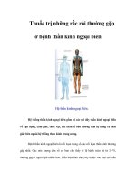
Figure 1
Treatment algorithm for closed fracture with associated nerve injury.
L. Randall Mohler, MD, and Douglas P. Hanel, MD
Volume 14, Number 1, January 2006 35
neously begin to do so in the first
few months. For those who do not,
electrodiagnostic studies—obtained
at 6 and 12 weeks—are a helpful ad-
junct.
5,17,18
The studies done at 6
weeks serve as a baseline for re-
examination and document the se-
verity of neurologic injury. A typical
finding is the identification of fibril-
lation potentials, positive sharp
waves, and monophasic action po-
tentials of short duration.
Repeat electrodiagnostic testing
of patients that does not demon-
strate clinical recovery at 12 weeks
will likely identify two subsets of pa-
tients. One group will demonstrate
improved nerve function with larger
polyphasic motor unit action poten-
tials of longer duration compared
with those in the 6-week study. This
is evidence that some degree of spon-
taneous recovery may be expected.
The degree to which the recovery
will occur is not predictable. In cases
in which the recovery is limited, ten-
don transfers are required to improve
function. The timing of these trans-
fers is not well defined. Although in-
jured nerves recover over 24 to 36
months, the degree of functional re-
covery is well established within 18
months of injury.
The second group will show few
signs of recovery, with fibrillation
potentials, positive sharp waves, and
diminished or absent small motor
unit potentials remaining the domi-
nant features of the study. This sec-
ond group is further divided into
three subgroups: children showing
little evidence of recovery, adults
with radial nerve deficits, and adults
with injuries other than of the radi-
al nerve. Children, defined by skele-
tal immaturity, should be observed
for a total of 9 months before explo-
ration or reconstruction because of
the reported 95% rate of recovery
within this timeframe.
5,10-12
Those
children who do not recover are
treated with tendon transfers.
Adults with radial nerve palsies
may be expected to recover within 6
months in >90% of cases. For adults
who do not recover, the authors of
two studies
16,19
propose that explora-
tion and repair remains a viable op-
tion for up to 6 months after injury.
This point remains controversial,
however, and the general opinion,
although undocumented, is that in-
stead of exploring the ner ve, sur-
geons should consider tendon trans-
fers for radial nerve palsies rather
than nerve repair. The rationale is
that tendon transfers in this setting
provide equal or better functional re-
covery than do radial nerve repairs
performed 6 months postinjury.
Patients in the third subgroup
include adults with nerve injuries
other than of the radial nerve. As
mentioned, Roganovic
16
demon-
strated that certain nerves have bet-
ter recovery potential that others.
For instance, the femoral and mus-
culocutaneous nerves demonstrate
excellent recovery potential when
repaired within 24 weeks of injury,
whereas the median, ulnar, and tib-
ial nerves demonstrate moderate re-
covery when repaired within the
same timeframe; all five nerves
demonstrate very little recovery
when repaired later than 24 weeks.
The peroneal nerve has very little re-
covery potential no matter when it
is explored or repaired.
Early exploration presents a co-
nundrum: the likelihood of finding a
repairable lesion in the setting of
closed fractures is unlikely, especial-
ly in the lower extremities, while
the likelihood of notable recovery,
should a repairable lesion be found,
is markedly diminished when repair
is delayed >4 months.
16
In other
words, the delayed exploration may
be unnecessary. With this in mind,
the surgeon is likely to discover a
nerve in anatomic, but not physio-
logic, continuity; he or she is then
faced with the difficult decision of
choosing between neurolysis, exci-
sion and repair, or doing no more
than ensuring that there are no com-
pressing fascial bands or bony frag-
ments, or a fracture callus, along the
course of the nerve. Physical conti-
nuity does not ensure spontaneous
recovery, nor does complete loss of
distal function preclude spontane-
ous recovery.
Intraoperative nerve stimulation
may assist with these decisions. The
intraoperative technique of Kline
and Happel,
20
of stimulating the in-
volved nerve proximally and record-
ing the nerve action potential distal
to the injured segment, is useful in
this difficult situation. They found
that, for patients in whom only neu-
rolysis was performed, good func-
tional recovery occurred in 93% of
nerves that had a recordable nerve
action potential distal to the lesion.
When resection of the injured seg-
ment was based on the absence of a
recordable nerve action potential
distal to the lesion, histologic exam-
ination uniformly confir med a le-
sion with poor potential for useful
recovery without repair.
These studies are not simply the
application of a nerve stimulator
proximal to a zone of injury, delivery
of an electrical stimulus, and obser-
vation of a twitching muscle distally.
Little information is gained from this
technique, especially when there is
no distal response. Intraoperative
nerve evaluation is the montage of in-
formation gained from somato-
sensory evoked potentials, elec-
tromyography, and nerve conduction
velocity studies. Obtaining this infor-
mation is technically demanding and
requires intraoperative collaboration
with a trusted neurophysiologist-
electrodiagnostician.
20,21
The mechan-
ics of this diagnostic technique con-
sist of placing recording electrodes
proximally (at the mastoid, seventh
cervical region, and contralateral
scalp), exposing the injured nerve, and
stimulating throughout the zone of
injury. Hook electrodes are placed
proximal to the zone of injury and the
effects of stimulation recorded prox-
imally. The electrodes are advanced
at 1-cm increments, and the stimu-
lation response is measured. The
point at which there is loss of soma-
tosensory evoked potential response
Closed Fractures Complicated by Peripheral Nerve Injury
36 Journal of the American Academy of Orthopaedic Surgeons
designates the transition between
functioning and nonfunctioning
nerves.
In further studies, stimulating the
nerve at a constant point proximal-
ly and measuring the response at
1-cm increments allows recording of
nerve compound action potentials.
The presence of nerve compound ac-
tion potentials resulting from in-
trafield stimulation demonstrates
regenerating nerve fibers, which are
an indication that further recovery
will occur and that intraneural dis-
section should be limited. Similarly,
by placing an electrode in a target
muscle and stimulating the injured
nerve motor, compound action po-
tentials reflect the response of a large
group of motor units, also a positive
finding. Nerve conduction velocities
are measured across the zone of inju-
ry and specific areas of slowing are
noted.
The intraoperative interpretation
of this information requires peri-
operative coordination, a dialogue
between the neurophysiologist and
the surgeon, and a clearly defined set
of goals for dealing with the infor-
mation provided. Most importantly,
these studies require time and a pa-
tient surgeon who is willing to listen
to the recommendations of, and re-
peat the studies requested by, the
electrodiagnostician. This coordinat-
ed effort, however, can at times pro-
vide equivocal data. In such cases,
the surgeon must make the difficult
decision of resection and grafting or
of leaving the neuroma in situ. In
most equivocal cases, we leave the
nerve intact, choosing further obser-
vation and appropriate tendon trans-
fers when the nerve fails to recover.
Summary
Closed fractures are occasionally
complicated by peripheral nerve in-
jury. The number of cases is limited
and incidence is sporadic, making
longitudinal research difficult. Most
patients recover without surgery.
Those who fail to show signs of re-
covery at 6 months, either clinically
or with electrodiagnotic testing,
should undergo exploration. Base-
line electrodiagnostic studies are
made 6 weeks postinjury; when
there is no sign of nerve recovery,
studies are repeated at 12 weeks.
Adults who show evidence of recov-
ery should continue to be observed.
Adults without evidence of recovery
and with radial nerve injury should
undergo repeat nerve studies and ul-
timately, if necessary, exploration,
repair, and/or tendon transfers. The
same procedure should be followed
in adults with injuries other than
those of the radial nerve. Exploration
in skeletally immature children,
whether exhibiting evidence of re-
covery or not, should be delayed for
9 months.
References
1. Noble J, Munro CA, Prasad VS, Midha
R: Analysis of upper and lower ex-
tremity peripheral nerve injuries in a
population of patients with multiple
injuries. J Trauma 1998;45:116-122.
2. de Laat EA, Visser CP, Coene LN,
Pahlplatz PV, Tavy DL: Nerve lesions
in primary shoulder dislocations and
humeral neck fractures: A prospective
clinical and EMG study. J Bone Joint
Surg Br 1994;76:381-383.
3. Postacchini F, Morace GB: Fractures
of the humer us associated with paral-
ysis of the radial nerve. Ital J Orthop
Traumatol 1988;14:455-464.
4. Pollock FH, Drake D, Bovill EG, Day
L, Trafton PG: Treatment of radial
neuropathy associated with fractures
of the humerus. J Bone Joint Surg Am
1981;63:239-243.
5. Shah JJ, Bhatti NA: Radial nerve paral-
ysis associated with fractures of the
humerus: A review of 62 cases. Clin
Orthop Relat Res 1983;172:171-176.
6. Sonneveld GJ, Patka P, van Mourik
JC, Broere G: Treatmentof fractures of
the shaft of the humerus accompanied
by paralysis of the radial nerve.
Injury 1987;18:404-406.
7. Böstman O, Bakalim G, Vainionpaa S,
Wilppula E, Patiala H, Rokkanen P:
Radial palsy in shaft fracture of the
humerus. Acta Orthop Scand 1986;
57:316-319.
8. Böstman O, Bakalim G, Vainionpaa S,
Wilppula E, Patiala H, Rokkanen P:
Immediate radial nerve palsy compli-
cating fracture of the shaft of the hu-
merus: When is early exploration jus-
tified? Injury 1985;16:499-502.
9. Whitson RO: Relation of the radial
nerve to the shaft of the humerus.
J Bone Joint Surg Am 1954;36:85-88.
10. McGraw JJ, Akbarnia BA, Hanel DP,
Keppler L, Burdge RE: Neurological
complications resulting from supra-
condylar fractures of the humerus in
children. J Pediatr Orthop 1986;6:
647-650.
11. Cramer KE, Green NE, Devito DP: In-
cidence of anterior interosseous nerve
palsy in supracondylar humerus frac-
tures in children. J Pediatr Orthop
1993;13:502-505.
12. Dormans JP, Squillante R, Sharf H:
Acute neurovascular complications
with supracondylar humerus frac-
tures in children. J Hand Surg [Am]
1995;20:1-4.
13. Brown IC, Zinar DM: Traumatic and
iatrogenic neurological complica-
tions after supracondylar humerus
fractures in children. J Pediatr
Orthop 1995;15:440-443.
14. Mehlman CT, Strub WM, Roy DR,
Wall EJ, Crawford AH: The effect of
surgical timing on the perioperative
complications of treatment of supra-
condylar humeral fractures in chil-
dren. J Bone Joint Surg Am 2001;83:
323-327.
15. Ring D, Chin K, Jupiter JB: Radial
nerve palsy associated with high-
energy humeral shaft fractures.
J Hand Surg [Am] 2004;29:144-147.
16. Roganovic Z: Factors influencing the
outcome of nerve repair. Vojnosanit
Pregl 1998;55:119-131.
17. Robinson LR: Traumatic injury to pe-
ripheral nerves. Muscle Nerve 2000;
23:863-873.
18. Robinson LR: Role of neurophysiolog-
ic evaluation in diagnosis. JAm
Acad Orthop Surg 2000;8:190-199.
19. Amillo S, Barrios RH, Martinez-Peric
R, Losada JI: Surgical treatment of the
radial nerve lesions associated with
fractures of the humerus. J Orthop
Trauma 1993;7:211-215.
20. Kline DG, Happel LT: Penfield Lec-
ture: A quarter century’s experience
with intraoperative nerve action po-
tential recording. Can J Neurol Sci
1993;20:3-10.
21. Slimp JC: Intraoperative monitoring
of nerve repairs. Hand Clin 2000;16:
25-36.
L. Randall Mohler, MD, and Douglas P. Hanel, MD
Volume 14, Number 1, January 2006 37