Báo cáo sinh học: " In vivo transcriptional targeting into the retinal vasculature using recombinant baculovirus carrying the human flt-1 promoter" potx
Bạn đang xem bản rút gọn của tài liệu. Xem và tải ngay bản đầy đủ của tài liệu tại đây (630.69 KB, 12 trang )
BioMed Central
Page 1 of 12
(page number not for citation purposes)
Virology Journal
Open Access
Research
In vivo transcriptional targeting into the retinal vasculature using
recombinant baculovirus carrying the human flt-1 promoter
Agustín Luz-Madrigal
1
, Carmen Clapp
2
, Jorge Aranda
2
and Luis Vaca*
1
Address:
1
Departamento de Biología Celular, Instituto de Fisiología Celular, Universidad Nacional Autónoma de México (UNAM), Ciudad
Universitaria, México D.F. 04510, México and
2
Instituto de Neurobiología, UNAM-Juriquilla, Querétaro, Qro México, 76001, México
Email: Agustín Luz-Madrigal - ; Carmen Clapp - ; Jorge Aranda - ;
Luis Vaca* -
* Corresponding author
Abstract
Background: Endothelial cells are a target for gene therapy because they are implicated in a
number of vascular diseases. Recombinant baculovirus have emerged as novel gene delivery
vectors. However, there is no information available concerning the use of endothelial-specific
promoters in the context of the baculovirus genome. In the present study, we have generated a
recombinant baculovirus containing the human flt-1 promoter (BacFLT-GFP) driving the expression
of the green fluorescent protein. Transcriptional gene targeting was analyzed in vitro in different
mammalian cell lines and in vivo in adult rat retinal vasculature.
Results: BacFLT-GFP evoked the highest levels of expression in the endothelial cell line BUVEC-
E6E7-1, similar to those reached by recombinant baculovirus carrying the CMV promoter (112%
relative to BacCMV-GFP, n = 4). Interestingly, BacFLT-GFP directed high levels of expression in rat
glioma C6 and in human glioblastoma CH235 cells (34.78% and 47.86% relative to BacCMV-GFP,
respectively). Histone deacetylase inhibitors such as butyrate or trichostatin A enhanced the
transcriptional activity of both BacCMV-GFP and BacFLT-GFP. Thus, in this study histone
deacetylation appears to be a central mechanism for the silencing of baculovirus, independently of
the promoter utilized. In vivo transcriptional targeting was demonstrated in adult rat retinal
vasculature by intravitreal delivery of BacFLT-GFP and immunohistochemical staining with von
Willebrand factor (vWF). Analysis by fluorescence microscopy and deconvolved three-dimensional
confocal microscopy of retinal whole mounts obtained after 3 days of baculovirus injection showed
that most GFP-expressing cells localized to the inner limiting membrane (ILM) and ganglion cell
layer (GCL) and colocalize with vWF (70%, n = 10) in blood vessels, confirming the endothelial
phenotype of the transduced cells.
Conclusion: Taken together, our results indicate that the restricted expression in endothelial cells
mediated by the flt-1 promoter is not affected by the context of the baculovirus genome and
demonstrate the potential of using recombinant baculovirus for transcriptional targeted gene
expression into the eye vasculature.
Published: 18 September 2007
Virology Journal 2007, 4:88 doi:10.1186/1743-422X-4-88
Received: 21 June 2007
Accepted: 18 September 2007
This article is available from: />© 2007 Agustín et al; licensee BioMed Central Ltd.
This is an Open Access article distributed under the terms of the Creative Commons Attribution License ( />),
which permits unrestricted use, distribution, and reproduction in any medium, provided the original work is properly cited.
Virology Journal 2007, 4:88 />Page 2 of 12
(page number not for citation purposes)
Background
Local delivery of genes to vascular wall is a promising
approach for the treatment of a number of vascular disor-
ders [1]. As a target organ for gene transfer, the vasculature
has several unique features such as a large surface area and
easy accessibility. The architecture of the normal vessel
wall is relatively simple consisting of three main cell types
(endothelial cells, smooth muscle cells, and fibroblasts)
and the transgene products may be secreted locally to
achieve an autocrine-paracrine effect or into the blood-
stream for a systemic effect. Within the vasculature,
endothelial cells are the main target for gene therapy
because they are closely related with disease process such
as inflammation, atherosclerosis, systemic and pulmo-
nary hypertension, cerebrovascular disease, and in angio-
genesis-related disorders [1]. Moreover, tumor
angiogenesis is crucial for the progression and metastasis
of cancer [2]. Therefore, tumor vascular targeting therapy
could represent an effective therapeutic strategy to sup-
press both primary tumor growth and tumor metastasis
[2].
Viral vectors have been used extensively in vascular gene
transfer; adenoviral vectors being the most commonly
used system [3]. Other vector systems include adeno-asso-
ciated virus (AAV) and lentiviral vectors [4]. Although
these vectors have demonstrated the transfer of genetic
material for its expression in endothelial cells, the main
limitations are associated with inflammatory reactions
due to the pre-existing immunity to human virus [4,5]. To
address this problem, the use of recombinant viruses of
non-human origin as gene therapy vectors has been sug-
gested [6].
Recently, recombinant baculovirus derived mainly from
Autographa californica multiple nuclear polyhedrosis virus
(AcMNPV) have emerged as a novel and safer system to
transfer genes for its expression into a wide variety of
mammalian cells [7]. Since the first studies made by two
different groups, showing the ability of baculovirus to
transfer genes in mammalian cells derived from hepatic
origin [8,9], the list of mammalian cells susceptible to
transduction by recombinant baculovirus has increased in
the last few years [7].
Transcriptional targeting using cellular tissue-specific reg-
ulatory sequences has been demonstrated as a powerful
strategy to restrict gene expression to a particular cell type
in various tissues, including liver, smooth muscle and
heart [10,11]. Moreover, utilization of tumor/tissue-spe-
cific promoters can reduce toxicity, increase safety, and
improve the therapeutic index [12,13].
The human transmembrane fms-like tyrosine kinase (Flt-
1) is one of the receptors for vascular endothelial growth
factor (VEGF) [14]. Flt-1 is expressed specifically in
endothelium and is likely to play a role in tumor angio-
genesis and embryonic vascularization [15]. Morishita et
al., demonstrated that a 1-kb DNA fragment of the 5'-
flanking region of human flt-1 gene (region from -748 to
+284 bp) is involved in endothelial-specific gene expres-
sion [16]. So far, there is no information available con-
cerning the use of endothelial-specific promoters in the
context of the baculovirus genome. Furthermore, only
two reports show to this date in vivo transcriptional gene
targeting by recombinant baculovirus.
In this study, we produced a recombinant baculovirus
(BacFLT-GFP) containing the human flt-1 promoter driv-
ing the expression of the green fluorescent protein (GFP)
and evaluated the maintenance of endothelial-specific
gene expression after in vitro transduction of different
mammalian cell lines. We also demonstrated in vivo tran-
scriptional targeting into the rat retinal vasculature by
immunoflurescence staining after intravitreal delivery of
BacFLT-GFP. Three-dimensional (3-D) confocal recon-
struction studies of retinas from animals injected with
BacFLT-GFP showed for the first time the selective target-
ing to blood vessels of a baculovirus vector.
Results
Transduction susceptibility mediated by recombinant
baculovirus in mammalian cells
To compare the selectivity and the levels of expression
mediated by the endothelial specific baculovirus (BacFLT-
GFP), we generated a recombinant baculovirus containing
a 761-bp DNA fragment of the cytomegalovirus (CMV)
promoter driving the expression of GFP (Figure 1a). This
promoter was selected because it drives high levels of
expression into mammalian cells from different tissues.
We included the immortalized bovine umbilical vein
endothelial cell line BUVEC-E6E7-1, which retains
endothelial cell characteristics and has been utilized to
investigate the action of regulatory factors of vascular
endothelium [17]. The cells were transduced at a multi-
plicity of infection (MOI) of 100 in the presence or
absence of 5 mM butyrate, a histone deacetylase inhibitor.
The percentage of GFP positive (GFP+) cells and the levels
of expression, represented by the mean fluorescence
intensity (MFI), were analyzed 48 h after transduction by
flow cytometry. In all cases mock-transduced cells (cells
treated with medium alone and butyrate) were used as
control for background fluorescence. In the experiments
performed without the addition of butyrate (Figure 1b,c)
the hepatocarcinoma cell line HepG2 was the most sus-
ceptible to transduction, with 89.2 ± 2.3% GFP positive
cells, followed by the rat glioma cell line C6, 57.7 ± 23%;
the bovine umbilical vein endothelial cell line BUVEC-
E6E7-1, 24.2% ± 6.5%; the human embryonic kidney cell
Virology Journal 2007, 4:88 />Page 3 of 12
(page number not for citation purposes)
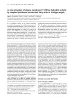
Transduction susceptibility mediated by BacCMV-GFP in mammalian cellsFigure 1
Transduction susceptibility mediated by BacCMV-GFP in mammalian cells. 10
5
cells were transduced at a multiplic-
ity of infection (MOI) of 100 with BacCMV-GFP. (a) Drawing showing the cassette structure of BacCMV-GFP. Abbreviations:
CMV, cytomegalovirus immediately-early promoter/enhancer (nucleotides -655 to +106); EGFP, enhanced green-fluorescent
protein; BGHpA, bovine growth hormone poly adenylation sequence. (b) Representative histograms obtained by flow cytome-
try after 48 h post-transduction. The percentage of GFP+ cells is reported in the insets, and was calculated by subtracting the
background obtained with mock transduced cells (see Methods). Numbers in the inset refer to the percentage of GFP+ cells ±
SD without (above) or with (below) treatment with butyrate. (c) Percentage of GFP+ cells determined by FACS analysis in
either the presence or absence of butyrate. *P < 0.01, **P < 0.05, ***P = 0.7244 versus cells non-treated with butyrate. NS =
non significant. d) Levels of expression of GFP and induction ration (indicated on the right) as determined by the mean fluores-
cence intensity in the presence or absence of butyrate. Values are means ± SD of four independent experiments.
a
b
c
d
Mean fluorescence
1 10 100 1000 10000
C6
HepG2
HEK293
RINm5F
BUVEC
Induction
ratio
10.1±3.6
5.9 0.9±
3.9 0.6±
33.4 5.3±
92.6 2.8±
Mock
-Butyrate
+Butyrate
060
0
Events
GFP+
RINm5F
14.4±3.5%
59.6±11.3%
60
GFP+
BUVEC
24.2±6.5%
66.8±9.6%
Events
10
0
10
1
10
3
10
4
10
2
10
0
10
1
10
3
10
4
10
2
GFP+
060
Events
060
Events
C6
57.7±23%
63.5±13%
60
Events
GFP+
0
HEK293
19.4±7.5%
66.2±17.7%
10
0
10
1
10
3
10
4
10
2
10
0
10
1
10
3
10
4
10
2
10
0
10
1
10
3
10
4
10
2
HepG2
GFP+
89.2±2.3%
94.0±0.6%
- Butyrate
+Butyrate
Percentage of GFP + cells
0
20
40
60
80
100
C6
HepG2
HEK293
RINm5F
BUVEC
**
*
*
BacCMV-GFP
EGFP
+106655
-
BGHpA
CMV-IE promoter
-
Butyrate
+
Butyrate
**
NS
A luz et al. Fig. 1
Virology Journal 2007, 4:88 />Page 4 of 12
(page number not for citation purposes)
line HEK-293, 19.4 ± 7.5% and the rat insulinoma cell
line RIN-m5F, 14.4 ± 3.5%.
Modification of the accessibility of the transcription
machinery to gene promoters is one of the underlying
mechanisms regulating gene expression in mammalian
cells [18]. Although several mechanisms are involved in
the regulation of this process, the status of the histone
acetylation/deacetylation and DNA methylation/demeth-
ylation seems to be of major importance [18,19]. Previous
reports have suggested that epigenetic regulation seems to
influence the transduction efficiency mediated by recom-
binant baculovirus in mammalian cells [20,21]. There-
fore, we treated the cells with 5 mM butyrate, which has
been used to reactivate transgene expression by inhibiting
the chromatin-remodeling of histone deacetylases
(HDAC), which results in an increase of transcriptional
activity of the promoter [22]. Treatment with butyrate
(Figure 1b,c) dramatically reactivated the expression in
almost all cell lines, except in C6 in which there was no
significant difference from control (63.5 ± 13% and 57.7
± 23% of GFP+ cells for control and butyrate, respectively;
P = 0.7244, n = 4). In agreement to previous reports
[20,21], levels of expression evaluated by the MFI were
increased by addition of butyrate (Figure 1d). Particularly,
the cell line RIN-m5F was the most susceptible to reactiva-
tion by butyrate (92.6 ± 2.8-Fold), followed by BUVEC-
E6E7-1, and HEK-293 cells (33.4 ± 5.3 and 10.1 ± 3.6-
Fold, respectively). In an attempt to reactivate gene
expression by DNA methylation inhibitors, we treated the
cells with 5'aza-2'deoxycytidine (5'aza-C) [23], however
there was not a significant difference between the levels of
expression reached by control untreated cells and treated
with 30 µM of 5'aza-C (data not shown). These results
suggest that in these cell lines including BUVEC-E6E7-1
cells, there is a strong repression of the transgene expres-
sion most likely due to histone deacetylation. In contrast,
HepG2 and C6 were less susceptible to reactivation (5.9 ±
0.9 and 3.9 ± 0.6-fold, respectively) indicating that in
these cell lines histone deacetylation does not play a criti-
cal role in gene silencing the baculovirus genome.
Baculovirus containing the human Flt-1 promoter drives
endothelial-specific gene expression in vitro
We generated a novel recombinant baculovirus with the
human flt-1 promoter driving the expression of GFP (Bac-
FLT-GFP, Figure 2a) to test its selectivity and efficiency as
a vascular vector for gene therapy.
We first assessed the efficiency of transduction and levels
of expression mediated by BacFLT-GFP in the immortal-
ized bovine umbilical vein endothelial cell line BUVEC-
E6E7-1 in the presence of 5 mM butyrate. Analysis by flow
cytometry 48 h after transduction with BacFLT-GFP (MOI
of 100) showed a large number of GFP+ cells (60.34%, n
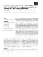
Specificity and levels of gene expression mediated by BacFLT-GFPFigure 2
Specificity and levels of gene expression mediated by
BacFLT-GFP. (a) Drawing showing the cassette structure
of BacFLT-GFP. Abbreviations: hFlt-1, promoter sequence of
the human flt-1 gene (nucleotides -748 to +284 bp); EGFP,
enhanced green-fluorescent protein; BGHpA, bovine growth
hormone poly adenylation sequence. (b) (Left panel), Repre-
sentative histogram obtained by flow cytometry 48 h after
transduction of BUVEC-E6E7-1 cells with 100 MOI of Bac-
FLT-GFP and 5 mM of butyrate. The percentage of GFP+
cells is reported in the inset, and was calculated by subtract-
ing the background from mock transduced cells (see Meth-
ods). (Right panel) Levels of expression obtained with
BacCMV-GFP and BacFLT-GFP in BUVEC-E6E7-1 cells, rep-
resentative of four independent experiments, mean ± SD. (c)
Mean fluorescence intensity (MFI) measured 48 h after trans-
duction with BacFLT-GFP relative to BacCMV-GFP. The per-
centage of GFP+ cells are indicate above of each bar. Cells
were transduced with 100 MOI of BacFLT-GFP or BacCMV-
GFP. Data are from four independent experiments ± SD.
a
BacFLT-GFP
+284-748
EGFP
BGHpA
hFlt-1 promoter
b
c
060
Events
10
0
10
1
10
2
10
4
10
3
GFP+
BUVEC
60.34%
Fluorescence
BacCMV-GFP
BacFLT-GFP
0
100
200
300
400
500
nrsecMea fluo e c n e
P=0.197
0
C6
HepG2
H29EK 3
RNm5FI
BU ECV
C25H3
Pe ta ercen g
ea v to c M
r l ti e Ba C V-GFP
20
40
60
80
100
120
140
34.78%
47.86%
5.32%
3.6%
1.98%
112.54%
A luz et al. Fig. 2
Virology Journal 2007, 4:88 />Page 5 of 12
(page number not for citation purposes)
= 4), similar to that obtained with BacCMV-GFP (Figure
2b). Moreover, there is not significant difference between
the levels of expression reached by BacFLT-GFP and Bac-
CMV-GFP at the same MOI (Figure 2b right panel, P =
0.197, n = 4). Similar results were obtained by Nicklin et
al., in which recombinant adenoviral vector containing
the same promoter sequence produced levels of expres-
sion comparable to those evoked by an adenovirus con-
taining the CMV promoter in human umbilical vein
(HUVEC) and human saphenous vein endothelial cells
(HSVEC) [24].
We next analyzed levels of expression in non-endothelial
cells to evaluate in vitro cell specificity (Figure 2c). Since
the efficiency of gene transfer differs among cell types, the
levels of expression (i.e. MFI) for each cell line were nor-
malized using BacCMV-GFP as positive control.
The highest level of expression with BacFLT-GFP was
observed in BUVEC-E6E7-1 cells (112 ± 6.2% relative to
BacCMV-GFP, n = 4). Importantly, the levels of expression
were severely reduced in other non-endothelial cell types
including HepG2, HEK293 and RINm5F. For example,
GFP expression in HepG2 cells was only 5.32 ± 0.37%, of
that obtained with BacCMV-GFP, demonstrating the low
activity of flt-1 promoter in non-endothelial cells (Figure
2c). Furthermore, BacFLT-GFP produced high levels of
expression in the rat glioma C6 and in the human gliob-
lastoma CH235 cells (34.78 ± 4.6, 47.86 ± 7.84, respec-
tively). In agreement with our results, Flt-1 and KDR
(both receptors for VEGF) have been identified on malig-
nant cells from human CNS, breast, prostate cancer and in
cell lines derived from these tumors [15,25]. Furthermore,
RT-PCR studies of C6 and CH235 cells showed high levels
of expression of the Flt-1 receptor mRNA (data not
shown).
In summary, these results indicate that the flt-1 promoter
retains its transcriptional selectivity in the context of bac-
ulovirus genome in vitro.
Histone deacetylase inhibitors reactivate the expression
mediated by recombinant |baculovirus containing the
human Flt-1 promoter
To analyze whether HDAC inhibitors can also improve
recombinant baculovirus-mediated gene expression
under the control of flt-1 promoter, BUVEC-E6E7-1 cells
were transduced with BacFLT-GFP at an MOI of 100 in the
presence of different concentrations of butyrate or TSA
(Figure 3a). The GFP+ cells and MFI were examined by
flow cytometry 48 h post-transduction. Treatment with
both HDAC inhibitors clearly improved gene expression
in terms of GFP+ cells in a dose-dependent manner (Fig-
ure 3a). The number of GFP+ cells was increased from
8.64% in untreated control cells to 78.25% in cells treated
with 15 mM butyrate. Butyrate inhibits HDAC but also
has a number of unrelated effects [22]. To determine
whether the inhibition of histone deacetylation was the
major contributor to enhanced GFP expression, cells were
treated with TSA, which is a more potent and selective
inhibitor of deacetylases [26]. TSA significantly enhanced
transgene expression in a dose-dependent manner, rang-
ing from 15.85% in untreated control cells to 53.04%
GFP+ in cells treated with 50 nM TSA (Figure 3a, right
panel). In both cases (butyrate or TSA) the range of fluo-
rescence intensities in the population is spread over
approximately three orders of magnitude, suggesting dif-
ferent levels of expression within the cells. Improvement
of gene expression after treatment with butyrate or TSA
was also evidenced by MFI (Figure 3b). The average level
of induction was 81.68-fold and 61.69-fold for 15 mM
butyrate and 50 nM TSA, respectively. There were no sig-
nificant differences in expression at concentrations
between 5 and 10 mM of butyrate or 25 and 30 nM of TSA
(Figure 3a,b). Higher concentrations of butyrate or TSA
administrated for 48 h resulted in toxic effects in terms of
viable cell number evaluated by trypan blue exclusion.
We performed transient transfections in BUVEC-E6E7-1
cells using the transfer plasmid pBlueFLT-GFP (see Meth-
ods), to determine whether the baculovirus genome is
involved in the silencing of gene expression (Figure 3c).
Transfected cells treated with butyrate or TSA did not
show any significant reactivation of gene expression com-
pared with untreated control cells using this construct,
even at the highest butyrate or TSA concentrations tested
in this study.
Therefore, all these results demonstrate that transgene
expression mediated by recombinant baculovirus con-
taining the human flt-1 promoter is enhanced by the addi-
tion of HDAC inhibitors, and the viral genome or proteins
coupled to the DNA from the virus are implicated in the
silencing of gene expression, since no effect of HDAC
inhibitors was observed when the original plasmid used
to generate the recombinant baculovirus was directly
transfected to drive GFP expression. Moreover, in this
study the effect of HDAC inhibitors was independent of
the two promoters used.
In vivo endothelial-specific gene expression by baculovirus
vectors containing the human Flt-1 promoter
In order to determine whether the endothelial-specific
gene expression mediated by BacFLT-GFP is retained in
vivo, we selected the eye as a target organ for gene delivery
because it is a closed system clearly separated from the sys-
temic circulation, facilitating the delivery of the vector.
Furthermore, the blood retinal barrier (BRB) separates the
retina from blood, which contains inhibitory factors (e.g.
complement) [27]. These characteristic is particularly rel-
Virology Journal 2007, 4:88 />Page 6 of 12
(page number not for citation purposes)
evant to baculovirus gene transfer, since complement has
been clearly implicated in the inactivation of in vivo
applied recombinant baculovirus [28]. Moreover, trans-
gene expression in rat retina after intravitreal delivery of
recombinant baculovirus has been previously demon-
strated, showing that viral particles are able to diffuse
through the vitreous body reaching the retina, with maxi-
mal GFP expression 2–3 days after injection [29,30].
Based on these considerations, ten microliters of virus
(BacFLT-GFP) solution concentrated by ultracentrifuga-
tion [approximately 1 × 10
7
plaque forming units (PFU)
of viral particles] or vehicle as a control were injected into
the vitreous cavity of rat eyes. Three days after virus injec-
tion, the rats were sacrificed and the retinas were quickly
dissected, fixed, and analyzed by fluorescence microscopy
using a GFP filter set. Intravitreal delivery of viral particles
resulted in strong reporter gene expression, with most
GFP-expressing cells almost exclusively localized in the
inner limiting membrane (ILM) and ganglion cell layer
(GCL). In order to successfully identify the phenotype of
the GFP+ cells, we performed immunohistochemical
staining showing that most GFP-expressing cells (70%, n
= 10) react with antibodies to the endothelial specific
marker vWF [31], (Figure 4b,c,d,f) confirming the
endothelial phenotype of the in vivo transduced cells. In
contrast, control eyes injected with vehicle alone did not
show positive signal for GFP (Figure 4f,g).
To further confirm that our baculovirus vector was driving
GFP expression in endothelial cells, we performed tridi-
mensional confocal reconstructions of retinal sections
from control and BacFLT-GFP treated animals. Figure 4i
Effect of histone deacetylase inhibitors in the expression mediated by BacFLT-GFPFigure 3
Effect of histone deacetylase inhibitors in the expression mediated by BacFLT-GFP. (a) Representative histograms
obtained by flow cytometry 48 h after transduction of BUVEC-E6E7-1 cells with 100 MOI of BacFLT-GFP and treated with
increasing concentrations (shown in the figure) of butyrate or trichostatin A (TSA). The percentage of GFP+ cells (shown
inside green rectangles) is reported in the insets, and was calculated by subtracting the background from mock transduced
cells. (b) Fold induction in GFP expression mediated by BacFLT-GFP with increasing concentrations of butyrate or TSA. (c)
Fold induction of the expression of GFP in transfected cells with the transfer plasmid pBlueFLT-GFP (see Methods) and treated
with increasing concentrations of butyrate or TSA. Results from four independent experiments ± SD. NS = non significant.
0
20
40
60
80
0 10 25 30 50
TSA (nM)
BacFLT-GFP
Fold
nducI tion
1.2
1.0
0.2
0.4
0.6
0.8
0
0 10 25 30
TSA (nM)
Plasmid
Fold
nducti
Ion
GFP Fluorescence
Number of events
600
GFP+
8.64%
41.12%
600
67.29%
600
75.37%
600
10
0
10
1
10
3
10
4
10
2
78.25%
600
0
1
5
10
Bu y ate (m )tr M
15
GFP+
15.85%
27.63%
44.77%
49.07%
53.04%
0
10
25
30
TSA (nM)
50
10 10 10 1010
01 342
600600600600600
a
0
20
40
60
80
100
0 1 5 10 15
Butyrate (mM)
BacFLT-GFP
Fold
Induction
b
2.5
2.0
1.5
1.0
0.5
0
0 1 5 10
Butyrate (mM)
Plasmid
Fold
nducItion
c
NS
NS
A luz et al. Fig. 3
Virology Journal 2007, 4:88 />Page 7 of 12
(page number not for citation purposes)
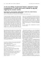
BacFLT-GFP directs endothelial-specific gene expression into the retina vasculatureFigure 4
BacFLT-GFP directs endothelial-specific gene expression into the retina vasculature. 10 µl of BacFLT-GFP virus
solution (approximately 1 × 10
7
PFU of viral particles) or vehicle (PBS) were injected into the rat eye vitreous chamber. Three
days after the virus injection, the rats were sacrificed and the retinas processed for immunohistochemistry using an antibody
against von Willebrand factor (red) and DAPI (blue) to highlight nuclei (see Methods). Representative retinas from eyes
injected with 1 × 10
7
pfu of BacFLT-GFP (a, b, c, d) or vehicle (PBS) (e, f, g, h). The same sections were evaluated by phase-con-
trast microscopy (a, e) and by anti-von Willebrand factor antibodies coupled to TRITC (red) and GFP green fluorescence.
Merge images illustrate the colocalization of red and green fluorescence (in yellow) in capillary-like structures (big white rec-
tangles in c, d, g) obtained from the areas in small white rectangle insets from b, f. Confocal three-dimensional reconstruction
images of retinas injected with vehicle (h) or with BacFLT-GFP (i). In red is shown the localization of von Willebrand factor
(red) within characteristic blood vessel structures. The colocalization of red (vWF) and green (GFP) fluorescence is shown in
yellow (white arrowheads in i). (j) Percentage of vWF and GFP pixel co-localization in retinas injected with BacFLT-GFP, (n =
10 animals). Abbreviations: RPE, retinal pigment epithelium GCL, ganglion cell layer; INL, inner nuclear layer; and ONL, outer
nuclear layer; ILM inner limiting membrane.
0
20
40
60
80
100
F%G P-
cp o- ocalization
ixel l
vWF
BacFLT-GFP
j
n=10
10 µm12 µm
e
f
g
h
i
50 µm
BacFLT-GFPControl
RPE
ONL
INL
GCL
ILM
A luz et al. Fig. 4
RPE
ONL
INL
GCL
ILM
a
b
c
d
50 µm
vWF
GFP
DAPI
vWF
GFP
DAPI
vWF
GFP
vWF
GFP
Virology Journal 2007, 4:88 />Page 8 of 12
(page number not for citation purposes)
illustrates tridimensional projections of the confocal
reconstructions clearly showing the blood vessel pattern
of GFP expression. Blood vessels were not only recognized
for their morphology, but also by immunoassaying with a
TRITC-coupled anti- vWF antibody (shown in red). Addi-
tional file 1 illustrates the three-dimensional reconstruc-
tions (rotating vessels) obtained with retinas from mock-
injected animals and animals injected with BacFLT-GFP.
These data shows the selective labeling of blood vessels, or
portions of them with our recombinant baculovirus.
Discussion
In the present study, we have demonstrated that recom-
binant baculovirus containing the 1-kb region from the
human flt-1 promoter is able to drive transcriptional gene
targeting in vitro and in vivo in the retinal vasculature. The
immortalized bovine umbilical vein endothelial cell line
BUVEC-E6E7-1 and other non-endothelial cell types (i.e.
C6, HepG2, HEK293, RINm5F) were susceptible to Bac-
CMV-GFP vector-mediated gene transfer. Of all cell lines
explored, the highest levels of expression were observed
with the hepatocarcinoma cell line HepG2, as previously
reported [8,9]. In agreement with the results recently pub-
lished by Wang et al., we also demonstrate that rat glioma
cell line C6 is highly susceptible to transduction (> 50%
GFP+) by this recombinant baculovirus [32].
In addition, our results show that the endothelial cell line
BUVEC-E6E7-1 appeared to be more susceptible to trans-
duction than the human carcinoma/endothelial cell-like
ECV-304, in which 21% of the cells were transduced at a
MOI of 1000. Such differences could be due to different
conditions in which the vectors were titered [33].
The susceptibility of transduction demonstrated in RIN-
m5F cells, corroborates the results observed by Ma et al.,
in which pancreatic islet cells were efficiently transduced
by recombinant baculovirus carrying a construct similar
to the one reported here [34].
Reversible acetylation of the histone tails by histone acety-
lases (HATs)/histone deacetylases (HDACs) is one of the
best studied posttranslational modifications of histones,
correlating with transcriptional activation/repression. We
have shown here that the treatment of cells with butyrate,
an histone deacetylase inhibitor enhanced the transcrip-
tional activity mediated by both BacCMV-GFP or BacFLT-
GFP. However, histone hyperacetylation is one of many
cellular changes induced by butyrate. Therefore, to con-
firm whether histone acetylation is involved in reactiva-
tion of transgene expression, we also tested the potent and
selective HDAC inhibitor, TSA. To date 17 human genes
have been identified encoding HDACs, 11 of these genes,
members of so-called clase I and II, are inhibited by TSA
[35]. Treatment with TSA reactivates GFP expression
mediated by BacFLT-GFP at concentrations as low as 10
nM, and maximal effects were observed at 50 nM, corre-
sponding to a concentration 10,000 fold smaller than the
concentration of butyrate utilized to reach similar effects
(81.68-fold and 61.69-fFold for 15 mM butyrate and 50
nM TSA, respectively). To determine whether DNA meth-
ylation is also implicated in baculovirus silencing, 5'aza-
C, which is known to interfere with the process of DNA
methylation, was added to a concentration of 25 µM
alone or in combination with butyrate or TSA. We did not
observe a significant increase of transgene expression with
5'aza-C (data not shown). Therefore, DNA methylation
does not appear to play an important role in baculovirus-
mediated gene transfer silencing. In contrast, histone
deacetylation appears to be a general mechanism for
silencing baculovirus independently of the promoter or
the gene utilized (we have observed reactivation of beta-
galactosidase with butyrate or trichostatin A as well, data
not shown).
Therefore our results suggest an association between the
episomal baculovirus DNA and mammalian histones to
resemble nucleosomes. A recently published work dem-
onstrates the association between the episomal adeno-
associated virus (AAV) vector genome and the acetylated
histone H3 [36]. Thus, a similar mechanism may direct
the promoter and transgene-independent silencing of
recombinant baculovirus observed here.
Although we do not know the exact mechanism(s) by
which TSA or butyrate reactivates transgene expression, it
is possible that nuclear spatial positioning can influence
the levels of expression observed in the mammalian cells
transduced by BacCMV-GFP or BacFLT-GFP. Reposition-
ing of baculovirus genome after HDACs inhibition is
something we are currently exploring.
A number of reports have shown that viral sequences
including viral regulatory elements can interfere with het-
erologous promoters used to drive transgene expression
and may impair tissue-specific or inducible transgene
expression [37-39]. Our data demonstrate that the
restricted expression in endothelial cells mediated by the
flt-1 promoter is not affected by the context of the baculo-
virus genome or by the mechanisms of silencing, since
BacFLT-GFP evoked the highest levels of expression in
BUVEC-E6E7-1 (112 ± 6.2% relative to BacCMV-GFP, n =
4), compared to the other cell lines. Interestingly, we have
obtained efficient GFP expression in BUVEC-E6E7-1 using
100 MOI of BacFLT-GFP, and previous studies have
shown that endothelial cells transduced with recom-
binant adenovirus required MOI of 500 to achieve similar
levels of expression in HUVEC and HSVEC [24].
Virology Journal 2007, 4:88 />Page 9 of 12
(page number not for citation purposes)
We took advantage of intravitreous injection, in order to
analyze in vivo endothelial-specific gene expression medi-
ated by BacFLT-GFP into the retinal vasculature. Our
results show that the majority of GFP+ cells were found at
the inner limiting membrane (ILM) and ganglion cell
layer (GCL), a somewhat expected result since these two
structures are in closer apposition to the area of injection
(the vitreous). Consistent with this finding, VEGFR1 and
VEGFR2 mRNA and Flt-1 protein have been localized to
the inner nuclear and in the ganglion cell layer of the rat
retina [40,41].
By immunohistochemical staining with the von Wille-
brand factor (vWF), we demonstrated by fluorescence
microscopy and three-dimentional confocal analyses that
most of the GFP positive cells are labeled with this
endothelial cell specific marker, and localize in blood ves-
sels, showing the specificity of gene expression in vivo.
We have observed direct GFP fluorescence in retina slices
transduced with BacFLT-GFP even in the absence of his-
tone deacetylase inhibitors; this is probably due to the
strong expression driven by the human Flt-1 promoter,
which can overcome the silencing effect of histone
deacetylases.
Transgene expression in retina after intravitreous body
injection of recombinant baculovirus has been previously
documented [29]. In this study, a recombinant baculovi-
rus carrying GFP under the control of CMV promoter was
subretinally injected, resulting in widespread transgene
expression in the corneal endothelium, lens, the retinal
inner nuclear layer, GCL, and retinal pigment epithelial
(RPE). Unlike the previously mentioned study, the
endothelium-selective expression observed in our results
originates from the use of the human flt-1 promoter.
Conclusion
In summary, our study indicates that specific gene expres-
sion in the vascular endothelium mediated by the human
flt-1 promoter is retained in the context of the baculovirus
genome. However, the reactivation of transgene expres-
sion by histone deacetylase inhibitors such as TSA or
butyrate, suggest that baculovirus genome forms a chro-
matin-like structure assembled into nucleosomes after the
viral genome is delivered into mammalian cells. Future
experiments will address the nature of the proteins that
affect the silencing of baculovirus vectors in mammalian
cells. Finally, this study provides the proof of principle of
baculovirus mediated gene transfer to endothelial cells in
vivo and suggest the possibility of using this recombinant
baculovirus for targeted gene expression into the retinal
vasculature.
Methods
Construction of transfer plasmids
pBlueCMV was generated as follows, briefly a HindIII-
SmaI 1.1 kb fragment from pcDNA 3.1(+) (Invitrogen,
San Diego, CA) containing the polyadenylation sequence
of the Bovine Growth Hormone was inserted into HindIII-
SmaI digested pBlueScript (+) (Stratagene, La Jolla, CA) to
produce pBSpolyA. A fragment EcoRI-BamHI from
pBSpolyA was cloned in EcoRI-BglII sites of pBlueBac 4.0
(Invitrogene, San Diego, CA) in the opposite orientation
to the polyhedrin promoter to produce pBB4polyA.
Finally, a SalI-EcoRI 953 bp fragment which contains the
CMV was ligated into SalI-EcoRI sites of pBB4polyA to
generate pBlueCMV. This plasmid was designed to con-
tain a multiple cloning site to insert any cDNA under the
control CMV promoter. The transfer plasmid pBlueCMV-
GFP was constructed by ligation of a fragment BamHI-
NotI 730 pb containing the coding region GFP from
pEGFP-N1 (Clontech, Palo Alto, CA) into the KpnI-ApaI
sites of the pBlueCMV by blunt end ligation. The transfer
plasmid pBlueFLT-GFP was constructed by cloning a
BamHI-HindIII 1.0 kb fragment containing the human
fms-like tyrosine kinase-1 promoter (hFLT-1 promoter,
segment extending from -748 to +284) from the plasmid
p(-748/+284) [16] kindly provided by Dr. Andrew Baker
(University of Glasgow, Glasgow, UK) into BglII-HindIII
sites of pBlueCMV-GFP. The correct sequence of each
transfer plasmids were confirmed by DNA sequencing.
Production of recombinant baculovirus
The recombinant baculovirus BacCMV-GFP and BacFLT-
GFP, were generated by using the Bac-N-Blue Transfection
Kit according to the manufacturer's instructions (Invitro-
gen, Carlsbad, CA) and were plate-purified twice before to
large-scale production. To propagate the baculovirus Sf9
insect cells were infected (2 × 10
6
/ml) at a multiplicity of
infection of 0.1 and the viruses were purified as follows;
culture supernatants were harvested at 6 days after infec-
tion and cells debris was removed by centrifugation at
6,000 g for 15 min at 4°C. The supernatants obtained
were titered by plaque assay on Sf9 insect cells, stored at
4°C and used for in vitro experiments. In the experiments
for gene transfer in vivo, recombinant baculovirus were
concentrated by ultracentrifugation in a SW28 rotor
(Beckman) at 27,000 rpm for 60 min, resuspended in
phosphate-buffered saline (PBS) and loaded on 10–50%
(wt/vol) sucrose gradients, and was ultracentrifuged at
27,000 rpm for 60 min. The virus band was colleted and
diluted in PBS and was untracentrifuged at 27,000 rpm for
150 min in SW28 rotor. The virus pellet was resuspended
in PBS and titers were determined as above described.
Virology Journal 2007, 4:88 />Page 10 of 12
(page number not for citation purposes)
Cell culture and transduction with recombinant
baculoviruses
Insect Sf9 (Spodoptera frugiperda) cells were obtained from
Invitrogen (San Diego, CA) and were grown in Grace's
media (Sigma, St. Louis, MO) containing 10% (vol/vol)
of heat-inactivated fetal bovine serum (FBS) (Invitrogen,
Grand Island, NY), 1.0% of lactalbumin hydrolysate,
1.0% yeastolate, penicillin (100 U/ml), streptomycin
(100 U/ml) and 0.1% (vol/vol) pluronic F-68 (Invitrogen,
Grand Island, NY). Mammalian cell lines including
human (HepG2, HEK-293) and the rat cell lines (C6, RIN-
m5F) were purchased from American Type Culture Col-
lection (ATCC). The immortalized bovine umbilical vein
endothelial cell line BUVEC-E6E7-1, was grown as previ-
ously reported [17]. The human glioblastoma cell line
CH235, was provided by A. Gutierrez-Lopez (Instituto
Nacional de Rehabilitacion, Mexico, D.F.). All cells were
maintained in Dulbecco's modified Eagle's medium
(DMEM, Sigma, St Louis, MO), while RIN-m5F were
maintained in RPMI 1640 medium (Invitrogen, Carlsbad,
CA). All cultures were supplemented with 10% of heat-
inactivated fetal bovine serum (FBS) (Invitrogen,
Carlsbad, CA), glutamine 2 mM, 1% of antibiotics and
incubated at 37°C with 5% CO
2
in a humidity controlled
incubator (NUAIRE, Plymouth, MN). Different cell lines
were treated with recombinant baculovirus under similar
conditions. The day before transduction, cells were seeded
at 1 × 10
5
per well in six-well plates under conditions
above described. The next day the medium was removed,
replaced with virus inoculum diluted in Opti-MEM
medium (Invitrogen, Carlsbad, CA) to yield multiplicities
of infection indicated in the figure legends and incubated
during 4 h at 37°C. The inoculum was then replaced by 2
ml of fresh medium with or without sodium butyrate or
trichostatin A (TSA) (Sigma, St. Louis, MO) at different
concentrations above indicated. Cells were analyzed for
GFP expression by fluorescence microscopy and Fluores-
cence activated cell sorting (FACS) was performed 48 h
post-transduction.
Transient transfection experiments
BUVEC-E6E7-1 cells (2.5 × 10
5
) were transfected with 2.5
µg of the transfer plasmid pBlueFLT-GFP (see construc-
tion of transfer plasmids), using Lipofectamine plus (Inv-
itrogen, Carlsbad, CA) according to the manufacturer's
instructions. After transfection cells were treated with dif-
ferent concentrations of sodium butyrate or TSA and the
levels of GFP expression were analyzed 48 h after transfec-
tion by FACS.
Fluorescence-activated Cell Sorting
The percentage of transduced (GFP+) and mean fluores-
cence intensity (MFI) was assessed by flow cytometry
(FACSCalibur, BD Biosciences). Untreated cells were used
to adjust the number of GFP+ cells and mean fluorescence
intensity (10,000 events/sample). Acquisition and analy-
sis of FACS data were performed using CellQuest software
(BD Biosciences, Palo Alto, CA).
Animals
Animal care and treatment were according to the ARVO
"Statement for the Use of Animals in Ophthalmic and
Vision Research." Male Wistar rats (200–250 g) were anes-
thetized with 70% ketamine/30% xylazine (1 µl/g body
weight i.p.) for intravitreal injection of recombinant bac-
ulovirus.
In vivo Gene Transfer
For in vivo viral delivery, rats were anesthetized, and their
eyes were perforated with a 29-gauge needle to insert a
microsyringe (Hamilton, Reno, NV). 10 µl of the vitreous
body were extracted, immediately after this 10 µl (approx-
imately 1 × 10
7
PFU of viral particles) of recombinant bac-
ulovirus or vehicle (PBS) were injected into the vitreous
cavity.
Immunohistochemistry
Three days after the virus injection, the rats were sacrificed
and the retinas were quickly dissected and fixed in 4%
paraformaldehyde-PBS (pH = 7.4) for 6 hours and then
placed in 10% sucrose for 3 hours, 20% sucrose for over-
night, and 30% sucrose for 3 days. The tissue was then
embedded in optimal cutting temperature (OCT) com-
pound (Tissue-Tek; Sakura Finetek, Torrance, CA), and
sectioned at 12 µm. Retinal sections were blocked (0.05%
Triton X-100, 1% BSA and 1% NGS) and washed with PBS
(pH 7.4). Next, the sections were incubated overnight
with an antibody against von Willebrand factor (ZYMED
Laboratories, South San Francisco, CA) which specifically
stains endothelial cells. Retinal sections were washed and
incubated with a rabbit anti-goat TRITC conjugated anti-
body (Sigma-Aldrich USA, 1:100) and 4',6-diamino-2-
phenylindole (DAPI; Vector Labs) was added to label
nuclei. Immunofluorescence labeling was observed under
a microscope equipped with fluorescence illumination
(model BX60; Olympus, Lake Success, NY).)
Confocal microscopy and image deconvolution
Confocal experiments were performed in a FV1000
(Olympus, Japan). GFP fluorescence was obtained after
exciting the samples with 488 nm and reading the fluores-
cence at 540 nm. For the detection of TRITC the excitation
wavelength was 554 and emission was collected at 576
nm. Images were digitized and evaluated with Image Pro-
Plus 5.1 software (Media Cybernetics, San Diego, CA).
Blood vessel reconstruction was achieved by performing
optical slices of retina mounts taken 0.3 µm apart in stacks
of about 30 images. All images were obtained from a min-
imum of 2 slices from at least 2 different animals.
Virology Journal 2007, 4:88 />Page 11 of 12
(page number not for citation purposes)
Image stacks of 30 images obtained at 0.3 µm intervals
were deconvolved using Autodeblur-X-G-CF software
(Media Cybernetics, Inc. Silver Spring, MD). All images
were background subtracted prior to deconvolution.
Three-dimensional projections were performed with
ImagePro Plus v6 (Media Cybernetics, Inc. Silver Spring,
MD). Movies showing rotating images were produced
with Adobe Premier Pro CS3 (Adobe Systems Incorpo-
rated, San Jose, CA).
Statistical analysis
All data were analyzed using the MedCalc Software (Frank
Schoonjans, Belgium) by unpaired Student's t-test and are
shown as mean ± standard deviation (SD). Data were con-
sidered significant when P < 0.05. All experiments were
performed in triplicate and repeated on at least three inde-
pendent occasions.
Competing interests
The author(s) declare that they have no competing inter-
ests.
Authors' contributions
LMA carried out the production of recombinant baculovi-
rus, the experiments in vitro, immunohistochemistry and
drafted the manuscript. CC designed and supervised the in
vivo studies and data analyses. JA carried out the virus
injection and data analyses. LV conceived the study and
designed several of the experiments and complete the
manuscript. All authors read and approved the final man-
uscript.
Additional material
Acknowledgements
We thank Drs. L.T. Williams, A.H. Baker and S.A. Nicklin for providing the
plasmid p(-748/+284) and Alicia Sampieri for excellent technical assistance
and Alfonso Salgado for reviewing the manuscript. This research was sup-
ported by fellowships from CONACYT (169909) and Dirección General
de Estudios de Posgrado (DGEP) to A. Luz-Madrigal and grant No. 42469/
A from CONACyT to L.Vaca. The services from the Molecular Biology and
Microscopy units and from the Library at the Institute of Cellular Physiology
are greatly appreciated.
References
1. Gruchala M, Roy H, Bhardwaj S, Yla-Herttuala S: Gene therapy for
cardiovascular diseases. Curr Pharm Des 2004, 10(4):407-423.
2. Dutour A, Rigaud M: Tumor endothelial cells are targets for
selective therapies: in vitro and in vivo models to evaluate
antiangiogenic strategies. Anticancer Res 2005,
25(6B):3799-3807.
3. Baker AH, Kritz A, Work LM, Nicklin SA: Cell-selective viral gene
delivery vectors for the vasculature. Exp Physiol 2005,
90(1):27-31.
4. Liu Y, Deisseroth A: Tumor vascular targeting therapy with
viral vectors. Blood 2006, 107(8):3027-3033.
5. Thomas CE, Ehrhardt A, Kay MA: Progress and problems with
the use of viral vectors for gene therapy. Nat Rev Genet 2003,
4(5):346-358.
6. Loser P, Huser A, Hillgenberg M, Kumin D, Both GW, Hofmann C:
Advances in the development of non-human viral DNA-vec-
tors for gene delivery. Curr Gene Ther 2002, 2(2):161-171.
7. Kost TA, Condreay JP, Jarvis DL: Baculovirus as versatile vectors
for protein expression in insect and mammalian cells. Nat Bio-
technol 2005, 23(5):567-575.
8. Boyce FM, Bucher NL: Baculovirus-mediated gene transfer into
mammalian cells. Proc Natl Acad Sci U S A 1996, 93(6):2348-2352.
9. Hofmann C, Sandig V, Jennings G, Rudolph M, Schlag P, Strauss M:
Efficient gene transfer into human hepatocytes by baculovi-
rus vectors. Proc Natl Acad Sci U S A 1995, 92(22):10099-10103.
10. Kim S, Lin H, Barr E, Chu L, Leiden JM, Parmacek MS: Transcrip-
tional targeting of replication-defective adenovirus trans-
gene expression to smooth muscle cells in vivo. J Clin Invest
1997, 100(5):1006-1014.
11. Griscelli F, Gilardi-Hebenstreit P, Hanania N, Franz WM, Opolon P,
Perricaudet M, Ragot T: Heart-specific targeting of beta-galac-
tosidase by the ventricle-specific cardiac myosin light chain 2
promoter using adenovirus vectors. Hum Gene Ther 1998,
9(13):1919-1928.
12. Peng KW: Strategies for targeting therapeutic gene delivery.
Mol Med Today 1999, 5(10):448-453.
13. Saukkonen K, Hemminki A: Tissue-specific promoters for can-
cer gene therapy. Expert Opin Biol Ther 2004, 4(5):683-696.
14. Neufeld G, Cohen T, Gengrinovitch S, Poltorak Z: Vascular
endothelial growth factor (VEGF) and its receptors. Faseb J
1999, 13(1):9-22.
15. Shibuya M, Claesson-Welsh L: Signal transduction by VEGF
receptors in regulation of angiogenesis and lymphangiogen-
esis. Exp Cell Res 2006, 312(5):549-560.
16. Morishita K, Johnson DE, Williams LT: A novel promoter for vas-
cular endothelial growth factor receptor (flt-1) that confers
endothelial-specific gene expression. J Biol Chem 1995,
270(46):27948-27953.
17. Cajero-Juarez M, Avila B, Ochoa A, Garrido-Guerrero E, Varela-
Echavarria A, Martinez de la Escalera G, Clapp C: Immortalization
of bovine umbilical vein endothelial cells: a model for the
study of vascular endothelium. Eur J Cell Biol 2002, 81(1):1-8.
18. Strahl BD, Allis CD: The language of covalent histone modifica-
tions. Nature 2000, 403(6765):41-45.
19. Newell-Price J, Clark AJ, King P: DNA methylation and silencing
of gene expression. Trends Endocrinol Metab 2000, 11(4):142-148.
20. Sarkis C, Serguera C, Petres S, Buchet D, Ridet JL, Edelman L, Mallet
J: Efficient transduction of neural cells in vitro and in vivo by
a baculovirus-derived vector. Proc Natl Acad Sci U S A 2000,
97(26):14638-14643.
21. Spenger A, Ernst W, Condreay JP, Kost TA, Grabherr R: Influence
of promoter choice and trichostatin A treatment on expres-
sion of baculovirus delivered genes in mammalian cells. Pro-
tein Expr Purif 2004, 38(1):17-23.
22. Davie JR: Inhibition of histone deacetylase activity by
butyrate. J Nutr 2003, 133(7 Suppl):2485S-2493S.
23. Juttermann R, Li E, Jaenisch R: Toxicity of 5-aza-2'-deoxycytidine
to mammalian cells is mediated primarily by covalent trap-
ping of DNA methyltransferase rather than DNA demethyl-
ation. Proc Natl Acad Sci U S A 1994, 91(25):11797-11801.
Additional file 1
Three-dimensional reconstructions (rotating vessels) from retinas
obtained from mock-injected animals and animals injected with BacFLT-
GFP. A movie showing the rotating vessels obtained from mock-injected
retinas and transduced with recombinant baculovirus. Images collected
with a confocal microscope and deconvolved to reduce optical aberrations
and out of focus fluorescence (see Methods). Green shows GFP expression,
red von Willenbrand factor and in yellow is illustrated the co-localization
of both fluorescent markers.
Click here for file
[ />422X-4-88-S1.wmv]
Publish with Bio Med Central and every
scientist can read your work free of charge
"BioMed Central will be the most significant development for
disseminating the results of biomedical research in our lifetime."
Sir Paul Nurse, Cancer Research UK
Your research papers will be:
available free of charge to the entire biomedical community
peer reviewed and published immediately upon acceptance
cited in PubMed and archived on PubMed Central
yours — you keep the copyright
Submit your manuscript here:
/>BioMedcentral
Virology Journal 2007, 4:88 />Page 12 of 12
(page number not for citation purposes)
24. Nicklin SA, Reynolds PN, Brosnan MJ, White SJ, Curiel DT, Dominic-
zak AF, Baker AH: Analysis of cell-specific promoters for viral
gene therapy targeted at the vascular endothelium. Hyperten-
sion 2001, 38(1):65-70.
25. Steiner HH, Karcher S, Mueller MM, Nalbantis E, Kunze S, Herold-
Mende C: Autocrine pathways of the vascular endothelial
growth factor (VEGF) in glioblastoma multiforme: clinical
relevance of radiation-induced increase of VEGF levels. J
Neurooncol 2004, 66(1-2):129-138.
26. Yoshida M, Horinouchi S: Trichostatin and leptomycin. Inhibi-
tion of histone deacetylation and signal-dependent nuclear
export. Ann N Y Acad Sci 1999, 886:23-36.
27. Sakamoto T, Ikeda Y, Yonemitsu Y: Gene targeting to the retina.
Adv Drug Deliv Rev 2001, 52(1):93-102.
28. Hofmann C, Strauss M: Baculovirus-mediated gene transfer in
the presence of human serum or blood facilitated by inhibi-
tion of the complement system. Gene Ther 1998, 5(4):531-536.
29. Haeseleer F, Imanishi Y, Saperstein DA, Palczewski K: Gene trans-
fer mediated by recombinant baculovirus into mouse eye.
Invest Ophthalmol Vis Sci 2001, 42(13):3294-3300.
30. Li Y, Yang Y, Wang S: Neuronal gene transfer by baculovirus-
derived vectors accommodating a neurone-specific pro-
moter. Exp Physiol 2005, 90(1):39-44.
31. Blann AD: Plasma von Willebrand factor, thrombosis, and the
endothelium: the first 30 years. Thromb Haemost 2006,
95(1):49-55.
32. Wang CY, Li F, Yang Y, Guo HY, Wu CX, Wang S: Recombinant
baculovirus containing the diphtheria toxin a gene for malig-
nant glioma therapy. Cancer Res 2006, 66(11):5798-5806.
33. Airenne KJ, Hiltunen MO, Turunen MP, Turunen AM, Laitinen OH,
Kulomaa MS, Yla-Herttuala S: Baculovirus-mediated periadven-
titial gene transfer to rabbit carotid artery. Gene Ther 2000,
7(17):1499-1504.
34. Ma L, Tamarina N, Wang Y, Kuznetsov A, Patel N, Kending C, Hering
BJ, Philipson LH: Baculovirus-mediated gene transfer into pan-
creatic islet cells. Diabetes 2000, 49(12):1986-1991.
35. Johnstone RW: Histone-deacetylase inhibitors: novel drugs for
the treatment of cancer. Nat Rev Drug Discov 2002, 1(4):287-299.
36. Okada T, Uchibori R, Iwata-Okada M, Takahashi M, Nomoto T, Non-
aka-Sarukawa M, Ito T, Liu Y, Mizukami H, Kume A, Kobayashi E,
Ozawa K: A histone deacetylase inhibitor enhances recom-
binant adeno-associated virus-mediated gene expression in
tumor cells. Mol Ther 2006, 13(4):738-746.
37. Imler JL, Dupuit F, Chartier C, Accart N, Dieterle A, Schultz H,
Puchelle E, Pavirani A: Targeting cell-specific gene expression
with an adenovirus vector containing the lacZ gene under
the control of the CFTR promoter. Gene Ther 1996, 3(1):49-58.
38. Ring CJ, Harris JD, Hurst HC, Lemoine NR: Suicide gene expres-
sion induced in tumour cells transduced with recombinant
adenoviral, retroviral and plasmid vectors containing the
ERBB2 promoter. Gene Ther 1996, 3(12):1094-1103.
39. Shi Q, Wang Y, Worton R: Modulation of the specificity and
activity of a cellular promoter in an adenoviral vector. Hum
Gene Ther 1997, 8(4):403-410.
40. Kim I, Ryan AM, Rohan R, Amano S, Agular S, Miller JW, Adamis AP:
Constitutive expression of VEGF, VEGFR-1, and VEGFR-2 in
normal eyes. Invest Ophthalmol Vis Sci 1999, 40(9):2115-2121.
41. Shih SC, Ju M, Liu N, Smith LE: Selective stimulation of VEGFR-
1 prevents oxygen-induced retinal vascular degeneration in
retinopathy of prematurity. J Clin Invest 2003, 112(1):50-57.