Báo cáo sinh học: " Genetic incorporation of the protein transduction domain of Tat into Ad5 fiber enhances gene transfer efficacy" docx
Bạn đang xem bản rút gọn của tài liệu. Xem và tải ngay bản đầy đủ của tài liệu tại đây (1.67 MB, 11 trang )
BioMed Central
Page 1 of 11
(page number not for citation purposes)
Virology Journal
Open Access
Research
Genetic incorporation of the protein transduction domain of Tat
into Ad5 fiber enhances gene transfer efficacy
Tie Han
†1
, Yizhe Tang
†1
, Hideyo Ugai
1
, Leslie E Perry
1
, Gene P Siegal
2,4
,
Juan L Contreras
3
and Hongju Wu*
1,5,6
Address:
1
Division of Human Gene Therapy, Department of Medicine, University of Alabama at Birmingham, Birmingham, USA,
2
Division of
Human Gene Therapy, Departments of Pathology, University of Alabama at Birmingham, Birmingham, USA,
3
Division of Human Gene Therapy,
Departments of Surgery, University of Alabama at Birmingham, Birmingham, USA,
4
Division of Human Gene Therapy, Departments of Cell
Biology, University of Alabama at Birmingham, Birmingham, USA,
5
Division of Human Gene Therapy, Departments of Obstetrics and
Gynecology, University of Alabama at Birmingham, Birmingham, USA and
6
Gene Therapy Center, University of Alabama at Birmingham,
Birmingham, USA
Email: Tie Han - ; Yizhe Tang - ; Hideyo Ugai - ; Leslie E Perry - ;
Gene P Siegal - ; Juan L Contreras - ; Hongju Wu* -
* Corresponding author †Equal contributors
Abstract
Background: Human adenovirus serotype 5 (Ad5) has been widely explored as a gene delivery
vector for a variety of diseases. Many target cells, however, express low levels of Ad5 native
receptor, the Coxsackie-Adenovirus Receptor (CAR), and thus are resistant to Ad5 infection. The
Protein Transduction Domain of the HIV Tat protein, namely PTD
tat
, has been shown to mediate
protein transduction in a wide range of cells. We hypothesize that re-targeting Ad5 vector via the
PTD
tat
motif would improve the efficacy of Ad5-mediated gene delivery.
Results: In this study, we genetically incorporated the PTD
tat
motif into the knob domain of Ad5
fiber, and rescued the resultant viral vector, Ad5.PTD
tat
. Our data showed the modification did not
interfere with Ad5 binding to its native receptor CAR, suggesting Ad5 infection via the CAR
pathway is retained. In addition, we found that Ad5.PTD
tat
exhibited enhanced gene transfer efficacy
in all of the cell lines that we have tested, which included both low-CAR and high-CAR decorated
cells. Competitive inhibition assays suggested the enhanced infectivity of Ad5.PTD
tat
was mediated
by binding of the positively charged PTD
tat
peptide to the negatively charged epitopes on the cells'
surface. Furthermore, we investigated in vivo gene delivery efficacy of Ad5.PTD
tat
using
subcutaneous tumor models established with U118MG glioma cells, and found that Ad5.PTD
tat
exhibited enhanced gene transfer efficacy compared to unmodified Ad5 vector as analyzed by a
non-invasive fluorescence imaging technique.
Conclusion: Genetic incorporation of the PTD
tat
motif into Ad5 fiber allowed Ad5 vectors to
infect cells via an alternative PTD
tat
targeting motif while retaining the native CAR-mediated
infection pathway. The enhanced infectivity was demonstrated in both cultured cells and in in vivo
tumor models. Taken together, our study identifies a novel tropism expanded Ad5 vector that may
be useful for clinical gene therapy applications.
Published: 24 October 2007
Virology Journal 2007, 4:103 doi:10.1186/1743-422X-4-103
Received: 22 August 2007
Accepted: 24 October 2007
This article is available from: />© 2007 Han et al; licensee BioMed Central Ltd.
This is an Open Access article distributed under the terms of the Creative Commons Attribution License ( />),
which permits unrestricted use, distribution, and reproduction in any medium, provided the original work is properly cited.
Virology Journal 2007, 4:103 />Page 2 of 11
(page number not for citation purposes)
Background
Human adenovirus serotype 5 (Ad5) has been widely
exploited as a gene delivery vector, owing largely to its
superior gene delivery efficacy, minor pathological effect
on humans, and easy manipulation in vitro. Several prob-
lems, however, have been identified in the course of
development and application of Ad5-based gene therapy
protocols, one of which is the inefficient gene delivery
into target cells [1-3]. It is known that infection of Ad5 is
initiated by attachment of its capsid fiber protein to the
cell surface coxsackievirus adenovirus receptor (CAR),
which is followed by interaction of its penton base with α
v
integrins that triggers the internalization of the viruses [4-
7]. Many target cells, such as malignant tumor cells, are
found to express very low level of CAR, and thus are resist-
ant to Ad5 infection. Therefore, strategies to re-direct Ad5
infection via alternative receptors would be useful for
gene therapy applications.
Since fiber, the capsid protein extruding from the Ad vir-
ion surface, is an essential mediator of Ad5 infection, fiber
modification has been explored as a means to re-direct
Ad5 tropism [1]. Ad5 fiber is composed of an N-terminal
tail that is attached to a penton base on the virion surface,
a shaft domain consisting of 22 repeats of a 15-amino acid
residue motif, and a C-terminal globular domain, named
knob, which functions as a receptor binding domain.
Because of the essential role of the fiber knob domain in
mediating Ad5 infection, knob modification could be one
of the most effective ways to re-direct Ad5 tropism.
Indeed, both genetic and non-genetic strategies have been
shown to successfully retarget Ad5 vectors. For example,
bi-specific adapter proteins that bind both the knob
domain and an alternative receptor expressed on the sur-
face of the target cells have been employed to re-direct
Ad5 infection [8-11]. In addition, genetic incorporation
of RGD peptide and/or a polylysine epitope into the knob
domain allowed Ad5 to infect cells through alternative
receptors (cell surface integrins for RGD and negatively
charged epitopes such as heparan sulfate proteoglycans
for polylysine), thus greatly improving the gene delivery
efficacy Ad5 vectors in many target cells [12-15].
Protein transduction domains (PTD) or Cell Penetrating
Peptides (CPP) are a class of small peptides that can
traverse the plasma membrane of many, if not all, mam-
malian cells [16-20]. Among these peptides, the PTD of
the Tat protein (PTD
tat
) of human immunodeficiency
viruses types 1 and 2 (HIV-1 and HIV-2) has been one of
the most widely studied PTDs. PTD
tat
consists of 11 highly
basic amino acid residues, YGRKKRRQRRR [21,22]. The
mechanism of how PTD
tat
crosses the cell membrane has
been intensively studied, but controversies remain [23-
26]. Nonetheless, it is commonly agreed upon that the
interaction between the positive charge of the PTD
domain and the negative epitopes, in particular, the
heparan sulfate proteoglycans expressed on cell mem-
branes, plays an essential role in the internalization of
PTD
tat
fusion proteins [17,20,27]. Further studies suggest
that the interaction between PTD
tat
and heparan sulfate is
specified by both charge and structure of the peptide and
the proteoglycans [17,27-30].
Given the potential importance of the PTDs in drug deliv-
ery, much interest has been generated in exploiting this
system as a tool to deliver therapeutic molecules or parti-
cles into mammalian cells. PTDs have already been widely
used in the field of protein therapy whereby PTDs are
fused to the protein of interest, and used to deliver the het-
erologous protein into cultured cells [17,20,31]. Interest-
ingly, it has been demonstrated in several mouse studies
that PTD
tat
fusion proteins can be delivered into different
tissues in vivo following systemic administration, and
therapeutic benefits have been observed [32-35]. In addi-
tion, PTDs have been used to deliver other large molecules
or particles including plasmids, liposomes, nanoparticles,
phages and viruses, with variable efficiency [36-41]. In
these applications, PTDs were conjugated to the vehicle of
interest by incubation in coupling solutions. In other
words, the coating of the vehicle was not based on genetic
modification, but on ionic or other interactions between
the peptides and the vehicle.
Because of the potency of PTD
tat
in mediating cellular
uptake of small and large molecules, in this study, we
attempted to re-direct Ad5 infection via the PTD
tat
path-
way. Previous studies have demonstrated pre-treatment of
Ad particles with chemically synthesized PTDs or bi-spe-
cific adaptor proteins composed of the extracellular
domain of CAR and PTDs improved Ad infection [37,42].
Nonetheless, intrinsic to these non-genetic modification
strategies, the efficiency of retargeting depended on the
affinity and stability of protein-protein interactions, and
thus may be highly variable in different systems. In addi-
tion, a large amount of peptide or adaptor protein is seen
to be required for in vivo investigations. Our study was
designed to retarget Ad5 vectors to the PTD
tat
pathway
using a genetic capsid modification strategy. We geneti-
cally incorporated the sequences encoding the PTD
tat
pep-
tide into the 3' end of the Ad5 fiber gene, rescued the
modified viruses, and characterized them in detail. Our
data demonstrated that genetic modification of Ad5 fiber
with the PTD
tat
motif greatly improved the efficacy of gene
delivery in both cultured cells and in tumor models. Our
study thus identified a novel tropism expanded Ad5 vec-
tor that may be useful for clinical gene therapy applica-
tions, especially for applications involving gene delivery
into low-CAR expressing cells.
Virology Journal 2007, 4:103 />Page 3 of 11
(page number not for citation purposes)
Results
Development of PTD
tat
-modified Ad5 vector – Ad5.PTD
tat
As the receptor binding domain, the knob of the Ad5 fiber
has been shown to be an effective site for incorporating
foreign targeting motifs [12-15]. In this study, we geneti-
cally incorporated the PTD
tat
epitope into the C-terminal
end of the fiber knob domain (Fig. 1). The Ad5 genome
contains about 36 kilobases (kb) and is too large for direct
modification using conventional cloning techniques. To
achieve our goal, we therefore established a bacteria-based
homologous recombination system for Ad5 fiber modifi-
cation [15]. Using this system, the nucleotide sequences
encoding PTD
tat
were incorporated into the 3'end of the
fiber gene, immediately before the stop code. The modi-
fied Ad5 (Ad5.PTD
tat
) and the unmodified control (Ad5)
were both replication deficient as their E1 region, which is
essential for Ad5 replication, was replaced with a CMV
promoter-driven green fluorescence protein (GFP)
reporter gene. The viruses were rescued in 293 cells stably
expressing Ad-E1 genes, and purified with CsCl gradient
ultracentrifugation. The yield of Ad5.PTD
tat
total viral par-
ticles (VPs) and the ratio of VPs : plaque formation units
(pfu) were in the same range as that of unmodified Ad5
viruses, suggesting that the modification did not interfere
with virus formation (data not shown). The modification
was confirmed by both polymerase chain reaction (PCR)
and sequence analysis of the modified region of the viral
genome using viral DNA from purified Ad5 and Ad5.PTD-
tat
viruses (data not shown).
CAR-binding activity of Ad5.PTD
tat
Unmodified Ad5 viruses interact with their native receptor
CAR via the fiber knob domain. We thus examined
whether incorporation of PTD
tat
into the knob domain
interfered with the Ad5-CAR interaction. An enzyme-
linked immunosorbent assay (ELISA) was employed in
this regard. In the assay, Ad5.PTD
tat
or Ad5 viral particles
were immobilized in the wells of a 96-well maxi-sorp
plate, and incubated with varying amounts of recom-
binant extracellular domain of CAR (sCAR) protein. After
extensive washing, binding of sCAR to the viruses were
assessed with an anti-CAR antibody and corresponding
secondary antibody conjugated to alkaline phosphatase
(AP). The OD405 readings resulting from the color reac-
tion with an AP substrate correspond to the binding activ-
ity of sCAR to the viruses. As shown in Fig. 2, binding of
sCAR to Ad5.PTD
tat
is similar to that of unmodified Ad5,
suggesting the genetically modified vector Ad5.PTD
tat
maintained its ability to interact with the Ad5 native
receptor, CAR.
Cell-binding activities of Ad5.PTD
tat
The fiber knob domain of Ad is responsible for Ad5 bind-
ing to its target cells, which is the initial step in viral infec-
tion. Ad5.PTD
tat
was designed to re-direct Ad5 infection.
Ad5.PTD
tat
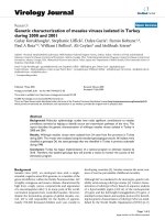
showed similar CAR-binding activity to unmodi-fied Ad5 vector in an ELISA-based binding assayFigure 2
Ad5.PTD
tat
showed similar CAR-binding activity to
unmodified Ad5 vector in an ELISA-based binding
assay. In the experiment, 10
9
VPs of each viral vector were
immobilized in the wells of a 96-well ELISA plate, and incu-
bated with increasing concentrations of recombinant sCAR
(extracellular domain of CAR, i.e. soluble CAR). The binding
activity was detected by AP activity conjugated to detection
antibodies.
0.0
0.1
0.2
0.3
0.4
0.5
0.6
0.7
0.8
0.9
1.0
0 50 100 150 200 250 300 350
sCAR concentration (ng/100µl)
OD405
Ad5
Ad5.PTD
tat
Diagram of PTD
tat
modified Ad5 vectorFigure 1
Diagram of PTD
tat
modified Ad5 vector. (A) PTD
tat
peptide incorporated into the fiber knob domain. (B) Struc-
tural diagram of Ad5 and Ad5.PTD
tat
vector. The PTD
tat
motif was incorporated at the C-terminal end of the fiber.
PTD
tat
peptide ( ): YGRKKRRQRRR
A
B
Ad5.PTD
tat
Ad5
Virology Journal 2007, 4:103 />Page 4 of 11
(page number not for citation purposes)
We thus examined whether PTD
tat
modification had any
effect on Ad5 binding to cells. To distinguish viruses
bound to cells from viruses internalized into the cells, we
performed a cell binding assay at 4°C since Ad internali-
zation occurs through receptor-mediated endocytosis
which is energy dependent, and is thus inhibited at 4°C
[5,7]. In the assay, Ad5.PTD
tat
or control Ad5 was incu-
bated with cells expressing different levels of CAR at 4°C
for 1 hour, and the bound viral particles were examined
by a quantitative PCR assay which assessed the viral
genome copies in the cell lysates. We found that Ad5.PTD-
tat
exhibited a significant higher cell-binding activity in
almost all of the cells we examined, including both high-
CAR and low-CAR containing cells. Shown in Fig. 3 are
results obtained in two representative cell lines: high-CAR
expressing Hela cells, and low-CAR expressing U118MG
cells [43,44].
Enhanced gene transfer efficacy of Ad5.PTD
tat
We further investigated the gene transfer efficacy of
Ad5.PTD
tat
in a variety of cultured cells using the reporter
GFP protein. Ad5.PTD
tat
vector or unmodified Ad5 was
used to infect cells at different multiplicities of infection
(MOIs). Two days after infection, we evaluated the trans-
gene expression using a fluorescent microscope and a flu-
orescent plate reader. We found that Ad5.PTD
tat
showed
more efficient gene delivery than unmodified Ad5 in all of
the cells tested (Fig. 4). In particular, Ad5.PTD
tat
exhibited
significantly higher gene transfer efficacy than unmodi-
fied Ad5 in the cells expressing low or medium levels of
CAR such as RD cells, U118MG cells, and D65MG cells
[43,44]. In high-CAR expressing cells that are readily
accessible to unmodified Ad5 vector, Ad5.PTD
tat
also
showed enhanced infectivity, presumably because
Ad5.PTD
tat
maintained the CAR-mediated infection path-
way while gaining extra targeting activity through the
PTD
tat
pathway (Fig. 4).
Identification of pathways mediating Ad5.PTD
tat
infection
Ad5.PTD
tat
showed enhanced gene delivery efficacy com-
pared to unmodified Ad5 vectors. To confirm that this
expanded tropism was mediated by the genetically incor-
porated targeting motif PTD
tat
, we performed a gene trans-
fer assay in the presence of competitive inhibitors. It has
been shown that the interaction between the positively
charged PTD
tat
and the negatively charged cell surface
epitopes such as heparan sulfate proteoglycans is essential
for PTD
tat
mediated protein transduction. Heparin, the
structural analogue of heparan sulfate, would thus be
expected to inhibit PTD
tat
mediated infection. In addition,
recombinant knob protein was used to block the native
CAR-mediated Ad5 infection because it compete with Ad5
vectors for cell surface CAR. In low-CAR containing
U118MG cells [44], due to the paucity of CAR, unmodi-
fied Ad5 showed poor gene transfer efficacy, and neither
knob nor heparin had any effect on Ad5-mediated trans-
gene expression (Fig. 5A). In contrast, Ad5.PTD
tat
exhib-
ited efficient gene delivery into U118MG cells, which was
completely inhibited by heparin, but not by the recom-
binant knob protein (Fig. 5A). These data demonstrated
PTD
tat
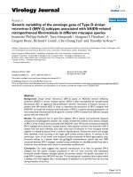
modification promoted Ad5 binding to cell surfacesFigure 3
PTD
tat
modification promoted Ad5 binding to cell
surfaces. Binding of Ad5 and Ad5.PTD
tat
were examined in
both high-CAR expressing Hela cells (A) and low-CAR
expressing U118MG cells (B) at 4°C. The amount of viruses
associated with the cells was determined by quantitative PCR
after DNA isolation from the cell lysate, and the viral copy
numbers were normalized to actin DNA in the samples. The
* indicates p < 0.05 and ** indicates p < 0.01 as analyzed by
the Student's t-test.
Hela cells
A
B
U118MG cells
0
50000
100000
150000
200000
250000
300000
Viral copy/ng actin DNA
Ad5 Ad5.PTD
tat
**
0
600000
Virus cop y /ng actin D N A
500000
300000
400000
200000
100000
*
Ad5 Ad5.PTD
tat
Virology Journal 2007, 4:103 />Page 5 of 11
(page number not for citation purposes)
Ad5.PTD
tat
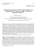
exhibited enhanced gene transfer efficacy in a variety of tumor cellsFigure 4
Ad5.PTD
tat
exhibited enhanced gene transfer efficacy in a variety of tumor cells. Gene transfer efficacy was evalu-
ated by use of a GFP reporter that was carried in the E1 region of each vector. In the assay, tumor cells expressing varying lev-
els of CAR were infected with either Ad5 or Ad5.PTD
tat
at an MOI of 100 or 500 VPs/cell, and GFP expression was examined
by fluorescence microscopy and a fluorescence plate reader. (A) Representative fluorescence images of low-CAR containing
cells (RD), medium-CAR containing cells (D65MG) and high-CAR expressing cells (Hela) that were infected with Ad5 or
Ad5.PTD
tat
at an MOI of 500 VPs/cell. (B) GFP expression in a variety of cells infected with either Ad5 or Ad5.PTD
tat
was quan-
tified using a fluorescence plate reader.
RD
10
100 500
MOI
10
4
10
5
10
3
10
2
A549
10
MOI
100 500
10
4
10
5
10
3
10
2
Hela
10
5
10
4
10
3
10
2
10
MOI 100 500
10
MOI 100 500
10
5
10
4
10
3
10
2
D65MG
Ad5
Ad5.PTD
tat
U118
10
10
4
10
5
100 500
MOI
10
3
10
2
Ad5
Ad5.PTD
tat
RD
D65MG
Hela
RD
D65MG
Hela
A
B
GFP intensity
GFP intensity
Virology Journal 2007, 4:103 />Page 6 of 11
(page number not for citation purposes)
Ad5.PTD
tat
infected low-CAR expressing cells mainly
through the incorporated PTD
tat
motif. In high-CAR con-
taining A549 cells [43], infection of unmodified Ad5 was
completely blocked by recombinant knob protein while
heparin had little effect, confirming that unmodified Ad5
mainly infected cells through the CAR pathway (Fig. 5B).
On the other hand, Ad5.PTD
tat
-mediated gene transfer
was partially blocked by either knob or heparin, but com-
pletely blocked in the presence of both knob and heparin,
suggesting Ad5.PTD
tat
could infect cells via both CAR and
the PTD
tat
motif (Fig. 5B).
In vivo gene transfer efficacy of Ad5.PTD
tat
We next examined whether the infectivity-enhanced vec-
tor Ad5.PTD
tat
could deliver enhanced gene transfer effi-
cacy in vivo. Since Ad5.PTD
tat
showed more profound
infectivity enhancement for low-CAR expressing tumor
cells in vitro, we assessed the in vivo gene delivery efficacy
of the Ad5 vectors using tumor models established with
low-CAR containing U118MG cells. After the tumors were
established subcutaneously in athymic (nude) mice, PBS,
unmodified Ad5, or Ad5.PTD
tat
vectors were injected into
the tumors. The gene delivery efficacy of each vector was
analyzed by non-invasive fluorescence imaging that
detected GFP expression in live mice. As shown in Fig. 6A,
Competitive inhibition assay showing the enhanced gene transfer efficacy of Ad5Figure 5
Competitive inhibition assay showing the enhanced gene transfer efficacy of Ad5.PTD
tat
was mediated by the
PTD
tat
motif. In this assay, recombinant knob protein (50 µg/ml) was used to block CAR-mediated viral infection, and heparin
(100 µg/ml) was used to block PTD
tat
mediated infection. Infections were performed at an MOI of 100 VPs/cell. (A) In low-
CAR expressing U118MG cells that were resistant to unmodified Ad5 vector, Ad5.PTD
tat
mediated efficient gene delivery and
the efficacy was completely inhibited by heparin, while recombinant knob had little effect, suggesting the enhanced infectivity of
Ad5.PTD
tat
in low-CAR expressing cells resulted from the PTD
tat
motif. (B) In high-CAR expressing A549 cells, Ad5.PTD
tat
mediated gene delivery was partially inhibited with either knob or heparin, while being completely inhibited in the presence of
both inhibitors, suggesting Ad5.PTD
tat
infected high-CAR expressing cells via both CAR and PTD
tat
pathways.
Virology Journal 2007, 4:103 />Page 7 of 11
(page number not for citation purposes)
Ad5.PTD
tat
-infected tumors showed more intensive green
fluorescence signals than Ad5-infected tumors, while no
signal was detected in PBS-injected tumors. Quantitative
analysis of the green fluorescence signals revealed that
Ad5.PTD
tat
-mediated GFP expression was significantly
higher than that of unmodified Ad5 vector in the tumors
(p < 0.01) (Fig. 6B). These data suggest the infectivity-
enhanced Ad5.PTD
tat
vector could be a useful vector for in
vivo gene delivery into tumors, which is essential for can-
cer gene therapy.
Discussion
In this study, we sought to improve the gene transfer effi-
cacy of Ad 5 vectors by genetic modification of the fiber
knob domain with a PTD
tat
motif. Our data demonstrated
the success of this strategy. The fiber modified Ad5 vector,
Ad5.PTD
tat
, not only exhibited enhanced gene delivery
efficiency of Ad5 vectors in low-CAR cells that are resistant
to unmodified Ad5 infection, but also in high-CAR cells
that are permissive to Ad5 infection. The enhanced infec-
tivity of Ad5.PTD
tat
was found to be mediated by targeting
of PTD
tat
to the negatively charged epitopes such as
heparan sulfate containing proteoglycans on cell surface.
In addition, we found PTD
tat
mediated Ad5.PTD
tat
infec-
tion is additive to native CAR-mediated infection as
assessed by competitive inhibition assays, which was not
unexpected since Ad5.PTD
tat
maintained full CAR-bind-
ing activity. More significantly, the enhanced gene deliv-
ery efficacy of Ad5.PTD
tat
was demonstrated in vivo using
low-CAR U118MG tumor models, and employment of a
recently developed non-invasive optical imaging system
PTD
tat
modification of Ad5 fiber enhanced in vivo gene delivery efficacy of the vectorFigure 6
PTD
tat
modification of Ad5 fiber enhanced in vivo gene delivery efficacy of the vector. In vivo gene delivery of
Ad5.PTD
tat
was examined using a non-invasive fluorescence imaging technique in low-CAR expressing tumor models. 10
10
VPs
of Ad5 or Ad5.PTD
tat
were injected into the subcutaneous U118MG tumors, and in vivo green fluorescence images were
acquired at different days post viral injection. (A) Representative in vivo images from PBS, Ad5, or Ad5.PTD
tat
injected mouse
tumor models at day 7 after vector administration. The colors representing different intensities of signal are shown on the
color bar. Ad5.PTD
tat
infection resulted in more intensive GFP signals than unmodified Ad5 vectors. (B) Quantitative analysis of
the GFP intensity in the tumor model of each group. The * marks significant differences (p < 0.01) as analyzed by the Student's
t-test.
PBS
Ad5
Ad5.PTD
tat
0
3.5×10
6
PBS Ad5
Ad5.PTD
tat
Total GFP intensity
3.0×10
6
2.5×10
6
2.0×10
6
1.5×10
6
1.0×10
6
5×10
5
*
A
B
Virology Journal 2007, 4:103 />Page 8 of 11
(page number not for citation purposes)
allowed us to visually detect the enhanced gene delivery in
vivo.
As a cell penetrating peptide, PTD
tat
is capable of travers-
ing the plasma membrane of mammalian cells. Since the
initial description that PTD
tat
is responsible for the ability
of the HIV Tat protein to enter mammalian cells, PTD
tat
has attracted tremendous interest as a drug delivery vehi-
cle [16-20]. Further interest has been stimulated by the
observation that PTDs can facilitate systemic delivery of
biologically active recombinant proteins in vivo [32-
35,37]. Since inefficient gene delivery into target cells has
been one of the major limitations in Ad5-mediated gene
therapy, in this study, we attempted to employ PTD
tat
pep-
tide to facilitate Ad5 mediated gene delivery. Employment
of PTDs to facilitate virus infection has been investigated
previously, but only using non-genetic methods [37,42].
In particular, chemically synthesized PTDs or bi-specific
adaptor proteins consisting of PTDs and the extracelluar
domain of CAR have been used to coat Ad vectors. These
strategies too resulted in enhanced gene delivery [37,42].
Compared to the non-genetic methods, our genetically
PTD
tat
modified vector has major advantages for two
major reasons: 1) genetic modification allows stable inter-
action between Ad5 and the PTD
tat
targeting epitope, thus
reducing the volatility associated with the affinity and sta-
bility of protein-protein interactions in the presence of
different environmental factors. This is critical especially
for in vivo applications; and 2) genetic modification does
not require production of peptides or fusion proteins
other than the viral vector, while large amounts of high
quality protein/peptide production is required for non-
genetic strategies (in addition to high quality production
of the viral vectors), which is especially important for in
vivo studies.
One issue associated with PTD
tat
-mediated protein deliv-
ery is the inefficient release of PTD
tat
fusion proteins from
the endosomal compartments [24,45-48]. It has been
demonstrated that a large proportion of the PTD
tat
fusion
protein remains trapped in non-cytosolic compartments
even though it is efficiently taken up by the cells. This
apparently would compromise the therapeutic effect of
the fusion protein. In our study, we examined the distri-
bution of Ad5.PTD
tat
particles in cells at various time
points (from 0.5 hour to 4 hours) following addition of
the viruses to the cells by immunofluorescent staining,
and found that the distribution of Ad5.PTD
tat
inside the
cells was similar to that of unmodified Ad5 vectors (data
not shown). This indicates endosomal trapping is not sig-
nificant, if any present at all, with Ad5.PTD
tat
infection of
cells. In addition, the enhanced gene delivery mediated by
Ad5.PTD
tat
confirmed that the virions were able to effi-
ciently escape the endosomal compartment.
The potential utility of the infectivity-enhanced Ad5.PTD-
tat
vector in cancer gene therapy was initially investigated
in this study using low-CAR expressing tumor models.
Indeed, many tumor cells have been shown to express
very low levels of CAR, which is partially responsible for
the low efficacy of Ad5 mediated cancer gene therapy in in
vivo studies, especially in clinical trials [1-3]. The ability of
Ad5.PTD
tat
to improve the gene delivery efficacy is attrib-
utable to the PTD
tat
motif, which binds to the negatively
charged motifs expressed on cell surface, in particular,
heparan sulfate containing proteoglycans that are widely
expressed in a variety of cells including tumor cells [49-
51]. In addition to cancer gene therapy, Ad5.PTD
tat
may
also be applied in other gene therapy applications where
infectivity-enhancement is beneficial. Infectivity-
enhanced vectors will not only allow efficient gene deliv-
ery into low-CAR target cells, but also allow use of a
reduced amount of viral vectors, thus reducing vector-
associated toxicity.
Previous studies have developed several other infectivity-
enhanced vectors, which include Ad5 vectors modified
with RGD, polylysine, or knobs from other Ad serotypes
[13-15,52]. Since each of the modified vectors uses a
unique extra targeting motif, the enhanced gene delivery
efficacy in a specific cell type depends on the expression of
individual receptors on its cell surface. Similar to PTD
tat
,
the polylysine epitope, which is composed of a stretch of
lysine residues, is highly basic, and can utilize heparan
sulfate as its receptor. Nonetheless, the interaction
between PTD
tat
and heparan sulfate is not only based on
ionic intereactions, but also on the specific structures of
the peptide and the proteoglycans [27-29]. Therefore, the
choice of an infectivity-enhanced vector needs to be deter-
mined for each specific application involving gene deliv-
ery enhancement.
Conclusion
Our data showed that a genetically modified Ad5 vector,
Ad5.PTD
tat
, maintained the ability to interact with its
native receptor CAR, and delivered transgenes into both
high-CAR and low-CAR cells more efficiently than the
unmodified Ad5 vector. Our data further showed
Ad5.PTD
tat
infected cells via both CAR and PTD
tat
path-
ways. More significantly, Ad5.PTD
tat
exhibited enhanced
gene delivery in vivo in a tumor model, and thus may be
useful for gene therapy applications involving low gene
delivery efficacy.
Methods
Cell culture
The human embryonic kidney 293 cells stably trans-
formed with Ad-E1 DNA, human lung carcinoma A549
cells, human cervix adenocarcinoma Hela cells, human
embryonic rhabdomyosarcoma RD cells, and human gli-
Virology Journal 2007, 4:103 />Page 9 of 11
(page number not for citation purposes)
oma D65MG and U118MG cells were all obtained from
the American Type Culture Collection (ATCC, Manassas,
VA). The 293 cells, A549 cells and U118MG cells were cul-
tured in Dulbecco's modified Eagle's medium/Ham's F12
medium (DMEM/F12) containing 10% fetal bovine
serum (FBS) and 2 mM L-glutamine. Hela cells were cul-
tured cultured in minimum essential Eagle medium
(MEM) containing 10% FBS and 2 mM L-glutamine. Both
RD and D65MG cells were cultured in DMEM containing
10% FBS and 2 mM L-glutamine. All of the cells were
maintained at 37°C in a 5% CO
2
humidified incubator.
Generation of the Ad5.PTD
tat
vector
Genetic modification of the Ad5 vector with PTD
tat
was
achieved using our previously established fiber modifica-
tion system [15]. In brief, the fiber shuttle vector contain-
ing a unique SnaBI restriction site immediately in front of
the stop code of the fiber gene, named pNEB.PK.SnaBI,
was used to generate a PTD
tat
modification. The sense and
antisense oligonucleotides encoding the PTD
tat
motif, 5'-
phos-ACT TTT TCA TAC ATT GCG CAA GAA GGC GGT
GGA GGG TAT GGC AGG AAG AAG CGG AGA CAG CGA
CGA AGA TAA TAA A-3' (sense) and 5'-phos-TTT ATT ATC
TTC GTC GCT GTC TCC GCT TCT TCC TGC CAT ACC
CTC CAC CGC CTT CTT GCG CAA TGT ATG AAA AAG T
-3' (antisense), were annealed and cloned into the fiber
shuttle vector pNEB.PK.SnaBI. This resulted in the fiber
modified shuttle vector pNEB.PK.PTD
tat
. In order to incor-
porate the modified fiber into an Ad5 genome,
pNEB.PK.PTD
tat
was linearized and recombined in
Escherichia coli (E. coli) BJ5183 with a linearized Ad5 back-
bone plasamid pVK50 that contained the CMV promoter
driven GFP reporter gene in its E1 region. After the posi-
tive recombinant plasmid, designated pAd5.PTD
tat
, was
identified, stable and high quality plasmid was obtained
from E. coli DH5α after re-transformation of the construct.
The modification was confirmed by sequencing analysis.
The modified virus Ad5.PTD
tat
was rescued and purified as
previously described [53]. In brief, the pAd5.PTD
tat
plas-
mid was digested with PacI (to release the viral genome),
purified, and transfected into 293 cells stably expressing
the complementary E1 genes. After the virus plaques
formed, they were amplified in 293 cells, and purified uti-
lizing a standard CsCl gradient protocol. The viral particle
(VP) titer was determined using a conversion factor of 1.1
× 10
12
VPs/ml per absorbance unit at 260 nm.
ELISA
The ELISA binding assay was performed essentially as
described [15]. In brief, 10
9
VPs of either Ad5 or Ad5.PTD-
tat
in 100 µl of 100 mM carbonate buffer (pH 9.5) was
immobilized in each well of a 96-well maxisorp plate
(Nunc, Roskilde, Denmark) by overnight incubation at
4°C. Following extensive washes with Tris-buffered saline
(TBS) containing 0.05% Tween 20 (TBS-Tween), and
blocking with 2% bovine serum albumin (BSA) in TBS-
Tween, the viruses were incubated with varying amounts
of purified recombinant sCAR. The binding of sCAR to the
viruses was detected by incubation with anti-CAR anti-
body (Santa Cruz Biotechnology Inc., Santa Cruz, CA),
followed by an AP-conjugated secondary antibody incu-
bation. AP activity reflecting the amount of bound sCAR
was determined using a color reaction with p-nitrophenyl
phosphate (Sigma, St. Louis, MO) as recommended by
the manufacturer. The absorbance at 405 nm (OD405)
was obtained using PowerWaveHT 340 microplate reader
(BioTek Instruments Inc., Winooski, VT).
Cell binding assay
Cells were cultured in 6-well plates until they were conflu-
ent. The plate was then cooled down on ice, and incu-
bated with Ad5 or Ad5.PTD
tat
at an MOI of 5000 VPs/cell
for one hour at 4°C. After washing cells twice with cold
phosphate buffered saline (PBS) on ice, the cells were col-
lected by incubation with Versene (0.53 mM EDTA). After
two more washes with PBS, the cells were lysed and proc-
essed to isolate DNA (Qiagen Inc., Valencia, CA). The viral
copy number in the DNA samples were obtained by quan-
titative PCR using primers designed for the E4 region of
adenoviral genome. The data were normalized against
actin DNA in each sample.
Gene transfer assay
Gene transfer efficacy of the viral vectors was assessed with
the use of GFP reporter. In the assay, cells were plated in
24-well plates with a density of 10
5
cells per well the day
before infection. Then the cells were infected with Ad5 or
Ad5.PTD
tat
at MOIs of 100 or 500 VPs/cell as described
previously [53]. Two days later, GFP expression was exam-
ined by fluorescence microscopy and quantified by a Syn-
ergy HT fluorescence plate reader (BioTek Instruments
Inc., Winooski, VT).
Competitive inhibition assays
Low-CAR U118MG cells or high-CAR A549 cells were
plated in 24-well plates at a density of 10
5
cells per well
the day before infection. Viruses equivalent to an MOI of
100 VPs/cell were used for each infection. To block cell
surface CAR, recombinant knob protein was pre-incu-
bated with cells at a final concentration of 50 µg/ml prior
to viral infection [54], and to block the PTD
tat
epitope, the
viruses were pre-incubated with 100 µg/ml of heparin
[15,54]. Two hours after infection, the cells were washed
with PBS, and refreshed with complete media containing
10% FBS. The cells were cultured for two days in the
humidified 37°C, 5% CO
2
incubator, and GFP micros-
copy was performed to examine the transgene expression.
Virology Journal 2007, 4:103 />Page 10 of 11
(page number not for citation purposes)
In vivo gene delivery
The subcutaneous tumors were established in athymic
nude mice using 1 × 10
7
U118MG cells per tumor per
mouse. After the tumors developed to ~0.5 cm in diame-
ter, PBS or 10
10
VPs of Ad5 or Ad.PTD
tat
were injected into
each tumor (n = 6). GFP expression was analyzed at 3, 7,
and 10 days post infection using a custom-built non-inva-
sive optical imaging system described previously [55]. The
mice were placed in the imaging chamber under anesthe-
sia with 3% isoflurane. Green fluorescence images were
acquired at f/8 with 20-second exposure using a combina-
tion of excitation filter HQ487/15× and emission filter
D535/30m (Chroma Technology, Rockingham, VT) sup-
ported by WinView32 software (Roper Scientific Inc.,
Trenton, NJ). All of the procedures involving animals
were approved by the Institutional Animal Care and Use
Committee of the University of Alabama at Birmingham
and performed according to their guidelines.
Competing interests
The author(s) declare that they have no competing inter-
ests.
Authors' contributions
TH participated in the generation and in vitro characteriza-
tion of the adenoviral vectors. YT carried out in vitro and
in vivo gene transfer assays. HU performed immunohisto-
chemistry studies. LEP participated in cell culture and
tumor model establishment. GPS helped in immunohis-
tochemical studies and in the preparation of the manu-
script. JLC assisted in the design of the study and
manuscript preparation. HW conceived of the study, par-
ticipated in its design and coordination, and drafted the
manuscript. All authors read and approved the final man-
uscript.
Acknowledgements
The authors thank Dr. Joel N. Glasgow for providing recombinant knob
protein and Minghui Wang for assistance in quantitative PCR analysis. This
work was supported by the NIH brain SPORE grant P50 CA097247 and the
Juvenile Diabetes Research Foundation grants 1-2005-71 and 5-2007-660.
References
1. Glasgow JN, Everts M, Curiel DT: Transductional targeting of
adenovirus vectors for gene therapy. Cancer Gene Ther 2006,
13(9):830-844.
2. Hedley SJ, Chen J, Mountz JD, Li J, Curiel DT, Korokhov N, Kovesdi
I: Targeted and shielded adenovectors for cancer therapy.
Cancer Immunol Immunother 2006, 55(11):1412-1419.
3. Rein DT, Breidenbach M, Curiel DT: Current developments in
adenovirus-based cancer gene therapy. Future Oncol 2006,
2(1):137-143.
4. Bai M, Harfe B, Freimuth P: Mutations that alter an Arg-Gly-Asp
(RGD) sequence in the adenovirus type 2 penton base pro-
tein abolish its cell-rounding activity and delay virus repro-
duction in flat cells. J Virol 1993, 67(9):5198-5205.
5. Bergelson JM, Cunningham JA, Droguett G, Kurt-Jones EA, Krithivas
A, Hong JS, Horwitz MS, Crowell RL, Finberg RW: Isolation of a
common receptor for Coxsackie B viruses and adenoviruses
2 and 5. Science 1997, 275(5304):1320-1323.
6. Louis N, Fender P, Barge A, Kitts P, Chroboczek J: Cell-binding
domain of adenovirus serotype 2 fiber. J Virol 1994,
68(6):4104-4106.
7. Wickham TJ, Mathias P, Cheresh DA, Nemerow GR: Integrins
alpha v beta 3 and alpha v beta 5 promote adenovirus inter-
nalization but not virus attachment. Cell 1993, 73(2):309-319.
8. Dmitriev I, Kashentseva E, Rogers BE, Krasnykh V, Curiel DT: Ecto-
domain of coxsackievirus and adenovirus receptor geneti-
cally fused to epidermal growth factor mediates adenovirus
targeting to epidermal growth factor receptor-positive cells.
J Virol 2000, 74(15):6875-6884.
9. Li HJ, Everts M, Pereboeva L, Komarova S, Idan A, Curiel DT, Her-
schman HR: Adenovirus tumor targeting and hepatic untar-
geting by a coxsackie/adenovirus receptor ectodomain anti-
carcinoembryonic antigen bispecific adapter. Cancer Res 2007,
67(11):5354-5361.
10. Tang Y, Han T, Everts M, Zhu ZB, Gillespie GY, Curiel DT, Wu H:
Directing adenovirus across the blood-brain barrier via
melanotransferrin (P97) transcytosis pathway in an in vitro
model. Gene Ther 2007, 14(6):523-532.
11. Watkins SJ, Mesyanzhinov VV, Kurochkina LP, Hawkins RE: The
'adenobody' approach to viral targeting: specific and
enhanced adenoviral gene delivery. Gene Ther 1997,
4(10):1004-1012.
12. Belousova N, Krendelchtchikova V, Curiel DT, Krasnykh V: Modula-
tion of adenovirus vector tropism via incorporation of
polypeptide ligands into the fiber protein. J Virol 2002,
76(17):8621-8631.
13. Dmitriev I, Krasnykh V, Miller CR, Wang M, Kashentseva E, Mikheeva
G, Belousova N, Curiel DT: An adenovirus vector with geneti-
cally modified fibers demonstrates expanded tropism via uti-
lization of a coxsackievirus and adenovirus receptor-
independent cell entry mechanism. J Virol 1998,
72(12):9706-9713.
14. Wickham TJ, Roelvink PW, Brough DE, Kovesdi I: Adenovirus tar-
geted to heparan-containing receptors increases its gene
delivery efficiency to multiple cell types. Nat Biotechnol 1996,
14(11):1570-1573.
15. Wu H, Seki T, Dmitriev I, Uil T, Kashentseva E, Han T, Curiel DT:
Double modification of adenovirus fiber with RGD and poly-
lysine motifs improves coxsackievirus-adenovirus receptor-
independent gene transfer efficiency. Hum Gene Ther 2002,
13(13):1647-1653.
16. Deshayes S, Morris MC, Divita G, Heitz F: Cell-penetrating pep-
tides: tools for intracellular delivery of therapeutics. Cell Mol
Life Sci 2005, 62(16):1839-1849.
17. Fittipaldi A, Giacca M: Transcellular protein transduction using
the Tat protein of HIV-1. Adv Drug Deliv Rev 2005, 57(4):597-608.
18. Joliot A, Prochiantz A: Transduction peptides: from technology
to physiology. Nat Cell Biol 2004, 6(3):189-196.
19. Snyder EL, Dowdy SF: Cell penetrating peptides in drug deliv-
ery. Pharm Res 2004, 21(3):389-393.
20. Wadia JS, Dowdy SF: Transmembrane delivery of protein and
peptide drugs by TAT-mediated transduction in the treat-
ment of cancer. Adv Drug Deliv Rev 2005, 57(4):579-596.
21. Ruben S, Perkins A, Purcell R, Joung K, Sia R, Burghoff R, Haseltine
WA, Rosen CA: Structural and functional characterization of
human immunodeficiency virus tat protein. J Virol 1989,
63(1):1-8.
22. Vives E, Brodin P, Lebleu B: A truncated HIV-1 Tat protein basic
domain rapidly translocates through the plasma membrane
and accumulates in the cell nucleus. J Biol Chem 1997,
272(25):16010-16017.
23. Ferrari A, Pellegrini V, Arcangeli C, Fittipaldi A, Giacca M, Beltram F:
Caveolae-mediated internalization of extracellular HIV-1 tat
fusion proteins visualized in real time. Mol Ther 2003,
8(2):284-294.
24. Fischer R, Kohler K, Fotin-Mleczek M, Brock R: A stepwise dissec-
tion of the intracellular fate of cationic cell-penetrating pep-
tides. J Biol Chem 2004, 279(13):12625-12635.
25. Fittipaldi A, Ferrari A, Zoppe M, Arcangeli C, Pellegrini V, Beltram F,
Giacca M: Cell membrane lipid rafts mediate caveolar endo-
cytosis of HIV-1 Tat fusion proteins. J Biol Chem 2003,
278(36):34141-34149.
26. Richard JP, Melikov K, Vives E, Ramos C, Verbeure B, Gait MJ, Cher-
nomordik LV, Lebleu B: Cell-penetrating peptides. A reevalua-
Publish with BioMed Central and every
scientist can read your work free of charge
"BioMed Central will be the most significant development for
disseminating the results of biomedical research in our lifetime."
Sir Paul Nurse, Cancer Research UK
Your research papers will be:
available free of charge to the entire biomedical community
peer reviewed and published immediately upon acceptance
cited in PubMed and archived on PubMed Central
yours — you keep the copyright
Submit your manuscript here:
/>BioMedcentral
Virology Journal 2007, 4:103 />Page 11 of 11
(page number not for citation purposes)
tion of the mechanism of cellular uptake. J Biol Chem 2003,
278(1):585-590.
27. Tyagi M, Rusnati M, Presta M, Giacca M: Internalization of HIV-1
tat requires cell surface heparan sulfate proteoglycans. J Biol
Chem 2001, 276(5):3254-3261.
28. Maccarana M, Casu B, Lindahl U: Minimal sequence in heparin/
heparan sulfate required for binding of basic fibroblast
growth factor. J Biol Chem 1993, 268(32):23898-23905.
29. Rusnati M, Tulipano G, Spillmann D, Tanghetti E, Oreste P, Zoppetti
G, Giacca M, Presta M: Multiple interactions of HIV-I Tat pro-
tein with size-defined heparin oligosaccharides. J Biol Chem
1999, 274(40):28198-28205.
30. Spillmann D, Witt D, Lindahl U: Defining the interleukin-8-bind-
ing domain of heparan sulfate. J Biol Chem 1998,
273(25):15487-15493.
31. Nagahara H, Vocero-Akbani AM, Snyder EL, Ho A, Latham DG, Lissy
NA, Becker-Hapak M, Ezhevsky SA, Dowdy SF: Transduction of
full-length TAT fusion proteins into mammalian cells: TAT-
p27Kip1 induces cell migration. Nat Med 1998,
4(12):1449-1452.
32. Asoh S, Ohsawa I, Mori T, Katsura K, Hiraide T, Katayama Y, Kimura
M, Ozaki D, Yamagata K, Ohta S: Protection against ischemic
brain injury by protein therapeutics. Proc Natl Acad Sci U S A
2002, 99(26):17107-17112.
33. Cao G, Pei W, Ge H, Liang Q, Luo Y, Sharp FR, Lu A, Ran R, Graham
SH, Chen J: In Vivo Delivery of a Bcl-xL Fusion Protein Con-
taining the TAT Protein Transduction Domain Protects
against Ischemic Brain Injury and Neuronal Apoptosis. J Neu-
rosci 2002, 22(13):5423-5431.
34. Orii KO, Grubb JH, Vogler C, Levy B, Tan Y, Markova K, Davidson
BL, Mao Q, Orii T, Kondo N, Sly WS: Defining the pathway for
Tat-mediated delivery of beta-glucuronidase in cultured
cells and MPS VII mice. Mol Ther 2005, 12(2):345-352.
35. Schwarze SR, Ho A, Vocero-Akbani A, Dowdy SF: In vivo protein
transduction: delivery of a biologically active protein into the
mouse. Science 1999, 285(5433):1569-1572.
36. Eguchi A, Akuta T, Okuyama H, Senda T, Yokoi H, Inokuchi H, Fujita
S, Hayakawa T, Takeda K, Hasegawa M, Nakanishi M: Protein trans-
duction domain of HIV-1 Tat protein promotes efficient
delivery of DNA into mammalian cells. J Biol Chem 2001,
276(28):26204-26210.
37. Gratton JP, Yu J, Griffith JW, Babbitt RW, Scotland RS, Hickey R,
Giordano FJ, Sessa WC: Cell-permeable peptides improve cel-
lular uptake and therapeutic gene delivery of replication-
deficient viruses in cells and in vivo. Nat Med 2003,
9(3):357-362.
38. Ignatovich IA, Dizhe EB, Pavlotskaya AV, Akifiev BN, Burov SV, Orlov
SV, Perevozchikov AP: Complexes of plasmid DNA with basic
domain 47-57 of the HIV-1 Tat protein are transferred to
mammalian cells by endocytosis-mediated pathways. J Biol
Chem 2003, 278(43):42625-42636.
39. Lewin M, Carlesso N, Tung CH, Tang XW, Cory D, Scadden DT,
Weissleder R: Tat peptide-derivatized magnetic nanoparticles
allow in vivo tracking and recovery of progenitor cells. Nat
Biotechnol 2000, 18(4):410-414.
40. Sandgren S, Cheng F, Belting M: Nuclear targeting of macromo-
lecular polyanions by an HIV-Tat derived peptide. Role for
cell-surface proteoglycans. J Biol Chem 2002,
277(41):38877-38883.
41. Torchilin VP, Rammohan R, Weissig V, Levchenko TS: TAT peptide
on the surface of liposomes affords their efficient intracellu-
lar delivery even at low temperature and in the presence of
metabolic inhibitors. Proc Natl Acad Sci U S A 2001,
98(15):8786-8791.
42. Kuhnel F, Schulte B, Wirth T, Woller N, Schafers S, Zender L, Manns
M, Kubicka S: Protein transduction domains fused to virus
receptors improve cellular virus uptake and enhance oncol-
ysis by tumor-specific replicating vectors. J Virol 2004,
78(24):13743-13754.
43. Seki T, Dmitriev I, Suzuki K, Kashentseva E, Takayama K, Rots M, Uil
T, Wu H, Wang M, Curiel DT: Fiber shaft extension in combina-
tion with HI loop ligands augments infectivity for CAR-nega-
tive tumor targets but does not enhance hepatotropism in
vivo. Gene Ther 2002, 9(16):1101-1108.
44. Van Houdt WJ, Wu H, Glasgow JN, Lamfers ML, Dirven CM, Gillespie
GY, Curiel DT, Haviv YS: Gene delivery into malignant glioma
by infectivity-enhanced adenovirus: in vivo versus in vitro
models. Neuro Oncol 2007, 9(3):280-290.
45. Albarran B, To R, Stayton PS: A TAT-streptavidin fusion protein
directs uptake of biotinylated cargo into mammalian cells.
Protein Eng Des Sel 2005, 18(3):147-152.
46. Al-Taei S, Penning NA, Simpson JC, Futaki S, Takeuchi T, Nakase I,
Jones AT: Intracellular traffic and fate of protein transduction
domains HIV-1 TAT peptide and octaarginine. Implications
for their utilization as drug delivery vectors. Bioconjug Chem
2006, 17(1):90-100.
47. Loison F, Nizard P, Sourisseau T, Le Goff P, Debure L, Le Drean Y,
Michel D: A ubiquitin-based assay for the cytosolic uptake of
protein transduction domains. Mol Ther 2005, 11(2):205-214.
48. Wadia JS, Stan RV, Dowdy SF: Transducible TAT-HA fusogenic
peptide enhances escape of TAT-fusion proteins after lipid
raft macropinocytosis. Nat Med 2004, 10(3):310-315.
49. Blackhall FH, Merry CL, Davies EJ, Jayson GC: Heparan sulfate pro-
teoglycans and cancer. Br J Cancer 2001, 85(8):1094-1098.
50. Davies EJ, Blackhall FH, Shanks JH, David G, McGown AT, Swindell R,
Slade RJ, Martin-Hirsch P, Gallagher JT, Jayson GC: Distribution
and clinical significance of heparan sulfate proteoglycans in
ovarian cancer. Clin Cancer Res 2004, 10(15):5178-5186.
51. Steck PA, Moser RP, Bruner JM, Liang L, Freidman AN, Hwang TL,
Yung WK: Altered expression and distribution of heparan sul-
fate proteoglycans in human gliomas. Cancer Res 1989,
49(8):2096-2103.
52. Kanerva A, Mikheeva GV, Krasnykh V, Coolidge CJ, Lam JT, Mahas-
reshti PJ, Barker SD, Straughn M, Barnes MN, Alvarez RD, Hemminki
A, Curiel DT: Targeting adenovirus to the serotype 3 receptor
increases gene transfer efficiency to ovarian cancer cells. Clin
Cancer Res 2002, 8(1):275-280.
53. Wu H, Dmitriev I, Kashentseva E, Seki T, Wang M, Curiel DT: Con-
struction and characterization of adenovirus serotype 5
packaged by serotype 3 hexon. J Virol 2002,
76(24):12775-12782.
54. Glasgow JN, Kremer EJ, Hemminki A, Siegal GP, Douglas JT, Curiel
DT: An adenovirus vector with a chimeric fiber derived from
canine adenovirus type 2 displays novel tropism. Virology 2004,
324(1):103-116.
55. Le LP, Le HN, Dmitriev IP, Davydova JG, Gavrikova T, Yamamoto S,
Curiel DT, Yamamoto M: Dynamic monitoring of oncolytic ade-
novirus in vivo by genetic capsid labeling. J Natl Cancer Inst
2006, 98(3):203-214.