Báo cáo sinh học: "The combined transduction of copper, zincsuperoxide dismutase and catalase mediated by cell-penetrating peptide, PEP-1, to protect myocardium from ischemia-reperfusion injury" ppt
Bạn đang xem bản rút gọn của tài liệu. Xem và tải ngay bản đầy đủ của tài liệu tại đây (5.89 MB, 11 trang )
RESEARC H Open Access
The combined transduction of copper, zinc-
superoxide dismutase and catalase mediated by
cell-penetrating peptide, PEP-1, to protect
myocardium from ischemia-reperfusion injury
Guang-Qing Huang
1,2
, Jia-Ning Wang
1,4*
, Jun-Ming Tang
1,3*
, Lei Zhang
1
, Fei Zheng
1
, Jian-Ye Yang
1
, Ling-Yun Guo
1
,
Xia Kong
1
, Yong-Zhang Huang
1
, Yong Liu
2
and Shi-You Chen
1,4
Abstract
Background: Our previous studies indicate that either PEP-1-superoxide dismutase 1 (SOD1) or PEP-1-catalase
(CAT) fusion proteins protects myocardium from ischemia-reperfusion-induced injury in rats. The aim of this study
is to explore whether combined use of PEP-1-SOD1 and PEP-1-CAT enhances their protective effects.
Methods: SOD1, PEP-1-SOD1, CAT or PEP-1-CAT fusion proteins were prepared and purified by genetic
engineering. In vitro and in vivo effects of these proteins on cell apoptosis and the protection of myocardium after
ischemia-reperfusion injury were measured. Embryo cardiac myocyte H9c2 cells were used for the in vitro studies.
In vitro cellular injury was determined by the expression of lactate dehydrogenase (LDH). Cell apoptosis was
quantitatively assessed with Annexin V and PI double staining by Flow cytometry. In vivo, rat left anterior
descending coronary artery (LAD) was ligated for one hour followed by two hours of reperfusion. Hemodynamics
was then measured. Myocardial infarct size was evaluated by TTC staining. Serum levels of myocardial markers,
creatine kinase-MB (CK-MB) and cTnT were quantified by ELISA. Bcl-2 and Bax expression in left ventricl e
myocardium were analyzed by western blot.
Results: In vitro, PEP-1-SOD1 or PEP-1-CAT inhibited LDH release and apoptosis rate of H9c2 cells. Combined
transduction of PEP-1-SOD1 and PEP-1-CAT, however, further reduced the LDH level and apoptosis rate. In vivo,
combined usage of PEP-1-SOD1 and PEP-1-CAT produced a greater effect than individual proteins on the
reduction of CK-MB, cTnT, apoptosis rate, lipoxidation end product malondialdehyde, and the infarct size of
myocardium. Functionally, the combination of these two proteins further increased left ventricle systolic pressure,
but decreased left ventricle end-diastolic pressure.
Conclusion: This study provided a basis for the treatment or prevention of myocardial ischemia-reperfusion injury
with the combined usage of PEP-1-SOD1 and PEP-1-CAT fusion proteins.
Introduction
Ischemic heart disease, especially acute myocardial
infarction (AMI), a primary myocardial disease charac-
terized by the loss of cardiomyocytes and the increase of
fibroblasts, i s an important c ause of heart f ailure. Early
reperfusion is an absolute prerequisite for the survival of
ischemic myocardium. However, reperfusion has been
referred as the “ double-edged sword” because repe rfu-
sion itself may lead to accelerated and additional myo-
car dial injury beyond that generated by ischemia, which
results in a spectrum of reperfusion-associat ed patholo-
gies, collectively called reperfusion injury [1].
Several mechanisms have been proposed to cause
reperfusion injury including formation of oxygen fr ee
radicals (OFR), calcium overload, neutrophils-mediated
myocardial and endothelial injury, progressive decline in
* Correspondence: ;
1
Institute of Clinical Medicine and Department of Cardiology, Renmin
Hospital, Hubei University of Medicine, Shiyan, Hubei 442000, China
Full list of author information is available at the end of the article
Huang et al. Journal of Translational Medicine 2011, 9:73
/>© 2011 Huang et al; licensee BioMed Central Ltd. This is an Open Access article distributed under the terms of the Creative Commons
Attribution License (http://creati vecommons.org/licenses/by/2.0), which permits unrestricted use, distribution, and reproduction in
any medium, provid ed the original wor k is properly cited.
microvascular flow to the reperfused myocardium, or
depletion of the high-energy phosphate store [2].
Among these factors, overproduction of OFR during the
first few minutes of reperfusion is considered as a key
event. About 25% of cell death in cardiomyocytes after
reperfusion of a cute myocardial infarction is caused by
reperfusion injury [3]. OFR includes superoxide anion
(O
2
-
), hydroxyl radical (OH
-
), hydrogen peroxide
(H
2
O
2
), etc. Excessive OFR causes cell DNA breakage,
degeneration, and lipid peroxidation, ultimately leading
to cell death. The key antioxidant enzymes, including
superoxide dismutase (SOD), catalase (CAT) and glu-
tathione peroxidase (GPx), provide a defense system
against oxidative stress by removing the OFR, thus pro-
tecting cells from oxidative damage [4,5]. However, the
endogenous antioxidant activity is severely damaged
after ischemia-reperfusion which makes the myocardium
extremely vulnerable to OFR [6]. Moreover, exogenous
SOD1 and CAT can not be delivered into living cells
because of the poor permeability and selectivity of the
cell membrane, which has limited its usage in protecting
cells/tissues from oxidative stress damage.
There is a growing effort to circumvent these pro-
blems by designing strategies to deliver full-length pro-
teins into a large number of cells. Morris Group [7]
designed and synthesized a new type of cell penetrating
peptide PEP-1, which consists of three domains: a
hydrophobic tryptophan rich motif (KETWWETWW-
TEW), a spacer (SQP), and a hydrophilic lysine-rich
domain (KKKRKV). Pep-1 is cationic and adopts amphi-
pathic a-helical structure on the membrane. These char-
acteristics are similar to those of cationic antimicrobial
peptides involved in host innate immunity, s uggesting
that PEP-1 can kill microbes [8-10]. In addition, Park’s
study [11] shows that PEP-1 has antichlamydial activity.
More importantly, many studies have demonstrated the
successful delivery of full-length PEP-1 fusion proteins
into cultured cells and the nervous system by protein
transduction technology including EGFP, b-Gal, full-
length specific antibodies, human copper chaperone for
Cu, Zn-SOD, CAT and SOD [7,12,13]. Our previous
studies indicate that PEP-1-SOD1 or PEP-1-CAT fusion
proteins can be transduced into myocardial tissues to
protect myocardium from ischemia-reperfusion-induced
injury in rats [12,14]. Cu, Zn- superoxide dismuta se (Cu,
Zn-SOD, also called SOD1) only catalyzes the dismuta-
tion of O
2
-
into H
2
O
2
and O
2
. Elimination of H
2
O
2
requires the endogenous CAT or GPx activity, which
removes H
2
O
2
by breaking it down into H
2
OandO
2
,
thus preventing the generation of OH
-
. Therefore, we
hypothesize that combination of PEP-1-SOD1 and PEP-
1-CATfusionproteinsismoreusefulinpreventing
myocardium from ischemia-reperfusion injury.
Materials and methods
The present st udy conformed to the Guide for the Care
and Use of Laboratory Animals published by the US
National Institutes of Health (NIH Publication number
85-23, revised 1985). The animal use protocol was
approved by the Institutional Animal Care and Use
Committee of Hubei University of Medicine.
Expression, purification and transduction of PEP-1-SOD1
and PEP-1-CAT
Four prokaryotic expression plasmids with His-tag,
pET15b-SOD1-His, pET15b-PEP -1-SOD1-His, pET15b-
CAT-His, and pET15b-PEP-1-CAT-His were con-
structed by the TA-cloning method. The recombinant
plasmids were transformed into E.coli BL21 (DE3)
(Novagen, USA). The transformed bacteria were grown
in 100 ml LB medium at 3 7°C to an OD600 value of
0.5-1.0 and induced with 0.5 mM isopro pyl-b-D-thioga-
lactosid e (IPTG) (Promega, USA) at 25°C for 12 h. Bac-
teria were lysed by sonication at 4°C in a bin ding buffer
(5 mM imidazole, 500 mM NaCl, 20 mM Tris-HCl, pH
7.9). To purify the recombinant fusion proteins, cell
lysates were loaded onto a Ni
2+
-nitrilotriacetic a cid
sepharose affinity column (Qiagen, USA) under native
conditions. After the column was washed with 10
volumes of the binding buffer and 6 volumes of wash
buffer (60 mM imidazole, 500 mM NaCl, 20 mM Tris-
HCl, pH 7.9), the fusion proteins were eluted using an
eluting buffer (1 M imidazole, 500 mM NaCl, 20 mM
Tris-HCl, pH 7.9). The fusion-protein-containing frac-
tions were combined, and the salts were removed using
a PD-10 column. Protein concentrat ions were measured
by the Bradford method [15].
Cell culture
H9c2 cells, derived from embryonic heart tissue (Ameri-
can Type Culture Collection, Manassas, VA), were cul-
tured in Dulbecco ’ s modified Eagle’smedium(DMEM,
Invitrogen) with 5 g/L glucose supplemented with 15%
(v/v) fetal bovine serum (FBS, Hangzhou Sijiqing Bio lo-
gical Engineering Materials Co. Ltd., China). Cells were
routinely grown to subconfluency (> 90% by visual esti-
mate) in 75 cm
2
flasks at 37°C in a humidified atmo-
sphere of 5% CO
2
prior to passage and seeding for
experiments.
Transduction of PEP-1-SOD1 and PEP-1-CAT fusion
protein into H9c2 cells
H9c2 cells were grown to confluence on 25 cm
2
flasks
and pretreated with PEP-1- SOD1-His or PEP-1-CAT-
Hisatdifferentdoses(0.5~2.0μM)for15min~72h.
The cells were then washed with phosphate-buffered
saline (PBS) and treated with trypsin- EDTA followed by
Huang et al. Journal of Translational Medicine 2011, 9:73
/>Page 2 of 11
lysate preparation for western blot or enzyme activity
assay. The SOD and CAT activity were measured using
SOD and CAT kits by following the manufacturer’s pro-
tocols (JianCheng Bioengineering Institute, China).
Immunocytochemistry
To directly visualize the transduction of PEP-1-SOD1
and PEP-1-CAT fusion protein into H9c2 cells, cells
were treated with 2 μM of control SOD1, purified PEP-1-
SOD1, CAT, or PEP-1-CAT. After 1 or 6 h of incubation
at 37°C, the cells were washed twice with 1 × PBS and
fixed with 4% paraformaldehyde for 15 min at room tem-
perature. Immunocytochemis try was performed by incu-
bation with specific primary a ntibodies: rabbit anti-
polyhistidine (diluted 1:200) (Santa Cruz Biotechnology,
USA) or mouse anti-Troponin T (diluted 1:200) (Santa
Cruz Biotechnology, USA) at 4°C overnight. Cells were
then incubat ed with TRITC-conjugated rat anti-rabbi t Ig
G (diluted 1:250) or FITC-c onjugated goat anti-mouse Ig
G (diluted 1:250) at 25°C for 2 h. Nuclei were stained
with DAPI (Sigma, USA). The immunoreactions were
observed under a fluorescent microscope (Nikon, Japan).
Hypoxia-reoxygenation treatment of H9c2 Cells
For the protective effect of combined pretreatment of
PEP-1-SOD1 and PEP-1-CAT on H9c2 cells, cells were
pretreated with or without PEP-1-SOD1 (2 μM) for 1 h
or PEP-1-CAT (2 μM) for 6 h. Then, medium was chan-
ged to DMEM containing 1 g/L glucose and 1% FBS.
Cells were cultured in a humidified hypoxia chamber
(Stem Cell Technology, USA) and flushed with 9 5% N
2
+5%CO
2
to achieve 0.1% oxygen environment. The
sealed chamber was placed into a 37°C incubator for 21
h. After hypoxia incubation, the cells were reoxygenized
with fresh medium and incubation in 95% air + 5% CO
2
for 6 h [16]. Control cells were kept in normoxic cond i-
tions for the corresponding times. The supernatants and
cells were collected respectively after treatment.
Annexin V and propidium iodide (PI) binding assay
To measure H9c2 cell apoptosis after hypoxia-reoxy-
genation treatment, we labeled the cells with Annexin V
and PI fluorescein (Bender MedSystems , Austria). Cells
were washed with 1×PBS, and suspended in 200 μl
1×binding buffer (10 mM HE PES pH 7.4, 140 mM
NaCl, 2.5 mM CaCl
2
)/1×10
6
/L cells. Cells were then
incubated with Annexin V (1:20) for 3 min followed by
PI for 15 min. The apoptosis rate was evaluated by Flow
cytometry.
Transduction of PEP-1-SOD1 and/or PEP-1-CAT in rat
myocardium and ischemia-reperfusion injury
To observe whether transduced PEP-1-SOD1 and PEP-1-
CAT protect myocardial ischemia-reperfusion injury in
vivo, we e stablished the model of myocardial ischemia-
reperfusion injury in rats. 240-280 g male Sprague-Daw-
ley rats we re obtained from the Experiment Animal Cen-
ter at Hubei University of Medicine and housed at an
appropriate temperature (25°C) and relative humidity
(55%) with a fixed 12 h light/dark cycle and free access to
food and water. The animals were randomly divided into
five groups as follows: sham-operated group, ischemia-
reperfusion injury group (I/R), PEP-1-SOD1 pretreat-
ment (2 mg/Kg), PEP-1-CAT pretreatment (2 mg/Kg),
and PEP-1-SOD1 (2 mg/Kg) + PEP-1-CAT (2 mg/Kg)
pretreatment (n = 20 for each group). The animals were
anesthetized with 10% chloral hydras (250 mg/kg, i.p.)
and ventilated during the LAD coronary artery ligation.
Surgery was performed under sterile conditions. One
hour after pretreatment with PEP-1-SOD1 and/or PEP-1-
CAT (i.p.), the left anterior descending coronary artery
(LAD) was ligated for one hour followed by two hours of
reperfusion as described previously [12,14].
Measurement of creatine kinase (CK), CK-MB activity,
cardiac troponin T (cTnT), and malondialdehyde (MDA)
levels
Rat serum was obtained after centrifugation of blood
samples at 3,500 rpm for 15 min. CK activities were
measured by spectrophotometry at 340 nm [12]. Malon-
dialdehyde (MDA), an end product of peroxidation of
cell membrane lipids caused by OFR, is considered as a
reliable marker of cardiomyocyte oxidative damage.
MDA level w as determined by measuring chromogen
generation from the reaction of MDA with 2-thiobarbi-
turic acid. The CK and MDA biochemical analyses were
performed using commercial kits (J ianCheng Bioengi-
neering I nstitute, China). CK-MB activity and cTnT
levels were quantified by ELISA (Rapidbio, USA).
Western blot
Rat hearts w ere transected along the LAD ligature to
separate ischemic tissue and remote myocardium. Heart
tissues were lysed in lyses buffer. Western blot analysis
was performed using procedures established in our
laboratory. The following antibodies were used: rabbit
anti-Bax (Santa Cruz Biotechnology), mouse anti-Bcl-2
(Santa Cruz Biotechnology), and peroxidase-conjugated
secondary a ntibody (Sigma). The bands were visualized
using the enhanced chemiluminescence (Sigma).
Measurement of hemodynamics
Hemodynamic measurement was performed as
described previou sly [12,14]. Briefly, after 2 hours of
reperfusion, left carotid artery and femoral artery were
exposed. Two catheters filled with heparinized (10 U/
ml) saline solution were connected to a Statham pres-
sure transducer (Gould, Saddle Brook, USA). The
Huang et al. Journal of Translational Medicine 2011, 9:73
/>Page 3 of 11
carotid arterial catheter was advanced into the left ven-
tricle to record ventricular pressure for 3~5 min. The
femoral artery catheter was inserted into an isolated
femoral artery to monitor hemodynamics. Hemody-
namic parameters were monitored simultaneously and
recorded on a thermal pen-writing recorder (RJG-4122,
Nihon Kohden, Japan) and on an FM magnetic tape
recorder (RM-7000, Sony, Japan).
Evaluation of infarct Size
Six or seven hearts in each group were used for this
experiment. After 2 hours of reperfusion, the hearts were
removed and treated with K-H buffer at room tempera-
ture for 3 minutes, and then frozen at -20°C for 1 h fol-
lowe d by transverse sectioning into 4 parts (thickness, 2-
5 mm). Sections were incubated in 1% 2, 3, 5-triphenylte-
trazolium chloride (TTC) at 37°C for 15 minutes. TTC
did not stain the infarcted myocardium, thus showing
whiteincolorwhilenon-ischemicmyocardiumwas
stained by TTC and showed brick-red in color. In the
ischemia-reperfusion hearts, the left ventricle was at risk
of infarction, the total and infarcted areas of left ventricle
were measured using planimeter in a double-blinded
manner. The volumes of the infarcted zone were calcu-
lated by multiplying the planimetered areas by slice
thickn ess. Infarcted volume was expressed as th e percen-
tage of left ventricular volume for each heart.
Statistical analysis
All data are expressed as means ± SD. Differences
between g roups were determined with unpaired Student
t-test and one-way analysis of variance followed by a
Newman-Keuls post hoc test. Probability values of P <
0.05 were considered to be significant.
Result
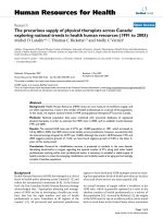
Expression and purification of PEP-1-SOD1 and PEP-1-CAT
fusion protein
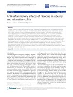
pET15b-SOD1-His, pET15b-PEP -1-SOD1-His, pET15b-
CAT-His and pET15b-PEP-1-CAT-His were successfully
expressed and purified as shown in Figure 1A. The
results indicated that the purified p roteins had the cor-
rect molecular mass: i.e., SOD1, 22 KDa; PEP-1-SOD1,
26 KDa, CAT and PEP-1-CAT: 69 KDa. In addition,
their enzyme activities were 356.98 U/mg, 355.54 U/mg,
3.18×10
3
U/g, 3.22×10
3
U/g, respectively. These data
suggest that fusion proteins PEP-1-SOD1-His or PEP-1-
CAT-His had the similar enzymatic activities as the wild
type SOD1 or CAT.
Transduction of PEP-1-SOD1 or PEP-1-CAT into H9c2 cell
The subcellular transduction of PEP-1-SOD1 or PEP-1-
CAT fusion protein into H9c2 cells was confirmed by
direct fluore scence analysis. As shown in Figure 1B,
almost all cultured cells were transduced with PEP-1-
SOD1 or PEP-1-CAT fusion pro teins. However, the red
fluorescent signa ls were not detected in cells treated
with control SOD1 or CAT.
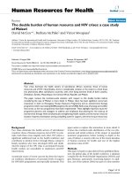
To further investigate the transduction efficiency of
PEP-1-SOD1 and PEP-1-CAT fusion proteins, we incu-
bated H9c2 with 2 μM of PEP-1-SOD1 or PEP-1-CAT
fusion proteins in cell culture medium at different time
intervals, and analyzed the cellular fusion protein levels
by western blotting. The intracellular fusion proteins
were detected within 15 min and gradually increased
until 60 min (PEP-1-SOD1) or 360 mi n (PEP-1-CAT)
(Figure 2a and 2A). Moreover, the fusion proteins were
transduced into H9c2 cells in a dose-depende nt manner
(Figure2,b,c,B,C).ThewildtypeSOD1orCATwas
not transduced into the cells (Figure 2)
It is essential that transduced PEP-1-SOD1 or PEP-1-
CAT fusion proteins in cells retai n their e nzymatic
activity. Therefore, we detected the SOD1 or catalase
activities. As s hown in Figure 2, the enzymatic activity
of S OD1 or CAT in transduced cells increased in a
dose- and time-dependent manner. Nearly seven
(SOD1) or five fold (CAT) increase was observed in
groups treated with PEP-1-SOD1 (Figure 2, d, e, f) or
PEP-1-CAT (2 μM) (F igure 2, D. E, F), but not with the
control SOD1 or CAT. These results demonstrate that
the PEP-1-SOD1 or PEP-1-CAT fusion proteins were
not only able to be transduce d into H9c2 cells, but also
was the transduced proteins able to retain their enzy-
matic activities for at least 48 h.
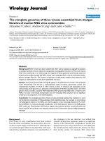
PEP-1-SOD1 and PEP-1-CAT decreased LDH levels and
inhibited H9c2 cell apoptosis in vitro
LDH level is an indicator of cellular injury. Compared to
H/R group, LDH levels were decreased in PEP-1-SOD1
or PEP-1-CAT-treated groups . However, the reduction
of LDH levels was greater in the groups with both PEP-
1-SOD1 and PEP-1-CAT, as compared to individual
protein-treated groups (Figure 3A).
In the normoxia environment, H9c2 cells apoptosis
was not significantly different among pretreatment with
PEP-1-SOD1 and/or PEP-1-CAT a nd control group.
However, apopt osis rate of the control cells increased to
83.8% after treated with hypoxia-reoxygenation. The
apoptosis was significantly reduced in cells treated with
PEP-1-SOD1 or PEP-1-CAT. Combined use of PEP-1-
SOD1 and PEP-1-CAT further inhibited the apoptosis
(Figure 3B).
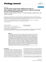
PEP-1-SOD1 and PEP-1-CAT suppressed CK, CK-MB, cTnT
and MDA levels in vivo
The animal survival rate after surgery in different groups
was as follows: 100% in sham group, 57.7% in I/R group,
66.1% in PEP-1-SOD1+PEP- 1-CAT group, 61.5% in
Huang et al. Journal of Translational Medicine 2011, 9:73
/>Page 4 of 11
PEP-1-CAT group, and 59.3% in PEP-1-SOD1 group.
The activiti es of s erum CK, CK-MB and cTnT were
used to monitor the myocardial dam age. MDA levels
reflect cardiomyocyte oxidative damage. Compared to
the sham group, CK, CK-MB activity, cTnT and MDA
levels were markedly increased du e to ischemia-r eperfu-
sion injury, but decreased after PEP-1-SOD1 or PEP-1-
CAT treatment. Importantly, combined usage of PEP-1-
SOD1 and PEP-1-CAT further suppressed CK, CK-M B
activity, cTnT and MDA levels (Figure 4).
PEP-1-SOD1 and PEP-1-CAT altered the expression of
apoptosis proteins in vivo
Bcl-2, an anti-apoptotic protein, promotes cell growth,
while Bax, a pro-apop totic pro tein memb er of Bcl-2
family, accelerates apoptosis. Western blot analysis
showed that Bcl-2 expression was markedly increased,
while Ba x expression was markedly decreased in PEP-1-
SOD1 or PEP-1-CAT-treated hearts (P <0.05),ascom-
pared to I/R group (P < 0.05). Bcl-2 expression was
further increased by the treatment with both PEP-1-
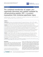
Figure 1 Transduction of purified PEP-1-SOD1 or PEP-1-CAT into H9c2 cells. A: purific ation of PEP-1-SOD1-His and PEP-1-CAT-His fusion
proteins. Purified fusion proteins were analyzed by western blot with rabbit anti-polyhistidine antibody. B: H9c2 cells were treated with 2 μM
purified His-tagged PEP-1-SOD1, wild type SOD1, PEP-1-CAT, or wild type CAT proteins for 6 h. Cells were incubated with rabbit-anti-
polyhistidine and mouse-anti Troponin T (cardiomyocyte marker) antibodies (cTnT), and then visualized with fluorescent microscopy. Red
fluorescent signals represent TRITC-labeled His-tag of SOD1 or CAT, Green fluorescent signals represent FITC-labeled Troponin T; Blue fluorescent
signals represent DAPI-labeled nuclei.
Huang et al. Journal of Translational Medicine 2011, 9:73
/>Page 5 of 11
SOD1 and PEP-1-CAT although Bax expression seems
no significant changes. However, Bcl-2/Bax ratio in
PEP-1-SOD1 and PEP-1-CAT-treated groups was signif-
icantly larger than the treatment with individual pro-
teins ( Figur e 5). These data suggest that combination of
PEP-1-SOD1 and P EP-1-CAT further i nhibited ische-
mia-reperfusion-induced apoptosis.
PEP-1-SOD1 and PEP-1-CAT decreased infarct size and
improved left ventricular (LV) function
To investigate whether the transduced PEP-1-SOD1 and
PEP-1-CAT fusion proteins are biologically active in
vivo, we measured the effects of PE P-1-SOD1 and PEP-
1-CAT on myocardial i nfarct size with 1% TTC staining
of the rat hearts with myocardial ischemia-reperfusion
Figure 2 Transduction and enzyme activities of PEP-1-SOD1 and PEP-1- CAT fusion proteins in H9c2 cells.(a-c):Timeanddose-
dependent transduction of PEP-1-SOD1. Control or 2 μM SOD1 was added into the culture medium for 15~60 min (a-Short time course); 0.5~2
μM PEP-1-SOD1 or control SOD1 was added to the culture medium for 1 h (b-dose dependent); or cells pretreated with 2 μM PEP-1-SOD1 were
incubated for different times (1~48 h) (c-longer time course). Western blots were performed using anti-His antibody. (A~C): Time and dose-
dependent transduction of PEP-1-CAT. 2 μM PEP-1-CAT or control CAT was added into the culture medium for 15~360 min (A-shorter time
course); 0.5~2 μM PEP-1-CAT or control CAT was added to the culture medium for 6 h (B-dose-dependent); or cells pretreated with 2 μM PEP-1-
CAT were incubated for different times (6~72 h) (C-longer time course). Western blots were performed using anti-His antibody. (d~f): Enzymatic
activity of PEP-1-SOD1. (D~F): Enzyme activity of PEP-1-CAT. Results are mean ± SD, n = 5, *P < 0.01,
#,$
P < 0.05 vs control (CTL) group in each
individual set of experiments.
Huang et al. Journal of Translational Medicine 2011, 9:73
/>Page 6 of 11
Figure 3 Effect of PEP-1-SOD1 and PEP-1-CAT on LDH level and apoptosis rate. (A) Effect of PEP-1-SOD1 and PEP-1-CAT on LDH level. *P <
0.01 vs. control (CTL) group;
$
P < 0.01 vs H/R group;
#
P < 0.01 vs PEP-1-SOD1 group;
@
P < 0.01 vs PEP-1-CAT group (n = 5). (B) Effect of PEP-1-
SOD1 and PEP-1-CAT on apoptosis of H9c2 cells under hypoxia-reoxygenation injury. H9c2 cells were pretreated with PEP-1-CAT for 6 h and/or
PEP-1-SOD1 for 1 h. The cells were then placed in a normoxia environment for 27 h or in hypoxia chamber for 21 h followed by 6 h of
reoxygenation. Apoptosis was measured by staining the cells with Annexin V and PI followed by Flow cytometry. The apoptosis rates are shown.
Figure 4 Effects of PEP-1-SOD1 and PEP-1-CAT on CK, CK-MB, cTnT and MDA content after myocardial ischemia-reperfusion. CK activity (A)
and MDA levels (D) were measured as described in Materials and Methods. CK-MB activity (B) and cTnT (C) levels were quantified by ELISA. *P <0.01
and
!
P < 0.05 vs sham group;
#
P <0.01and
$
P < 0.05 vs I/R group;
&
P <0.05vsPEP-1-SOD1group;
@
P < 0.01 vs PEP-1-CAT group. n = 6.
Huang et al. Journal of Translational Medicine 2011, 9:73
/>Page 7 of 11
injury. Compared to I/R group (48.56 ± 4.63%), infarct
size were reduced in rats pretreated with PEP-1-SOD1
(27.14 ± 4.10%) or PEP-1-CAT (30.12 ± 4.78%). The
combined usage of both PEP-1-SOD1 and PEP-1-CAT
had a much greater effect on the reduction of the necro-
tic area (20.38 ± 3.86%) (Figure 6). These results ind i-
cate that combination of PEP-1-SOD1 and PEP-1-CAT
can more effectively decrease infarct size.
In vivo hemodynamic measurements showed that the
LV function was significantly improved in hearts treated
with PEP-1-SOD1 or PEP-1-CAT compared to I/R
hearts. Treatment with both PEP-1-SOD1 and PEP-1-
CAT resulted in a larger increase of LVSP and ± dp/
dt
max
, and a lower LVEDP compared to PEP-1-SOD1 or
PEP-1-CAT individually-treated hearts (Figure 7).
Discussion
Ischemic heart disease, a maj or cause of mortali ty in
developed countries, is characterized by interrupted
blood s upply to the myocardium that leads to tissue
necrosis. The treatment of this condition allows the
rapid return of blood flow to the ischemic zones of the
myocardium. However, reperfusion may cause further
complications such as decreased cardiac contractile
function and arrhythmias. Therefore, the developments
of cardioprotective agents, which may delay the onset
of necrosis during ischemia-reperfusion, lessen the
necrotic tissue mass, improve myocardial function and
decrease the incidence of arrhythmias is of great clini-
cal relevance. The exact cellular mechanisms of ische-
mia-reperfusion injury are still a question of debate,
however, among several other mediators, superoxide
(O
2
-
), nitric oxide (NO), and peroxynitrite (ONOO
-
)
play a major role in ischemia-reperfusion injury
[17,18]. Furthermore, there is good evidence that OFR,
such as superoxide anions, hydroxyl radicals and
hydrogen peroxide, mediate pathphysiology of human
diseases [19-21]. OFR also prompts vascular smooth
muscle cell migration and proliferation causing intimal
hyperplasia and remodeling, eventually leading to
artery restenosis after arterial balloon angioplasty [22].
A few studies suggest that there is continuous OFR-
mediated oxidative stress injury after acute myocardial
infarction treated by percutaneous coronary interven-
tion (PCI) [23]. Another study shows that overexpres-
sion of Cu/Zn-SOD and/or catalase in ApoE-deficient
mice suppresses benzo (a) pyrene-accelerated athero-
sclerosis [24]. Gene therapy is considered to be a pro-
mising approach, but some key problems of gene
therapy have not be en fundamenta lly resolv ed, includ-
ing the efficiency of gene transfer, control of gene
expression, effectiveness, security. Therefore, it is
important to find new ways to modify antioxidant
enzyme for the efficient introduction into cells.
Figure 5 Effect of PEP-1-SOD1 and PEP-1-CAT on Bcl-2 and Bax expression. Bcl-2 and Bax expression was detected by Western blot as
described in Materials and Methods (A), and quantified by normalization to tubulin (B and C). Bcl-2/Bax ratio (D) was calculated by dividing the
normalized expression of Bcl-2 by Bax. *P < 0.01 and
#
P < 0.05 vs I/R group;
&
P < 0.05 vs PEP-1-SOD1 group;
$
P < 0.01 vs PEP-1-CAT group. n = 6.
Huang et al. Journal of Translational Medicine 2011, 9:73
/>Page 8 of 11
The antioxida nt enzymes ( SOD1 and CAT) ha ve the
potential to prevent OFR-mediated tissue damage, but
they cannot fre ely pass the cell membrane, whi ch limits
their applications. In this study, the human SOD1 and
CAT gene were fused with a PEP-1 peptide to produce
PEP-1-SOD1 and PEP-1-CAT fusion proteins. These
fusion proteins can be transduced into cells and main-
tain their enzymatic activities. Our in vitro studies
demonstrate that PEP-1-SOD1 and PEP-1-CAT together
generate greater inhibitory e ffects than individual pro-
teins on LDH release and apop tosis r ates in cardiomyo-
cyte H9c2 cells. The lev els of LDH in the sup ernata nts
and the apoptosis of cells were indicators of hypoxia-
reoxygenation injury [25,26].
Our previous studies have shownthatapplicationof
PEP-1-SOD1 or PEP-1-CAT can tranduced into myo-
cardium and protected against myocardial ischemia-
reperfusion injury in rats [12,14]. To examine the com-
bined effect of PEP-1-SOD1 and PEP-1-CAT, we applied
both of PEP-1-SOD1 and PEP -1-CAT to rats with
myocardial ischemia-reperfu sion injury. CK CK- MB and
cTnT are widely present in the cytoplasm of myocardial
cell s, and elevation of serum CK, CK-MB and cTnT are
reliable indicators of myocardium injury [12,27]. MDA
can be detected at a very early time of an injury, and is
a reliable marker of myocardium oxidative damage.
PEP-1-SOD1 or PEP-1-CAT reduced the increase of
serum CK, CK-MB, cTnT and myocardial MDA levels
caused by myocardial ischemia-reperfusion injury. How-
ever, combination of PEP-1-SOD1 and PEP-1-CAT
resulted in a greater reduction of CK, CK-MB, cTnT
and MDA, in dicating that PEP-1-SOD1 and PEP-1-CAT
cooperatively protected heart against ischemia -reperfu-
sion injury by removing OFR.
Ischemia-reperfusion injury induces myocardial apopto-
sis [28,29]. OFR produced during the reperfusion turns on
mitochondrial apoptosis pathway, which is considered to
be the major mechanism of cardiomyocyte apoptosis
[29,30]. Ligation of the left anterior descending coronary
artery in dogs for 1 hour then 6~72 hours reperfusion
Figure 6 PEP-1-SOD1 and PEP-1-CAT fusion proteins reduced myocardial infarction size. (A) TTC-stained myocardium 2 h after reperfusion.
(B) Infarction size in each group. The infarcted volume was expressed as a percentage of left ventricular volume for each heart. *P < 0.01 vs
sham group;
#
P < 0.01 vs I/R group;
@
P < 0.05 vs PEP-1-SOD1 group;
$
P < 0.01 vs PEP-1-CAT group. n = 6.
Huang et al. Journal of Translational Medicine 2011, 9:73
/>Page 9 of 11
have resulted in a reduction Bcl-2 and increase of Bax
expression with the corresponding myocardial apoptosis
and myocardial infarction [31]. These data suggest that
the levels of regional myocardial expression of Bcl-2 and
Bax after myocardial ischemia-reperfusion re flect the
severity of cardiomyocyte apoptosis. Increased Bcl-2/Bax
ratio may reduce cardiomyocyte apoptosis. PEP-1-SOD1
or PEP-1-CAT incr eased of Bcl-2 and Bcl-2/Bax r atio
while reduced Bax levels. Combination of PEP-1-SOD1
and PEP-1-CAT further in crease d Bcl-2 level and Bcl-2/
Bax ratio, suggesting that PEP-1-SOD1 and PEP-1-CAT
combination may better prevent heart from myocardial
ischemia-reperfusion-induced injury.
In addition, myocardial infarction area is correlated
with the exercise tolerance capability. The smaller the
infarction area, the better quality of life. Infarction areas
in hearts treated with both PEP-1-SOD1 and PEP-1-CAT
were significantly decreased compared to PEP-1-SOD1
or PEP-1-CAT-treated alone. Functionally, LVSP and ±
dp/ dt
max
were better improved, and LVDEP was further
reduced with both PEP-1-SOD1 and PEP-1-CAT. Impor-
tantly, the LV function was also better improved with
combined treatment of PEP-1-SOD1 and PEP-1-CAT.
The greater protective effects of combined use of PEP-
1-SOD1 and PEP-1-CAT against myocardial ischemia-
reperfusion injury are due to the combined function of
SOD1 and CAT. PEP-1-SOD1 or PEP-1-CAT alone can
onl y remove part of OFR. Combination of PEP-1-SOD1
andPEP-1-CATnotonlymoreeffectivelyandcomple-
tely remove O
2
-
or H
2
O
2
, thus produce more oxygen to
prevent oxygen deficiency, but also reduce myocardial
apoptosis by blocking apoptotic factors such as CK, CK-
MB, cTnT, LDH, MDA, which minimize the myocardial
infarction, leading to an improved LV function.
Conclusion
Combination of PEP-1-SOD1 and PEP-1-CAT fusion
proteins can more efficiently protect against ischemia-
Figure 7 Effect of PEP-1-SOD1 and PEP-1-CAT on hemodynamics. 2 h after reperfusion, LV function was measured as described in Materials
and Methods. LVSP-left ventricle systolic pressure (A); LVEDP-left ventricle end-diastolic pressure (B); ± dp/dt
max
-rate of the rise or fall of left
ventricular pressure (C and D). *P < 0.01 vs sham group;
#
P < 0.05 and
$
P < 0.01 vs I/R group;
&
P < 0.05 and
%
P < 0.01 vs PEP-1-SOD1 group;
@
P
< 0.01 vs PEP-1-CAT group. N = 6.
Huang et al. Journal of Translational Medicine 2011, 9:73
/>Page 10 of 11
reperfusion-induced myocardial injury than PEP-1-
SOD1 or PEP-1-CAT alone, which provides a basis for
using PEP-1-SOD1 and PEP-1-CAT together to prevent
myocardial ischemia-reperfusion injury. This study pro-
vides valuable information for myocardial protection in
acute myocardial infarction after percutaneo us coronary
intervention, cardiopulmonary bypass or heart
transplantation.
Acknowledgements
This study was supported by grants from National natural Science
Foundation of China (30700306 to J.M.T), Hubei Education Department
Science Foundation (Q200524003 and T200811 to J.N.W), and National
Institutes of Health (HL093429 and HL107526 to S.Y.C)
Author details
1
Institute of Clinical Medicine and Department of Cardiology, Renmin
Hospital, Hubei University of Medicine, Shiyan, Hubei 442000, China.
2
Department of Critical Care Medicine, Renmin Hospital, Hubei University of
Medicine, Shiyan, Hubei 442000, China.
3
Department of Physiology and Key
Lab of human Embryonic Stem Cell of Hubei Province, Hubei University of
Medicine, Hubei 442000, China.
4
Department of Physiology & Pharmacology,
The University of Georgia, Athens, GA 30602, USA.
Authors’ contributions
GQH designed and performed the experiments, collected the data and
analyzed the results. JNW and JMT participated in the experimental design
and interpretation of the results. LZ performed some of the in vitro
experiments. FZ carried out Western blot. JYY participated in animal
experiments. LYG made fusion protein and evaluated the apoptosis by Flow
Cytometry. YL and SYC analyzed the results and help writing the manuscript.
All the authors have read and approved the final manuscript.
Competing interests
The authors declare that they have no competing interests.
Received: 22 January 2011 Accepted: 21 May 2011
Published: 21 May 2011
References
1. DM Yellon, GF Baxter, Protecting the ischemic and reperfused myocardium
in acute myocardial infarction: distant dream or near reality? Heart. 4,
381–387 (2000)
2. IL Zweier, P Kuppusamy, R Williams, BK Rayburn, D Smith, ML Weisfeldt, JT
Flaherty, Measurement and characterization of postischemic free radical
generation in the isolated perfused heart. Biol Chem. 32, 18890–18895
(1989)
3. RA Kloner, R Bolli, E Marban, L Reinlib, E Braunwald, Medical and cellular
implications of stunning hibernation and preconditioning: an NHLBI
workshop. Circulation. 18, 1848–1867 (1998)
4. JM Mates, Effects of antioxidant enzymes in the molecular control of
reactive oxygen species toxicology. Toxicology. 1-3,83–104 (2000)
5. B Halliwell, JM Gutteridge, Role of free radicals and catalytic metal ions in
human disease: an overview. Methods Enzymol. 186,1–85 (1990)
6. K Vijayasarathy, K Shanthi Naidu, BKS Sastry, Melatonin metabolite 6-
Sulfatoxymelatonin, Cu/Zn superoxide dismutase, oxidized LDL and
malondialdehyde in unstable angina. Int J Cardiol. 144, 315–317 (2010).
doi:10.1016/j.ijcard.2009.03.004
7. MC Morris, J Depollier, J Mery, F Heitz, G Divita, A peptide carrier for the
delivery of biologically active proteins into mammalian cells. Nat
Biotechnol. 12, 1173–1176 (2001)
8. M Zasloff, Antimicrobial peptides of multicellular organisms. Nature. 415,
389–395 (2002). doi:10.1038/415389a
9. RE Hancock, MG Scott, The role of antimicrobial peptides in animal
defenses. Proc Natl Acad Sci. 97, 8856–8861 (2000). doi:10.1073/
pnas.97.16.8856
10. RE Hancock, G Diamond, The role of cationic antimicrobial peptides in
innate host defences. Trends Microbiol. 8, 402–410 (2000). doi:10.1016/
S0966-842X(00)01823-0
11. N Park, K Yamanaka, D Tran, P Chandrangsu, JC Akers, JC de Leon, NS
Morrissette, ME Selsted, M Tan, The cell-penetrating peptide, Pep-1, has
activity against intracellular chlamydial growth but not extracellular forms
of Chlamydia trachomatis. J Antimicrob Chemother. 63, 115–23 (2009)
12. YE Zhang, JN Wang, JM Tang, LY Guo, JY Yang, YZ Huang, Y Tan, SZ Fu, X
Kong, F Zheng, In Vivo Protein Transduction: Delivery of PEP-1-SOD1 Fusion
Protein into Myocardium Efficiently Protects against Ischemic Insult. Mol
Cells. 2, 159–166 (2009)
13. DW Kim, HJ Jeong, HW Kang, MJ Shin, EJ Sohn, MJ Kim, EH Ahn, JJ An, SH
Jang, KY Yoo, MH Won, TC Kang, IK Hwang, OS Kwon, SW Cho, J Park, WS
Eum, SY Choi, Transduced human PEP-1-catalase fusion protein attenuates
ischemic neuronal damage. Free Radic Biol Med. 7, 941–952 (2009)
14. YJ Zhang, JN Wang, JM Tang, YZ Huang, JY Yang, LY Guo, Protective effect
of preconditioning with PEP-1-CAT fusion protein against myocardial
ischemia-reperfusion injury in rats. Nan Fang Yi Ke Da Xue Xue Bao. 12,
2429–2432 (2009)
15. M Bradford, A rapid and sensitive method for the quantitation of
microgram quantities of protein utilizing the principle of protein-dye
binding. Anal biochem. 72, 248–54 (1976). doi:10.1016/0003-2697(76)90527-
3
16. EJ Shin, K Schram, XL Zheng, G Sweeney, Leptin Attenuates Hypoxia/
Reoxygenation-Induced Activation of the Intrinsic Pathway of Apoptosis in
Rat H9c2 Cells. J Cell Physiol. 2, 490–497 (2009)
17. WH Lee, JS Gounarides, ES Roos, MS Wolin, Influence of peroxynitrite on
energy metabolism and cardiac function in a rat ischemia-reperfusion
model. Am J Physiol Heart Circ Physiol. 4, H1385–95 (2003)
18. G Szabó, S Bährle, Role of nitrosative stress and poly(ADP-ribose)
polymerase activation in myocardial reperfusion injury. Curr Vasc Pharmacol.
3, 215–20 (2005). doi:10.2174/1570161054368599
19. JW Stephens, SC Bain, SE Humphries, Gene-environment interaction and
oxidative stress in cardiovascular disease. Atherosclerosis. 2, 229–238 (2008)
20. AH Schapira, Oxidative stress in Parkinson’s disease. Neuropathol Appl
Neurobiol. 1,3–9 (1995)
21. JM Forbes, MT Coughlan, ME Cooper, Oxidative stress as a major culprit in
kidney disease in diabetes. Diabetes. 6, 1446–1454 (2008)
22. KK Griendling, GA FitzGerald, Oxidative stress and cardiovascular injury: Part
II: animal and human studies. Circulation. 17, 2034–2040 (2003)
23. Nikolic-Heitzler, F Tatzber, N Vrkic, N Bulj, S Borovic, W Wonisch, BM Sunko,
N Zarkovic, Persistent oxidative stress after myocardial infarction treated by
percutaneous coronary intervention. Tohoku J Exp Med. 3, 247–255 (2006)
24. H Yang, L Zhou, Z Wang, LJ Roberts, X Lin, Y Zhao, Z Guo, Over-expression
of antioxidant enzymes in ApoE-deficient mice suppresses benzo(a)pyrene-
accelerated atherosclerosis. Atherosclerosis. 1,51–58 (2009)
25. M Bienengraeber, C Ozcan, A Terzic, Stable transfection of UCP1 confers
resistance to hypoxia/reoxygenation in a heart-derived cell line. J Mol Cell
Cardiol. 7, 861–865 (2003)
26. YH Woo, MM Waye, SK Tsui, ST Yeung, CH Cheng, Andrographolide Up-
Regulates Cellular-Reduced Glutathione Level and Protects Cardiomyocytes
against Hypoxia/Reoxygenation Injury. J Pharmacol Exp Ther. 1, 226–235
(2008)
27. LD Hillis, E Braunwald, Myocardial ischemia (second of three parts). New
Rngl Med. 18, 1014–1019 (1977)
28. RA Gottlieb, KO Burleson, RA Kloner, BM Babior, RL Engler, Reperfusion
injury induces apoptosis in rabbit cardiomyocytes. J Clin Invest. 4,
1621–1628 (1994)
29. F Correa, V Soto, C Zazueta, Mitochondrial permeability transition relevance
for apoptotic triggering in the post-ischemic heart. Int J Biochem Cell Biol.
4, 787–798 (2007)
30. MP Mattson, G Kroemer, Mitochondria in cell death: novel targets for
neuronprotection and cardioprotection. Trends Mol Med. 5, 196–205 (2003)
31. ZQ Zhao, DA Vetez, NP Wang, KO Hewan-Lowe, M Nakamura, RA Guyton, J
Vinten-Johansen, Progressively developed myocardial Apoptosis cell death
during late phase of reperfusion. Apoptosis. 4, 279–90 (2001)
doi:10.1186/1479-5876-9-73
Cite this article as: Huang et al.: The combined transduction of copper,
zinc-superoxide dismutase and catalase mediated by cell-penetrating
peptide, PEP-1, to protect myocardium from ischemia-reperfusion
injury. Journal of Translational Medicine 2011 9:73.
Huang et al. Journal of Translational Medicine 2011, 9:73
/>Page 11 of 11