Nanotechnology and the Environment - Chapter 9 ppt
Bạn đang xem bản rút gọn của tài liệu. Xem và tải ngay bản đầy đủ của tài liệu tại đây (612.61 KB, 32 trang )
© 2009 by Taylor & Francis Group, LLC
193
9
Toxicology and
Risk Assessment
Chris E. Mackay and Jane Hamblen
AMEC Earth & Environmental
Toxicologicalriskassessment,acommontoolinregulatoryscience,projectsorchar-
acterizes the potential and extent for a given situation to result in a dened adverse
effect. It usually involves a consideration of an exposure rate, which is then com-
paredtoaraterelatedtoagiventoxicresponse.Risk,then,isquantiedbasedon
thepossibilityorprobabilityoftheexposureratemeetingorexceedingtheratethat
causes toxicity.
Bothexposureandresponsedependonanagent’schemistryrelativetoitsenvi-
ronmentaltransport,distribution,andfatewithinthetargetorganism(pharmacoki-
netics),anditsabilitytoelicitanadverseresponseatoneormoresitesorreceptors
(activity). Any change in the chemical disposition of an agent that affects exposure,
pharmacokinetics, or activity inevitably will alter the projections of potential adverse
effect and thereby the risk.
CONTENTS
9.1 Risk Assessment and Nanomaterials 194
9.1.1 Effects of Steric Hindrance 194
9.1.2 Inammatory and Immune-Based Mechanisms 195
9.1.3 Critical Variables 195
9.2 Exposure and Effects through Ingestion 196
9.2.1 Diffusion 196
9.2.2 Endocytosis 199
9.3 Exposure and Effects through Dermal Absorption 200
9.4 Exposure and Effects through Inhalation 201
9.4.1 Mechanisms for Adsorption and Removal 201
9.4.2 Case Study: Inhalation of Carbon Nanotubes 205
9.4.2.1 Pulmonary Toxicology 205
9.4.2.2 Risk Assessment 207
9.6 Known Toxicity of Nanomaterials 209
9.7 Conclusions 220
9.8 List of Symbols 220
References 221
© 2009 by Taylor & Francis Group, LLC
194 Nanotechnology and the Environment
Ananomaterialisaparticulatemanifestationofoneormoreidentiablechemi-
calscombinedasaninsolubleentityinitsmediumoftransport.Becausecovalent
interactionswouldnegatetheparticle’sidentityasananomaterial,interactionswith
thesuspendingmediumusuallyinvolveonlyweakorCoulombforces.Bydenition,
nanomaterialsrangeinsizefrom1to100nanometers(nm).Theuniquenessofnano
-
m
at
erials is based on the fact that they present an environmentally or toxicologi-
cal
lyreactiveentitywithamulti-atomicormulti-molecularsurfaceassociatedwith
non-surface constituents. The surface properties of these particles often differ from
their molecular form with regard to photo- and electrochemistry as well as reactive
thermodynamics [1]. Furthermore, their size imparts to nanomaterials a potential
for environmental and pharmacokinetic distributions that differ from both larger
particulate and smaller molecular forms. These departures can signicantly impact
theriskassessmentbyalteringorevennegatinginherentassumptionsregardingboth
exposure and toxicological response.
Atthetimeofpublicationofthiswork,theunderstandingoftheactualexposure
andtoxicologyofspecicnanomaterialswasstillinitsinfancy.Toaidintheprogress
of risk assessment for nanomaterials in the environment, this chapter concentrates
rstonaspectsoftheassessmentprocessthatwouldbespecicanduniquetonano
-
mat
erials,andsecondonhowtointegratetheseconsiderationswithinariskpara-
di
gm useful for the evaluation of human and ecological safety. (Note that Section 9.8
lists the symbols used in the mathematical models in these discussions.) The chapter
concludes with a brief review of the current knowledge base.
9.1 RISK ASSESSMENT AND NANOMATERIALS
Risk assessment is the quantitative analysis intended to predict the magnitude of a
responseastheresultofanevent.Inthiscase,theeventisthepresentationofanano-
mat
erial at a given rate or concentration, and the response is a physiological impair-
me
nt within a dened receptor. This type of toxicological risk assessment originated
in medical and clinical practices. Its use has since expanded to quantify situations
involving matters ranging from product safety to environmental pollution.
Applicationoftoxicologicalriskassessmenttonanomaterialswillnotrequirea
signicantchangeinthestandardparadigms.However,itwillentailnewconsider
-
ati
onsthatpreviouslywereeitherinsignicantorcouldbereasonablygeneralized
usingconservativeorequilibrium-basedassumptions.Fornanomaterials,suchgen
-
er
alizations could be extremely imprecise. Hence, considerations such as partition-
independent penetration, inammatory and sensitivity reactions, and disequilibrium
dynamics will be required to accurately quantify risk.
9.1.1 EFFECTS OF STERIC HINDRANCE
Nanomaterials,likeultraneparticles,donotnecessarilyfollowthesametoxico-
logical paradigms as molecular toxicants. Differing routes and altered potential for
absorption can result in different exposures. The toxicological response to particulate
toxicants may not always follow the concentration gradient because of steric limita
-
ti
ons resulting from the particle size. Steric limitations arise when a physiological
© 2009 by Taylor & Francis Group, LLC
Toxicology and Risk Assessment 195
barrier retards or prohibits the movement of the material, regardless of the concen-
tration gradient. Therefore, a nanoparticulate form of a material may have no effect,
whereas a molecular form may invoke toxicity simply because the larger nanopar-
ti
culateformcannotreachthesiteofaction.Conversely,stericinhibitiontotrans-
po
rt may cause a nanomaterial to accumulate in a particular physiological region,
resultinginauniquetoxicologicalresponse.Forexample,amoleculartoxicantthat
causes systemic toxicity may, when in nanoparticulate form, cause only toxicity at
thepointofenvironmentalcontactbecauseofstericinhibitiontoabsorptionofthe
nanoparticle. However, risk assessment must consider variations in response. Many
ofthephysiologicalbarrierstoparticulateexposure,absorption,andevenresponse,
tendtovarygreatlywithinthegeneralpopulation.Thismayresultfromphysiologi
-
ca
l
conditions (age, disease state, etc.), co-exposure to other environmental factors,
and/orgeneticpredispositions.Asaresult,itwillbeimportanttoquantitativelycon-
si
derthisvariabilitywhenselectingtoxicendpointsandpredictingtheproportional
response of the exposed population in any risk assessment.
9.1.2 INFLAMMATORY AND IMMUNE-BASED MECHANISMS
Thegeneralunderstandingofthetoxicityofnanomaterialsisstillevolvingwith,in
some cases, surprising results. Initial research shows that inammatory and immune-
based mechanisms of toxicity may be particularly important for nanomaterials. For
example,themostsignicanttoxicitycurrentlyattributabletoananomaterialresults
from exposure to single-walled carbon nanotubes. Such exposure can cause pul
-
monary inammation manifesting in granuloma and brosis. The relative impor-
ta
nceofinammatoryandimmunogenicresponsescansignicantlycomplicaterisk
assessment because such responses, as an adverse effect, vary widely within the
generalpopulation.Thesametoxicantexposurecouldelicitresponsesindifferent
peoplerangingfromnoeffecttolifethreatening.
Intrapopulationvariabilityconfoundsattemptstoquantifytheprobabilityand
magnitude of immunogenic or inammatory response. Sensitivity may not only vary
withgenotype,butalsowithfactorssuchasageandexposurehistory.Thusitisvery
difcult to predict. The
a priori identication o
fsensitivesub-populationswillbe
challenging and may require the development of screening methods not currently
employed in environmental risk assessment. The signicance of this variability will
depend on the relative prevalence of a predisposition to response within the general
population.Currentadvancesintoxicogenomicswillprovidethebasisforcharacter
-
iz
ingsub-populationsensitivitiesandislikelytobecomeasignicantconsideration
intheriskassessmentofnanomaterialexposure.
9.1.3 CRITICAL VARIABLES
Thetoxicityofananomaterial,aswithanyagent,dependsonthechemicalproper-
tiesthatdetermineitspotentialinteractionswithvariouscellulartargetsinanorgan-
is
m.Deningexposureasthepresentationofthepotentialtoxicanttothetarget
organism at the environmental boundary (ex integument),
thetoxicitythencanbe
considered as the intersecting functions of absorption, distribution, response (which
is the combination of damage and repair relative to homeostasis), metabolism, and
© 2009 by Taylor & Francis Group, LLC
196 Nanotechnology and the Environment
elimination.Themanifestationofatoxicresponseoftenvarieswiththerouteof
exposure,dependingmoreontheamount,barrierstoabsorbance,andtransportof
thetoxicantthanontheactualactivityofthetoxicantitself.Examiningtoxicity
basedonroutesofexposureisolatesthedifferentialresponsesandsegregatessub-
populations with respect to activities incurring exposure and in terms of an easily
measurable dose factor. The principal routes of exposure considered here are oral
ingestion, dermal absorption, and inhalation.
9.2 EXPOSURE AND EFFECTS THROUGH INGESTION
Ingestionandinhalation,ratherthanabsorptionthroughtheskin,arethemostlikely
method of direct exposure to nanoparticles. (See Section 9.4 on inhalation exposure.)
There are two important considerations in assessing the risk related to the ingestion
ofnanomaterials.Therstisthepotentialdirecttoxicityresultingfromcontactwith
the digestive epithelium. The second is the potential for the nanomaterial to enter
the blood circulation (central compartment) via the digestive tract and thereby be
systemically distributed.
Increasingthesizeofacompoundorparticledecreasesitsabilitytocrossa
cellular barrier. This can result from steric hindrance (the particle is too large to
physically t through a pore or space) or thermodynamics (the rate of movement is
tooslowtobeofconsequence).
Theepitheliumofthedigestivetractcontainstightjunctionsthatlimitthesize
of materials that can pass between cells to enter the central compartment. Particles
withaneffectivediametergreaterthan4nmcannotpassbetweenthecells[2]and
therefore must undergo cellular transport, either passively or actively. Active trans
-
port, via channel transport or endocytosis, is subject to the limited capacity of the
celltotransportmaterial.Passivetransportisdrivenbythediffusiongradientandis
subjecttothepermeabilityofinterveningmembranes.Passivecellulartransportcan
be considered a two-step chemical reaction. First, a particle dissolved in digestive
uidspartitionsanddissolvesinthecell’slipidbilayermembrane.Second,thepar-
ti
cle partitions and dissolves in the cytosolic medium. This process also is subject to
thermodynamiclimitations.Topredicttherateofabsorptionforananomaterialwith
avariablesizeandsurfacebehaviorrequiresthatthistwo-stepreactionbebroken
into its components.
9.2.1 DIFFUSION
Theintroductionofamoleculeintothelipidbilayerisanendothermicprocess.The
energynecessarytoinitiatetheprocessisprovidedbythecombinedpartitiongradi-
ent(i.e.,differentialafnityofasoluteforanaqueousvs.non-aqueousmedium)and
concentration gradient, and is released once the compound leaves the membrane.
The larger the compound, the more energy is necessary for it to transfer from the
aqueousphaseintothelipidphaseofthebilayer.Thismaybeconsideredinterms
oftheprobabilityofaholeforminginthebilayerlargeenoughtoaccommodatethe
compound:thelargerthecompounds,thelowertheprobabilityanappropriatesized
© 2009 by Taylor & Francis Group, LLC
Toxicology and Risk Assessment 197
hole will be formed to accommodate the nanomaterial, and the slower its passage
into the membrane.
LiebandStein[3]describedamodelfordeterminingthediffusionrateofmaterials
through a bilayer based on size-dependent steric hindrance. Briey, the permeability of
thebilayertoagivencompound(P)istheproductofthepartitioncoefcientofasolute
relative to the aqueous medium (k
mem
) andthediffusioncoefcientofthemembrane
(D
mem
)relativetothediffusiondistanceormembranethickness(d
mem
)asfollows:
P
kD
d
mem mem
mem
"
·
(9.1)
Hence:
D
P
k
d
mem
mem
mem
" ·
(9.2)
where d
mem
is constant regardless of solute. Therefore, the effect of molecular size
can be isolated from molecular volume (V) as the empirical relation of D
mem
vs. V
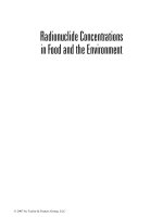
(Figure 9.1 [4]) with the following relation:
DD
mem mem
VmV
"
"0
10
()
S
(9.3)
Combining the two equations above, the slope of this relation (m
v
)canthenbe
applied to determine the theoretical permeability (P) assuming a molecular volume
of zero (P
V=0
).
FIGURE 9.1 Size correction relation (m
v
) applied to determine molecular permeability (P)
from the theoretical zero-volume permeability (P
v=0
).
© 2009 by Taylor & Francis Group, LLC
198 Nanotechnology and the Environment
P
kD
d
P
V
mem mem
V
mem
mV
v
"
"
""
0
0
10·
()
(9.4)
LiebandStein[3]showedthatlogP
V=0
correlates with log k
ow
with a slope of
0.0546. This allows for the description of the overall permeability in terms of vol-
umeandpartitionasfollows:
PP
P
P
VmV
V
mV
k
ow
"
"
"
"
"
0
0
0 0546
10
10
10
1
()
()
.log
S
S
00
()mV
S
(9.5)
Thus, the initial inux rate (J
mem
) can be determined as follows:
JD
dC
dx
DPdx
dn
dt
DA
dC
dx
dn
mem mem
mem
mem mem
"
"
"
·
ddt
PA dC
mem
" ·
(9.6)
where n isthenumberofparticles,A
mem
is the membrane surface area available for
absorption, and dC/dx istheconcentrationgradient.
Thediffusionmodel,asparameterized,predictsthetrans-membraneuxfrom
extracellular to intracellular spaces within the digestive epithelium. This, however,
is expected to be initially faster than diffusion from the intercellular to the central
compartment because: (1) while the permeability P isnotlikelytodiffersignicantly
across the epithelial cells, the microvilli on the exterior of the digestive epithelium
dramatically increase the cellular surface area (A
mem
); and (2) the initial concentra-
tion gradient from the digestive tract to the intracellular compartment is greater than
thegradientfromtheepitheliumtothecentralcompartment.
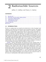
To predict transport kinetics from the digestive tract to the central compartment,
themembranediffusionmodelmustbecoupledintoathree-compartmentmodel
(Figure9.2)toisolatetherate-limitingstepasfollows:
dn
dt
PA C C
dn
dt
PA C
GI IC GI
CC CC
1
2
"
"
[] []
·[ ] [CC
CI
]
(9.7)
© 2009 by Taylor & Francis Group, LLC
Toxicology and Risk Assessment 199
where:
dn
1
/
dt
=Rateofsoluteuxfromgastrointestinal(GI)tracttoGIepithelial
cell
dn
2
/
dt
= Rate of solute ux from GI epithelial cell to the central
compartment
A
GI
=CellularsurfacepresentedtotheGItract
A
CC
= Cellular surface presented to the central compartment
[C]
GI
=SoluteconcentrationwithintheGItract
[C]
IC
=SoluteconcentrationintheGIepitheliumcell
[C]
CC
=Soluteconcentrationwithinthecentralcompartment
WhiledataareavailabletodeterminetherelationsofD
mem
vs. V and P
v=0
vs. k
ow
,
one problem with this approach in relation to nanomaterials is the lack of compa-
rabledatarelatedtothepermeabilitytomaterialsinanappropriatesizerange.While
rst principal thermodynamics suggests that if the original relations are accurate,
the relation between P and V
should hold through the nanoparticle range; the relation
between P
V=0
and k
ow
is in fact a structure/activity relationship and may not be valid
in extrapolation to such large particle sizes. This data gap must be lled to under-
standthepotentialabsorptionandhencetoxicityofingestednanomaterials.
9.2.2 ENDOCYTOSIS
Endocytosisreferstotheprocessofcellulartransportwithoutrequiringtransmem-
branediffusion.Itusuallyinvolvestheactivationofamembranereceptorthatresults
intheinvaginationandseparationofamembranevesselwithinwhichtheactivating
materialiscontained.Thecell,ineffect,engulfstheparticle.Fornanomaterials,
FIGURE 9.2 Time course of diffusion equilibrium across the intestinal epithelium.
© 2009 by Taylor & Francis Group, LLC
200 Nanotechnology and the Environment
endocytosismaybethemostimportanttransportmechanismbecauseofthepredicted
low diffusion rates for materials with volume on the order of hundreds to thousands
of cubic nanometers. Endocytosis tends to follow the concentration gradient, in that
high exogenous particle concentrations result in high rates of endocytotic transport.
However, the capability to initiate endocytosis is chemical and cell-specic, and the
kinetics do not follow a diffusion relation. This necessitates the use of specic empir
-
i
c
al expressions for the derivation of P that cannot be derived thermodynamically.
Nanoparticles have been shown to be transported by endocytosis into the central
compartmentwithasizecut-offofabout300nm[5].Itisknownthatparticulate
matteristransportedfromtheintestinallumenintothelymphaticsystemviaPeyer’s
patches that contain specialized endocytes called M-cells. Uptake via the intestinal
epithelium or intestinal lymphatic tissue results from an induced cellular response
andthereforewouldbeexpectedtovarybynanomaterialsize,partitioncharacteris
-
ti
cs, and charge distribution.
Fewdatadescribethepotentialforultraneornanomaterialstoimpactthegas-
tr
ointestinaltract.Particulatemetalsinhighconcentrationscandisrupttheuidbal-
ance in the colon. Some evidence indicates that ultrane particles may be involved
in inammatory conditions such as irritable bowel syndrome and Crohn’s disease
[6].However,ageneticpredispositionappearstoberequiredfortheconditionto
manifest itself, thereby making population-based generalizations difcult in risk
assessment.Nanoparticlesofzinchavereportedlyinducedbothcontactandsys
-
t
e
mictoxicityuponingestion[7].However,itisunclearwhethertheseareparticle
effectsortheresultofzincdissolutionfromtheparticlesurface.
9.3 EXPOSURE AND EFFECTS THROUGH DERMAL ABSORPTION
Todate,nospecicreportshaveindicateddermaltoxicityresultingfromexposureto
an identied nanoproduct. However, ultrane metal particles have been known to cause
contact dermatitis, as have polyaromatic hydrocarbon-contaminated soots [8, 9].
Reportedly,nanoparticlesoftitaniumoxide[10],transitionmetals[11],liposomes
[12],andfunctionalizedfullerenes[13]canpenetratethroughtheouterlayersofthe
skin (stratum corneum) into the viable epidermis and dermis. The rates and amounts
varywiththematerialaswellasthehealthofthereceptor.Conditionssuchasage,
site of exposure, and certain chronic disease conditions mediate the rate and extent
of penetration. Secondary exposure factors such as vehicle, pH, and even humidity
can dramatically affect particulate penetration [14]. Past research on particle pen
-
etration has involved the movement of particles through the stratum corneum via
impromptu channels formed between the subsequent layers [15]. The thickness and
permeabilityofstratumcorneumvarieswithlocationonanindividual.Hairfollicles
alsomayactasaconduitforthemovementofmaterialsfromtheenvironmentinto
thedermallayers.Similartothestratumcorneum,hairfolliclesarealsoprotected
byahornylayer,althoughittendstobethinnerthanthatpresentonsurfaceskin
[14]. Studies with micro-scale titanium dioxide (TiO
2
) particles indicate penetration
of the epidermal layers with the greatest concentrations clustered about the hair fol-
li
cles [10].
© 2009 by Taylor & Francis Group, LLC
Toxicology and Risk Assessment 201
In risk assessment, dermal penetration follows the concentration gradient. How-
ever, the penetration of the stratum corneum is extremely rate limiting. As a result,
an attenuating gradient forms across this layer. Studies with polysaccharide mic-
roparticles demonstrated this gradient with almost no subdermal penetration [16].
The gradient is difcult to model based on the multifactorial nature of the diffusion
dynamics. Furthermore, particulate matter that does reach the epidermal and dermal
layers is subject to phagocytosis by Langerhans cells and other macrophages, which
results in transport to the lymphatic system rather than the central compartment.
Whilelimitingsystemicexposure,lymphatictransportmayresultininammation
andhypersensitizationreactionsnotimmediatelyassociatedwiththepointofcon-
tact with the causative nanomaterial [17].
9.4 EXPOSURE AND EFFECTS THROUGH INHALATION
Generally, most of the work regarding exposure to nanomaterials derives from con-
cernsrelatedtotheinhalationofultraneparticlesfoundincertainoccupational
settings, as well as ultrane aerosols resulting from combustion. Scientists have spe-
cically linked serious chronic diseases to the inhalation of ultrane particles. These
diseases include Clara cell carcinomas (polycyclic aromatic hydrocarbons), meso-
thelioma (asbestos), and berylliosis (beryllium). General syndromes associated with
exposures to aerosols include black lung (coal), emphysema (combustion products),
and metal fume fever (zinc, tin, and other transition metals).
Relatively stable aerosols consist of a suspension of nonvolatile particles ranging
from 10 nm to 25 micrometers (μm). Typically, aerosol particles less than 500 nm
depositwithapatternmorelikethatofagasthanaparticulatesuspension.Hence,
diffusiongovernsdepositionandcanbeexpectedtooccurthroughouttherespira-
tory tract, including the alveoli. Deposition depends on the adherence and residence
time of the nanoparticles. Particles between 500 nm and 25 μm demonstrate a slow
depositionalpatternwherethemajoritymaybedepositedintheupperairway,but
some penetrate to the deep lung. Particles larger than 25 μm tend to be deposited
throughgravitationaldepositionandwillsettleinthenasopharyngealregionwhere
theowvelocityisreduced[18].
9.4.1 MECHANISMS FOR ADSORPTION AND REMOVAL
The ux rate (J) from the inhaled atmosphere to the respiratory epithelium can be pre-
dictedthroughamodicationofFick’slawofdiffusion,whichisexpressedasfollows:
JD
dc
dx
"
(9.8)
where dc is the concentration gradient, dx isthedistanceacrosstheconcentration
gradient, and D isthediffusioncoefcient.Inthecaseofinhalation,theseparation
distanceisafunctionofthesizeandshapeoftheairspace.Becauseaninhaled
nanomaterial is distributed within the three-dimensional air space, concentration
requires integration over the lateral and longitudinal directions based on the con-
centrationgradientrelativetoagivenlocationalongtheairway.Thisusuallycanbe
© 2009 by Taylor & Francis Group, LLC
202 Nanotechnology and the Environment
simpliedbyassumingtheairwayiscomposedofaseriesofrelativelyuniformpas-
sages(nasal,pharyngeal,tracheal,bronchi,bronchioles,andalveoli).Withanintrin-
sically constant surface area (A) and radius (r
a
)withineachgrouping,uxdynamics
(dn/dt,wheren is the number of particles) can be expressed based on the area of a
given passage as follows:
dn
dt
DA
dc
dx
x
r
a
"
"
µ
4
0
U (9.9)
SubstitutingtheStokes-Einsteinequation,therelationcanbeexpressedasasolvable
expression as follows:
dn
dt
kT
r
A
dc
dx
p
x
r
"
"
µ
2
3
0
M
(9.10)
where k is the Boltzmann constant, T istheabsolutetemperature,M is the viscosity
of the aerosol, and r
p
istheradiusofthenanoparticle.
Thediffusionofananomaterialfromgaseoussuspensiontotheepithelium
involvesnotonlyachangeinlocation,butalsoachangeinstatefromaerosolto
hydrosol within the mucous layer of the pulmonary airways. Usually, the concentra-
tion gradient, dc/dx, needs to be modied to account for the differential fugacity
between the two states. However, nanoparticles have a low escaping tendency
because of their high relative masses. Because nanomaterials contacting the muco-
sallayerwillnotsignicantlyreturntothegaseousaerosol,diffusiontransportis,in
effect,oneway,suchthattheintegralofdc/dx = 1. Furthermore, because of the rate
of ventilation and turbulence, the cross-sectional gradient within the airway can, for
the most part, be ignored. With these two assumptions, the concentration gradient
can be simplied to the differential concentration between that suspended in the air
streamandthatsuspendedinthemucosallayer.
Thelinearnatureoftheairwaymeansthatatanypoint(y), the concentration is
equivalent to the initial concentration ([C
0
]),minustheintegralofthemateriallost
inthepreviousairwayasfollows:
dn
dt
kT
r
AC
dn
dy
p
Y
y
Y
"
©
«
ª
ª
¹
»
º
º
"
µ
2
3
0
0
M
(9.11)
Notethattheintegralisbasedonthelineartransportofairandwilldifferbased
on whether the ventilation is in inhalation or exhalation. Furthermore, the air ow
velocity (v-)placesaconstraintondy, and by implication A
Y
, by the amount of sur-
faceareaexposedperunittimeasfollows:
© 2009 by Taylor & Francis Group, LLC
Toxicology and Risk Assessment 203
ACSvdt
Hence
dn
dt
kT
r
CS vdt C
dn
dy
YY
p
Y
y
"
"
:
2
3
0
M
""
µ
©
«
ª
ª
¹
»
º
º
0
Y
(9.12)
where CS
Y
is the cross-sectional area of the airway at point y,anddy is the inni-
tesimal of the change in position within the airway. Note that the area is expressed
asacross-sectionratherthanasafunctionofradius.Thisisbecausethepresence
of processes (i.e., projecting portions of bone or tissue), particularly in the naso-
pharyngeal region, can greatly increase the potential surface area of exposure per
unittime.However,inthepulmonaryregionofthelungs,wheretheairwaysare
relatively smooth, the exposed area per unit time can be expressed in terms of πr
2
a
dy.
Figure 9.3 shows examples of projected deposition rates based on mass and ber
numbersforthebronchioles.Asshown,thedepositionrateincreaseswithconcentra-
tion and decreases with particle size (diameter).
Direct solution of this relation is difcult because of the heterogeneity of the
mammalian airway. Predictions of absorption rates usually involve the construction
ofathree-dimensionalpassagemodelthatsegmentsdifferentialregionsoftheair-
way based on similar diffusion properties. These models generally indicate that the
number of particles deposited is inversely proportional to the size of the particle [19].
Therefore,thesmallertheparticle,thelargertheamountabsorbedastheresultof
higherratesofdiffusion.Althoughcounter-intuitive,therelationalsosuggeststhat
thefastertheairvelocity,thehighertherateofabsorption.Butnotethatthisresults
from the increase in surface area exposure per unit time, which decreases the lon-
gitudinal gradient, thereby allowing higher concentrations in deeper regions of the
airway.
Uponadsorptiontotheliningoftheairways,particulatematterissuspendedina
complex mixture called the tracheobronchial mucus. Produced by both submucosal
and epithelial secretory cells, the mucus comprises a mixture of glycoproteins and
electrolytes within an aqueous matrix. The viscosity of the mucus varies throughout
therespiratorysystem,therebyalteringthediffusivityofnanoparticles.Themucous
layerinhumanscontinuesfromthelarynxtotheendoftherst-generationbronchi-
oles.Withinthealveoli,TypeIIcellsalsoproduceaproteinaceoussecretionsimilar
to mucus, but usually of a lower viscosity and higher water content.
Thepulmonarymucosaispartofaclearancesystemreferredtoastherespira-
toryconveyer.Thissystemofciliatedcells,whichlinesthebronchiolesandtrachea,
trapsinhaledparticulatesinmucusandsweepstheladenmucosalmaterialupand
outoftherespiratorytract.Ratesofmovementvaryfromabout0.6mm/mininthe
bronchioles to about 10 mm/min in the trachea region [20]. The respiratory conveyor
deposits most of the material in the esophagus, which may represent a signicant
exposureroutefortheingestionofnanomaterials.
Materials with a sufcient concentration gradient to reach the alveoli are not
directlysubjecttothemucosalconveyerbecausetherearenociliainthealveoli.
© 2009 by Taylor & Francis Group, LLC
204 Nanotechnology and the Environment
Three principal methods can clear nanomaterials from the alveoli. The rst is dif-
fusion based and involves the movement of nanomaterials through the Type I cells
intothevascularcapillarybedandthegeneralcirculation,wheretheyarethen
removedbybloodltration.Thesecondandthirdmethodsinvolveinitialphago
-
cy
tosis (engulfment) by resident macrophages. Macrophages can engulf insoluble
particlesfrommoleculardimensionsuptoabout1μmindiameter[21].Theladen
FIGURE 9.3 Depositional kinetics of nanomaterials within the human bronchioles stan-
dardized based on (a) concentration and (b) particulate number.
© 2009 by Taylor & Francis Group, LLC
Toxicology and Risk Assessment 205
macrophages then can migrate vertically to the bronchioles where they are entrained
in the mucosal conveyer and rapidly eliminated. Alternately, nanoparticles may
be subject to endocytosis by macrophages that migrate into the lymphatic system
wheretheyareclearedviathetracheobronchiallymphnodesortheblood.This
relativelyslowprocesssometimestakesmonthstoremoveparticulatematerialfrom
an exposed organism.
Themostimportantconsiderationsinassessingtheriskfromexposuretonano
-
mat
erials in aerosols are the size of the particles and the rates of exposure relative to
the rates of response. From the discussion above, it is apparent that dispersed nano-
mat
erials will deposit all along the airways, including the alveoli. However, nano-
mat
erials, particularly the current carbonaceous materials, are rarely encountered
ineithertheoccupationalorgeneralenvironmentasstabledispersals(seeChapter
6). The critical rate of exposure relates to the rate and magnitude of injury relative
totheratesofeliminationandrepair.Ifinjuryresultingfromexposureexceedsthe
airway’s repair capacity as the result of inefcient removal capacity, then it can be
expected that an adverse effect will ensue.
Inammation is the most common response to brous or particulate material.
Itresultsfromtheactivationofinherentdefensemechanismsmediatedbymac
-
ro
phages that, if over-stimulated, will result in localized cellular necrosis and loss
oflungfunctions.Acasestudyofthepotentialriskassociatedwithsingle-walled
carbon nanotubes follows.
9.4.2 CASE STUDY: INHALATION OF CARBON NANOTUBES
Single-walledcarbonnanotubes(SWCNTs)consistofasheetofarylcarbonrings
curved around on themselves so as to form a tube one layer thick. SWCNTs are typi-
cal
ly1to4nmindiameterandvaryfromasshortas50nmtolengthsinexcessof
2 μm. Carbon nanotubes possess extremely low charge afnity compared to that of
uidmediasuchasairandwater.Assuch,theytendtorapidlyformclumpsbybind
-
in
gtooneanother,particularlyalongtheirlongaxes.Thismanifestsatertiarystruc-
tu
re consisting of numerous SWCNTs in forms referred to as nanoropes. Nanoropes
will associate further into groups of nanoropes referred to as tangles and will con-
ti
nuetoassociateuntiltheunitsbecomesolargeastofalloutofuidsuspension.
9.4.2.1 Pulmonary Toxicology
Asofthedateofpublication,nohumanstudieswereavailablethatevaluatedthe
pulmonarytoxicityofSWCNTs.Furthermore,animaltestsfordirectinhalation
werenotavailableduetothepracticaldifcultiesinisolatingandcollectingenough
SWCNT particles to conduct these studies [22]. As such, almost all the current stud
-
iesarebasedoneitherin vitro designs u
sing tissue explants of cultured cell lines, or
exposures of whole animals using intratracheal instillation. The term “intratracheal
instillation” describes a technique where researchers inject a bolus dose of a SWCNT
suspension into the trachea of the test animal to distribute SWCNTs throughout the
pulmonary airway by aspiration. While the intratracheal instillation method has
technicallimitations,itisanacceptedscreeningtestforpulmonarytoxicity[22–24].
© 2009 by Taylor & Francis Group, LLC
206 Nanotechnology and the Environment
Threeintratrachealinstillationstudieshaveexaminedthepulmonarytoxicityof
SWCNTs [23–25].
Lametal.[23]instilledmicewithasingletreatmentof0,0.1,or0.5mg/mouse
SWCNTsuspensionina50μLbuffer(equaltoapproximately3.94×10
6
and 1.96
×10
7
ber units per mouse, respectively). Four animals per dose group were eutha-
nized7daysafterthesingletreatment;veanimalsperdosegroupwereeuthanized
90 days post treatment. Lam et al. reported dose-dependent lesions, primarily inter-
stitial granulomas, in both the 7- and 90-day groups. The lesions were more promi-
nentinthe90-dayanimals.Micealsoweretreatedwithquartzandcarbonblack
(whose size range included nanoparticles). Minimal inammation was observed in
mice treated with carbon black, and moderate inammation was observed in mice
treatedwithquartz.Lametal.reportedthatthequartz-inducedtoxicitywasless
severe than lesions induced by SWCNTs.
Inasecondstudy,Warheitetal.[25]instilledratswithSWCNTsat0,1or5
mg/kg(approximately9.79×10
6
and 4.90 × 10
7
ber units per rat, respectively). The
researchteameuthanizedandexaminedanimals1,7,30,or90daysafterasingle
treatment. Granulomas were present after 1 month but the lesions were neither dose
dependentnortimedependent.Toxicitywasnotreportedinratsthatweretreated
with graphite. Based on the results, Warheit et al. Concluded that “granulomatous
reactionwasanonspecicresponsetoinstilledaggregatesofSWCNTsandthe
resultsmaynothavephysiologicalrelevance,andmayberelatedtotheinstillationof
a bolus of agglomerated nanotubes.” Lam et al. [22] postulated that this lack of dose
andtimedependencereportedbyWarheitetal.[25]mightbeduetoasignicant
portionoftheinstilledbolusdosenotreachingthealveolarregion.
Shévedovaetal.[24]conductedathirdstudyinanattempttoresolvethediffer-
ences.Inthisstudy,micewereinstilledwithaSWCNTsuspensionthathadbeen
highlypuriedtoremovemetals.MicewereadministeredSWCNT,carbonblack,or
quartzat0,10,20,or40μgpermouse(approximately3.92×10
5
,7.84×10
5
,and1.57
×10
6
ber units per mouse, respectively). Animals were euthanized at 1, 3, 7, 28, or
60 days following a single treatment. Acute pulmonary inammation, granulomas,
andbrosiswerereported.Thepulmonarytoxicitywasbothdoseandtimedepen-
dent.SimilartothestudiesconductedbyLametal.[23]andWarheitetal.[25],gran-
ulomaswereobservedatthesiteofdepositionofSWCNTaggregates,butuniqueto
this study was the dose- and time-dependent interstitial brosis in pulmonary regions
away from the sites of deposition. These data indicate brosis induced by dispersed
SWCNTs.Neithercarbonblacknorquartzproducedgranulomasorbrosis.
Tianetal.[26]reportedthatSWNCTsinducedthestrongestadverseeffectout
of ve nano-sized carbon materials tested on cultured human broblast survival. The
orderoftoxicityfromleasttomosttoxicwasasfollows:carbongraphite<multi-wall
carbon nanotubes (MWCNTs) < carbon black < activated carbon < SWCNT. Dis-
persedSWCNTsweremoretoxicthanunrenedSWCNTs,whichtendedtogroup
together in tangles, creating larger and less harmful brous units.
The results of these animal studies indicate that SWCNTs can induce inam-
matorypulmonarytoxicityintheformofgranulomasthatcanresultinbrosisif
theyreachthedeeplungtissue.Toxicityintheupperairwayismitigatedbyshort
residencetimesresultingfromtheirrapidremoval.
© 2009 by Taylor & Francis Group, LLC
Toxicology and Risk Assessment 207
9.4.2.2 Risk Assessment
AssessingtheriskassociatedwithSWCNTsrequirestwoseparateconsiderations:
(1)theabilityofamaterialtoreachasensitivesiteofaction,and(2)thetypeand
magnitudeoftheresultantresponseatthesensitivesite.Currentstudiesbasedon
intratracheal installation indicate that the sensitive site of action for SWCNTs is the
deep lung tissue — specically the respiratory bronchioles and alveoli. Indigenous
macrophages engulf SWCNTs that reach this part of the pulmonary airway. This
phagocytosis apparently results in an inammation cascade similar to that seen in
silicosis, which appears to manifest as a long-term or chronic condition because the
macrophagesbearingSWCNTsdonotmigrateintotheupperairwaysasisseen
with materials such as particulate graphite [25]. Chronic inammation in the lower
airwaywillresultindamagetotheunderlyingepitheliumandthegenerationof
scar tissue often referred to as brosis. Widespread damage throughout the lower
airways will reduce gas transfer signicantly and a condition akin to emphysema
candevelop.Furthermore,chronicinammationsofthistypehavebeenassociated
with the promotion of hyperplasias that have the potential to become cancerous [27].
However,itmustbecautionedthatthisisnotnecessarilythecase,andthereiscur-
rently no evidence that exposure to SWCNTs will result in either cancer initiation
or promotion.
ExposureoftheupperairwaystoSWCNTsislesstoxicologicallysignicant
fortworeasons.First,theresidencetimeoftheSWCNTsismuchshorterbecause
particlesthatimpactwithinthenasopharyngeal,tracheal,orbronchialregionsofthe
airwayarerapidlyremovedviathepulmonarymucousconveyer.Therefore,inam-
mationappearstobetransient(<2hr).Second,becausetheupperairwayisnotthe
siteofsignicantgastransfer,itcomprisesathickerandmorerobustepithelium
with greater regenerative capacity and therefore is less likely to manifest signicant
brosis [28].
Consequently, the greatest potential hazard to individuals working with SWCNTs
apparentlywouldstemfromexposuretomaterialscapableofdepositingwithinthe
deep lung tissue. Materials depositing within the upper airway may be acutely toxic
athighconcentrationsbutwillnotlikelyrepresentaserioushealthissueatorbelow
exposure concentration limits established to protect the deep lung.
Initial indications from histological studies indicate that inammation does not
dependdirectlyonthesizeoftheSWCNTberimpactingthepulmonarytissue
[24,25,29].Rather,itisthenumberanddistributionoftheimpactsthatresultsin
the overall toxic response. As such, the classic risk approach of quantifying toxicity
usingthemassdoseperunittimeorunitbodymassmaynotbeappropriate.Rather,
to capture the dose response, one must quantify the exposure in terms of number of
berunitsperunittime,whereaberunitisdenedasanyindependentSWCNT,
SWCNT rope, or SWCNT tangle.
Ofthecurrentanimalstudiesdescribedabove,thestudyperformedbyShevedova
etal.[24]providesthebesttoxicologicalcharacterizationandquanticationtoderive
exposureguidelines.Usingtheendpointofaveragealveolarthicknessasameasure
of induced brosis, Shevedova et al. found that a single exposure concentration of
3.92×10
5
berspermousehadnoeffectateither28or60dayspostexposure.
© 2009 by Taylor & Francis Group, LLC
208 Nanotechnology and the Environment
Toconvertthistoahumanexposure,theconcentrationinthemousemustbe
scaledtoahuman.GiventhatthemousemassinthestudybyShevedovaetal.[24]
wasreportedtobe20.3g,itispossibletoestimatethetotallungvolume(V
tot
)asthe
sumofthetidalvolume(i.e.,theamountofairpassinginandoutofthelungduring
normal resting breath; V
T
)andtheanatomicaldeadspace
*
(V
D
)forthemouseanda
75-kg human using the algometric scaling equations of Linstedt and Schaffer [30]
as follows:
Mouse W kg
VW mL
V
Tkg
D
(. ):
.
"
" "
0 0203
6 60 0 129
101
"" "
"
"
2 20 0 0430
0 172
101
.
(
.
WmL
VmL
Human W
kg
tot
775 0
6 60 517
220
101
1
.):
.
.
.
.
kg
VW mL
VW
Tkg
Dkg
" "
"
001
172
689
"
"
mL
VmL
tot
9.13)
Absolute pulmonary surface area (SA)isdifculttodeterminebecauseofthe
irregular geometry. However, by assuming proportional scaling to the total pulmo-
nary volume between the mouse and human, the relative surface area for the human
andthemousecanbescaledasfollows:
V
V
SA
SA
tot human
tot mouse
human
mous
©
«
ª
¹
»
º
"
23/
ee
" 252
(9.14)
Withanareascalingfactorof252humantomouse,asafedoseof3.92×10
5
ber
unitspermousecanbeextrapolatedto9.88×10
7
bers per person. This level can be
consideredanot-to-exceedbodyburdenforberslessthan5μmineffectivediam-
eter,whichisthetypicaluppersizelimitformaterialsthatarecapableofreaching
thedeeplung.
Mulleretal.[31]reportedtheclearancefromthedeeplungforMWCNTsasa
constant for elimination (k
G
)of0.01daysorahalf-lifeof69.3days.Thisassumed
an inherent interaction between the MWCNT and the pulmonary physiology, and is
* Anatomical dead space (VD): the volume of the conducting airways from the external environment (at
thenoseandmouth)downtothelevelatwhichinspiredgasexchangesoxygenandcarbondioxidewith
pulmonarycapillaryblood;formerlypresumedtoextenddowntothebeginningofalveolarepithelium
in the respiratory bronchioles, but more recent evidence indicates that effective gas exchange extends
somedistanceupthethicker-walledconductingairwaysbecauseofrapidlongitudinalmixing.
© 2009 by Taylor & Francis Group, LLC
Toxicology and Risk Assessment 209
therefore not a scalable value. Using this k
G
,anallowabledailyexposurerate[C]
G
can
be determined as follows:
[] []·
,[ ]
CC e
where t day C
o
kt
G
G
G
"
1
= 1 = 99.83 10
5
w fibers per day
(9.15)
BasedonstudyresultsreportedbytheU.S.EPA[32],theventilationratefor
an adult undertaking medium activity is 1.02 m
3
/hr.Thisequatestoanexposure
volume of 8.16 m
3
per8-hrday.Therefore,toensurethatthetotallungburdendoes
not exceed 9.88 × 10
7
bers, the 8-hr time-weighted concentration cannot exceed
1.20×10
5
bers/m
3
, and the maximum 1-hr exposure should not exceed 9.64 × 10
5
bers/m
3
forberswithaneffectivediameterlessthan5μm.
Currently, no published physiological or epidemiological studies describe the
effectofSWCNTinhalationinhumans.Theavailablestudieswereperformedin
rodents.Itisassumedinthisanalysisthatasafelevelinrodentsequatestoasafe
level in humans. Other studies with inammatory brous material appear to indicate
thatthecross-speciescomparisonsarevalid[33].However,itremainsanuncertainty
if this relation will hold true for SWCNTs.
Histological examination of the lesions associated with SWCNTs in the deep
lungsuggeststhatthedegreeofgranulomaformationandresultinginammationis
independent of the amount of SWCNT within the granulomas [34]. This is similar,
withinlimits,toobservationswithotherbrousinammatoryagentssuchasasbes-
to
s[33].However,thisqualitativeobservationhasnotbeentesteddirectly.Itmaybe
that larger SWCNT tangles have a greater inammatory potential than smaller ones.
Ifthisisthecase,however,thedifferencesinmagnitudeareofanorderthattheydid
notpresentobvioushistologicaldifferencesinthecurrentavailablestudies.
Further uncertainty exists in the derivation of the SWCNT elimination rate
basedontwoobservationsbyMulleretal.[31]atanexposureratedifferentfromthat
usedasthetoxicitythreshold.Scalingtheexposuretotheprojectedriskthreshold
requiredanassumptionofrst-orderkinetics.Regressionoftheone-doseobserva-
ti
onssuggestsstronglythattheeliminationdoesfollowrst-orderkinetics.However,
ithasnotbeenrepeatedordemonstratedforotherSWCNTexposurerates.Itiscur-
rentlyanassumptionandthereforerepresentsanuncertaintyinthisderivation.
9.6 KNOWN TOXICITY OF NANOMATERIALS
Thestudyofthetoxicityofnanomaterialsisinitsinfancyandtheliteratureisgrow-
ingrapidly.Itisusefultoexaminetheliteraturetodateforthesixtypesofnanoma-
terials that are the focus of this book to understand the types of effects that might
occur. Table 9.1 offers a brief review of the literature.
AsshowninTable9.1,anumberofstudieshaveinvestigatedthepotentialhuman
health implications associated with exposure to nanomaterials. Although many of
theresultsareverypreliminary,thereareindicationsthatthesixmajorengineered
nanomaterialscanelicitanoxidativestressresponseincertainbiologicaltestsys-
te
ms [37, 39, 54–56]. This is seen as measured indication of cell membrane damage,
210 Nanotechnology and the Environment
TABLE 9.1
Effects of Nanomaterials or Nanoparticles on Mammalian Species
Species Particle
a
Category
Size or
Diameter Exposure or Dose Endpoint(s) Effect(s) Commentary Ref.
Human
mesothelioma and
rodent broblast
cell lines
Nanoparticle TiO
2
8nm
0, 3.75, 7.5, and 15 Rg/
ml for 6-day periods
and 0, 7.5, 15, and 30
Rg/mL for 3 days
exposure
Cytotoxicity Weak cytotoxic effects Both cell lines showed less
response after 6 days of
exposure compared to 3 days
of exposure. This might be
due to initial stress of the
nanoparticles, and then
detoxication of the particles
and cell culture viability
recovers.
[35]
Rats TiO
2
particles
Nanoscale TiO
2
rods
Nanoscale TiO
2
dots
300 nm (rutile
type) 200 nm ×
35 nm (anatase
type)10 nm
(anatase type)
1 or 5 mg/kg
intratracheally
instilled in
phosphate-buffered
saline; evaluated at
24 hr, 1 week, 1
month, and 3 months
post instillation
Oxidative stress/
cytotoxicity; lung
histopathology
TiO
2
particles, dots, and rods
caused transient inammatory
and cytotoxic effects observed
at 24 hr post exposure, but
effects were not sustained.
Instilled quartz particles
caused sustained dose-
dependent inammation as
well as lung tissue damage
consistent with pulmonary
brosis development.
Results indicate that nanoscale
particles might not be more
cytotoxic to lung compared to
larger-sized particles. In
addition, results demonstrate
that surface area might not be
associated with pulmonary
toxicity of nanoscale
particles.
[36]
Syrian hamster
embryo broblast
cells
Ul
trane TiO
2
≤20 nm Cells treated with 0.5,
1.0, 5, or 10 Rg/cm2
for 12, 24, 48, 66, or
72 hr
Oxidative stress/
cytotoxicity
A signicant dose
(concentration)-dependent
increase in micronuclei
formation between 0.5 and
5.0 Rg/cm
2
(measurement of
chromosomal change); cell
death (apoptosis) observed
after 24, 48, and 72 hr.
Results support the mechanism
of cell death from exposure to
nanoparticles: particles react
with cell membranes, in turn
generate reactive oxygen
species (ROS); the oxidative
stress leads to cell toxicity.
[37]
© 2009 by Taylor & Francis Group, LLC
Toxicology and Risk Assessment 211
Mice (C57B1/6) TiO
2
2–5 nm 0.77 or 7.22 mg/m
3
(acute exposure, 4
hr); 8.8 mg/m
3
(subacute exposure, 4
hr/day for 10 days)
Oxidative stress/
cytotoxicity;
lung
histopathology
Acute exposure: at the high
concentration, BAL uid
signicantly increased; other
parameters did not show
inammation. Subacute
exposure: alveolar
macrophages elevated in mice
necropsied at weeks 0, 1, and
2 post exposure, not elevated
at week 3 post exposure.
Minimal inammatory
response likely reects a
surface area threshold;
anything below this threshold
will cause little or no
inammatory response.
[38]
Mouse microglia
cells
Nanosize T
iO
2
(Degussa P25)
826–2368 nm 5–120 ppm for 6 or 18
hr
Oxidative stress Signicant release of ROS
occurred at 60 min post
exposure at concentrations
≥20 ppm; cell death not
observed at all concentrations
Results demonstrate that TiO
2
can stimulate microglia to
produce ROS; however,
microglia remained viable.
Further study to understand if
the ROS translates into neural
damage in situ.
[39]
Human red blood
cells
TiO
2
, anatase 0.02–0.03 m 5 μg/mL; incubated 4–
24 hours
Red blood cells TiO
2
aggregates with a
diameter ≤0.2 μm were taken
up by red blood cells; larger
aggregates were stuck to
surface of cell membrane.
Results suggest that
nanoparticles may penetrate
red blood cells by a
mechanism other than
phagocytosis and endocytosis
[40]
Mouse microglia; rat
dopaminergic
neurons; and
embryonic rat
striatum
TiO
2
Diameter of
aggregates: 800
to 1900 nm (at 30
min); 770 nm (at
2 hr)
2.5–120 ppm Neurotoxicity of
nerve cells
(microglia,
neuron)
Cytotoxicity reported for
microglia and striatum; TiO
2
did not cause toxicity to the
dopaminergic neurons.
Results suggest that the
neurotoxicity of TiO
2
is
mediated through microglia-
generated ROS.
[41]
© 2009 by Taylor & Francis Group, LLC
212 Nanotechnology and the Environment
TABLE 9.1 (CONTINUED)
Effects of Nanomaterials or Nanoparticles on Mammalian Species
Species Particle
a
Category
Size or
Diameter Exposure or Dose Endpoint(s) Effect(s) Commentary Ref.
Mouse
spermatogonial
stem cell line
Nanoscale silver 15 nm 5, 10, 25, 50, and 10
μg/mL
Cytotoxicity;
mitochondrial
function, cell
morphology, cell
membrane
leakage, and cell
death
Silver nanoparticles were the
most cytotoxic of the
compounds tested. Cytotoxic
effects were dose-dependent.
The spermatogonial cell line
was chosen for this study to
evaluate the toxicity of
nanoparticles on the male
germline. Results of this study
suggest that the cell line is a
good model.
[42]
Human epithelial
cells
Nanoparticle
carbon
black, Fine carbon
black, Titanium
dioxide,
Nanoparticle TiO
2
14.3 nm
260 nm
250 nm
29 nm
31.25–200 μg/mL Cytotoxicity Inammatory response The highly toxic nature and
reactive surface chemistry of
the carbon black
nanoparticles very likely
induced the type II cell line to
release pro-inammatory
mediators that can potentially
induce migration of
macrophages.
[43]
© 2009 by Taylor & Francis Group, LLC
Toxicology and Risk Assessment 213
Mice (LDLR/KO) Carbon black 120.7 nm Endotracheal
dispersion of 1 mg
per animal per week
for 10 weeks or
intratracheal
dispersion of air and
1 mg per animal per
week for 10 weeks.
Diets were also
controlled for 0 or
0.51% cholesterol.
Acute study
performed: animals
fed 0.51% cholesterol
diet for 3 days and a
single 1 mg/animal
dose of carbon black.
Aorta/circulatory
system
Aortic lipid-rich lesions were
reported in mice receiving the
0.51% diet with and without
carbon black exposure. No
lesions in mice receiving the
0.0% diet. Greatest amount of
lesions were reported in
0.51% group with carbon
black exposure.
Results indicate that
respiratory exposure to
carbon black might accelerate
the development of
atherosclerosis and be
associated with
cardiovascular adverse
effects.
[44]
Rat aortic smooth
muscle cells
SWCNT 10–15
nm 0.0–0.1 mg/mL added
to cells and incubated
for periods of 1, 2.5,
and 3.5 days
Cytotoxicity Unltered SWCNT media: cell
growth not affected after 1
day; decrease in cell growth at
2.5 days for concentrations
from 0–0.05 mg/mL; ltered
SWCNT (removal of SWCNT
aggregates): increase in cell
number for concentrations 0–
0.05 mg/mL; growth
inhibition at 0.1 mg/mL dose
for both ltered and unltered
SWCNT.
Results indicate that aggregates
affect cell growth, but not
solely responsible for the
cytotoxicity.
[45]
© 2009 by Taylor & Francis Group, LLC
214 Nanotechnology and the Environment
TABLE 9.1 (CONTINUED)
Effects of Nanomaterials or Nanoparticles on Mammalian Species
Species Particle
a
Category
Size or
Diameter Exposure or Dose Endpoint(s) Effect(s) Commentary Ref.
Human leukemic
cells
Single- and multi-
walled carbon
nanotubes; 3
different samples by
different synthesis:
Sample 1 —
MWCNTs
(synthesized by an
electric discharge)
Sample 2 — 50%
MW CNTs + 30%
SWCNTs Sample 3
— MWCNTs
(purchased)
Sample 1: 10–50
nm
Sample 2: 10–40
nm
Sample 3: 110–
170 nm
25 μg/ml of each of the
three samples
Oxidative stress/
cytotoxicity
Cytotoxic effects not observed;
cell growth rate reduced
With the lack of cytotoxicity,
the decrease in cell growth
might be a result of the
carbon nanotubes affecting
the cell cycle directly.
[46]
Rat (in vivo)
peritoneal
macrophages (in
vitro)
Multi-walled
carbon
nanotubes (CNT)
and ground CNT
9.7 nm (CNT)
11.3 nm (ground
CNT)
0.5, 2, or 5 mg/animal
intratracheally
instilled (in vivo)
Pulmonary
toxicity:
inammatory
response; brotic
response
In vivo results: inammatory
response and dose-dependent
pulmonary brosis for both
CNT and ground CNT, effects
more pronounced with ground
CNT. In vitro results: ground
CNT increased macrophage
production.
Results indicate that multi-wall
carbon nanotubes are capable
of eliciting inammatory and
brotic response in lungs
[31]
© 2009 by Taylor & Francis Group, LLC
Toxicology and Risk Assessment 215
Mice Single-walled carbon
nanotubes
1–4 nm Acute: 10–40 μg/
mouse, single
intrapharyngeal
instillation (sacriced
at 1, 7, 28, and 56
days post exposure)
Chronic: 20 μg/
mouse, pharyngeal
aspiration, once every
other week for 8
weeks (mice in this
group are bred with
elevated cholesterol
levels and were fed a
high-fat diet).
Aortic
mitochondria
(oxidative stress
assays)
Aortic mitochondrial DNA
damage at 7, 28, and 60 days
post exposure. Increase in
atherosclerosis plaque
formation reported in
chronically treated mice.
Results indicate that
respiratory exposure to
SWCNTs might accelerate
the development of
atherosclerosis and be
associated with
cardiovascular adverse
effects.
[47]
Guinea pig alveolar
macrophages
SWCNTs,
MWCNT10,
and
fullerene (C60)
1.4 nm (SWCNT)
10–20 nm
(MWCNT10)
C60 — not
provided
SWCNT and C60: 0,
1.41, 2.82, 5.65,
11.30, 28.20, 56.50,
113.00, and 226.0 μg/
cm
2
MWCNT10: 0,
1.41, 2.82, 5.65,
11.30, and 22.60
μg/cm
2
Cytotoxicity SWCNT: high cytotoxicity at
lowest dose; for MWCNT10,
cytotoxic effects but at the
highest dose. C60 did not
induce cytotoxicity at any
concentration.
Results suggest that toxicity of
nanomaterials increases with
an increase in surface area.
[34]
Mice (B6C3F
1
) SWCNT, Carbon
black
1 nm Single dose of 0, 0.1,
or 0.5 mg/mouse
intratracheally
instilled; euthanized
7 and 90 days post
treatment
Cytotoxicity/lung SWCNT: Dose-dependent
epithelioid granulomas,
interstitial inammation,
peribronchial inammation,
necrosis. Lesions more
persistent and pronounced in
90-day group. Carbon black:
no lung adverse effects.
Results suggest that toxicity of
nanomaterials increases with
an increase in surface area.
[23]
© 2009 by Taylor & Francis Group, LLC
216 Nanotechnology and the Environment
TABLE 9.1 (CONTINUED)
Effects of Nanomaterials or Nanoparticles on Mammalian Species
Species Particle
a
Category
Size or
Diameter Exposure or Dose Endpoint(s) Effect(s) Commentary Ref.
Rat Single-walled carbon
nanotube (SWCNT)
Diameters < 2 nm,
with lengths
ranging from 0.5
to 40 μm and a
purity > 90%.
Oropharyngeal
aspiration of 2 mg/
kg-bw. Evaluated
(bronchoalveolar
lavage) 1 and 21 days
post exposure
Lung–lung
histopathology,
brogenic
potential, cell
proliferation, and
growth factor
mRNAs.
Exposure
biomarker
SWCNT did not cause lung
inammation, but induced the
formation of small, focal
interstital brotic lesions in
the alveolar region of rat
lungs.
Of greatest interest — unique
intercellular carbon structures
composed of SWCNT-bridged
lung macrophages. These
bridges offer an easily
identiable exposure
biomarker.
[48]
C57BL/6 Mice SWCNT 1–4
nm 0, 10, 20, or 40 μg/
mouse; pharyngeal
aspiration. Animals
euthanized at 1, 3, 7,
28, and 60 days post
exposure
Oxidative stress/
cytotoxicity lung
Acute inammation with early
onset, progressive brosis and
granulomas. Dose-dependent
increase in oxidative stress
biomarkers. Functional
respiratory deciencies and
decreased bacterial clearance
also observed.
Results support in vitro studies. [24]
Human broblasts SWCNT
MWCNT
Carbon
black
Activated carbon
Carbon graphite
2nm
200 nm
25 nm
50 nm
500 nm
0.8, 1.61, 3.125, 6.25,
12.5, 25, 50, and 100
μg/mL for 1 to 5 days
Cytotoxicity Cellular apoptosis/necrosis.
SWCNTs induced strongest
cellular apoptosis/necrosis.
Rened SWCNTs are more
toxic than unrened
counterpart.
Results suggest that toxicity of
nanomaterials increases with
increase in surface area.
[26]
© 2009 by Taylor & Francis Group, LLC
Toxicology and Risk Assessment 217
Rats SWCNT 30 nm 1 or 5 mg/kg
intratracheally
instilled; evaluated 24
hr, 1 week, 1 month,
and 3 months post
exposure
Cytotoxicity/lung Transient inammatory and cell
injury effects, non-dose-
dependent series of multi-
focal granulomas.
Physiological relevance of
these ndings should be
determined by conducting an
inhalation toxicity study.
[25]
Mouse L929
brosarcoma
Rat
C6 glioma
U251 human glioma
C60 fullerence
C60(OH)
n
—
polyhydroxylated
fullerene
100 nm
<5 nm
1 or 1000 (g/mL Oxidative
stress/cytotoxicity
Cytotoxic action reached
maximum after 6 hr with C60;
minimal cytotoxicity for the
same time period for
C60(OH)
n
; Reactive oxygen
species (ROS) not produced in
cells treated with C60(OH)
n
;
rapid increase of ROS in cells
treated with C60.
Results demonstrate that C60
is at least three orders of
magnitude more toxic than
C60(OH)
n
.
[49]
Rats C60
fullerenes 160 ± 50 nm 0.2, 0.4, 1.5, or 3.0
mg/kg intratracheally
instilled; lung tissue
evaluated 1 day, 1
week, 1 month, and 3
months post
instillation
Oxidant and
glutathione
endpoints;
bronchoalveolar
lavage (BAL)
uid biomarkers;
lung tissue
Transient inammatory and cell
injury at 1 day post exposure,
not different from controls at
other post exposure times;
BAL biomarkers increased in
1.5 and 3.0 mg/kg dose
groups at 1 day and 3 months
post exposure; no adverse
lung tissue effects at 3 months
post exposure at any dose.
Results not consistent with
results reported for in vitro
studies; such ndings
highlight the difculty in
extrapolating in vitro effects
to in vivo effects.
[50]
© 2009 by Taylor & Francis Group, LLC