Lasers Applications in Science and Industry Part 9 docx
Bạn đang xem bản rút gọn của tài liệu. Xem và tải ngay bản đầy đủ của tài liệu tại đây (5.22 MB, 20 trang )
8
Pulsed-Laser Ablation of Au Foil
in Primary Alcohols Influenced
by Direct Current
Karolína Šišková
Dept. of Physical Chemistry, RCPTM,
Palacky University in Olomouc
Czech Republic
1. Introduction
Almost two decades ago, Henglein pioneered the application of laser pulses for the
synthesis of nanoparticles (Amendola & Meneghetti, 2009, as cited in Henglein et al., 1993).
Since that the pulsed-laser ablation process of a foil performed in liquids is one of the top-
down processes of nanomaterials generation. In a nutshell, laser pulses are focused into a
metallic target immersed in a particular liquid producing thus nanoparticles dispersions
(Amendola & Meneghetti, 2009; Georgiou & Koubenakis 2003; Zhigilei & Garrison, 1999).
Noble metal nanoparticles are frequently formed by this approach because of a well-known
fact that the as-prepared nanoparticle solutions do not contain any by-products and
chemicals remaining from usual bottom-up approaches such as chemical syntheses. Hence,
pulsed-laser ablation constitutes a “green” technique of nanoparticles formation.
There are several other benefits which make pulsed-laser ablation process attractive. One of
them lies in the choice of ablation medium which is usually determined by a further usage
of noble metal nanoparticles. So far, numerous papers have been written about pulsed-laser
ablation performed in water and in aqueous solutions of simple ions (e.g. Procházka et al,
1997; Srnová et al, 1998; Šišková et al, 2008), surfactants (e.g. Fong et al, 2010), organic
molecules (e.g. Darroudi et al, 2011; Kabashin et al, 2003; Mafune et al, 2002; Šišková et al,
2007, 2008, 2011), or even DNA (Takeda et al, 2005). In the literature, there have also been
published pulsed-laser ablation processes of metallic foils performed in ionic liquids
(Wender et al., 2011), or in a wide range of organic solvents, such as chloroform
(Compagninni et al., 2002; Mortier et al, 2003; Šišková et al, 2010), toluene (Amendola et al.,
2005), tetrahydrofurane (Amendola et al., 2007), dimethylsulfoxide (Amendola et al., 2007),
N,N-dimethylformamid (Amendola et al., 2007), acetonitrile (Amendola et al., 2007), acetone
(Burakov et al., 2005, 2010; Boyer et al., 2010; Tarasenko et al, 2005), primary alcohols
(Burakov et al, 2010; Compagnini et al, 2002; Simakin et al, 2004; Werner et al, 2008).
Another substantial advantage of pulsed-laser ablation process is the possibility to choose
(at least in principle) laser wavelength, pulse duration (ns, ps, fs), energy per pulse, and
fluence (energy per area). All these parameters distinctly influence the final nanoparticles
size, shape, uniformity, and their production efficiency. The reader is referred to the
appropriate literature for more details, namely concerning the other advantages and
Lasers – Applications in Science and Industry
152
disadvantages of the pulsed-laser ablation process in conjunction with the parameters (e.g.
Amendola & Meneghetti, 2009; Franklin & Thareja 2004; Semerok et al., 1999; Sobhan et al.,
2010; Tsuji et al., 2004).
Laser pulses can be applied not only for the generation, but also for the size reduction and
reshaping of noble metal nanoparticles, the process known as nanoparticles fragmentation
(Dammer et al, 2007; Kamat et al., 1998; Kurita et al., 1998; Link et al., 1999; Mafune et al,
2001, 2002; Peng et al., 2005; Shoji et al., 2008; Šmejkal et al., 2003, 2004; Takami et al., 1999;
Werner et al., 2010; Yamada et al, 2006, 2007). Laser-pulses induced nanoparticles
fragmentation has been described by two possible mechanisms so far: (i) coulomb explosion
due to the sequential photo-ejection of electrons during the absorption of a single laser pulse
(Link & El-Sayed, 2003; Yamada et al., 2006), and/or (ii) vaporization of particles due to the
heating, induced by photon absorption, to a temperature higher than the boiling threshold
(Franklin & Thareja 2004; Inasawa et al., 2006; Kurita et al., 1998; Takami et al., 1999).
Similarly as in the case of pulsed-laser ablation process, particles fragmentation strongly
depends on laser wavelength, pulse duration (ns, ps, fs), energy per pulse, and fluence. For
instance, Au nanoparticles with the maximum of extinction at 520 nm can be efficiently
fragmented by using the nanosecond laser pulses of 532 nm wavelength using reasonable
values of fluence (Amendola & Meneghetti, 2009).
In the past three decades, nanoparticles have gained an increasing attention due to their
unique optical, electrical, and magnetic properties which differ from bulk materials
(Roduner, 2006). In particular, it has been demonstrated that noble metal nanoparticles (Ag,
Au, Cu) possess surface plasmons which are responsible for enhanced light scattering and
absorption (Le Ru & Etchegoin, 2008). This characteristic property of noble metal
nanoparticles is fully exploited in surface-enhanced Raman scattering (SERS) spectroscopy.
Recently, noble metal nanoparticles have also been employed in cancer diagnosis and
therapy (Jain et al., 2007) as well as in photovoltaic devices (Atwater, H.A. & Polman A.,
2010; Kim et al., 2008; Morfa et al., 2008; Tong et al. 2008).
According to a particular exploitation, either liquid dispersions of nanoparticles, or
nanoparticles deposited on a substrate are preferentially required. Noble metal
nanoparticles can be deposited on a particular substrate by several different ways
depending on the force which is responsible for nanoparticles assembling. Roughly divided,
nanoparticles assembling can be directed by molecular interactions, or by external fields as
reviewed in more details in (Grzelczak et al., 2010). An elegant method is to allow self-
assembling of nanoparticles exploiting spontaneous processes (Rabani et al., 2003; Siskova et
al., 2011).
When molecular interactions are intended to be exploited for nanoparticles assembling,
either substrate or nanoparticles have to be suitably modified by a surface modifier which
enables the mutual interaction between nanoparticles and substrates. As an excellent
example, the modification by amino- and/or mercapto-alkylsiloxane, or porphyrins can be
referenced (Buining et al., 1997; Doron et al., 1995; Grabar et al., 1996; Šloufová-Srnová &
Vlčková, 2002; Sládková et al., 2006). Obviously, surface modifications may be useful in or,
on the contrary, disable some applications because they change electrical and optical
properties of nanoparticles as well as of substrates (Carrara et al., 2004; de Boer et al., 2005;
Durston et al., 1998; Rotello, 2004; Schnippering et al., 2007; Wu et al., 2009). Therefore,
many research groups look for other types of nanoparticles assembling. One of many
Pulsed-Laser Ablation of Au Foil in Primary Alcohols Influenced by Direct Current
153
possibilities is the electrophoretic deposition technique which is based on the fact that
charged nanoparticles are driven to and deposit on a substrate`s surface when an electric
field is applied perpendicular to the substrate (Zhitomirsky et al., 2002).
Recently, a few papers have appeared using electrophoresis for the deposition of noble
metal nanoparticles on substrates intended for particular purposes. ZnO nanorod arrays
have been decorated by electrophoretically deposited Au nanoparticles (He et al., 2010).
Such Au nanoparticle-ZnO nanorod arrays have exhibited an excellent surface-enhanced
Raman scattering performance and enabled the detection at a single molecule level (He et
al., 2010). Another electrophoretic deposition of Au nanoparticles performed in acetone has
been motivated by the effort to prepare a SALDI (surface-assisted laser desorption
ionization) substrate (Tsuji et al., 2011). Kim and co. used electrodeposited Au nanoparticles
for electrochromic coloration (Nah et al., 2007). In another study (Yang et al., 2009), it has
turned out that electrophoresis carried out for a long time (14 hours) can even lead to a
preferential growth of nanoparticles on a substrate resulting in nanoplates. By changing the
parameters of electrophoresis, namely the current density, the morphologies and structures
of the obtained films can be easily controlled and tuned (Yang et al., 2009).
This chapter deals with Au nanoparticles prepared by pulsed-laser ablation process
exploiting nanosecond laser pulses of 532 nm wavelength, performed in primary alcohols
while direct current passes simultaneously through the ablation medium. Due to the
charges present on the surface of arising Au nanoparticles, they are moved toward
electrodes where they deposit. We assume the impact of simultaneous electrophoresis on
the outcomes of pulsed-laser ablation, i.e., on the resulting nanoparticles dispersions. This
point has never been addressed yet. Although electrophoresis of nanoparticles formed by
pulsed-laser ablation process, however, using femtosecond laser pulses and aqueous
environment, have been investigated by Barcikowski group (Menendez-Manjon et al., 2009),
the authors focussed mainly on the velocities of nanoparticles using laser scattering
velocimetry and on the surface patterning of metal target induced by the impact of a train of
femtosecond laser pulses. In contrast, a complete characterization of Au nanoparticles
solutions gets attention in this chapter.
Moreover, the chapter reports brand new results concerning not only the as-prepared
solutions of Au nanoparticles influenced by direct current, but also microscopic and
spectroscopic characteristics of three selected types of substrates which Au nanoparticles are
deposited on due to electrophoresis.
Last, but not least, a possible elucidation of the influence of direct current value on the
mechanism of Au nanoparticles generation by the pulsed-laser ablation process combined
with electrophoretic deposition and performed in primary alcohols is suggested.
2. Experimental
2.1 Materials
Ethanol and butanol of UV-spectroscopy grade purchased from Fluka were used. Cleaning
of a pure Au foil (99.99%, Aldrich) and ablation cell by washing in piranha solution
(H
2
O
2
:H
2
SO
4
, 1:1) was carried out. The latter was also washed with aqua regia
(HNO
3
:HCl, 1:3) in order to remove any residual Au nanoparticles from the previous
experiments. Indium-tin-oxide (ITO) and fluorine-tin-oxide (FTO) coated glass substrates
purchased from Aldrich were ultrasonicated in acetone (p.a., Penta) and dried by nitrogen
flow prior to their use as electrodes in the course of the simultaneous pulsed-laser
Lasers – Applications in Science and Industry
154
ablation and electrophoretic deposition process. Alternatively, freshly cleaved highly
oriented pyrolytic graphite plates (HOPG, purchased from RMI, Lazne Bohdanec, Czech
Republic) were employed as electrodes.
2.2 Simultaneous pulsed-laser ablation and electrophoretic deposition
Homemade experimental setup for the simultaneous pulsed-laser ablation and electrophoretic
deposition process is depicted in Scheme 1. Cylindrical glass ablation cell with a teflon cover
was equipped with (i) two glass tubes allowing inert gas (Ar, 99.999%) to come in and leave,
(ii) two electrode holders connected with a power supply, and (iii) a Au foil holder. Inert
atmosphere is employed in order to increase the yield of nanoparticles which has been
demonstrated in the literature (Werner et al., 2008). Laser pulses provided by Q-switched
Nd/YAG laser system (Continuum Surellite I), wavelength 532 nm (the second harmonic)
with the repetition rate of 10 Hz, effective diameter of a pseudo-Gaussian spot of 5 mm, and
pulse width (FWHM) of 6 ns were used for the pulsed-laser ablation of the Au foil immersed
in primary alcohols (100 mL). Pulsed-laser beam passed through ca. 8 mm column of a
primary alcohol solution before hitting the Au target. Lenses (plano-convex, BK7, 25 mm in
diameter) of 250 mm focal length were used to focus the pulsed-laser beam. The Au foil was
irradiated for 6 min by a train of laser pulses of the 105 mJ/pulse energy as determined by a
volume absorber powermeter PS-V-103 (Gentec Inc.). Simultaneously with the pulsed-laser
ablation, electrophoresis took place, i.e. direct electric current (controlled by an ampere-meter)
passed through the ablation medium due to the immersed electrodes (3 cm distant). Two
values of direct current were employed, 10 A and 17 A (the applied voltage was set
accordingly). The experiments have been performed at least 3 times.
2.3 Instrumentation
UV-visible extinction spectra of Au nanoparticle solutions in a 1 cm cuvette as well as of the
selected substrates with electrophoretically deposited Au nanoparticles were recorded on a
Scheme 1. Depiction of experimental setup for simultaneous pulsed-laser ablation and
electrophoresis.
Pulsed-Laser Ablation of Au Foil in Primary Alcohols Influenced by Direct Current
155
double-beam spectrophotometer (Perkin-Elmer Lambda 950). Zeta-potentials were
measured by means of Zetasizer Nano series (Malvern Instruments). Transmission electron
microscopy (TEM) was used for the characterization of sizes of Au nanoparticles dispersed
in alcoholic solutions after the simultaneous pulsed-laser ablation and electrophoresis. TEM
imaging of dried drops of the Au nanoparticle solutions deposited on a carbon-coated Cu
grid was performed using a JEOL-JEM200CX microscope. Scanning electron microscopy
(SEM) was employed for the characterization of ITO- and/or FTO-coated glass substrates.
SEM images were recorded on a SEM microscope Quanta 200 FEI. HOPG substrates were
measured on Ntegra scanning tunnelling microscope (STM). Mechanically clipped Pt/Ir tip
was approached toward a sample until a set tunnel current was detected. All STM
experiments were done under ambient conditions. STM images were recorded and treated
by using Nova 1.0.26 software provided by NT-MDT.
3. Results and discussion
Our choice of Au target, primary alcohols, and the other parameters for the combined
pulsed-laser ablation and electrophoretic deposition (PLA+EPD) process has been
influenced by several good reasons. First of all, Au nanoparticles are preferred by many
applications as it has been well documented in Introduction. Furthermore, they do not
undergo surface oxidation as easily as Ag and/or Cu nanoparticles (Muto et al., 2007).
Primary alcohols as ablation medium have been chosen because of a good stability of Au
nanoparticles in ethanol and other aliphatic alcohols as reported in the literature many times
(Amendola et al., 2006, 2007; Amendola & Meneghetti, 2009; Compagnini et al., 2002, 2003).
Laser pulses of nanosecond time duration have been rather used because the occurrence of
explosive boiling or other photomechanical ablation mechanisms is suppressed in
comparison to the situation when using femtosecond pulses (Amendola & Meneghetti,
2009). The 532 nm wavelength has been employed in our study owing to the fact that a
narrow particle size distribution can be obtained due to an efficient Au nanoparticles
fragmentation accompanying their generation (by pulsed-laser ablation) at this wavelength.
The selection of substrate types serving as electrodes is given by possible applications of Au
nanoparticles-modified substrates in photovoltaic devices. Therefore, indium-tin-oxide
(ITO) and/or fluorine-tin-oxide (FTO) coated glass substrates have been used. On the
contrary, highly oriented pyrolytic graphite (HOPG) plates serving as electrodes in the
PLA+EPD process have been employed with the aim to investigate the influence of the
surface roughness on the character of electrodeposited Au nanoparticles, thus, HOPG has
been chosen for a purely scientific reason.
3.1 Au nanoparticles solutions resulting from PLA+EPD process
In general, Au nanoparticles posses surface plasmon (collective oscillations of free electrons)
resonances in the visible region of the electromagnetic spectrum. The position of the
maximum of surface plasmon extinction (i.e., absorption + scattering) strongly depends on
the nanoparticle size, shape, surrounding, and aggregation state (Rotello, 2004). Thus,
measurements of extinction spectra of Au nanoparticle dispersions can serve as a first tool of
their characterization. However, this characterization is insufficient since it does not report
solely about one feature of nanoparticles. Therefore, transmission electron microscopy
(TEM) has to be used as well in order to visualize Au nanoparticles and to distinguish
Lasers – Applications in Science and Industry
156
between influences of shape and/or size on extinction spectrum for instance. Another
important feature of nanoparticles in solutions is their zeta-potential which enables to
predict their stability in solutions, their aggregation state. Obviously, the combination of all
three measurements can fully characterize the Au nanoparticle alcoholic solutions resulting
from the PLA+EPD process.
Figure 1 shows UV-visible extinction spectra of Au nanoparticles generated by the
PLA+EPD process in ethanol. Two distinct values of direct current, 10 and 17 μA, have been
allowed to pass through the ethanolic ablation medium. These Au nanoparticles solutions
are labelled from now on as Au10 and Au17 according to the passing direct current values.
Fig. 1. UV-vis extinction spectra of Au nanoparticles generated by PLA+EPD process in
ethanol while direct current of 10 μA and/or 17 μA passed through.
The maximum of surface plasmon extinction of Au10 is located at 522 nm, while that of
Au17 is placed at 517 nm - Figure 1. Considering that all the other conditions, except for the
direct current value, are the same (duration of PLA+EPD, laser fluence, experimental setup,
etc.), and taking into account Mie theory (Rotello, 2004), the average nanoparticle size of
Au17 could be smaller than that of Au10. This assumption is corroborated by particle size
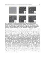
distribution (PSD) determined on the basis of TEM imaging – Figure 2. While Au10 contains
the nanoparticles of 7.3 ± 3.1 nm in diameter (Figures 2A,B), nanoparticles of 4.0 ± 0.9 nm in
diameter are encountered in Au17 (Figures 2C,D).
Interestingly, the optical density of Au10 is slightly higher than that obtained for Au17
(Figure 1) which can be related to a lower concentration of nanoparticles in Au17 solution.
The decrease of Au nanoparticles concentration in Au17 solution is most probably caused by
a higher amount of electro-deposited Au nanoparticles on electrode surface when the direct
current of 17 μA is passed through the ablation medium. This hypothesis will be discussed
in the next section.
Zeta potentials of Au nanoparticles ethanolic solutions have been measured and are
presented in Table 1. Both types of solutions, Au10 and Au17, reveal values below -30 mV
which indicates stable nanoparticle dispersions.
300 400 500 600 700
10 A
17 A
522
517
Extinction
Wavelength [nm]
Pulsed-Laser Ablation of Au Foil in Primary Alcohols Influenced by Direct Current
157
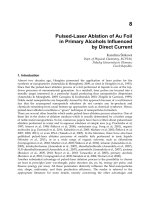
Fig. 2. TEM images (A, C) and appropriate PSD (B, D) of Au nanoparticles formed by
PLA+EPD process while 10 μA (A, B) and/or 17 μA (C, D) passing through ethanolic
ablation medium.
System label Solvent DC [μA] Zeta potential [mV]
Au10 Ethanol 10 -37.4 ± 2.0
Au17 Ethanol 17 -42.6 ± 0.8
Au10B Butanol 10 -10.8 ± 1.1
Au17B Butanol 17 -12.9 ± 1.2
Table 1. Zeta potentials determined for ethanolic as well as butanolic Au nanoparticle
solutions. DC means direct current.
In the next step, the PLA+EPD process has been performed in butanol. The resulting Au
nanoparticles solutions are entitled as Au10B and Au17B when direct current of 10 μA and
17 μA passed through the butanolic ablation medium, respectively. The values of zeta
potentials of these systems are presented in Table 1. They indicate rather unstable Au
nanoparticles solutions since the values are above -30 mV and below 30 mV. The differences
in zeta potential values of Au10, Au17, and Au10B, Au17B can be ascribed to different
Lasers – Applications in Science and Industry
158
dielectric constants of solvents: ethanol possess the value of 24.3, while butanol 17.1
(Sýkora, 1976).
UV-visible extinction spectra of Au10B and Au17B solutions are shown in Figure 3. Both
systems manifest themselves by a well pronounced surface plasmon extinction band with
the maximum located at 526 nm indicating thus similar sizes of Au nanoparticles. This idea
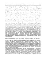
has been confirmed by PSD based on TEM imaging, presented in Figure 4. Au nanoparticles
in Au10B solution reveal sizes of 4.9 ± 1.2 nm and in Au17B sizes of 5.2 ± 1.7 nm
in diameter.
Fig. 3. UV-vis extinction spectra of Au nanoparticles generated by PLA+EPD process in
butanol while direct current of 10 μA and/or 17 μA allowed to pass through.
Similarly as in the case of ethanolic Au nanoparticle solutions, the concentrations of Au
nanoparticles appear to be slightly higher in Au10B than in Au17B solution. The reason will
be discussed in the next section.
To sum up, it can be concluded that Au nanoparticles of controlled sizes dispersed in
ethanol can be prepared by changing the direct current passing through the ethanolic
ablation medium during the PLA+EPD process. In contrast, the same factor (direct current
value) does not induce any changes in the average size of Au nanoparticles when formed by
the PLA+EPD process in butanol. Considering the zeta potential values of ethanolic and
butanolic Au nanoparticles solutions, this result is fully understandable since the higher the
zeta potential value, the stronger effect of applied electric field on the generated
nanoparticles. The longer aliphatic chain of butanol induces smaller zeta potential values of
generated Au nanoparticles and, consequently, the effect of direct current passing during
the PLA+EPD process is decreased.
Furthermore, ethanolic Au nanoparticles solutions can be prepared with a narrower particle
size distribution when the direct current of 17 μA instead of 10 μA employed. On the
contrary, the dispersity of butanolic Au nanoparticles solutions is almost negligibly
influenced. Obviously, the length of primary alcohols has a distinct effect on the average
size of Au nanoparticles generated by the PLA+EPD process.
300 400 500 600 700
17 A
10 A
526
526
Extinction
Wavelength [nm]
Pulsed-Laser Ablation of Au Foil in Primary Alcohols Influenced by Direct Current
159
Fig. 4. TEM images (A, C) and appropriate PSD (B, D) of Au nanoparticles formed by
PLA+EPD process while 10 μA (A, B) and/or 17 μA (C, D) passing through butanolic
ablation medium.
3.2 Substrates with electrophoretically-deposited Au nanoparticles
In this section, three types of substrates serving as electrodes in the PLA+EPD process will
be characterized by means of microscopic techniques and visible absorption spectroscopy.
With respect to the negative values of zeta potential of generated Au nanoparticles in both
primary alcohols, they are preferentially deposited on anodes.
3.2.1 ITO-coated glass substrates
SEM images of the ITO-coated glass substrates modified by electrodeposited Au
nanoparticles during the PLA+EPD process performed in ethanol are shown in Figure 5.
Comparing the SEM images of substrates in Figure 5A (10 μA direct current) and 5B (17 μA
direct current), a higher surface coverage of substrates by Au nanoparticles is observed at
higher current values than at the lower one. This microscopic observation goes hand in hand
with the fact deduced from the UV-visible extinction spectra of Au nanoparticles solutions
(discussed in the previous section): the final concentration of Au17 solution is lower than
that of Au10 solution. The reason for this difference lies in a larger amount of Au
nanoparticles being deposited under the higher than the lower current value and, as a
consequence, a decrease of Au nanoparticles concentration in Au17 solution being
determined.
Lasers – Applications in Science and Industry
160
Fig. 5. SEM images (A, B) and particular differential visible extinction spectra (C, D) of ITO-
coated glass substrates modified by Au nanoparticles electrodeposited at 10 μA (A, C)
and/or 17 μA (B, D) during PLA+EPD process performed in ethanol.
Furthermore, Au nanoparticle aggregates are frequently encountered under both direct
current values (Figures 5A and 5B). The aggregation can be also derived from the measured
visible extinction spectra of the two discussed substrate samples, presented in Figure 5C and
5D. The differential extinction spectra have been obtained by the subtraction of the
extinction spectrum of an unmodified ITO-coated glass substrate from that of a
nanoparticles-modified ITO-coated glass substrate. The position of the maximum located at
around 610 nm (Figure 5C) reports about aggregated Au nanoparticles on the substrates
modified under 10 μA. In the case of Au nanoparticles deposited on ITO-coated glass
substrates under 17 μA, there is even no distinct maximum of extinction band (Figure 5D)
indicating thus an extensive aggregation of Au nanoparticles.
The same type of experiments using ITO-coated glass substrates as electrodes in the
PLA+EPD process has been performed in butanol. The resulting SEM morphologies and
differential visible extinction spectra are shown in Figure 6. Comparing Figures 6A (10 μA
direct current) and 6B (17 μA direct current), a slightly higher amount of Au nanoparticles
can be seen on ITO-coated glass substrates when a higher current value used. This is quite
similar result to that observed in ethanolic systems. However, regarding the absolute counts
of electro-deposited Au nanoparticles, the substrates from ethanolic solutions are generally
Pulsed-Laser Ablation of Au Foil in Primary Alcohols Influenced by Direct Current
161
more covered by Au nanoparticles than that obtained in butanolic solutions. As it has been
already stated above, the higher zeta potential values of Au nanoparticles in ethanolic
solutions are most probably responsible for this result. Furthermore, Au nanoparticles are
more evenly dispersed on ITO-coated glass substrates immersed in butanolic than in
ethanolic solutions. This can be related to the effect of aliphatic chain length.
The differential extinction spectra, shown in Figures 6C and 6D, reveal a distinct band with
the maximum positioned at 575 nm when the lower, and at 615 nm when the higher current
values employed. The positions of the maxima of surface plasmon extinction bands correlate
with the microscopic observation presented in Figures 6A and 6B. Indeed, the higher the
surface coverage of substrates by Au nanoparticles, the more intense and red-shifted surface
plasmon extinction observed. In comparison to the extinction spectra of substrates
immersed in ethanolic solutions during the PLA+EPD process, the surface plasmon
extinction band is well-developed at both current values exploited for the PLA+EPD process
performed in butanol. Thus, regarding the aggregation of Au nanoparticles electro-
deposited on ITO-coated glass substrates, it is less pronounced in butanolic than in ethanolic
samples. Again, the same result evidenced by two independent methods, microscopic and
spectroscopic one.
Fig. 6. SEM images (A, B) and particular differential visible extinction spectra (C, D) of ITO-
coated glass substrates modified by Au nanoparticles electro-deposited at 10 μA (A, C)
and/or 17 μA (B, D) during PLA+EPD process performed in butanol.
Lasers – Applications in Science and Industry
162
3.2.2 FTO-coated glass substrates
As it has been already evidenced in the previous section, there are strong effects of direct
current value and the type of alcohol on the final coverage of a substrate by Au
nanoparticles. In order to investigate if there is any additional influence of substrate
roughness, FTO-coated glass substrates have been used as electrodes during the PLA+EPD
process performed in ethanol at both values of direct current, 10 μA as well as 17 μA.
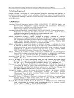
In Figure 7A, the SEM image of a cleaned bare FTO-coated glass substrate surface is shown.
Obviously, the surface of a FTO-coated glass substrate is very rough with plates and crystals
being of sizes of hundreds of nanometers. Taking into account that the local current density
can be very different on the edges of a plate and/or crystal, an inhomogeneous distribution
of Au nanoparticles and their aggregates on FTO-coated glass substrate can be awaited.
Figures 7B and 7C depict the SEM images of Au nanoparticles-modified FTO-coated glass
substrates when the lower and the higher electric field applied, respectively. Mutually
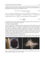
Fig. 7. SEM images of (A) cleaned bare FTO-coated glass substrate, (B, D) Au nanoparticles-
modified FTO-coated glass substrate when 10 μA allowed to pass through the ethanolic
ablation medium, and (C) Au nanoparticles-modified FTO-coated glass substrate when
17 μA used. Higher magnification is intentionally shown in (D).
Pulsed-Laser Ablation of Au Foil in Primary Alcohols Influenced by Direct Current
163
compared, there is seen the effect of the direct current value, i.e., with an increasing current
a higher surface coverage by Au nanoparticles is observed. When compared to ITO-coated
glass substrates serving as electrodes under otherwise the same experimental conditions
(SEM images presented in Figures 5A and 5B), the surface of FTO-coated glass substrates is
covered even more randomly by Au nanoparticles and their aggregates. These results
indicate that the surface roughness of a substrate does play an important role in the course
of electrophoretic deposition of Au nanoparticles. The assumption about the inhomogenity
of electric field, made a few lines above, is well documented by a characteristic SEM image
in Figure 7D revealing a preferential deposition of Au nanoparticles on the edges of plates
and crystals of a FTO-coated glass substrate.
3.2.3 HOPG substrates
Considering the results of the two previous sections, it can be hypothesized that a substrate
with a very smooth surface, such as HOPG for instance, could lead to homogeneously
dispersed electrodeposited Au nanoparticles since the current density will be homogeneous
everywhere on the substrate surface. In order to prove this hypothesis, the PLA+EPD
process performed in ethanol while 17 μA passed through has been chosen because under
these conditions, the highest degree of aggregation of electrodeposited Au nanoparticles
and inhomogenity in surface coverage were observed as shown in the two previous sections.
With respect to the fact that Au nanoparticles in the selected system are tiny (around 4 nm
in diameter), another microscopic technique than SEM has to be employed in order to
visualize isolated nanoparticles on HOPG substrates. Scanning electron microscopy (STM)
can fulfil this task when appropriate measuring conditions met (Durston et al., 1998; Wang
et al., 2000).
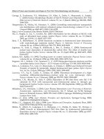
Figure 8 shows topographic as well as tunnelling current images of a freshly cleaved HOPG
surface without and with electrodeposited Au nanoparticles. Smoothness of HOPG surface
is well evidenced in Figure 8A where the value along z axis (perpendicular to the plane of
the image) stays well below 1 nm. The values of tunnelling current below 0.2 pA have been
recorded on a freshly cleaved HOPG substrate measured under ambient conditions, in air
and at the room temperature – Figure 8B. Under the same conditions, STM measurements of
a HOPG substrate which served as the anode during the PLA+EPD process have been
undertaken and one of the resulting topographic images together with its tunnelling current
values are shown in Figures 8C and 8D, respectively. Evidently, isolated Au nanoparticles
are randomly, however, quite homogeneously dispersed on the surface of a HOPG plate
(Figure 8C), the value of 6 nm along z axis is not surpassed. It is worth noting that
tunnelling current exceeds 0.4 nA (Figure 8D), which is the value of more than three orders
of magnitude higher than on a bare HOPG substrate (Figure 8B). This can be related to the
presence of Au nanoparticles.
It is known that HOPG substrate can contain terraces and steps as observed in Figure 8C.
Hypothetically, the edges of these terraces and steps could be the places of a locally higher
electrical density, hence, more electrodeposited Au nanoparticles could be awaited to occur
on these edges. However, this was not the case as evidenced in Figure 8C.
Figure 9 shows topography and tunnelling current image of a smaller flat surface area (200 x
200 nm
2
) on a HOPG substrate decorated with electrodeposited Au nanoparticles. At this
place it should be noticed that the bias voltage of +0.1 V has been applied between the
measured HOPG substrate and the tip during STM imaging. This is a sufficiently low value
to suppress any unwanted manipulation of Au nanoparticles (Durston et al., 1998).
Lasers – Applications in Science and Industry
164
Fig. 8. Topographic (A, C) and tunnelling current (B, D) images of HOPG substrate serving
as cathode (A, B) or anode (C, D) in PLA+EPD process while 17 μA passed through
ethanolic ablation medium. Dimensions of scans are 500x500 nm
2
.
Fig. 9. 200 x 200 nm
2
scan of HOPG substrate with electrodeposited Au nanoparticles due to
PLA+EPD process while 17 μA passed through ethanolic ablation medium:
(A) Topography, (B) tunnelling current image.
Pulsed-Laser Ablation of Au Foil in Primary Alcohols Influenced by Direct Current
165
Therefore, on the basis of these results, it can be concluded that the substrate roughness in
the range of hundreds of nanometers (the case of FTO-coated glass substrate) distinctly
impedes the homogeneous coverage of substrate by electrodeposited Au nanoparticles. On
the contrary, the surface roughness being below 1 nm does not hamper a homogeneous
distribution of electrodeposited Au nanoparticles.
3.3 Possible elucidation of direct current value influence on mechanism of Au
nanoparticles generation during PLA+EPD process in primary alcohols
Since the final stages of Au nanoparticles solutions and/or the electrodeposited Au
nanoparticles on different substrates have been investigated, it cannot be unambiguously
stated the exact formation mechanism of Au nanoparticles during the PLA+EPD process.
However, it can be hypothesized the influence of direct current value on the mechanism of
Au nanoparticles generation by the PLA+EPD process in comparison to a generally adopted
mechanism of pulsed-laser ablation itself.
The prevailing formation mechanism of nanoparticles by a classical pulsed-laser ablation
process implies the generation of a plasma plume followed by its cooling (Amendola &
Meneghetti, 2009; Tsuji et al., 2004). The former step is nothing else than the vaporization of
the part of a target which was attacked by the focused beam of laser pulses. During the
second step (plasma plume cooling), the formation of nanoparticles nuclei starts. The
driving force for the nucleation is the supersaturation in the plasma plume (Amendola &.
Meneghetti, 2009). Subsequently, the nuclei grow and coalesce into the sizes of resulting
nanoparticles. This last step strongly depends on the polarity of solvents, the presence
and/or the absence of simple ions or adsorbing species which may stabilize nanoparticles of
a particular size.
Under the assumption that our pulsed-laser ablation process in the selected solvent (e.g.
ethanol or butanol) is repeatedly performed in the same way and under otherwise the same
experimental conditions, the value of the applied electric field can induce changes rather in
the step of nuclei growth and coalescence than during the plasma plume generation and/or
the nucleation process. As it has been pointed out in section 3.1, ethanol possesses a higher
dielectric constant than butanol which means that ethanol is more easily polarized by an
increasing electric field than butanol. This implies that a further nuclei growth and
coalescence is possibly hindered in the case of 17 μA direct current value passing through
the ethanolic ablation medium when pulsed-laser ablation takes place. Basically, charged
nanoparticles of smaller sizes in diameter can be efficiently stabilized in a more polarized
solvent, i.e., in our case, at a higher current value passing through the ethanolic ablation
medium. Thus, a smaller average particle size is observed in Au17 than in Au10 solutions.
Nevertheless, this hypothesis needs a further experimental support which is beyond the
scope of this chapter.
4. Conclusions
The application of nanosecond laser pulses of 532 nm laser wavelength for the generation of
Au nanoparticles in primary alcohols (ethanol and butanol) has been discussed and the
impact of a direct current passing simultaneously through the ablation medium during the
pulsed-laser ablation process has been determined. On the basis of a complete
characterization of Au nanoparticles solutions, it has been concluded that the average size of
Lasers – Applications in Science and Industry
166
Au nanoparticles can be influenced by the type of an alcoholic ablation medium as well as
by the direct current value (the latter induces changes only in the case of ethanol).
Moreover, the length of aliphatic chains in the two selected alcohols affects the character of
coverage of ITO-coated glass substrates by Au nanoparticles; more evenly dispersed electro-
deposited Au nanoparticles have been encountered in the butanolic ablation medium. In
contrast, aggregates of Au nanoparticles have been observed when the ethanolic ablation
medium used. The amount of electrodeposited Au nanoparticles is generally higher in
ethanol than in butanol which can be related to the differences in zeta potential values of Au
nanoparticles. The surface roughness of substrates has appeared to be another very
important parameter influencing the final characteristic coverage of substrates by Au
nanoparticles generated by the PLA+EPD process. An excellent correlation between
microscopic and spectroscopic results has been demonstrated. Finally, a possible
explanation of the influence of direct current value on the mechanism of Au nanoparticles
generation during the PLA+EPD process has been proposed.
5. Acknowledgment
The author thanks to Mrs. Jiřina Hromádková for SEM and TEM imaging. Financial support
by GAČR P108/11/P657 is gratefully acknowledged.
6. References
Amendola, V., Rizzi, G.A., Polizzi, S. & Meneghetti, M. (2005). Synthesis of gold
nanoparticles by laser ablation in toluene: quenching and recovery of the surface
plasmon absorption. The Journal of Physical Chemistry B, Vol. 109, No. 49, pp. 23125-
23128, ISSN 1520-6106 print / ISSN 1520-5207 online
Amendola, V., Polizzi, S. & Meneghetti, M. (2007). Free silver nanoparticles synthesized by
laser ablation in organic solvents and their easy functionalization. Langmuir, Vol.
23, No. 12, pp. 6766-6770, ISSN 0743-7463 print / ISSN 1520-5827 online
Amendola, V. & Meneghetti, M. (2009). Laser ablation synthesis in solution and size
manipulation of noble metal nanoparticles. Physical Chemistry Chemical Physics, Vol.
11, pp. 3805-3821, ISSN 1463-9076 print / ISSN 1463-9084 online
Atwater, H.A. & Polman A. (2010). Plasmonics for improved photovoltaic devices. Nature
Materials, Vol. 9, pp. 205-213, ISSN 1476-1122 print / ISSN 1476-4660 online
Boyer, P., Menard, D. & Meunier, M. (2010). Nanoclustered Co-Au particles fabricated by
femtosecond laser fragmentation in liquids. J. Phys. Chem. C, Vol. 114, No. 32, pp.
13497-13500, ISSN 1932-7447 print / ISSN 1932-7455 online
Buining, PA, Humbel, BM, Phillipse, AP & Verkleij, AJ. (1997). Preparation of functional
silane-stabilized gold colloids in the (sub)nanometer size range. Langmuir, Vol. 13,
No. 15, pp. 3921-3926, ISSN 0743-7463 print / ISSN 1520-5827 online
Burakov, V.S., Tarasenko, N.V., Butsen, A.V., Rozantsev, V.A. & Nedelko, M.I. (2005).
Formation of nanoparticles during double-pulse laser ablation of metals in liquids.
Eur. Phys. J. Appl. Phys., Vol. 30, pp. 107-112, ISSN 0021-8979 print / ISSN 1089-7550
online
Pulsed-Laser Ablation of Au Foil in Primary Alcohols Influenced by Direct Current
167
Burakov, V.S., Butsen, A.V. & Tarasenko, N.V. (2010). Laser-induced plasmas in liquids for
nanoparticle synthesis. Journal of Applied Spectroscopy, Vol. 77, No. 3, pp. 386-393,
ISSN 0021-9037 print / ISSN 1573-8647 online
Carrara, M., Kakkassery, J.J., Abid, J.P. & Fermin, D.J. (2004). Modulation of the work
function in layer-by-layer assembly of metal nanoparticles and poly-L-lysine on
modified Au surfaces. Chem. Phys. Chem., Vol. 5, pp. 571-575, ISSN 1439-7641
Compagninni, G., Scalisi, A.A. & Puglisi, O. (2002). Ablation of noble metals in liquids: a
method to obtain nanoparticles in a thin polymeric film. Phys. Chem. Chem. Phys.,
Vol. 4, pp. 2787-2791, ISSN 1463-9076 print / ISSN 1463-9084 online
Dammer, O., Vlckova, B., Slouf, M. & Pfleger, J. (2007). Interaction of high-power laser
pulses with monodisperse gold particles. Materials Science and Engineering B, Vol.
140, pp. 138-146, ISSN 0921-5107
Darroudi, M., Ahmad, M. B., Zamiri, R., Abdullah, A.H., Ibrahim, N.A., Shameli, K. &
Husin, M.S. (2011). Preparation and characterization of gelatine mediated silver
nanoparticles by laser ablation. Journal of Alloys and Compounds, Vol. 509, pp. 1301-
1304, ISSN 0925-8388
de Boer, B., Hadipour, A., Mandoc, M.M., van Woudenbergh, T. & Blom, P.W.M. (2005).
Tuning of metal work functions with self-assembled monolayers. Adv.Mater.,Vol.
17, No. 5, pp. 621-625, ISSN 0935-9648 print / ISSN 1521-4095 online
Doron, A., Katz, E. & Willner, I. (1995). Organization of Au colloids as monolayer films onto
ITO glass surfaces: application of the metal colloid films as base interfaces to
construct redox-active monolayers. Langmuir, Vol. 11, No. 4, 1313-1317, ISSN 0743-
7463 print / ISSN 1520-5827 online
Durston, P.J., Palmer, R.E. & Wilcoxon, J.P. (1998). Manipulation of passivated gold clusters
on graphite with the scanning tunneling microscope. Appl. Phys. Lett., Vol. 72, No.
2, pp. 176-178, ISSN 0003-6951 print / ISSN 1077-3118 online
Fong, Y.Y., Gascooke, J. R., Visser, B.R., Metha, G.F. & Buntine, M.A. (2010). Laser-based
formation and properties of gold nanoparticles in aqueous solution: formation
kinetics and surfactant-modified particle size distributions. J. Phys. Chem. C, Vol.
114, No. 38, pp. 15931-15940, ISSN 1932-7447 print / ISSN 1932-7455 online
Franklin, S.R. & Thareja, R.K. (2004). Simplified model to account for dependence of ablation
parameters on temperature and phase of the ablated material. Applied Surface
Science, Vol. 222, pp. 293-306, ISSN 0169-4332
Georgiou, S.& Koubenakis, A. (2003). Laser-induced material ejection from model molecular
solids and liquids: mechanisms, implications, and applications. Chemical Reviews,
Vol. 103, No. 2, pp.349-393, ISSN 0009-2665 print / ISSN 1520-6890 online
Grabar, K.C., Allison, K.J., Baker, B.E., Bright, R.M., Brown, K.R., Freeman, R.G., Fox, A.P.,
Keating, Ch.D., Musick, M.D. & Natan, M.J. (1996). Two-dimensional arrays of
colloidal gold particles: a flexible approach to macroscopic metal surfaces.
Langmuir, Vol. 12, No. 10, 2353-2361, ISSN 0743-7463 print / ISSN 1520-5827 online
Grzelczak, M., Vermant, J., Furst, E.M. & Liz-Marzan, L.M. (2010). Directed self-assembly of
nanoparticles. ACS Nano, Vol. 4, No. 7, pp. 3591-3605, ISSN 1936-0851 print / ISSN
1936-086X online
Lasers – Applications in Science and Industry
168
He, H., Cai, W., Lin, Y. & Chen, B. Surface decoration of ZnO nanorod arrays by
electrophoresis in the Au colloidal solution prepared by laser ablation in water.
Langmuir, Vol. 26, No. 11, 8925-8932, ISSN 0743-7463 print / ISSN 1520-5827 online
Inasawa, S., Sugiyama, M., Noda, S. & Yamaguchi, Y. (2006). Spectroscopic study of laser-
induced phase transition of gold nanoparticles on nanosecond time scales and
longer. J. Phys. Chem. B, Vol. 110, No. 7, pp. 3114-3119, ISSN 1520-6106 print / ISSN
1520-5207 online
Jain, P.K., El-Sayed, I.H. & El-Sayed M.A. (2007). Au nanoparticles target cancer. Nanotoday,
Vol. 2, No. 1, pp. 18-28, ISSN 1748-0132
Kabashin, A.V., Meunier, M., Kingston, Ch. & Luong, J.H.T. (2003). Fabrication and
characterization of gold nanoparticles by femtosecond laser ablation in an aqueous
solution of cyclodextrins. J.Phys.Chem. B, Vol. 107, No. 19, pp. 4527-4531, ISSN 1520-
6106 print / ISSN 1520-5207 online
Kamat, P.V., Flumiani, M. & Hartland, G.V. (1998). Picosecond dynamics of silver
nanoclusters. Photoejection of electrons and fragmentation. J. Phys. Chem. B, Vol.
102, No. 17, pp. 3123-3128, ISSN 1520-6106 print / ISSN 1520-5207 online
Kim, S.S., Na, S.I., Kim, D.Y. & Nah, Y.Ch. (2008). Plasmon enhanced performance of
organic solar cells using electrodeposited Ag nanoparticles. Appl. Phys. Lett., Vol.
93, 073307, ISSN 0003-6951 print / ISSN 1077-3118 online
Kurita, H., Takami, A. & Koda, S. (1998). Size reduction of gold particles in aqueous solution
by pulsed laser. Appl.Phys.Lett., vol. 72, No. 7, pp. 789-791, ISSN 0003-6951 print /
ISSN 1077-3118 online
Le Ru, E. & Etchegoin, P. (2008). Principles of Suface-Enhanced Raman Spectroscopy and related
plasmonic effects, Elsevier Science, ISBN 0444527796, Amsterdam, The Netherlands
Link, S., Burda, C. Mohamed, M.B., Nikoobakht, B. & El-Sayed, M.A. (1999). Laser
photothermal melting and fragmentation of gold nanorods: energy and laser pulse-
width dependence. J. Phys. Chem. A,Vol. 103, No. 9, pp. 1165-1170, ISSN 1089-5639
print / ISSN 1520-5215 online
Link, S & El-Sayed, MA. (2003). Otpical properties and ultrafast dynamics in metallic
nanocrystals. Annu. Rev. Phys. Chem., Vol. 54, pp. 331-366, ISSN 0066-426X
Mafune, F., Kohno, Jy., Takeda, Y. & Kondow, T. (2001). Dissociation and aggregation of
gold nanoparticles under laser irradiation. J. Phys. Chem. B, Vol. 105, No. 38, pp.
9050-9056, ISSN 1520-6106 print / ISSN 1520-5207 online
Mafune, F., Kohno, Jy., Takeda, Y. & Kondow, T. (2002). Full physical preparation of sized-
selected gold nanoparticles in solution: laser ablation and laser-induced size
control. J. Phys. Chem. B, Vol. 106, No. 31, pp. 7575-7577, ISSN 1520-6106 print /
ISSN 1520-5207 online
Menendez-Manjon, A., Jakobi, J., Schwabe, K., Krauss, J.K. & Barcikowski, S. (2009).
Mobility of nanoparticles generated by femtosecond laser ablation in liquids and its
application to surface patterning. JLMN-Journal of Laser Micro/Nanoengineering, Vol.
4, No. 2, pp. 95-99, ISSN 1880-0688
Morfa, A.J., Rowlen, K.L., Reilly III, T.H., Romero, M.J. & van de Lagemaat, J. (2008).
Plasmon-enhanced solar energy conversion in organic bulk heterojunction
Pulsed-Laser Ablation of Au Foil in Primary Alcohols Influenced by Direct Current
169
photovoltaics. Appl. Phys. Lett., Vol. 92, 013504, ISSN 0003-6951 print / ISSN 1077-
3118 online
Mortier, T., Verbiest, T. & Persoons, A. (2003). Laser ablation of gold in chloroform solutions
of cetyltrimethylammoniumbromide. Chem.Phys.Lett., Vol. 382, pp. 650-653, ISSN
0009-2614
Muto, H., Yamada, K., Miyajima, K. & Mafune, F. (2007). Estimation of surface oxide on
surfactant-free gold nanoparticles laser-ablated in water. J.Phys.Chem. C, Vol. 111,
No. 46, pp. 17221-17226, ISSN 1932-7447 print / ISSN 1932-7455 online
Nah, Y.Ch., Kim, S.S., Park, J.H. & Kim, D.Y. (2007). Electrochromic coloration of MEH-PPV
films by electrodeposited Au nanoparticles. Electrochemical and Solid-State Letters,
Vol. 10, No. 1, pp. J12-J14, ISSN 1099-0062
Peng, Z., Walther, T. & Kleinermanns, K. (2005). Photofragmentation of phase-transferred
gold nanoparticles by intense pulsed laser light. J.Phys.Chem.B, Vol. 109, No. 33, pp.
15735-15740, ISSN 1520-6106 print / ISSN 1520-5207 online
Procházka, M., Mojzeš, M., Štěpánek, J., Vlčková, B. & Turpin, P.Y. (1997). Probing
applications of laser-ablated Ag colloids in SERS spectroscopy: improvement of
ablation procedure and SERS spectral testing. Anal. Chem., Vol. 69, No. 24, pp. 5103-
5108, ISSN 0003-2700 print / ISSN 1520-6882 online
Roduner, E. (2006). Nanoscopic Materials: Size-dependent Phenomena, The Royal Society of
Chemistry, ISBN-13: 978-0-85404-857-1, Dorchester, Dorset, UK
Semerok, A., Chaleard, C., Detalle, V., Lacour, J.L., Mauchien, P., Meynadier, P., Nouvellon,
C., Salle, B., Palianov, P., Perdrix, M. & Petite, G. (1999). Experimental
investigations of laser ablation efficiency of pure metals with femto, pico and
nanosecond pulses. Applied Surface Science, Vol. 138-139, pp. 311-314, ISSN 0169-
4332
Shoji, M., Miyajima, K. & Mafune, F. (2008). Ionization of gold nanoparticles in solution by
pulse laser excitation as studied by mass spectrometric detection of gold cluster
ions. J. Phys. Chem. C, Vol. 112, No. 6, pp. 1929-1932, ISSN 1932-7447 print / ISSN
1932-7455 online
Simakin, A.V., Voronov, V.V., Kirichenko, N.A. & Shafeev, G.A. (2004). Nanoparticles
produced by laser ablation of solids in liquid environment. Appl. Phys. A, Vol. 79,
pp. 1127-1132, ISSN 0947-8396 print / ISSN 1432-0630 online
Sládková, M., Vlčková, B., Mojzeš, P., Šlouf, M., Naudin & C. LeBourdon, G. (2006). Probing
strong optical fields in compact aggregates of silver nanoparticles by SERRS of
protoporphyrin IX. Faraday Discuss., Vol. 132, pp. 121-134, ISSN 0301-7249 print /
ISSN 1364-5498 online
Sobhan, MA, Ams, M, Withford, MJ & Goldys, EM. (2010). Ultrafast laser ablative
generation of gold nanoparticles: the influence of pulse energy, repetition
frequency and spot size. J. Nanopart. Res., Vol. 12, pp. 2831-2842, ISSN 1388-0764
print / ISSN 1572-896X online
Srnová, I., Procházka, M., Vlčková, B., Štěpánek, J. & Malý, P. (1998). Surface-enhanced
Raman scattering-active systems prepared from Ag colloids laser-ablated in
chemically modified aqueous media. Langmuir, Vol. 14, No. 16, 4666-4670, ISSN
0743-7463 print / ISSN 1520-5827 online
Lasers – Applications in Science and Industry
170
Sýkora, V. (1976). Chemicko-analyticke tabulky. SNTL, ISBN 80-03-00049-1, Prague, Czech
Republic
Sylvestre, J.P., Poulin, S., Kabashin, A.V., Sacher, E., Meunier, M. & Luong, J.H.T. (2004).
Surface chemistry of gold nanoparticles produced by laser ablation in aqueous
media. J.Phys.Chem. B, Vol. 108, pp. 16864-16869, ISSN 1520-6106 print / ISSN 1520-
5207 online
Rabani, E., Reichman, D.R., Geissler, P.L. & Brus, L.E. (2003). Drying-mediated self-assembly
of nanoparticles Nature, vol. 426, pp. 271-274, , ISSN 1545-0740
Rotello, V. (Ed.). (2004). Nanoparticles: Building blocks for nanotechnology. Kluwer
Academi/Plenum Publishers, ISBN 0-306-48287-8, New York, USA
Schnippering, M., Carrara, M., Foelske, A., Kotz, R. & Fermin, D.J. (2007). Electronic
properties of Ag nanoparticle arrays. A Kelvin probe and high resolution XPS
study. Phys.Chem.Chem.Phys., Vol. 9, pp. 725-730, ISSN 1463-9076 print / ISSN 1463-
9084 online
Šišková, K., Vlčková, B., Turpin, P.Y., Fayet, C. Hromádková, J. & Šlouf, M. (2007). Effect of
citrate ions on laser ablation of Ag foil in aqueous medium. Journal of Physics:
Conference Series, Vol. 59, pp. 202-205, ISSN 1742-6588 print / ISSN 1742-6596 online
Šišková, K., Vlčková, B., Turpin, P.Y., Thorel, A. & Grosjean, A. (2008). Porphyrins as SERRS
spectral probes of chemically functionalized Ag nanoparticles. Vibrational
Spectroscopy, Vol. 48, pp. 44-52, ISSN 0924-2031
Šišková, K., Pfleger, J. & Procházka, M. (2010). Stabilization of Au nanoparticles prepared by
laser ablation in chloroform with free-base porphyrin molecules. Applied Surface
Science, Vol. 256, pp. 2979-2987, ISSN 0169-4332
Šišková, K., Vlčková, B., Turpin, P.Y., Thorel, A. & Procházka, M. (2011). Laser ablation of
silver in aqueous solutions of organic species: probing Ag nanoparticle-adsorbate
systems evolution by surface-enhanced Raman and surface plasmon extinction
spectra. J. Phys. Chem. C, Vol. 115, pp. 5404-5412, ISSN 1932-7447 print / ISSN 1932-
7455 online
Šišková, K., Šafářová, K., Seo, J.H., Zbořil, R. & Mashlan, M. (2011) Non-chemical approach
toward 2D self-assemblies of Ag nanoparticles via cold plasma treatment of
substrates. Nanotechnology, Vol. 22, 275601 (7pp) NANO/381585/PAP, ISSN 0957-
4484 print / ISSN 1361-6528 online
Šloufová-Srnová, I. & Vlčková, B. (2002) Two-dimensional assembling of Au nanoparticles
mediated by tetrapyridylporphine molecules. NanoLetters, Vol. 2, No. 2, 121-125,
ISSN 1530-6984 print / ISSN 1530-6992 online
Šmejkal, P., Šišková, K., Vlčková, B., Pfleger, J., Šloufová, I., Šlouf, M. & Mojzeš, P. (2003).
Characterization and surface-enhanced Raman spectral probing of silver hydrosols
prepared by two-wavelength laser ablation and fragmentation. Spectrochimica Acta
A, Vol. 59, pp. 2321-2329, ISSN 1386-1425
Šmejkal, P., Pfleger, J., Šišková, K., Vlčková, B., Dammer,O. & Šlouf, M. (2004). In-situ study
of Ag nanoparticle hydrosol optical spectra evolution during laser
ablation/fragmentation. Appl. Phys. A, Vol. 79, pp. 1307-1309, ISSN 0947-8396 print
/ ISSN 1432-0630 online