Lasers Applications in Science and Industry Part 11 potx
Bạn đang xem bản rút gọn của tài liệu. Xem và tải ngay bản đầy đủ của tài liệu tại đây (939.42 KB, 20 trang )
Part 3
Biological Applications
10
Laser Pulse Application in IVF
Carrie Bedient, Pallavi Khanna and Nina Desai
Cleveland Clinic Foundation
U.S.A
1. Introduction
In-vitro fertilization (IVF) involves the culture and manipulation of gametes and embryos
within a laboratory environment. IVF procedures are channeled towards enhancing
fertilization and assisting the normal developmental physiology of the growing embryo to
increase implantation potential, culminating in the birth of a healthy baby. Laser and its
selective application to various steps in the IVF process is an area of growing interest.
In this chapter, we review the use of laser technology in the field of assisted reproduction as
well as in stem cell research. The first step in the IVF process involves fertilization of the
oocyte. For this to occur, sperm must penetrate the outer membrane known as the “zona
pellucida” which surrounds the egg. This natural barrier prevents the entry of multiple
sperm. Often it is necessary to assist fertilization by directly injecting a single sperm into the
oocyte, a technique known as Intracytoplasmic Sperm Injection (ICSI). Laser pulse has been
utilized to immobilize the human sperm tail before ICSI and in assisting the injection
technique by creating a hole in the zona (laser assisted ICSI). Once successfully fertilized, the
resulting embryo undergoes successive cell divisions. To implant on the uterine wall, the
embryo must escape from the surrounding zona, a process known as hatching. Laser
assisted hatching has been employed to create a controlled opening of the zona and facilitate
embryo implantation after transfer to the patient’s uterus. Zona opening through use of a
laser pulse has also been used to extract a single cell from the growing embryo for
preimplantation genetic diagnosis (PGD). Another application of the laser in reproductive
biology has been cellular microsurgery. Embryonic stem cells can be isolated from a
blastocyst stage embryo by selective ablation of trophectodermal cells, leaving behind the
stem cell source material. More recently, laser has been used to induce fluid loss from the
blastocyst stage embryo before cryopreservation. We discuss this novel application of laser
and our own work with artificially collapsing blastocysts before freezing to reduce ice
crystal damage.
This article also documents the evolution of laser pulse in IVF from the first generation of
lasers with UV range wavelengths to the newer generation of lasers with emissions in the
infrared range. Design characteristics for the ideal laser pulse for clinical IVF use are
presented. Finally, safety considerations as regards laser usage at such early stages of
development and potential risks to the newborn are discussed. The current FDA
classification and approved devices are also reviewed.
Numerous engineering devices have been used in biomanipulation and a thorough
understanding of both the disciplines of biology and engineering is imperative to develop
Lasers – Applications in Science and Industry
194
an efficient system for handling biological materials. Lab procedures used during IVF
involve some of the newest innnovations in medical technology, which may be attributed to
the constant pressure to increase accuracy and efficiency in completing procedures. Among
these innovations is laser technology. With the replacement of mechanical manipulation by
laser pulse, interuser variability may be lessened and consistently high laboratory standards
may be maintained.
In vitro fertilization (IVF) is one of several treatment options used in assisted reproduction.
It involves an interplay of diagnostic tests, hormonal supplementation, surgery and
laboratory techniques to help the subfertile couple achieve a pregnancy resulting in a
healthy baby. When a couple approaches the physician with the issue of subfertility, they
undergo a series of tests to determine the cause of subfertility and the optimal assisted
reproductive technique for their clinical situation. Causes of infertility may include lack of
eggs (oocytes), lack of sperm, inability of egg and sperm to meet due to blocked fallopian
tubes, inability to grow or implant in the uterus, or an unknown etiology.
In a typical IVF procedure, oocytes are harvested from the ovary after hormonal ovarian
stimulation. A sperm sample is collected from the male partner and washed from
surrounding semen. Alternatively, sperm is surgically retrieved from the testis or
epididymis. The oocytes are allowed to naturally fertilize in a Petri dish by co-incubation
with sperm. If the sperm count or motility is compromised, the insemination step is carried
out by direct injection of each oocyte with a single sperm using a glass needle. This
specialized procedure is known as ICSI (Intracytoplasmic Sperm Injection). If fertilization
occurs, a zygote forms. The zygote divides, undergoing cell cleavage, and forms an embryo.
The cells within the embryo continue rapidly dividing over the 4-6 day culture interval,
ultimately arranging in a distinct pattern to become a blastocyst. The blastocyst consists of a
peripheral layer of cells called the “trophectoderm” and a discrete grouping of cells known
as the inner cell mass (ICM) that will eventually form the fetus (Figure 1). The developing
embryo is protected by an outer shell of protein called the “zona pellucida” until it is large
enough to break free during a process known as “hatching”, in preparation for implantation
into the uterine wall.
Couples will have multiple embryos developing simultaneously in culture. Each embryo is
evaluated throughout its growth process. On the day of transfer 1-3 embryos are selected
from the laboratory dish and transferred to the patient’s uterus. This transfer may occur on
day 3 or day 5 after fertilization. Any additional embryos that are appropriately developed
are frozen for possible later transfer. Selection of embryos most likely to implant and lead to
a viable pregnancy is generally based on embryo morphology.
While some applications of lasers in IVF remain research topics, others have been
successfully employed in clinical practice. Laser assisted ICSI is used to aid fertilization.
Laser assisted hatching has been employed to create a controlled opening of the zona and
facilitate embryo implantation after transfer to the patient’s uterus. Zona opening through
use of a laser pulse has also been used to extract a single cell from the growing embryo for
preimplantation genetic diagnosis (PGD) and screen for genetic disorders prior to transfer.
Another application of the laser in reproductive biology has been cellular microsurgery.
Embryonic stem cells can be isolated from a blastocyst stage embryo by selective ablation of
trophectodermal cells, leaving behind the stem cell source material.
When first approaching the application of lasers to reproductive medicine, concerns were
raised as regards the safety profile and class of lasers to be used. Given the delicate stage of
human development at the time of fertilization, the major concerns regarding the use of
Laser Pulse Application in IVF
195
laser at earlier stages have been DNA damage, failed embryo development and possible
congenital disorders. These concerns primarily centered on laser wavelength, heat
generation and the amount of manipulation required of the fragile embryos. The primary
aim of this review is to assimilate the significance and limitations of laser technology in the
fast growing field of IVF and to outline the technical details to be considered when dealing
with laser pulses in reproductive technology.
Fig. 1. Egg fertilization and development
2. History of lasers in IVF
Laser technology has been used in Assisted Reproductive Technology since the 1980s (Ebner
et al., 2005). Laser pulse has found wide application in IVF technology, particularly when
efficient and precise manipulation is of paramount importance (Taylor et al., 2010).
Two general types of laser systems exist: contact and noncontact. Noncontact lasers do not
require additional physical manipulation of the embryo. Laser beams travel through the
objective lenses and only microscope stage movement is required to adjust embryo position
(Tadir et al., 1989, 1990, 1991). In contrast, contact laser systems require direct contact
between the laser and embryo, usually with either glass or an optical fiber (Neev et al., 1992).
This increases the likelihood of trauma to the embryo. Distance also affects damage – a
greater distance from the embryo to the laser will result in a larger hole in the embryo, even
if the difference in distance is only between the top and bottom of culture dish (Taylor et al.,
2010). Contact lasers also require use of a medium different than routine culture media in
order to affect the most efficient energy transfer.
The first generation of lasers to be used in IVF included argon fluoride (ArF), Xenon
chloride (XeCl), krypton fluoride (KrF), nitrogen and Nd:YAG lasers. The Nd:YAG laser
(1064 nm) was the first non-contact laser used in reproductive technologies. Initial use was
primarily for spermatozoa manipulation via optical trapping. Applications were then
expanded to add a potassium-titanyl-phosphate crystal in order to create a hole in the zona
pellucida to assist hatching (Tadir et al.,1989, 1990, 1991). Excimer lasers under development
around the same time period function by temporarily exciting rare earth gasses. After
comparing Nd:YAG lasers with the ArF (193 nm) excimer laser, the 193 nm was found to
produce a more uniform, smooth tunnel in the zona pellucida (Palanker et al. 1991). Similar
findings were noted with the XeCl (308 nm) excimer laser (Neev et al., 1992). Many excimer
Lasers – Applications in Science and Industry
196
lasers, including KrF (248 nm), and nitrogen lasers (337 nm) function at a wavelength in the
UV spectrum. Ultraviolet wavelengths are close to the absorption wavelength of DNA (260
nm). As a result, these lasers are minimally used in reproductive technologies due to concern
for mutagenic effects (Green et al., 1987; Hammadeh et al., 2011; Kochevar et al., 1989).
The next generation of lasers were designed to circumvent dangers of UV wavelength and
cytotoxicity by emitting wavelengths in the infrared region (>800 nm) (Ebner et al., 2005).
The first of the newer generation of lasers to be used in IVF was the 2.9 um pulsed
erbium:yttrium-aluminum-garnet laser (Er:YAG) (Feichtinger et al., 1992). This device’s use
is limited by the need for constant contact with the embryo, as well as limitations due to
interactions with the liquid media (Rink et al., 1996). The next development was the
holmium:yttrium-scandium-gallium-garnet laser (Ho:YSGG) with 2.1 um emission. In order
to retain the beneficial effect of the infrared emission wavelength with this laser, the
embryos require additional manipulation on a quartz slide, offsetting the advantages
obtained by a safer wavelength (Schiewe et al., 1995).
Currently, the 1.48 um diode wavelength indium-gallium-arsenic-phosphorus (InGaAsP)
semiconductor laser is used in IVF. It is a non contact laser, has a safer wavelength and
produces consistent results in the form of uniform, smooth edged tunnels (Rink et al., 1996).
This diode laser is delivered through a complex arrangement, requiring 3 mirrors and 3
lenses. A continuous laser beam is emitted and collimated by a microscope objective, and
then paired with a visible beam. These pass through a mirror which reflects the invisible
beam and is partially transparent to the 670 nm wavelength. Both beams are then directed
through the primary microscope objective lens and to the desired object. The variability is
less than 1 um, showing excellent reproducibility. Use of this laser does not require
additional manipulation of the embryo or pose threat to DNA integrity by damaging
radiation (Rink et al., 1996).
3. Laser characteristics for IVF
Lasers in IVF have a wide variety of applications, however, the desirable characteristics of
the laser used are similar across those applications. During laser targeting, the embryo’s
unique culture environment must remain consistent at all times to optimize the potential for
a viable pregnancy. To that end, any laser used in the IVF laboratory must be very precise,
extremely consistent with reproducible results and integrate well into the equipment
required for routine IVF. In addition, it must not pose any additional threat to the integrity
of the embryo. This includes an infrared wavelength to avoid direct chromosomal damage.
It also helps when a non-contact mode is employed to avoid any unnecessary manipulation
of the fragile embryo. Contact mode lasers requiring glass pipettes (UV wavelength) or
quartz fibers (infrared wavelengths) add a layer of complexity with respect to additional
manipulation of the embryo (Hammadeh et al, 2011). Similarly, no additional changes or
alternations of media should be made to avoid undue stress on the embryo’s environment,
which should be kept at a physiologic pH of 7.2 and at 37 degrees Celsius at all times to
optimize growth (Douglas-Hamilton & Conia, 2001 as cited in Al-Katanani et al., 2002). This
limits use to lasers which will not produce a thermal effect on the media containing the
embryo, which is impacted by the laser’s power, number of shots required, pulse length and
irradiation time. Ease of use and speed of a technique also contribute to maintaining an
appropriate environment for the embryo in that a faster procedure exposes the embryo to a
hostile environment for a much shorter period of time.
Laser Pulse Application in IVF
197
Lasers have three characteristics directly impacting embryos: wavelength, power and pulse
length. Wavelengths used in IVF tend to remain above 750 nm, in the infrared region, to
avoid mutagenic effects on DNA (Kochevar et al., 1989; Taylor et al., 2010). The amount of
power in a single laser remains constant but impacts the diameter of the hole created as well
as the amount of heat emitted in the process, with higher power translating to larger
diameter and increased heat (Taylor et al., 2010). Different lasers may each have a different
power. A similar scenario exists with pulse length, which can vary from 20 ms to >1,000 us.
A longer pulse length also correlates with a larger hole (Rink et al., 1996). Focusing the beam
waist on a target provides a larger diameter of tunnel as well (Neev et al.,1992).
Beyond the physical characteristics of the laser itself are secondary characteristics and
limitations impacting embryo use. For example, the mineral oil overlay may adhere to
optical fibers in a contact mode laser, absorb additional heat and thus expand, moving the
embryo and disrupting the path of the laser beam (Neev et al. 1992). The optical fibers used
must be sterilized, as well as the micropipette tips, expensive disposable equipment leading
to increased costs. Additional instruments used for manipulation introduce increased cost
and possible damage to the embryo in the form of contamination and constant physical
contact.
4. Applications of laser in IVF
Since the discovery of laser in 1960s, it has found application in many fields. The accuracy,
versatility and spatial focusing potential have helped it to find a wide application in the
Fig. 2. Applications of lasers in IVF
Lasers – Applications in Science and Industry
198
medical arena. The applications of laser in IVF may be classified into diagnostic and
interventional use for the ease of discussion (Figure 2). Diagnostic techniques include
assessing the strength of the zona pellucida and pre-implantation genetic diagnosis.
Interventional or therapeutic techniques involve manipulating individual gametes with
oocyte enucleation and sperm immobilization, aiding fertilization and development with
laser assisted ICSI and assisted hatching. Additional material may be obtained with stem
cell derivation and cellular microsurgery. Embryos are optimized for freezing with
blastocoele collapse. Regardless of the specific procedure, lasers provide an excellent
method for precise intracellular surgery (Raabe et al., 2009).
4.1 Diagnostic techniques
4.1.1 Assessing the zona pellucida
The zona pellucida is the hard protein coat surrounding and protecting the genetic material
carried within the egg. This layer is approximately 15-20 um thick and must be breached in
order for the sperm to make contact with the egg. In vivo, entry of the sperm initiates a
reaction to ensure no other sperm obtains access to the egg and further hardens the protein
layer to protect the zygote as it travels to the uterus. The proteinaceous coating must
ultimately thin to allow the embryo to break out of the shell and implant in the uterine
lining, or endometrium. Studies using laser pulses have determined the extent to which
the zona hardens during the period from oocyte to blastocyst (Montag et al., 2000b) and
further identify which embryos may need assistance with sperm entry or hatching. Zona
hardness is greater during in vitro culture as compared with in vivo growth. Montag et al.
(2000b) and Inoue & Wolf (1975a) have shown that identical laser pulses create larger
holes ranging from 13-17 um in the zona at earlier stages (oocyte, zygote) as compared to
more advanced stages of development (morula, blastocyst) where holes are smaller at 10-
13 um. Also, larger holes were created in blastocysts cultured in vivo when compared
with in vitro grown blastocysts, suggesting zona hardening during culture (Montag et al.,
2000b; Rink et al., 1996).
4.1.2 Pre-implantation genetic diagnosis
Pre-implantation genetic diagnosis (PGD) is the analysis of genetic material from the
developing embryo prior to transfer to the uterus. This can be done on the oocyte/zygote by
extracting a polar body or on the 8-cell embryo by extracting a single cell or blastomere.
Once genetic material has been obtained it may be analyzed for genetic abnormalities.
Screening of oocytes and embryos for common chromosome abnormalities, such as trisomy
21, can improve pregnancy rates and reduce miscarriage rates. Some couples may be
interested in screening for specific genetic problems typically severe or lethal conditions,
carried by one or both partners, in order to avoid having an affected child.
4.1.2.1 Polar body biopsy
During oocyte maturation to the metaphase II stage and also after fertilization, duplicated
genetic material is extruded as polar bodies. The polar body can provide helpful
information by reflecting the maternal genetic material contained in that egg. (Clement-
Sengewald et al., 2002; Verlinsky et al., 1990). Abnormal oocytes with genetic defects can be
selectively excluded (Clement-Sengewald et al., 2002). Genetic assessment of the unfertilized
egg permits women who would not consider discarding an affected embryo due to personal
beliefs to be screened for age related aneuploidy or hereditary chromosomal defects. It may
Laser Pulse Application in IVF
199
also be performed in countries where it is illegal to perform blastomere biopsy to genetically
screen embryos (Dawson et al., 2005; Clement-Sengewald et al., 2002; Montag et al., 2004).
The polar body is located in the perivitelline space directly under the zona pellucida and
outside of the oocyte. It can be extracted by traversing the zona. Prior to the introduction of
lasers, biopsy was typically done by degradation of the zona pellucida with Tyrode’s acid,
after which a capillary tube would be used to aspirate the polar body. This technique was
highly variable, led to inconsistent opening size and could easily lead to further damage or
loss of cells. It also requires changing culture media and increasing the risk of
contamination. Alternatively to acid, mechanical biopsy could be performed with sharp
glass instruments, again introducing possibility for structural damage or alteration during
the manipulations (Clement-Sengewald et al., 2002; Dawson et al., 2005; Ebner et al., 2005).
Regardless of the method used, the oocyte must remain intact to continue development and
the polar body must allow adequate, undamaged material for genetic analysis.
When polar body biopsy is performed using lasers, a pulse is directed at the region of zona
pellucida nearest the polar body. In a description by Montag et al. (1998) two pulses of 14 ms
are given by a 1.48 um non contact laser, creating an opening of approximately 14-20 um.
The material is then extracted with a blunt capillary, avoiding potential damage to the
oocyte with a sharp instrument, and the entire procedure is completed in just a few minutes
(Montag et al., 1998). A similar procedure has been described by Clement-Sengewald et al.
using a nitrogen 337 nm laser and a Nd:YAG laser (Clement-Sengewald et al., 2002). That
same group described extraction of the polar body using optical tweezers (Nd:YAG, 1064
nm) and laser (nitrogen, 337 nm) pressure catapulting to collect the polar body, further
eliminating a source of contamination by introduction of another pipette. To catapult the
polar body, it was mounted to a membrane on a slide with the inner cap of a microfuge
tube placed next to it. One pulse of the laser was aimed at the membrane, freeing it to
catapult onto the nearby tube cap (Clement-Sengewald et al., 2002; Schutze & Lahr, 1998).
Oocyte recovery rates were only 67% in humans following this complete laser extraction
method. An improved blastocyst survival rate was noted when access was obtained via
laser as compared with acid solution, further strengthening the argument for laser use
(Dawson et al., 2005).
4.1.2.2 Blastomere biopsy
Blastomere biopsy is similar to polar body biopsy in that both techniques require careful
extraction of genetic material from a very delicate structure followed by genetic screening.
This procedure is also performed to facilitate selection of the embryo most likely to
establish a viable pregnancy with healthy offspring. Blastomere biopsy becomes relevant
at a later stage in development, after fertilization. Couples opt for this technique typically
when one or both parents carry a hereditary genetic defect they want to avoid passing to
children (Vela et al., 2009) or in cases of advanced maternal age to screen against
aneuploid embryos.
Until the introduction of laser assisted opening of the zona, blastomere biopsy was
performed by zona drilling with an acid tyrodes solution (Talansky & Gordon, 1986, as
cited in Malter & Cohen, 1989). The embryo is immobilized and held in place while acid
in a microcapillary tube is gently blown against the zona until it starts to dissolve. The
acid is then aspirated and the embryo is quickly rinsed to remove traces of acid. The
technique requires speed and expertise so as not to injure the embryo. The hole size can
often be variable.
Lasers – Applications in Science and Industry
200
The procedure for a blastomere biopsy using laser is similar to PGD with a polar body.
Laser pulse(s) are utilized to create a hole in the zona pellucida, through which a blastomere
is removed (Taylor et al., 2010). Analysis of laser pulse length in generating a hole for
blastomere extraction showed longer pulse duration (0.604 ms vs. 1.010 ms) produced larger
hole sizes (10.5 nm vs. 16.5 nm, respectively) (Taylor et al., 2010). However, Taylor et al.
found no difference in number of blastomeres lysed for a given pulse duration. They did
find a difference in number of blastomeres required to be obtained in each group. The
longer pulse duration group was noted to require additional blastomere biopsy. These
results were impacted by half of the affected embryos originating from the same patient
with poor quality embryos and cannot clearly be attributed to laser use.
Studies comparing embryos after laser assisted biopsy to untreated embryos showed no
adverse effects of treatment and similar hatching and development rates (Joris et al., 2003).
When performed with human embryos, pregnancy rates after laser blastomere biopsy are
comparable to mechanical blastomere biopsy (Schopper et al., 1999). Comparison of
blastomeres obtained during acid and laser mediated biopsies showed laser biopsy
generated more intact blastomeres (Joris et al., 2003).
4.2 Interventional techniques
4.2.1 Laser assisted ICSI
With male factor infertility, it is often necessary to assist fertilization by directly injecting a
single sperm in to the oocyte, a technique known as Intracytoplasmic Sperm Injection (ICSI).
The limited number of viable or motile sperm decreases chances of fertilization and a
successful pregnancy using the conventional oocyte insemination technique. ICSI is
performed by aspirating a sperm into a sharp glass needle (5 um in diameter), perforating
the oocyte’s zona and depositing the sperm into the ooplasm (Palermo et al., 1992).
Deformation of the oocyte during the injection process can trigger oocyte degeneration
either as a result of egg fragility or due to force required to traverse the membrane (Rienzi et
al., 2001, 2004; Abdelmassih et al., 2002; Palermo et al., 1996). Damage to the oocyte also
occurs by disturbing the spindle apparatus, damaging the oocyte cytoskeleton, introducing
harmful materials or by removal of cytoplasm during the injection procedure (Moser et al.,
2004; Hardarson et al., 2000; Tsai et al, 2000; Dumoulin et al., 2001).
Laser assisted zona drilling prior to ICSI can be used to increase the likelihood of successful
fertilization (Palanker et al., 1991). This may be done with a 193 nm ArF laser, which was
shown to drill very precise holes without undesired damage to the zona pellucida (Palanker
et al., 1991). A 1.48 um diode laser can also be used to assist with ICSI (Rienzi et al., 2001,
2004). A small channel of 5-10 um in diameter is drilled using low energy pulses of less than
2 milliseconds duration, taking care to leave the innermost layer of zona intact. The ICSI
injection pipette is introduced through this channel to deliver the previously immobilized
sperm (Rienzi et al.,
2001, 2004; Abdelmassih et al., 2002). Prior to laser assistance, this
technique was limited by operator skill and a non standardized tunnel size, potentially
leading to polyspermy or loss of genetic material (Rink et al, 1996). Laser assisted ICSI
provides a less traumatic method to create an opening in the zona pellucida for the purpose
of sperm microinjection, leading to decreased breakdown of oocyte membrane (5% vs. 37%,
Abdelmassih et al., 2002) and increased oocyte preservation, 97% vs. 85%, after ICSI (Rienzi
et al., 2004). The type of laser used is in infrared range and is not absorbed by nucleotides
and is considered safer than its counterparts (Ebner et al., 2005; Kochevar et al., 1989). The
decreased force necessary in penetrating the egg with the ICSI needle in entry may also
Laser Pulse Application in IVF
201
preserve embryo quality (Rienzi et al., 2001; Nagy et al., 2001) and has been shown to
improve embryo quality and survival, even when using poor quality oocytes (Abdelmassih
et al., 2002). To ensure even less traumatic manipulation, sperm may be injected into the
oocyte through a laser drilled hole using optical tweezers to achieve fertilization (Clement-
Sengewald et al., 1996, 2002).
Ultimately, to establish a pregnancy the embryo must “hatch” out of zona and implant on
the uterine wall. A potential drawback to laser assisted ICSI is that the thinning of the zona
may result in duplicate hatching sites. This allows the embryo to escape via two openings,
resulting in either degeneration or twinning. The theoretical concern is that the embryo
would hatch through the site created during assisted hatching but also through the ICSI site
as well (Abdelmassih et al., 2002). Moser et al. (Moser et al., 2004) discovered thinning the
zona pellucida instead of completely opening it eliminated the concern for a second opening
and incidentally improved blastulation rates through that site as well.
4.2.2 Sperm immobilization & selection
Sperm immobilization is critical when performing ICSI. The beating of the sperm tail in
the oocyte after injection can cause damage. Typically during ICSI, the sperm tail is
positioned under the glass microcapillary injection needle. The needle is brought down
and across the tail causing it to break and immobilizing the sperm (Palermo et al, 1992;
Nijs et al, 1996; Vanderzwalmen et al., 1996; Yanagida et al., 2001). Fertilization rates are
also closely linked to sperm immobilization, increasing from 54% to 68% (Vanderzwalmen
et al., 1996). Disruption of the sperm membrane aids the release of sperm factors
important in oocyte activation (Dozortsev et al., 1997). Low level laser pulse can also be
used to immobilize sperm, without affecting viability (Montag et al., 1998, 2000d, 2009;
Rienzi et al, 2004; Tadir et al., 1990).
A rather unique application of laser is to identify and select viable sperm for ICSI. Usually
motility is used as an indicator of living sperm. However in severe male factor cases such as
asthenozoospermia, no motile sperm may be evident. This makes it very difficult to identify
and select viable sperm for ICSI. A single laser pulse applied at the tip of a sperm’s tail can
aid in distinguishing living non-motile sperm from dead sperm. The tail of a viable sperm
will curl, whereas the nonviable sperm will not respond to the laser pulse. Fertilization rates
would be expected to be correspondingly higher if better sperm are selected for the injection
(Montag et al., 2000d, 2009). An alternative method for manipulating sperm includes optical
trapping. Optical trapping uses a single beam non contact laser to move sperm during after
immobilization or during ICSI (Clement-Sengewald et al., 1996, 2002; Tadir et al., 1991). The
optical tweezers can hold actively moving sperm and determine their velocity (Clement-
Sengewald
et al., 2002; Tadir et al., 1991). Lasers used in optical trapping may be either
infrared or ultraviolet (Clement-Sengewald et al., 2002; Tadir 1989. Advantages of this
technique include ease, no requirement for sophisticated micromanipulation skills or
additional expensive disposable equipment. The capacity for the optical tweezers to
determine velocity permits studies of medications on motility (Tadir et al., 1989). It may also
be used for polar body extraction or chromosomal manipulation (Tadir et al., 1991).
Disadvantages include increased exposure time of the embryo to lasers, possible ultraviolet
exposure depending on wavelength utilized and a potential adverse effect on the sperm
(Tadir et al., 1989).
Lasers – Applications in Science and Industry
202
4.2.3 Assisted hatching
To establish a successful pregnancy, the developing embryo must break out of its shell (zona
pellucida) on day 5 or 6 by a process known as hatching. Once the embryo is hatched, it may
implant on the endometrium and begin to grow but if it is unable to hatch, the pregnancy
will not continue. Various factors contribute to failed hatching and implantation – increased
maternal age, decreased egg quality, poor embryo and zona morphology to name a few, and
the exact cause of failed hatching is unknown (Balaban et al., 2002). An increase in zona
hardness has also been implicated during in vitro fertilization (Inoue & Wolf, 1975;
Montag et al., 2000; Balaban et al., 2002). The physiologic mechanism leading to hatching is
likely different in vivo than in vitro, with in vitro embryos hatching when a critical cell
number has been reached. This is compared with hatching independently of cell mass in
vivo, likely related to lytic enzymes found in vivo (Montag et al., 2000a). It has become
relatively common practice to facilitate the hatching of blastocysts by creating an artificial
opening in the zona pellucida either by mechanical, chemical or optical methods,
although the exact population benefiting most from this procedure is yet to be determined
(Hammadeh et al., 2011). Assisted hatching has been proposed to be potentially more
beneficial in patients over 40, with thicker zonae or poor prognosis patients (Balaban et al.,
2002; De Vos & Van Steirteghem, 2000; Hammadeh et al., 2011; Sagoskin et al., 2007;
Lanzendorf et al., 1998).
In the late 1980s, Cohen et al. mechanically opened the zona pellucida, achieving higher
implantation rates. Since that time, multiple methods have been proposed to facilitate
hatching (De Vos & Van Steirteghem, 2000; Cohen et al., 1990). Zona drilling uses Tyrode’s
acid solutions to create a defect in the zona (Malter & Cohen 1989; Ebner et al., 2005; Neev et
al., 1992; Balaban et al., 2002; De Vos & Van Steirteghem, 2000), whereas mechanical hatching
utilizes a microneedle to slice off a thin piece of the zona (Malter & Cohen 1989; Ebner et al.,
2005; Balaban et al., 2002; De Vos & Van Steirteghem, 2000). Enzymatic hatching using
pronase to generally thin the zona pellucida is also an accepted method of assisted hatching
(Balaban et al., 2002; Fong et al., 1998). Direct comparison of hatching methods is challenging
due to inter-operator variability, differing depths of zona penetration and heterogeneous
patient populations.
Laser provides an alternate means to facilitate hatching, and is faster and easier than other
methods (Balaban et al., 2002). The 2.94 um Er:YAG laser has been used for assisted hatching
with a significant increase in pregnancy rates (Antinori et al., 1996). The laser was deemed
safe for clinical use after trials in animal models (Obruca et al., 1994, as cited in Obruca et al.,
1997). The 1.48 micron infrared diode laser beam has been more widely used in clinical IVF
labs as an efficient and simple method for embryo hatching. Multiple studies have
demonstrated its safety (Sagoskin et al., 2007; Lanzendorf et al.,
2007; Wong et al., 2003) as
well as efficacy when compared to acid hatching (Lanzendorf et al., 2007; Balaban et al., 2002;
Jones et al., 2006).
The optimal technique for laser assisted hatching is still being debated. The laser can be
used to thin a large area of the zona, partially hatch by creating an incomplete hole or
completely hatch by drilling completely through the zona (Figure 3). The number of shots
and duration of pulse exposure is also subject to discussion with investigators varying
parameters to achieve an appropriate tunnel size. Optimal hole size is as yet unclear,
although >10 um leads to improved results (Ebner et al., 2005). A study by Montag et al.,
found no evidence of impaired growth or adverse effects as a result of laser hatching
(Montag et al., 2000a). Advocates of partial hatching argue increased safety using this
Laser Pulse Application in IVF
203
method because the laser does not come in to direct contact with the embryo. Finally,
proponents of the zona thinning technique contend that overall thinning will avoid
inadequate hatching and be more likely to correspond with the natural hatch site due to a
larger area being ablated (Moser et al., 2004). Studies comparing multiple methods of
hatching yield inconclusive results and no definitive recommendations can be made. A
study comparing pulse intensity and number of pulses determined 50% intensity with 2
pulses was the optimal setting to increase blastocyst formation (Tinney et al., 2005) by
creating a complete hole rather than the less effective zona thinning. Specific settings to
achieve those results would be expected to vary based on the power of different lasers.
Mantoudis et al., 2001, compared the three methods of laser hatching and determined partial
hatching or thinning the zona is more effective. Implantation rates were 2.8%, 9.1% and 8.1%
in the complete hatching, partial hatching and zona thinning groups. Clinical pregnancy
rates were also significantly improved with 5.2%, 18.3% and 22.1%, respectively. Thinning in
this study ablated the zona around 25% of the embryo, leaving only the inner membrane of
the zona pellucida intact in that section. It is unclear what the diameter of the complete
hatch site was in this study. Another concerning trend in this study was 22% of pregnancies
were multiple pregnancies, more than typically seen (Mantoudis et al., 2001), which is not
unique to this trial (Hammadeh et al., 2011). In contrast to the findings of Mantoudis et al.,
Wong et al. found improved hatching rates with complete hatching compared to partial
hatching, 38% vs. 25%, respectively (Wong et al., 2003). Laser-assisted zona pellucida
thinning prior to ICSI resulted in decreased oocyte degeneration rates, better blastocyst
hatching rates and improved pregnancy rates after day 3 embryo transfer (Moser et al.,
2004). In this study embryos had their zona pellucida thinned by 50% via 5-6 laser pulses,
covering at most 70 um of zona. A trial by Balaban et al. compared assisted hatching by
laser, acid Tyrodes, pronase treatment and mechanical technique. These investigators
concluded that all methods were comparable based on the outcome parameters studied,
including implantation and pregnancy rates, multiple pregnancy rates and abortion rates
(Balaban et al., 2002). Additional studies comparing laser assisted hatching with acid drilling
showed no significant differences with respect to pregnancy rates (Lanzendorf et al., 2007;
1999; Jones et al., 2006).
Laser assisted hatching is generally well-accepted in IVF labs, allowing improved
standardization between operators (Lanzendorf et al., 2007; Jones et al., 2006). Children
followed to one year of age after an assisted pregnancy using laser assisted hatching were
Fig. 3. Assisted hatching
Lasers – Applications in Science and Industry
204
found to have no increase in congenital malformations (Kanyo & Konc, 2003). Other
pregnancies have also yielded healthy babies following laser assisted hatching (Lanzendorf
et al., 1998). The first 1.48 um laser to receive US FDA approval for clinical use in assisted
hatching was the ZILOS-tk in 2004. This was followed by the Octax laser in 2006 and the
Saturn Active Laser System in 2008.
4.2.4 Laser pulse blastocyst collapse
As the efficiency of embryo culture increases, supernumerary embryos are produced and
cryopreserved for transfer in a future cycle (Iwayama et al., 2010; Gardner et al., 1998). One
method of cryopreservation known as “vitrification” involves high molar concentrations of
cryoprotectants and rapid cooling of the embryo at rates of -20,000 C°/min (Desai et al.,
2011). This cooling technique is extremely effective for embryos at all stages. The high
cooling rate prevents ice crystal formation in cellular cytoplasm. Post-warming survival
rates have been high with this technique. Yet it was observed that well-developed and
expanded blastocysts had lower survival rates than the less mature blastocyst or the morula
stage embryo (Vanderzwalmen et al., 2002). The primary structural difference between the
early stage blastocyst or morula and the later stage blastocyst is the presence of a fluid filled
cavity in the expanded blastocyst, called a blastocoele.
Artificial shrinkage of the blastocyst to reduce fluid volume in the blastocoelic cavity before
freezing was investigated as a technique to increase survival and ultimately increase clinical
pregnancy and implantation rates (Vanderwalzmen et al., 2002). This has been carried out by
either mechanical puncture of the blastocyst cavity with a needle and withdrawal of fluid
(Vanderwalzmen et al., 2002), use of osmotic shock to draw out fluid (Iwayama et al., 2010)
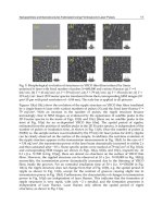
or by using laser pulses to collapse the blastocyst (Mukaida et al., 2006) (Figure 4). In
mechanical collapse, the inner cell mass of the blastocyst is positioned at 12 o’clock or 6
o’clock position. A glass micro needle is introduced into the cavity of the blastocoel and
then withdrawn, which results in collapse of the cavity over 30 seconds to 2 minutes
(Vanderwalzmen et al., 2002; Mukaida et al., 2006). During osmotic shock, the blastocyst is
passed through media with high concentrations of sucrose to essentially “dehydrate” the
embryo (Iwayama et al., 2010). For laser collapse, a short duration laser pulse directed at
the trophectoderm in a region away from the inner cell mass is delivered, shrinking the
cavity immediately without additional manipulation of the embryo (Mukaida et al., 2006).
No statistical difference was seen on comparison of mechanical versus laser shrinkage
(Mukaida et al., 2006), or with osmotic versus laser shrinkage (Iwayama et al., 2010),
although results were improved in both cases as compared to controls (Mukaida et al.,
2006; Iwayama et al., 2010). Human and mouse blastocsyts vitrified after mechanical or
laser collapse have fewer damaged cells than untreated controls and total blastomere
counts are higher after 24 hours of culture (Desai et al., 2008). The rate of re-expansion
after warming was also found to be higher (Desai et al., 2008). In this study, an OCTAX
1.48 uM laser was used to deliver a single shot 10 ms pulse to the junction of cells located
in the trophectoderm. The complete collapse of the blastocysts was seen within 2-4
minutes.
The major safety concern for use of laser is that the inner cell mass which ultimately
becomes the fetus will inadvertently be exposed to the laser pulse. At this time the FDA has
not approved this particular application of the laser in the U.S.
Laser Pulse Application in IVF
205
Fig. 4. (A) Blastocyst during mechanical collapse with ICSI needle. (B) Blastocyst
immediately after collapse, (C) Blastocyst rewarmed after laser collapse, (D) 3 hours after
rewarming, (E) after culture for 24 hours
4.2.5 Cellular microsurgery
Lasers may be used to remove material within the blastocyst that may prove detrimental to
its development. This detrimental material includes cellular fragments or necrotic
blastomeres. During embryo development it is possible to see cellular fragments appear.
This is a process that may lead to impaired development as cells are dividing and natural
planes are obstructed with fragments (Ebner et al., 2005; Alikani et al., 1999). Embryos with
higher levels of fragmentation were found to have decreased implantation and pregnancy
rates (Alikani et al., 1999; Sathananthan et al., 1990). Necrotic blastomeres are frequently
observed in cryopreserved embryos upon warming. Release of toxic metabolites from dying
cells may interfere with subsequent implantation. (Rienzi et al., 2002). The laser can be used
to create a small opening enabling extraction of fragments as well as dead cells. When
necrotic blastomeres are removed, cleavage and implantation rates improve, and pregnancy
rates increase from 17% to 45% (Rienzi et al., 2002).
Another type of microsurgery that is well suited to the laser technology is preparation of
zonae for the hemizona assay. The hemizona assay is used as a diagnostic tool to assess the
binding capacity of sperm to the oocyte zona and also as a research model to study the
effects of the environment or administered medications on the zona pellucida (Schopper et
al., 1999; Montag et al., 2000c, 2009). For this procedure, the test oocyte is sliced into two
sections, one to be used as the control and the other for the test treatment. A critical aspect of
maintaining the accuracy of the test is that the oocyte is evenly divided so comparisons can
be made. This bi-section can be accomplished using a mechanical technique or with the
laser. Consecutive adjacent laser shots can be used to drill a series of holes through an
oocyte immobilized using a micropipette (Montag et al., 2000c). A study comparing laser to
mechanical hemizona creation showed no difference in sperm binding between the two
Lasers – Applications in Science and Industry
206
methods, and the laser drilling produced very even, flat hemizonae (Montag 2000c). The
hemizona assay is performed more easily using lasers via mechanical techniques with a
microscalpel (Schopper et al., 1999).
The laser is particularly well suited for cellular microsurgery. The introduction of a laser
with a femtosecond pulse to be used as a laser scalpel may further increase the accuracy of
diagnostic and interventional procedures performed on the embryo (Rakityansky et al.,
2011). Biopsy of the trophectodermal cells of the blastocyst for pre-implantation genetic
screening is one possibility. Currently the ability to accurately deliver the laser pulse to a
very fine area and minimize heat transfer to adjacent cells has been a concern, limiting this
use of lasers to research. Lasers may also be further developed to aid in elucidating a
proteomic profile for embryos to help predict their success (Vela et al., 2009).
4.2.6 Stem cell derivation
Embryonic stem (ES) cell lines are derived from the inner cell mass of blastocysts. Once
isolated the inner cell mass can be used to establish pluripotent stem cell lines for use in
transplants and to study cellular differentiation (Turetsky et al., 2008). The ICMs from
embryos that have been diagnosed with genetic disorders after PGD screening are potential
source material for developing cell lines containing specific genetic conditions (i.e. cystic
fibrosis, hemophilia) for use in research. Mechanical dissection of the inner cell mass from
the trophectoderm is highly operator dependant, and chemical dissolution of the
trophectoderm with Tyrode’s acid subjects the inner cell mass to possible damage from the
corrosive fluid (Turetsky et al., 2008). Removal of the inner cell mass using several laser
pulses has been shown to be an effective and an easy method to extract this stem cell source
material from the blastocysts to establish ES cell lines (Turetsky et al., 2008; Tanaka et al.,
2006). Laser also facilitates ICM isolation for cryopreservation of the stem cell source
material (Desai et al., 2011).
4.2.7 Oocyte enucleation
Oocyte enucleation is similar to the process of dissecting the inner cell mass away from the
outer layer of cells in an embryo. It is more challenging in the sense that only one cell, the
oocyte, exists rather than the many cells in a blastocyst. Enucleation separates the nucleus of
the oocyte from the remaining cellular material, effectively removing all genetic potential
from the oocyte (Hirata et al., 2011). This is done to establish cell lines for research purposes
and to explore the genetic reprogramming potential of the oocyte cytoplasm (Hirata et al.,
2011; Malenko et al., 2009; Raabe et al., 2009). Once the nucleus with chromosomes is
removed using a micropipette, new genomic material from somatic cells is introduced in to
the enucleated oocyte (Malenko et al., 2009; Hirata et al., 2011). This may be done to develop
embryonic cell lines for future therapeutic use (Hirata et al., 2011). Using this procedure the
cytoplasm of the oocyte reprograms differentiated somatic chromosomes into embryonic
cells (Hirata et al., 2011). Women who may feel uncomfortable donating eggs for research or
therapeutic uses because the oocyte contains their genetic material may be more willing to
donate knowing their genome will be removed (Hirata et al., 2011).
The 1.48 uM diode laser may be used in conjunction with oocyte enucleation procedures in a
similar manner as with ICSI and assisted hatching. A small hole is drilled in the zona
pellucida, through which the nucleus is removed while leaving most of the cytoplasm (Li et
al., 2009). A picosecond pulsed 405 nm diode laser also effectively aids in enucleation with
Laser Pulse Application in IVF
207
extremely short pulse duration of 1-2 seconds. This laser has not been approved for use in
humans, however it is of interest due to its effect on intracellular structures (Raabe et al.,
2009). Although intracellular organelles were not found to be directly harmed following
irradiation, function during the cell division process was prolonged when compared to non-
irradiated cells. This indicates non-specific damage may have occurred (Raabe et al., 2009)
and cautions for judicious study of non-specific effects of irradiation in human embryology.
5. Safety & regulations
Lasers are currently considered by the Food and Drug Administration as a Class II device,
special controls. As a Class II device, lasers must go through more than the general control
measures regarding marketing and safety standards. They do not, however, have the
stringent requirements and prolonged approval process prior to marketing required of the
more highly regulated Class III devices. Class III devices are considered to be high risk, to
the level of supporting life or presenting an unreasonable risk of harm. Three lasers have
been approved by the FDA for use in reproductive technology: Saturn Active Laser System,
Octax Laser Shot System and Hamilton Thorne Zona Infrared Laser Optical System. These
lasers have only been approved for ablation of a small hole in the zone pellucida or thinning
of the zona pellucida in approved patients.
The use of lasers in reproductive technology, particularly with respect to embryology, has
stirred numerous concerns since its initial application. Areas of concern focus on the safety
of the procedure as related to embryos at the time of development and for the children those
embryos ultimately become. Primary aspects of laser function related to this issue are
wavelength, heat generation and direct injury to blastomeres or oocytes through additional
manipulation or imprecise beams.
The wavelength of lasers in reproductive technology falls into either the ultraviolet or
infrared spectrum. Those lasers that have ultraviolet wavelengths provoke concern for
possible mutagenic damage to embryonic DNA. The peak absorption rate of DNA is at 260
nm. Any laser with a wavelength in the UV range of the spectrum, 10-380 nm, increases
likelihood of genetic damage or cytotoxicity. This includes excimer lasers with wavelengths
at 193 nm, 308 nm and nitrogen 337 nm (Clement-Sengewald et al., 2002). Data collected
after zona drilling on mouse embryos with a 1.48 um laser found no significant differences
in DNA methylation or early gene expression (Peters et al., 2009; Kochevar, 1989).
Thermal damage occurs with absorption of heat by media surrounding the cells of interest.
This is particularly true of the Er:YAG laser, which has a wavelength in the infrared
spectrum but may pose a threat to cells by elevating the temperature of the culture media
while in use (Clement-Sengewald et al., 2002). Cells subjected to elevated temperatures may
produce heat shock proteins as a protective mechanism, particularly HSP70i. When
produced, these heat shock proteins help to stabilize other proteins and prevent apoptosis
(Al-Katanani & Hansen, 2002). In a study examining the production of heat shock protein
after 1.48 um laser drilling, no increase in levels of HSP70i were noted. Of note, the embryos
were exposed to larger doses of laser energy during experiments than during routine zona
drilling (Hartshorn et al., 2005). This lends credence to the belief that the 1.48 uM laser has
no immediate adverse effects on the embryo as a result of heat generation. Additionally,
embryos exposed to laser drilling continue to develop at the same, if not better, rates than
control embryos, and thus do not exhibit the retardation of growth seen if a cell is heat
shocked (Hartshorn et al., 2005). An associated problem lies within optimal laser settings for
Lasers – Applications in Science and Industry
208
a given procedure and the differing damage sustained by two routes to the same objective.
For example, although visible results and initial growth may be unchanged, the amount of
thermal spread anticipated to emerge from a lower power but longer duration pulse is
greater than a higher power but much shorter pulse (Taylor et al., 2010; Tucker et al., 2009).
This could lead to abnormal development later due to thermal spread (Tucker et al., 2009).
Although the peak temperature is much lower when a low powered laser is used, the
prolonged pulse time leads to more extensive heating of the media and cells within that
media (Tucker & Ball, 2009; Taylor et al., 2010). It is currently uncertain how this type of
thermal spread affects outermost blastomeres. A study examining oocyte lysis, cytogenic
development and oocyte development following polar body biopsy via laser determined no
deleterious effects were seen after the procedure (Hammoud et al., 2010).
Long term data on childrens’ health after use of the 1.48 um diode laser for zona opening
is still limited. A study by Kanyo and Konc (2003) found no increase in congenital
malformations after this procedure which is quite reassuring. As the use of laser
technology in reproductive medicine becomes more widespread, more long term studies
will be needed to evaluate both congenital defects and DNA abnormalities that may not
manifest until later in life.
6. Conclusions
Lasers are useful in IVF as an additional tool with which to perform delicate procedures.
The most commonly used laser in clinical IVF labs is the 1.48 um diode laser. This laser
appears to be relatively safe for polar body or blastomere biopsy, sperm manipulation,
drilling through the zona pellucida, stem cell derivation and cellular microsurgery. Laser
technology may make performance of these tasks faster and easier. Definitive
recommendations regarding whether or not to use lasers in reproductive technology are
lacking. No conclusive data exists regarding long term safety of laser assistance in
reproductive techniques and should be investigated more closely in the future.
7. References
Abdelmassih S., Cardoso, J., Abdelmassih V., Dias, J., Abdelmassih, R., & Z. Nagy. (2002)
Laser-assisted ICSI: a novel approach to obtain higher oocyte survival and embryo
quality rates. Human Reproduction, Vol. 17, No. 10, pp. 2694-99.
Al-Katanani, Y. & P. Hansen. (2002) Induced thermotolerance in bovine two-cell embryos
and the role of heat shock protein 70 in embryonic development. Molecular
Reproduction and Development, Vol. 62, pp. 174-80.
Alikani, M., Cohen, J., Tomkin, G., Garrisi, J., Mack, C., & R. Scott. (1999) Human embryo
fragmentation in vitro and its implications for pregnancy and implantation. Fertility
and Sterility, Vol. 7, No. 5, pp. 836-42.
Antinori, S., Panci, C., Selman, H., Caffa, B., Dani, G., & C. Versaci. (1996) Zona thinning
with the use of laser: a new approach to assisted hatching in humans. Human
Reproduction, Vol. 11, No. 3, pp. 590-94.
Balaban, B., Urman, B., Alatas, C., Mercan, R., Mumcu, A., & A. Isiklar. (2002) A comparison
of four different techniques of assisted hatching. Human Reproduction, Vol. 17, No.
5, pp. 1239-43.
Laser Pulse Application in IVF
209
Clement-Sengewald, A., Schutze, K., Ashkin A., Palma, G., Kerlen, G., & G. Brem. (1996)
Fertilization of bovine oocytes induced solely with combined laser microbeam and
optical tweezers. Journal of Assisted Reproduction and Genetics, Vol. 13, No. 3, pp. 259-
65.
Clement-Sengewald, A., Buchholz, T., Schutze, K., Berg, U. & F. Berg. (2002) Noncontact,
laser-mediated extraction of polar bodies for prefertilization genetic diagnosis.
Journal of Assisted Reproduction and Genetics, Vol. 19, No. 4, pp. 183-94.
Cohen, J., Elsner, C., Kort, H., Malter, H., Massey, J., Mayer, M. & K. Wiemer. (1990)
Impairment of the hatching process following IVF in the human and improvement
of implantation by assisting hatching using micromanipulation. Human
Reproduction, Vol. 5, No. 1, pp. 7-13.
Cohen, J., Garrisi, G., Congedo-Ferrara, T., Kieck, K., Schimmel, T., & R. Scott. (1997)
Cryopreservation of a single human spermatozoa. Human Reproduction, Vol. 12, No.
5, pp. 994-1001.
Dawson, A., Griesinger, G., & K. Diedrich. (2005) Screening oocytes by polar body biopsy.
Reproductive BioMedicine Online, Vol. 13, No 1, pp 104-9.
De Vos, A., & A. Van Steirteghem. (2000) Zona hardening, zona drilling and assisted
hatching: new achievements in assisted reproduction. Cells Tissues Organs, Vol. 166,
pp. 220-27.
Desai, N., Szeptycki, J., Scott, M., AbdelHafez, A., & J. Goldfarb. (2008) Artificial collapse of
blastocysts before vitrification: mechanical vs. laser technique and effect on
survival, cell number, and cell death in early and expanded blastocysts. Cell
Preservation Technology, Vol. 6, pp. 181-90.
Desai, N., Xu., J., Tsulaia, T., Szeptycki-Lawson, J., AbdelHafez, F., Goldfarb, J., & T.
Falcone. (2011) Vitrification of mouse embryo-derived ICM cells: a tool for
preserving embryonic stem cell potential? Journal of Assisted Reproductive Genetics,
Vol. 28, pp. 93-99.
Dozortsev, D., Qian, C., Ermilov, A., Rybouchkin, A., Sutter, P., & M. Dhont. (1997) Sperm-
associated oocyte-activating factor is released from the spermatozoa within 30
minutes after injection as a result of the sperm-oocyte interaction. Human
Reproduction, Vol. 12, No. 12, pp. 2792-96.
Dumoulin J., Coonen, E., Bras, M., Bergers-Janssen, J., Ignoul-Vanvulchelen, R., van Wissen,
L., Geraedts, J., & J. Evers. (2001) Embryo development and chromosomal
anomalies after ICSI: effect of the injection procedure. Human Reproduction, Vol. 16,
No. 2, pp. 306-12.
Ebner T., Moser M., & G. Tews. (2005) Possible applications of a non-contact 1.48 um
wavelength diode in assisted reproduction technologies. Human Reproduction
Update, Vol. 11, No. 4, pp 425-35.
Feichtinger, W., Strohmer, H., Fuhrberg, P,. Radivojevic, K., Antinori, S., Pepe, G., & C.
Versaci. (1992) Photoablation of oocyte zona pellucida by erbium-yag laser for in
vitro fertilization in severe male infertility. Lancet, Vol. 339, p. 811.
Fong, C., Bongso, A., Ng, S., Kumar, J., Trounson, A., & S. Ratnam. (1998) Blastocyst
transfer after enzymatic treatment of the zona pellucida: improving in-vitro
fertilization and understanding implantation. Human Reproduction, Vol. 13, No. 10,
pp. 2926-32.
Lasers – Applications in Science and Industry
210
Gardner, DK., Lane, M., Stevens, J, Schlenker, T., & W. Schoolcraft. (2000) Blastocyst score
affects implantation and pregnancy outcome: towards a single blastocyst transfer.
Fertility and Sterility, Vol. 73, No. 6, pp. 1155-58.
Hammadeh, M., Fischer-Hammadeh, C., & K. Ali. (2011) Assisted hatching in assisted
reproduction: a state of the art. Journal of Assisted Reproductive Genetics, Vol. 28, pp.
199-228.
Hammoud, I., Molina-Gomes, D., Albert, M., Bergere, M., Bailley, M., Wainer, R., Selva, J. &
F. Vailard. (2010) Are zona pellucida laser drilling and polar body biopsy safe for
in vitro matured oocytes? Journal of Assisted Reproductive Genetics, Vol. 27, pp. 423-
27.
Hardarson, T., Lundin, K., & L. Hamberger. (2000) The position of the metaphase II spindle
cannot be predicated by the location of the first polar body in the human oocyte.
Human Reproduction, Vol. 15, No. 6, pp. 1372-76.
Hartshorn, C., Anshelevich, A., & L. Wangh. (2005) Laser zona drilling does not induce
hsp70i transcription in blastomeres of eight-cell mouse embryos. Fertility and
Sterility, Vol. 84, No. 5, pp. 1547-50.
Hirata, S., Fukasawa, H., Wakayama, S., Wakayama, T., & K. Hoshi. (2011) Generation of
healthy cloned mice using enucleated cryopreserved oocytes. Cellular
Reprogramming, Vol. 13, No. 1, pp. 7-11.
Inoue, M., & D. Wolf. (1975) Comparative solubility properties of rat and hamster zonae
pellucidae. Biology of Reproduction, Vol. 12, pp 535-40.
Inoue, M., & D. Wolf. (1975) Fertilization-associated changes in the murine zona pellucida: a
time sequence study. Biology of Reproduction, Vol. 13, pp. 546-51.
Iwayama, H., Hochi, S., & M. Yamashita. (2010) In vitro and in vivo viability of human
blastocysts collapsed by laser pulse or osmotic shock prior to vitrification. Journal of
Assisted Reproductive Genetics, published online ahead of print December 2010.
Jones, A., Wright, G., Kort, H., Straub, R., & Z. Nagy. (2006) Comparison of laser-assisted
hatching and acidified Tyrode’s hatching by evaluation of blastocyst development
rates in sibling embryos: a prospective randomized trial. Fertility and Sterility, Vol.
85, No. 2, pp. 487-91.
Joris, H., De Vos, A., Janssens, R., Devroey, P., Liebaers, I., & I. Van Steirteghem. (2003)
Comparison of the results of human embryo biopsy and outcome of PGD after
zona drilling using acid Tyrode medium or a laser. Human Reproduction, Vol. 18,
No. 9, pp. 1896-1902.
Kanyo K. & J. Konc. (2003) A follow up study of children born after diode laser assisting
hatching. European Journal of Obstetrics and Gynecology, Vol. 110, pp. 176-80.
Kochevar, I., (1989) Cytotoxicity and mutagenicity of excimer laser radiation. Lasers in
Surgery and Medicine, Vol. 9, pp. 440-45.
Lanzendorf, S., Ratts, V., Moley, K., Goldstein, J., Dahan, M., & R. Odem. (2007) A
randomized, prospective study comparing laser-assisted hatching and assisted
hatching using acidified medium. Fertility and Sterility, Vol. 87, No. 6, pp. 1450-57.
Li, J., Liu, X., Wang, H., Zhang, S., Liu, F., Wang, X, & Y. Wang. (2009) Human embryos
derived by somatic cell nuclear transfer using an alternative enculeation approach.
Cloning and Stem Cells, Vol. 11, No. 1, pp. 39-50.
Malenko, G., Stepanov, O., Komissarov, A., Antipova, T., Pinyugina, M., & M. Prokofiev.
(2009) Efficiency of asynchronously in vitro-matured oocytes as recipients for