Báo cáo hóa học: "Adaptive robot training for the treatment of incoordination in Multiple Sclerosis" doc
Bạn đang xem bản rút gọn của tài liệu. Xem và tải ngay bản đầy đủ của tài liệu tại đây (570.88 KB, 11 trang )
RESEARC H Open Access
Adaptive robot training for the treatment of
incoordination in Multiple Sclerosis
Elena Vergaro
1*†
, Valentina Squeri
1,2†
, Giampaolo Brichetto
3
, Maura Casadio
1,2
, Pietro Morasso
1,2
, Claudio Solaro
4
,
Vittorio Sanguineti
1,2
Abstract
Background: Cerebellar symptoms are extremely disabling and are common in Multiple Sclerosis (MS) subjects.
In this feasibility study, we developed and tested a robot therapy protocol, aimed at the rehabilitation of
incoordination in MS subjects.
Methods: Eight subjects with clinically defined MS performed planar reaching movements while grasping the
handle of a robotic manipulandum, which generated forces that either reduced (error-reducing, ER) or enhanced
(error-enhancing, EE) the curvature of their movements, assessed at the beginning of each session. The protocol
was designed to adapt to the individual subjects’ impairments, as well as to improvements between sessions (if
any). Each subject went through a total of eight training sessions. To compare the effect of the two variants of the
training protocol (ER and EE), we used a cross-over design consisting of two blocks of sessions (four ER and four
EE; 2 sessions/week), separated by a 2-weeks rest period. The order of application of ER and EE exercises was
randomized across subjects. The primary ou tcome measure was the modification of the Nine Hole Peg Test (NHPT)
score. Other clinical scales and movement kinematics were taken as secondary outcomes.
Results: Most subjects revealed a preserved ability to adapt to the robot-generated forces. No significant
differences were observed in EE and ER training. However over sessions, subjects exhibited an average 24%
decrease in their NHPT score. The other clinical scales showed small improvements for at least some of the
subjects. After training, movements became smoother, and their curvature decreased significantly over sessions.
Conclusions: The results point to an improved coordination over sessions and suggest a potential benefit of a
short-term, customized, and adaptive robot therapy for MS subjects.
Background
Multiple Sclerosis (MS) is associated with a variety of
symptoms and functional deficits, in proportions that
change widely from patient to patient. About 30% of
subjects show functionally relevant cerebellar deficits
[1]. The most common symptoms are tremor [2,3] and
ataxia [4]. Cognitive deficits have been reported as well
[5]. Ataxia in particular implies an inability to perform
coordinated movements that involve multiple joints [6].
In these subjects, movements typically result in curved
trajectories and prolonged durations. All these symp-
toms are highly disabling and resistant to treatment.
Even though evidence for efficacy of rehabilitation
came from studies with subjects with chronic progres-
sive MS [7], there is growing evidence that subjects with
relapsing-remitting MS may benefit from rehabilitation
interventions [8]. Recent reviews suggest that exercise
therapy can be beneficial for subject s with MS [9] and
that multi-disciplinary rehabilitation programs may
improve their experience in terms of activity and partici-
pation, but cannot change the level of impairm ent [10] .
Due to the different degrees of impairments in different
MS su bjects, it is crucial that in these subjects the tim-
ing and mode of rehabilitation treatment are set
individually.
As regards cerebellar symptoms in MS subjects, there is
no conclusive evidence on the efficacy of neuro-rehabilita-
tion treatments [11]. Physiotherapy approaches have
resulted in small, short-term improvements in gait [12],
* Correspondence:
† Contributed equally
1
University of Genoa, Department of Informatics, Systems and
Telecommunications, Via Opera Pia 13, Genoa, Italy
Vergaro et al. Journal of NeuroEngineering and Rehabilitation 2010, 7:37
/>JNER
JOURNAL OF NEUROENGINEERING
AND REHABILITATION
© 2010 Vergaro et al; licensee BioMed Central Ltd. This is an Open Access article distributed under the terms of the Creative Commons
Attribution License ( which permits unrestr icted use, distribution, and reproduction in
any medium, provided the original work is properly cited.
balance [13,14] and arm [13] functions. Repetitive tran-
scranial magnetic stimulation (rTMS) on the motor cortex
has been reported [15] to induce a short-term improve-
ment in coordination. Coo ling of the limbs was reported
to decrease tremor, but not incoordination [16,17].
Robot therapy has been shown effective in promoting
the recovery of stroke subjects [18]. It is natural to won-
der if it can be of any use in MS subjects, in particular
those with cerebellar symptom s. Very few studies have
addressed the application of robot-assisted treatm ents to
MS subjects, targeting gait [19,20] and movements of
the upper limb [21].
A prerequisite for rehabilitation, either robot- or
therapist-assisted, is that subjects preserve their ability
to adapt to novel dynamic environments [22]. Recent
studies have demonstrated that MS subjects with no dis-
ability have a preserved capability of predicting the
effects of robot-generated forces [23]. Moreover, MS
subjects with severe impairment have at least a residual
capability for sensorimotor adaptation in arm [24] and
posture [25] control.
Cerebellar deficits have been associated with an inabil-
ity to adapt to novel dynamic environments [26,27].
These subjects may possibly ben efit from adaptive train-
ing protocols [28], i n which robots do not just assist
subject s while they practice movements but, rather, they
provide unfamiliar dynamic environments to which sub-
jects are required to adapt. These approaches have been
investigated in the rehabilitation of chronic stroke survi-
vors [29]: improvement is grea ter when robot-generated
forces are directed toward magnifying the original
movement errors (i.e. lateral deviation), with respect to
situations in which forces tend to reduce (and possibly
reverse) such errors.
In this study, we investigate a robotic approach to
neuro-motor rehabilitation of MS subjects that com-
bines, in the same protocol, the evaluation of motor per-
formance and the fine tuning of the training exercise.
More specifically, we developed a personalized adaptive
training protocol, where subjects are required to adapt
to dynamic environments that either enhance or oppose
(i.e., reduce or even reverse) the motor errors which
result from impaired coordination.
We specifically asked (i) which approach (error-enhan-
cing, error-reducing) would be more effective and, more
in general, (ii) whether robot therapy - more specifically,
adaptive training - could be beneficial to cerebella r MS
subjects.
Methods
Subjects
Eight subjects with clinically definite MS according to
McDonald criteria [30] participated in this study (3 M +
5 F, age 48 ± 14 - mean ± SD).
Inclusion criteria were both sexes, age older than 18
years, stable phase of t he disease, without relapses or a
worsening greater than 1 point at the Expanded Disabil-
ity Status Scale (EDSS) [31] score in the last three
months and with an EDSS lower than 7.5, presence of
cerebellar signs such as kinetic/intention tremor and
incoordination at the upper limb. In order to have sub-
jects with prevalent cerebellar deficits, we selected sub-
jects with Scripps’ Neurological Rating Scale (NRS) [32]
scores for the upper extremity (0: severe, 1: moderate, 3:
mild, 5: normal) greater or equal to 3 (mild) for sensory
and motor system deficits, and lower or equal to 3
(mild) for cerebellar deficits.
The exclusion criteria were previous utilization of
robot-therapy, spasticity (Ashworth scale score greater
than 1 evaluated at the elbow and shoulder), presence of
nystagmus, visual acuity less than 4 (out of 10), kidney
or liver disease and pregnancy; relapses within the last
three months, treatment with corticosteroids within the
previous th ree months, use o f ant i-epileptic drugs, ben-
zodiazepine, antidepressants, b-blockers, drugs for spas-
ticity initiated within the last two weeks, Mini-Mental
State Examination (MMSE) < 24.
Disease duration was 11 ± 6 years. Disability - quan-
tified by the EDSS - was 5 ± 1. The degree of impair-
ment of the motor, sensory and cerebellar systems, as
it relates to upper limb function, was assessed through
the ‘ arm’ portion of the Scripps’ NRS, separately for
the two arms. The same neur ologist examined all the
subjects. Detailed demographic in formation is reported
in Table 1.
The research conforms to the ethical standards laid
down in the 1964 Declara tion of H elsinki that protects
research subjects and was approved by the competent
Ethical Commitee. Each subject signe d a consent form
that conforms to these guidelines.
Task
Subjects sat on a chair, with their torso and wrist
restrained by means of suitable holders, and grasped the
handle of a planar robotic manipulandum [33] with
their most affected hand. The position of the seat was
also adjusted in such a way that, with the cursor point-
ing at the center of the workspace, the elbow and the
shoulder joints were flexed about 90° and 45°,
respectively.
We used an adaptive training paradigm, which was
previously shown effective in stroke subjects [28,29,34].
The task consisted of reaching movements in three dif-
ferent directions, starting from the same center position.
The targets were presented on a 19” LCD computer
screen, placed in front of the subjects, about 1 m away,
at eye level. Targets were displayed as round green cir-
cles (diameter 1 cm) against a black background. The
Vergaro et al. Journal of NeuroEngineering and Rehabilitation 2010, 7:37
/>Page 2 of 11
current position of the hand was also continuously dis-
played, as a yellow circle (diame ter 0.5 cm). The nom-
inal amplitude of the movements (distance of the targets
fromthecenterposition)was10cm.Thesequenceof
target presentations alternated the central target and
one of the three peripheral targets (directions 30°, 150°,
270°), generated in random order.
To decrease movement variability, subjects were encour-
aged to keep an approximately constant timing. As reach-
ing movements are characterized by a bell-shaped velocity
profile [35], for each movement we estimated t he peak
value of hand speed, and provided a feedback/reward to
the subject if this value was comprised within the 0.25-
0.55 m/s range, which corresponds to a movement dura-
tion of 0.7-1.5 s. If the measured speed was smaller or
greater than the above range, the colour of the target was
changed to white or red, respectively.
The experiment was organized into epochs, each con-
sisting of the presentation of all three targets (one for
each direction), in random order. Each rehabilitation
session consisted of six phases:
(i) Familiarization (5 epochs, i.e. 15 movements). Sub-
jects became familiar with the manipulandum - which
did not generate forces - and with the task;
(ii) Baseline 1 (5 epochs, i.e. 15 movements). The
robot did not generate forces. For each target, we identi-
fied the subject’s ‘average’ trajectory, as the mean of all
five trajectories toward that target.
(iii) Robot Training (40 epo chs, i.e. 120 movements).
By means of an iterative procedure (see below) the
robot learned the forces necessary to generate lateral
perturbations (forces directed orthogonally with respect
to the trajectory) that, for each target direction, either
enhanced or decreased (and possibly reve rse) the lateral
deviation of the ‘average’ trajecto ries estimated during
the Baseline 1 phase (error-enhancing, EE, or error-
reducing sessions, ER, see below). To p revent subject
adaptation, the robot only generated forces in 1/4 of the
movements (selected randomly).
(iv) Baseline 2 (5 epochs, i.e. 15 movements). A sec-
ond unperturbed baseline phase, aimed at checking
whether the baseline pattern had changed.
(v) Subject trai ning (96 epochs, i.e. 288 movements).
Subjects were continuously exposed to the forces that
the robot had previously learned (force trials, i.e. move-
ments where force is turned on) with no more adjust-
ments. To monitor the progress of adaptation, i n the
last portion of this phase (last 56 epochs), in 1/8 of the
movements the force was unexpectedly removed (catch
trials). This fraction of catch trials on the total of move-
ments was chosen to provide enough information to
allow statistical analysis while avoiding, at the same
time, that adaptation occurs more slowly because of the
perceived uncertainty in the dynamic environment [36].
(vi) Wash-out (15 epochs, i.e. 45 movements). Forces
were turned off to assess the persistence of the induced
adaptation (if any).
Therefore, a complete session included 166 epochs (i.
e. 498 mov ements), and laste d approximately 60 min-
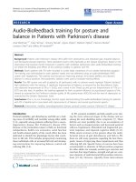
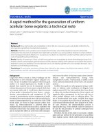
utes. Figure 1 (top) su mmarizes a schematic description
of the training protocol.
Robot Training procedure
An iter ative algorithm, similar to that proposed in [28],
was used to estimate and store the time profile of the
forces, to be generated by the robot during the subse-
quent Subject Training phase. The algorithm aims at
determining the forces that shift a subject’ s trajectory
toward a ‘reference’ trajectory, x
D
(t). The ‘reference’ tra-
jectory, x
D
(t), was defined as a ‘minimum jerk’ trajectory
passing through three points [37]: the center, the target
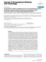
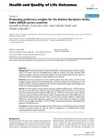
and a third via-point; see Figure 2.
We defined the via-point, placed at half the distance
from the starting point to the target, and shifted it later-
ally, of three times the maximum lateral deviation
observed in the average baseline trajectory. The ‘average’
trajectory was the ‘average’ of all trajectories in the same
direction during the Baseline 1.
Table 1 Clinical data for the experimental subjects
Subject Age
(y)
Sex Hand Disease Duration (y) Disease Course EDSS (0-10) MODE
S1 38 M R 14 RR 6.5 EE+ER
S2 41 F L 15 SP 3 EE+ER
S3 61 F R 3 SP 4 ER+EE
S4 42 F R 8 RR 4.5 ER+EE
S5 73 M L 4 SP 4.5 EE+ER
S6 34 F L 11 SP 5 ER+EE
S7 59 M R 20 SP 6.5 EE+ER
S8* 37 F R 4 SP 6 ER+EE
Total 48 ± 14 10 ± 6 5 ± 1
RR: relapsing-remitting; SP: secondary-progr essive. Subject 8 dropped out the study.
Vergaro et al. Journal of NeuroEngineering and Rehabilitation 2010, 7:37
/>Page 3 of 11
In error-enhancing (EE) sessions, the shift was on the
same side as the lateral deviation observed in the aver-
age trajectory. In error-reducing (ER) sessions, the s hift
was on the opposite side.
The force generated by the robot in direction d = 1 3,
F
d
(t), was only present during the initial 2/3 of the total
duration of the movement (estimated from that of the
‘average’ trajectory). This is because we were interested
in affecting the early portion of the movements, which
best reflects the operation of the feed-forward compo-
nent of control. Late portions of the trajectory are highly
variable, as they reflect the feedback corrections that are
likely due to errors in the early portion.
We initially set
Ft
d
1
0
()
=
for each t, and subsequent
movement repetitions were used to adjust the force
according to the following update rule [28], where d is
target direction (d = 1 3):
FtFt xtxt
d
n
d
n
d
D
d
n+
()
=
()
+
() ()
1
µ
·
(1)
The parameter μ is a learning rate, which was been
heuristically set in the range of 10-30 N/m. If μ is too
large, the robot training procedure becomes unstable, if
μ is too small convergence would take too long. In all
experiments, we used μ = 30 N/m.
As a c onsequence of this procedure, in EE sessions,
forces led to enhancing the lateral deviation of the base-
line trajectory. In contrast, in ER sessions, forces
opposed - reduced, and ultimately reversed - the initial
lateral deviation. For safety reasons, the forces generated
by the robot were limited to the ± 14 N range.
Study design
The rehabilitation protocol included a total of 8 ses-
sions. To compare the two variants of the robot therapy
treatment, we used a randomized double blind crossover
design. In four consecutive sessions (2 sessions/week),
subjects were trained with one type of error-enhancing
(EE) forces. In the remaining four sessions (2 sessions/
week), forces were error-reducing (ER). The order of
application o f the two treatments was randomized over
subjects - four subjects started with EE training, four
subjects started with ER training. The two treatment
periods were separated by a 2-weeks rest period.
Figure 1 (bottom) summarizes the study design.
Note that the forces used for training were calculated
at the beginning of each session. Therefore, the protocol
automatically adapted to the patient’s specific impair-
ment, as well as to the improvements - if any - t hat
occurred from session to session.
Subjects were blind with respect to the training pro-
tocol, in the sense that they did not receive a detailed
explanation of the modalities of generation of force by
the robot. Moreov er, each subject had pec uliar pat-
terns of incoordination and the applied forces were
highly direction-specific. Therefo re, it is unlikely that
they could d istinguish among either modality and that
they saw forces as something different than mere
perturbations.
Clinical testing included the evaluation of the follow-
ing clinical scales: EDSS and Functional Systems Score
[31], Scripps’ NRS [32], Ashworth scale [38], the Ataxia
Figure 1 Training protocol and study design. Top: Phases of the
training protocol: Baseline 1 (B1), Robot Training, Baseline 2 (B2),
Subject Training, Wash-out. The phases in which the robot
generates no forces (B1, B2, Wash-out) are indicated in white. Each
square corresponds to five epochs. Bottom: Overall study design,
indicating the treatment and rest periods and the times of
evaluation (T0-T4).
3
REFERENCE
MEAN
EE
ER
Figure 2 Desired trajectory construction. Maximum lateral
deviation (Δ) from the nominal path calculated after the evaluation
of the mean trajectory (grey). It is tripled (3Δ) and centered. The
corresponding point became the via-point for minimum-jerk
trajectory that enhance (black line) or reduce (black dotted line)
subject’s error.
Vergaro et al. Journal of NeuroEngineering and Rehabilitation 2010, 7:37
/>Page 4 of 11
and Tremor scales [39], the Nine-Hole Peg Test
(NHPT) [40], a Visual Analog Scale (VAS) for upper
limb tremor (0-10 score), a se lf-administered Tremor in
Activity of Daily Life (TADL) questionnaire [41]. Sub-
jects and the evaluating clinician were blind with respect
to the training protocol (ER or EE).
We made a total of four assessments, at T0 (baseline-
day 1), T1 (after 4 sessions - day 14), T2 (after the rest
period - day 28) and T3 (after 8 sessions - day 42).
We looked at both specific differences in the two
treatments and at the overall effect of robot treatment
over the whole duration of the trial.
Data Analysis
Hand trajectories were sampled at 100 Hz. The x and y
components were smoothed with a 4
th
order Savitzky-
Golay filter (window size 200 ms, equivalent cu t-off fre-
quency 6.6 Hz), which was also used to estimate the
first t hree time derivatives. We then estimated the fol-
lowing indicators:
- Lateral deviation of hand trajectory (root mean
square value).
- Movement duration, i.e. time elapsed between move-
ment onset and terminat ion; movement onset was iden-
tified as the first time instant when hand speed exceeds
a threshold (20% of peak speed); movement termination
was computed as the first time instant after onset in
which movement speed goes below the threshold.
- Symmetry: ratio between the durations of ac celera-
tion and deceleration phases.
- Jerk (Teulings’) index: root mean square of the jerk
(thir d time derivative of the trajectory), normalized with
respect to movement amplitude and duration [42].
Lateral deviation was also used to assess the subjects’
ability to adapt to the force patterns provided by the
robot.
Outcome measures
Asaprimaryoutcomemeasure,wetookthechangein
the Nine Hole Peg Test (NHPT) [40] , a quantitative scale
for distal upper limb function (the test involves the sub-
ject placing 9 dowels in 9 holes. Subjects are scored on
the amount of time it takes to place and remove all 9
pegs). The test was preceded by a familiarization phase to
extinguish learning effects. We took a 2 0% decrease as
the threshold for clinical significance [4 3,44]. Kinematic
(jerk index, lateral deviation, movement duration and
symmetry of the speed profile) and clinical indicators
(Scripps’ NRS, Ataxia score, VAS for upper limb tremor,
TADL) were taken as secondary outcome measures.
Statistical analysis
To compare the effects of the two tr eatments (EE and
ER), to account for the crossover design we analysed the
primary outcome measure by using a mixed-effect
model [13], with period (first, between T0 and T1, and
second, between T2 and T3) and treatment (EE or ER)
as f ixed factors, subject as random factor and the base-
line value at the start of the relevant period (i.e., T0 and
T2) as covariate. This adjustment allows us to reduce
the observed variation between the two groups of sub-
jects caused not by the treatment itself but by variation
of the clinical scale at the beginning of the therapy.
To test the overall effect of adaptive training, we com-
pared the primary outcome measures (change in the
clinical scores) between the baseline (T0) and the end of
the treatment (T3), irrespective of the training mode
(treatment).
As regards the kinematic indicators, we ran a
repeated-measures ANOVA with three factors: session
(early vs late, i.e. 1 vs 4), phase (baseline 1, baseline 2,
late wash-out - last 5 epochs) and treatment (EE, ER).
Significant period and session effects w ould indicate,
respectively, that subjects modify their behaviour within
and between sessions. To quantify w hether the session
effect was indeed an improvement, we also directly
compared (planned comparisons) session 1 and session
4, for the two treatments taken together and separately
for each training mode. As regards changes within one
session, to distinguish between the changes in perfor-
mance occurring during the Robot Training phase f rom
those occurring during the Subject Training phase, we
directly compared (planned comparisons) Baseline 1 and
Baseline 2 (effect of Robot Training), Baseline 2 and
Wash-out (effect of Subject Training) and finally Base-
line 1 and wash-out (overall phase effect).
Results
Seven subjects successfully completed the protocol. Sub-
jects were allowed to rest b etween consecutive blocks of
trials.However,noonedid,andinfactthetaskwas
well tolerated. Furthermore, there was no degradation of
performance at the end of the adaptation phase as com-
pared to the final portion of the wash-out phase. One
subject (S8) did not complete the second half of the
trial, for reasons unrelated to the study protocol. This
subject was excluded from all subsequent analyses.
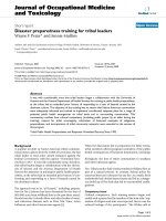
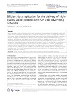
Figure 3 shows typical trajectories from the center
position to the three targets, during the different phases
of an error-enhancing (top) and an erro r-reducing ses-
sion (bottom).
As expected, the forces learned by the robot at the
end of the Robot Training ph ase reflect the average pat-
terns of curvature observed during the baseline phase.
Primary outcome
We first tested for differences in the training mode. We
found a significant effect of period (F(1,6) = 16.004; p =
Vergaro et al. Journal of NeuroEngineering and Rehabilitation 2010, 7:37
/>Page 5 of 11
0.00283). On average, the decrease in the NHPT score
was -9 ± 3 s in period 1 and -1 ± 3 s in period 2. How-
ever, we found no significant treatment and base line
effects. On average, the NHPT score decrease was -9 ±
5 s in period 1 of error-enhancing sessio ns, and -9 ± 5 s
in the same period of error-reducing sessions.
These results indicate that most of the improvement
occurs in period 1, irrespective of treatment type and
baseline value.
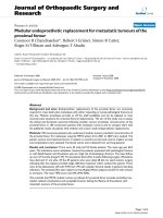
We then looked at the NHPT change from baseline
(T0) to the end of the treatment (T3), irrespective of the
training mode. In this case, the NHPT score decreased
from 61 ± 14 s to 48 ± 20 s, a 24% change (F(1,6) =
16.495, p = 0.00 7); see also Figure 4. In four subjects,
the improvement was greater than 20% (the threshold
for clinical significance). One subject displayed a 47%
change; no subjects showed significant worsening.
During the first four sessions, irrespective of the train-
ing mode, the average score decreased (F(1,6) = 6.7955,
p = 0.04021) from 61 ± 14 s to 52 ± 20 s (a 21%
change). A smaller decrease, from 49 ± 18 s to 48 ±
20 s (a 4% change) was observed during the last four
sessions. Although these results suggest a plateau effect
for the improvement in the NHPT score, subjects who
improved during period 1 exhibited an additional
improvement in period 2 (correlation between changes
in the two periods: 0.61); see Table 2.
Secondary outcome: clinical scales
The Ataxia score decreased from T0 and T3, irrespe c-
tive of the traini ng mode (F(1,6) = 6.1935, p = 0.04725).
The decrease occurred during the first four sessions
(F(1,6) = 10.500, p = 0.01768); no further decrease was
found in the late sessions. As regards tremor, the TADL
score decreased in the first four sessions, but only with
EE training (F(1,6) = 14.087, p = 0.00947); see Table 3.
Other clinical scales showed small improvements for at
least some of the subjects, but no significant effects
were observed.
Secondary outcome: changes in movement kinematics
We found no significant effects of Robot Training (base-
line 1 vs baseline 2). As regards the effect of Subject
Training (baseline 2 vs wash-out), we found a decrease
in the jerk index (F(1,6) = 13.632, p = 0.01018), i.e. after
Subject Training movements tend to be smoother - but
this same effect was no longer significant when consid-
ering baseline 1 vs late wash-out; see Figure 5.
Moreover, we found no significant improvements in
movement duration, speed profile symmetry and trajec-
tory curvature (as measured by the lateral deviation).
Overall, these results suggest that Subject Training con-
sistently increases movement smoothness, whereas mere
exercise - the Robot Training phase - does not have a
consistent effect. As regards the effect of session, we
found no significant effects for duration, speed profile
symmetry or the jerk index. However, we found a signif-
icant decrease in trajectory curvature (F(1,6) = 19.801,
p = 0.00433); see Figure 6.
Error-enhancing vs error-reducing training
In all indica tors the effect of the training m ode (EE vs
ER) was not significant except the TADL secondary
outcome that significantly decre ased only in EE train-
ing (F(1,6) = 14.087, p = 0.00947). Likewise, in no indi-
cator we found significant interactions between the
training mode and the other factors. Finally, as regards
trajectory curvature, we found that most of the
decrease occurred during the first block of four ses-
sions, irrespective of the training mode (F(1,6) =
17.767, p = 0.00559, sessions 1 vs 4; and F(1,6) =
8.6312, p = 0.02602, sessions 5 vs 8).
TRAININ
G
LEARNED EARLY LATE
BASELINE WASH−OUT
ENHANCEREDUCE
F
O
R
C
E TRAININ
G
Figure 3 Typical trajectories. Typical trajectories during an EE (top) and an ER (bottom) training session. From left to right: baseline trajectories,
learned forces, early and late training and late wash-out.
Vergaro et al. Journal of NeuroEngineering and Rehabilitation 2010, 7:37
/>Page 6 of 11
Force field adaptation
To assess the capability of adapting to the forces gener-
ated by the robot during the Sub ject Training phase, we
used a ‘learning index’ [26] that compared some signed
measure of execution error (here, maximum lateral
deviation) in movements where force is turned on (force
trials) and where force is turned off (catch trials). If
adaptation had occurred, the execution error observed
in early force trials should be negatively correlated with
the same motor error, observed in the late cat ch trials.
For each subject we displayed the error in ear ly force
trials versus the error in late catch trials. The results, for
each subject and for each training mode, are shown in
Figure 7.
Theslopesoftheregressionlinescanbeusedto
quantify the amount of adaptation. The estimat ed
slopes, as well as the corresponding correlation coeffi-
cients r are, -0.61 (S1, r = 0.80), -0.09 (S2, r = 0.01),
-0.46 (S3, r = 0.63), -0.41 (S4, r = 0.49), -0.19 (S5, r =
0.18), 0.30 (S6, r = 0.07), -0.14 (S7, r = 0.32). These
results suggest that five subjects display signs of adapta-
tion (negati ve slope, substantial correlation) to the force
generated by the robot. Two subjects have small correla-
tion, suggesting that little or no adaptation occurred.
Although the correlation was not significant, subjects
displaying a greater NHPT improvement were also
those displaying a greater amount of adaptation.
Discussion
In this feasibility study, we developed an adaptive robot
training technique, and applied it to MS subjects with
cerebellar symptoms, i.e. ataxia, tremor or both.
Adaptive robot training improves upper limb function
Across sessions, we found a significant decrease in the
NHPT score - a quantitative measure of arm-hand coor-
dination. Additional evidence for improved coordination
is provided by the decreases in the ataxia and tremor
scores (period 1, EE sessions only). Kinematic analysis of
motor performance supports these results. At the end of
a training session, movements become significantly
smoother. In addition, over sessions, the curvature of
movement trajectories decreases significantly.
The improved NHPT score is particularly remarkable,
as it suggests that the improved coordination may trans-
fer to tasks more related to activi ties of daily living [21].
In contrast, robot therapy in stroke subjects displays lit-
tle generalization to movements that had not been expli-
citly trained [45,46].
Most subjects showed a clear improvement in the first
four sessions, and only few improved further in the sec-
ond half of t he training protocol. However, improve-
ments in the first period predicted an additional
(smaller) improvement in the second period. This says
little on how many sessions could be appropriate to
maximize subjects’ benefit, but suggests that
T0 T1 T2 T3
20
30
40
50
60
70
80
90
NHPT [s]
TIME OF EVALUATION
S1
S2
S3
S4
S5
S6
S7
Figure 4 Nine Hole Peg Test. Change s in the Nine-Hole Peg Test
score for the seven subjects, during error-enhancing (dashed lines)
and error-reducing trials (solid lines).
Table 2 Changes in NHPT
NHPT [s] NHPT change [s]
Subject Sequence T0 T1 T2 T3 Period 1 (T1-T0) Period 2 (T3-T2) Overall (T3-T0)
S1 EE+ER 62 46 51 44 -16 -7 -18
S2 EE+ER 53 36 31 33 -17 2 -20
S3 ER+EE 42 32 32 31 -10 -1 -11
S4 ER+EE 55 38 32 29 -17 -3 -26
S5 EE+ER 83 75 73 73 -8 0 -10
S6 ER+EE 58 58 49 50 0 1 -8
S7 EE+ER 76 82 73 76 6 3 0
S8* ER+EE 57 61 NA NA 4 NA NA
Total 61 ± 14 52 ± 20 49 ± 19 48 ± 20 -9 ± 9 -1 ± 3 -13 ± 9
Vergaro et al. Journal of NeuroEngineering and Rehabilitation 2010, 7:37
/>Page 7 of 11
improvement in early sessions is predictive of a further
improvement.
Is the observed improvement due to the robot, or it is
just the effect of repeated exercise? Within a session,
improvements were only observed a fter Subject Train-
ing, whereas Robot Training - during which the robot
exerts no forces in 75% of the movements - did not
appear to have an effect. This observation points to a
specific within-session effect of the robot (robot-assisted
Subject Training phase) when compared to exercise
alone. These short- term effec ts, as well as adaptive pro-
cesses that occur at different time scales [47] may con-
tribute to the overall observed (between-session)
performance improvements.
Table 3 Changes in clinical scales
Subject Scripps’
NRS (5-0)
Ataxia (0-8) TADL (25-100) VAS tremor (0-10) Scripps’
NRS (5-0)
Ataxia (0-8) TADL (25-100) VAS tremor (0-10)
MSC MSC
S1 T0 5 5 1 5 37 5 T2 5 5 1 3 35 4.5
T1 5 5 3 3 32 3.5 T3 5 5 1 3 35 4
S2 T0 3 3 1 5 42 5 T2 3 3 3 3 40 4
T1 3 3 3 3 40 4 T3 3 5 3 3 40 3
S3 T0 3 5 3 3 45 5 T2 5 5 3 2 45 5
T1 5 5 3 2 45 5 T3 5 5 3 2 45 5
S4 T0 3 3 1 5 63 5 T2 5 3 1 3 62 4
T1 5 3 1 4 62 5 T3 5 3 3 2 60 4
S5 T0 5 3 3 3 47 8 T2 5 3 3 3 45 6
T1 5 3 3 3 47 7 T3 5 3 3 3 45 6
S6 T0 5 3 3 3 51 5 T2 5 3 3 3 45 5
T1 5 3 3 3 51 5 T3 5 3 3 3 45 5
S7 T0 5 5 1 3 44 6 T2 5 5 1 3 41 5
T1 5 5 1 2 44 5 T3 5 5 1 3 41 5
S8* T0 5 5 1 3 63 9 T2 - - -
T1 5 5 1 3 71 10 T3 - - -
MSC: Motor, Sensory and Cerebellar
T0 T1 T2 T3
0
20
40
60
TIME OF EVALUATION
JERK INDEX
B1
B2
WO
Figure 5 Jerk index. Changes in jerk index over sessions. The bars
represent the mean value of the indicator over subjects in the
baseline1 (B1), baseline2 (B2), wash-out phase (WO).
T0 T1 T2 T3
0
2
4
6
8
10
LATERAL DEVIATION [mm]
TIME OF EVALUATION
S1
S2
S3
S4
S5
S6
S7
Figure 6 Latera l deviation. Changes in lateral deviation over
sessions. Dashed lines indicate EE sessions and solid lines refer to
ER sessions.
Vergaro et al. Journal of NeuroEngineering and Rehabilitation 2010, 7:37
/>Page 8 of 11
Error-enhancing vs error-reducing training
Previous studies [29,34] on chronic stroke survivors sug-
gested that adaptation to error-enhancing perturbations
(error-enhancing t raining) can induce short-term
improvements of motor performance. In contrast, adap-
tation to perturbations that opposed the initial lateral
deviations (error-reducing training) induced a slight
worsening of performance [29].
In the prese nt study, we found no significant differ-
ences - neither short-term (within session) nor long-
term (between sessions) - between error-enhancing and
error-re ducing training. This may be due at least in part
to the small number of sessions and/or subjects.
Furthermore, as noted in the Methods, the ‘error-red u-
cing’ modality may a ctually tend to augment the error -
although in an opposite direction with respect to the
initial lateral deviation - so they may be no different in
terms of recovery.
Actually, the cited study on stroke subjects only
focusesonshort-term(onesession)effects,anditis
unclear what effect would be expected over multiple ses-
sions. Therefore, that study cannot be directly compared
to our findings. Nevertheless, the latter may indicate
that stroke survivors and MS subjects with cerebellar
symptoms have distinctly different modalities of
recovery.
In stroke subjects, recovery might be mostly driven by
motor errors, so that it would be greater and/or faster if
errors are amplified. Little is known about the mechan-
isms underlying functional recovery (if any) of M S sub-
jects with cerebellar symptoms. However, one possible
hypothesis is that in these subjects recovery may be
facilitated by exercises that challenge their ability to deal
with novel dynamic environments, for which the cere-
bellum plays an essential role [26,27]. As a consequence,
in these subjects recovery may not depend on the speci-
fic d ynamic environment to which to adapt but, rather,
on the mere task of adapting.
Further experiments are needed to test this working
hypothesis.
It should be noted that the cross-over study design as
a number of limitations. The effect of exercise during
the first period does not vanish during the 2-weeks rest
period. This is partly accounted for by the statistic pro-
cedures (performance at the beginning of treatment per-
iods taken as covariate), but existing differences in the
two treatment modalities as well as an interaction
among them cannot be completely ruled out. Additional
studies would be needed, involving more subjects and
two separate treatment groups.
MS subjects adapt to unfamiliar dynamic environments
In adaptive training, robots do not just assist subjects
while they practice movements (or resist to them) but,
rather, they provide unfamiliar dynamic environments,
to which subjects are required to adapt. Stroke subjects
are capable to adapt to these environments, and when
the latter are removed, after wash-out ot the after effects
they exhibit improved coordination [34]. These studies,
together with evidence of reorganization of the motor
cortex driven by motor skill learning [48] have sug-
gested that the neural processes associated with implicit
motor adaptation may reshape the sensorimotor map-
pings altered by stroke [49]. The same cortical reorgani-
zation occurs in subjects w ith early MS, and might
contribute to limit the consequences of irreversible tis-
sue damage in lesions and normal-appearing brain tissue
[50]. This would suggest that rehabilitation of MS sub-
jects should primarily aim a t facilitati ng the emergence/
−0.1 0 0.1
−0.1
0
0.1
S1
−0.1 0 0.1
−0.1
0
0.1
S6
−0.1 0 0.1
−0.1
0
0.1
S7
ERROR IN FORCE TRIALS [m]
−0.1 0 0.1
−0.1
0
0.1
S5
ERROR IN CATCH TRIALS [m]
−0.1 0 0.1
−0.1
0
0.1
S2
−0.1 0 0.1
−0.1
0
0.1
S3
−0.1 0 0.1
−0.1
0
0.1
S4
Figure 7 Motor adaptation by subject. From top to bottom:
subjects 1-7. Grey and black dots indicate ER and EE sessions
respectively. The grey line represents the regression line. Adaptation
is indicated by the negative correlation between the error in early
force trials and that in late catch trial.
Vergaro et al. Journal of NeuroEngineering and Rehabilitation 2010, 7:37
/>Page 9 of 11
reorganization of compensatory strategies. Adaptive
training seems an attractive way to promote such reor-
ganization and, consequently, par ticularly promising for
rehabilitation of MS subjects, who display different types
and degrees of deficit, often with a n important cerebel-
lar component.
This pilo t study provides new evidence that MS sub-
jects are able to adapt their arm movements when they
are e xposed to a robot-generated force field. More spe-
cifically, our results suggest that, when the robot inter-
acts with subjects performing movements, it is capable
to achieve a consistent pattern of force to either
enhance or reduce the subjects’ errors. A comparison of
the errors made during the early force trials and those
made during the late catch trials clearly demonstrated
that MS subjects are capable of adapting to both error-
enhancing and error-reducing force fields.
Conclusions
This study suggests that adaptive-type robot therapy
may be a useful and safe approach to improve cerebellar
symptoms in MS subjects.
In particular, the finding that six subjects (out of 7)
showed a clinically sig nificant improvement in NHPT in
pre-post analysis and an improved coordination is spe-
cially remarkable, as most medications and rehabilitation
approaches are little effective with cerebellar symptoms.
However, unlike stroke subjects, we could not observe
a clear difference in the effect of the two treatments
(error-enhancing, error-reducing). This may indicate a
different modality of recovery of these s ubjects with
respect to stroke survivors. While in stroke subjects
recovery is driven by motor errors, in MS cerebellar
subjects recovery may be triggered by the mere adaptive
training, irrespective of the specific perturbing environ-
ment). In fact, in our subjects the overall improvement
was associated with a preserved ability, within a session,
to adapt to unfamiliar dynamic environments.
We could not conclude on the ideal number and
duration of the treatment sessions. However, most of
the improvement occurred in the early exercise sessions
(period 1) and its magnitude was predictive of additional
improvements in later sessions (period 2).
The above conclusions need to be taken cautiously
because of the limited size of our sample, and should be
confirmed in a larger study. Nevertheless, this study
may represent a starting point toward designing novel
robot therapy approaches and to expand the range of
application of robots in neuromotor rehabilitation.
Acknowledgements
This work is partly supported by the Italian Multiple Sclerosis Foundation
(FISM) (R19/2004).
Author details
1
University of Genoa, Department of Informatics, Systems and
Telecommunications, Via Opera Pia 13, Genoa, Italy.
2
Italian Institute of
Technology, Via Morego 30, Genoa, Italy.
3
Department of Neuroscience,
Ophthalmology and Genetics, University of Genoa, Via A. De Toni 5, Genoa,
Italy.
4
Department of Neurology, ASL3 Genovese, Genoa, Italy.
Authors’ contributions
The overall design of the experiment was agreed by all authors after
extensive discussions. ViS and CS designed the overall study. ViS, MC and
PM defined the motor task. CS and GB selected the subjects and conducted
all clinical evaluations. EV and VaS programmed the robot, including the
Robot Training procedure, conducted all experiments and analyzed the data.
EV, VaS, and ViS performed the statistical analysis. ViS and CS wrote the
manuscript.
All authors read and approved the manuscript.
Competing interests
The authors declare that they have no competing interests.
Received: 6 August 2009 Accepted: 29 July 2010 Published: 29 July 2010
References
1. Thompson AJ: Progress in neurorehabilitation in multiple sclerosis. Curr
Opin Neurol 2002, 15:267-270.
2. Alusi SH, Worthington J, Glickman S, Bain PG: A study of tremor in
multiple sclerosis. Brain 2001, 124:720-730.
3. Koch M, Mostert J, Heersema D, De Keyser J: Tremor in multiple sclerosis.
J Neurol 2007, 254:133-145.
4. Hallett M, Berardelli A, Matheson J, Rothwell J, Marsden CD: Physiological
analysis of simple rapid movements in patients with cerebellar deficits.
J Neurol Neurosurg Psychiatry 1991, 54:124-133.
5. Valentino P, Cerasa A, Chiriaco C, Nistico R, Pirritano D, Gioia M, Lanza P,
Canino M, Del Giudice F, Gallo O, et al: Cognitive deficits in multiple
sclerosis patients with cerebellar symptoms. Mult Scler 2009, 15:854-859.
6. Bastian AJ, Martin TA, Keating JG, Thach WT: Cerebellar ataxia: abnormal
control of interaction torques across multiple joints. J Neurophysiol 1996,
76:492-509.
7. Freeman JA, Langdon DW, Hobart JC, Thompson AJ: The impact of
inpatient rehabilitation on progressive multiple sclerosis. Ann Neurol
1997, 42:236-244.
8. Liu C, Playford ED, Thompson AJ: Does neurorehabilitation have a role in
relapsing-remitting multiple sclerosis? J Neurol 2003, 250:1214-1218.
9. Rietberg MB, Brooks D, Uitdehaag BM, Kwakkel G: Exercise therapy for
multiple sclerosis. Cochrane Database Syst Rev 2005, CD003980.
10. Khan F, Turner-Stokes L, Ng L, Kilpatrick T: Multidisciplinary rehabilitation
for adults with multiple sclerosis. Cochrane Database Syst Rev 2007,
CD006036.
11. Mills R, Yap L, Young C: Treatment for ataxia in multiple sclerosis.
Cochrane Database Syst Rev 2007, CD005029.
12. Lord SE, Wade DT, Halligan PW: A comparison of two physiotherapy
treatment approaches to improve walking in multiple sclerosis: a pilot
randomized controlled study. Clin Rehabil 1998, 12:477-486.
13. Wiles CM, Newcombe RG, Fuller KJ, Shaw S, Furnival-Doran J, Pickersgill TP,
Morgan A: Controlled randomised crossover trial of the effects of
physiotherapy on mobility in chronic multiple sclerosis. J Neurol
Neurosurg Psychiatry 2001, 70:174-179.
14. Armutlu K, Karabudak R, Nurlu G: Physiotherapy approaches in the
treatment of ataxic multiple sclerosis: a pilot study. Neurorehabil Neural
Repair 2001, 15:203-211.
15. Koch G, Rossi S, Prosperetti C, Codeca C, Monteleone F, Petrosini L,
Bernardi G, Centonze D: Improvement of hand dexterity following motor
cortex rTMS in multiple sclerosis patients with cerebellar impairment.
Mult Scler 2008,
14:995-998.
16. Quintern J, Immisch I, Albrecht H, Pollmann W, Glasauer S, Straube A:
Influence of visual and proprioceptive afferences on upper limb ataxia
in patients with multiple sclerosis. J Neurol Sci 1999, 163:61-69.
17. Feys P, Helsen W, Liu X, Mooren D, Albrecht H, Nuttin B, Ketelaer P: Effects
of peripheral cooling on intention tremor in multiple sclerosis. J Neurol
Neurosurg Psychiatry 2005, 76:373-379.
Vergaro et al. Journal of NeuroEngineering and Rehabilitation 2010, 7:37
/>Page 10 of 11
18. Prange GB, Jannink MJ, Groothuis-Oudshoorn CG, Hermens HJ, Ijzerman MJ:
Systematic review of the effect of robot-aided therapy on recovery of
the hemiparetic arm after stroke. J Rehabil Res Dev 2006, 43:171-184.
19. Lo AC, Triche EW: Improving gait in multiple sclerosis using robot-
assisted, body weight supported treadmill training. Neurorehabil Neural
Repair 2008, 22:661-671.
20. Beer S, Aschbacher B, Manoglou D, Gamper E, Kool J, Kesselring J: Robot-
assisted gait training in multiple sclerosis: a pilot randomized trial. Mult
Scler 2008, 14 :231-236.
21. Carpinella I, Cattaneo D, Abuarqub S, Ferrarin M: Robot-based
rehabilitation of the upper limbs in multiple sclerosis: feasibility and
preliminary results. J Rehabil Med 2009.
22. Shadmehr R, Mussa-Ivaldi FA: Adaptive representation of dynamics during
learning of a motor task. J Neurosci 1994, 14:3208-3224.
23. Casadio M, Sanguineti V, Morasso P, Solaro C: Abnormal sensorimotor
control, but intact force field adaptation, in multiple sclerosis subjects
with no clinical disability. Mult Scler 2008, 14:330-342.
24. Leocani L, Comi E, Annovazzi P, Rovaris M, Rossi P, Cursi M, Comola M,
Martinelli V, Comi G: Impaired short-term motor learning in multiple
sclerosis: evidence from virtual reality. Neurorehabil Neural Repair 2007,
21:273-278.
25. Hatzitaki V, Koudouni A, Orologas A: Learning of a novel visuo-postural
co-ordination task in adults with multiple sclerosis. J Rehabil Med 2006,
38:295-301.
26. Smith MA, Shadmehr R: Intact ability to learn internal models of arm
dynamics in Huntington’s disease but not cerebellar degeneration.
J Neurophysiol 2005, 93:2809-2821.
27. Maschke M, Gomez CM, Ebner TJ, Konczak J: Hereditary cerebellar ataxia
progressively impairs force adaptation during goal-directed arm
movements. J Neurophysiol 2004, 91:230-238.
28. Patton JL, Mussa-Ivaldi FA: Robot-assisted adaptive training: custom force
fields for teaching movement patterns. IEEE Trans Biomed Eng 2004,
51:636-646.
29. Patton JL, Stoykov ME, Kovic M, Mussa-Ivaldi FA: Evaluation of robotic
training forces that either enhance or reduce error in chronic
hemiparetic stroke survivors. Exp Brain Res 2006, 168:368-383.
30. McDonald WI, Compston A, Edan G, Goodkin D, Hartung HP, Lublin FD,
McFarland HF, Paty DW, Polman CH, Reingold SC, et al: Recommended
diagnostic criteria for multiple sclerosis: guidelines from the
International Panel on the diagnosis of multiple sclerosis. Ann Neurol
2001, 50:121-127.
31. Kurtzke JF: Rating neurologic impairment in multiple sclerosis: an
expanded disability status scale (EDSS). Neurology
1983, 33:1444-1452.
32. Sipe JC, Knobler RL, Braheny SL, Rice GP, Panitch HS, Oldstone MB: A
neurologic rating scale (NRS) for use in multiple sclerosis. Neurology
1984, 34:1368-1372.
33. Casadio M, Morasso PG, Sanguineti V, Arrichiello V: Braccio di Ferro: a new
haptic workstation for neuromotor rehabilitation. Technol Health Care
2006, 13:1-20.
34. Patton JL, Kovic M, Mussa-Ivaldi FA: Custom-designed haptic training for
restoring reaching ability to individuals with poststroke hemiparesis.
J Rehabil Res Dev 2006, 43:643-656.
35. Morasso P: Spatial control of arm movements. Exp Brain Res 1981,
42:223-227.
36. Scheidt RA, Dingwell JB, Mussa-Ivaldi FA: Learning to move amid
uncertainty. J Neurophysiol 2001, 86:971-985.
37. Flash T, Hogan N: The coordination of arm movements: an
experimentally confirmed mathematical model. J Neurosci 1985,
5:1688-1703.
38. Ashworth B: Preliminary Trial of Carisoprodol in Multiple Sclerosis.
Practitioner 1964, 192:540-542.
39. Alusi SH, Worthington J, Glickman S, Bain PG: A study of tremor in
multiple sclerosis. Brain 2001, 124:720-730.
40. Mathiowetz V, Weber K, Kashman N, Volland G: Adult norms for the Nine-
Hole Peg Test of finger dexterity. Occup Ther J Res 1985, 5:24-28.
41. Bain PG, Findley LJ, Atchison P, Behari M, Vidailhet M, Gresty M,
Rothwell JC, Thompson PD, Marsden CD: Assessing tremor severity.
J Neurol Neurosurg Psychiatry 1993, 56:868-873.
42. Teulings HL, Contreras-Vidal JL, Stelmach GE, Adler CH: Parkinsonism
reduces coordination of finger, wrist, and arm fine motor control.
Experimental Neurology 1997, 146:159-170.
43. Vaney C, Vaney S, Wade DT: SaGAS, the Short and Graphic Ability Score:
an alternative scoring method for the motor components of the
Multiple Sclerosis Functional Composite. Mult Scler 2004, 10:231-242.
44. Schwid SR, Goodman AD, McDermott MP, Bever CF, Cook SD: Quantitative
functional measures in MS: what is a reliable change? Neurology 2002,
58:1294-1296.
45. Krebs HI, Hogan N, Aisen ML, Volpe BT: Robot-aided neurorehabilitation.
IEEE Trans Rehabil Eng 1998, 6:75-87.
46. Krebs HI, Hogan N, Volpe BT, Aisen ML, Edelstein L, Diels C: Overview of
clinical trials with MIT-MANUS: a robot-aided neuro-rehabilitation facility.
Technol Health Care 1999, 7:419-423.
47. Smith MA, Ghazizadeh A, Shadmehr R: Interacting adaptive processes
with different timescales underlie short-term motor learning. PLoS Biol
2006, 4:e179.
48. Nudo RJ: Adaptive plasticity in motor cortex: implications for
rehabilitation after brain injury. J Rehabil Med 2003, 7-10.
49. Reinkensmeyer DJ, Emken JL, Cramer SC: Robotics, motor learning, and
neurologic recovery. Annu Rev Biomed Eng 2004, 6:497-525.
50. Rocca MA, Colombo B, Falini A, Ghezzi A, Martinelli V, Scotti G, Comi G,
Filippi M: Cortical adaptation in patients with MS: a cross-sectional
functional MRI study of disease phenotypes. Lancet Neurol 2005,
4:618-626.
doi:10.1186/1743-0003-7-37
Cite this article as: Vergaro et al.: Adaptive robot training for the
treatment of incoordination in Multiple Sclerosis. Journal of
NeuroEngineering and Rehabilitation 2010 7:37.
Submit your next manuscript to BioMed Central
and take full advantage of:
• Convenient online submission
• Thorough peer review
• No space constraints or color figure charges
• Immediate publication on acceptance
• Inclusion in PubMed, CAS, Scopus and Google Scholar
• Research which is freely available for redistribution
Submit your manuscript at
www.biomedcentral.com/submit
Vergaro et al. Journal of NeuroEngineering and Rehabilitation 2010, 7:37
/>Page 11 of 11