báo cáo hóa học: " Mechanisms of human cerebellar dysmetria: experimental evidence and current conceptual bases" potx
Bạn đang xem bản rút gọn của tài liệu. Xem và tải ngay bản đầy đủ của tài liệu tại đây (1.4 MB, 18 trang )
Journal of NeuroEngineering and
Rehabilitation
Review
Mechanisms of human cerebellar dysmetria: experimental
evidence and current conceptual bases
Mario Manto
Address:
1
Laboratoire de Neurologie Expérimentale, FNRS-ULB, Bruxelles, Belgium
E-mail: Mario Manto* -
*Correspond ing author
Published: 13 April 2009 Received: 15 September 2008
Journal of NeuroEngineering and Rehabilitation 2009, 6:10 doi: 10.1186/1743-0003-6-10 Accepted: 13 April 2009
This article is available from: />© 2009 Manto; licens ee BioMed Central Ltd.
This is an Open Access article distributed under the term s of the Creative Commons Att ributio n License (
/>which permits unrestricted use, distribution, and reproduction in any medium, provided the original work is properly cited.
Abstract
The human cerebellum contains more neurons than any other region in the brain and is a major
actor in motor control. Cerebellar circuitry is unique by its stereotyped architecture and its
modular organization. Understanding the motor codes underlyi ng the organization of limb
movement and the rules of signal processing applied by the cerebellar circuits remains a major
challenge for the forthcoming decades. One of the cardinal deficits observed in cerebellar patients
is dysmetria, designating the inability to perform accurate movements. Patients overshoot
(hypermetria) or undershoot ( hypometria) the aimed tar get duri ng voluntar y goal- directed tasks.
The mechanisms of cerebellar dysmetria are reviewed, with an emphasis on the roles of cerebellar
pathways in controlling fundamental aspects of mov ement control such as anticipation, timing of
motor commands, sensorimotor synchronization, maintenance of sensorimotor associations and
tuning of the magnitudes of muscle activities. An overview of recent advances in our u nderstandi ng
of the contribution of cerebellar circuitry in the elaboration and shaping of motor commands is
provided, with a discussion on the relevant anatomy, the results of the neurophysiological studies,
and the compu tational model s which have been proposed to approach cerebellar function.
Optimal strategies are required to perform motion with
accuracy, given the highly complex non-linear biome-
chani cal f eatures of the human b ody, inclu ding the
muscles and joints, and the numerous interactions with
the environment. The central nervous system (CNS)
copes with noise and delays, which are inherent to
biology and also motion. The notion of noise in
biological signals includes both the input noise and
the internal noise [1,2]. Noise may also fluctuate w ith
time or according to a particular sensori-motor context.
Therefore, a high degree of adaptability and modifia-
bility in the operational mechanisms underlying motor
control is required, especially for learning procedures.
The cerebellum plays fundamental roles in action
control and motor learning [3]. Cerebellar circuitry
controls movement rate, smoothness, and coordination
aspects [4]. Several theories have been proposed these
last 4 decades, emerging mainly f rom the bioengineering
field. These computational theories take into account the
division of cerebellum in microcircuits and the con-
nectivity of the different cerebellar regions with the
motor/prefrontal cerebral cortex, the thalamus, the
brainstem and the spinal cord [5,6].
This review will focus on motor dysmetria of limbs, a
cardinal sign of cerebellar diseases. I examine the current
conceptual bases and the experimental findings. This
review does not analyze the literature of ocular re flexes/
oculomotor control and does not consider the mechan-
isms of gait ataxia. The neuropsychological deficits
observed in cerebellar patients ("cerebellar cognitive
Page 1 of 18
(page number not for citation p urposes)
BioMed Central
Open Access
affective syndrome", dysmetria of thought) have been
reviewed recently elsewhere [see [7]].
Definition of dysmetria
Dysmetria designates the lack of accuracy in voluntary
movements [8]. The most common form of errors in
metrics of motion is hypermetria, defined as the over-
shoot of an aimed target during voluntary movement
(Figure 1). Cerebell ar patients can al so exhibit an
undershoot or premature arrest before the target, called
hypometria. In some patients, both forms of dys metria are
present and in others hypermetria may be followed by
hypometria during an aberrant recovery following an
acute cerebellar lesion such as a cerebellar stroke.
Initiation of movement is often delayed in cerebellar
disorders[9,10].Thisiscommoninpatientsexhibiting
severe dysmetria associated with degenerative disorders
of the cerebellum. Cerebellar dysmetria occurs proxi-
mally and distally in upper and lower limbs, affects both
single-joint and multi-joint movements and is larger for
movements per formed as fast as possible (Figure 2).
Trajectories of cerebellar patients are characterized by an
increased curvature [11,12]. Trajectories of the wrist
during multi-joint re ach ing movem ents are abno rmall y
curved, with tendencies to move a joint at a time [13].
Dysmetria is often followed by corrective movements.
Unlike kinetic tremor, the second cardinal sign of a
cereb ell ar disease, hypermetria worsens when the mass
of the limb is increased. In ce rebellar hypermetria,
kinematic profiles of single-joint movements are o ften
asymmetrical, meaning that the deceleration peak is
higher than the acceleration peak, resulting in accelera-
tion/deceleration r atios lower than 1 (Figure 3). In
addition, acceleration time or deceleration time may
also be prolonged [10,14]. Moreover, dysmetria is
often associated with impaired rhythm generation and
increased variability in movement. Dysmetric
movements show an increased variability very early in
the movement trajectory, which is not influenced by
visual feedback [15]. However, the large errors near the
aimed target are increased in darkness. Despite the fact
that patients improve their performance under visual
guidance, the visual correction mechanism per se is
abnormal, with the end phase of the movement
prolonged and excessive deviations or directional
changes in the path [15]. Although hypermetr ic move-
ments are very suggestive of a cerebellar deficit, they are
not completely specific. They can be encountered in case
of thalamic lesion, for instance.
The anatomy and physiology of the cerebellum
The cerebellum is composed of a mantle of grey zone,
surrounding white matter i n which cerebellar nuclei are
embedded. Cerebellum is divided in 10 lobules (I-X).
Each region of the cerebellum has thus a unique
connectivity, despite the apparent homogeneous
cytoarchitecture [16]. Three main types of fibers enter
in the cerebellum: the climbing fibers, the mossy fibers
and t he diffusely distributed cholinergic/monoaminergic
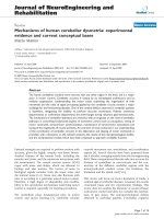
Figure 1
Cerebellar hypermetria. Superimposition of 9 fast wrist
flexion movements in a control subject [A] and a cerebellar
patient [B]. Movements (MVT) are accurate in A and are
hypermetric in B (overshoot of the target). Aimed target
(dotted lines) located at 0.4 rad from the start position
corresponding to a neutral position of the joint. The target is
visually displayed.
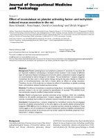
Figure 2
Effects of increasing velocities on kinematics of the
upper l imb pointing movements in a control s ubject
(upper panels) and a cerebellar patient (lower
panels). S ubjects are seated and comfortably restrained in
order to allow only shoulder and elbow movements. They
are asked to perform a verti cal poi nting movement towards a
fixed ta rget at various speeds. The target is located in front
of the subjects at a distance of 85% of total arm length. In the
patient, deficits in angular motion are enhanced with
increasing velocities, especially the increased angular motion
of elbow resulting in overshoot (hyperextension of the
elbow). Black lines: angular position of the elbow; grey lines:
angular position of the shoulder. Abbreviations: sh: shoulder
angle, elb: elbow angle.
Journal of NeuroEngineering and Rehabilitation 2009, 6:10 />Page 2 of 18
(page number not for citation p urposes)
afferents (Figure 4). Noteworthy, the inferior olive is the
single source of climbing fiber inputs to the cerebellum,
and houses cells with oscillatory properties [17]. By
contrast, mossy fibers arise from a large spectru m of
ipsilateral and contralateral sources.
Cerebellar cortex and microcomplexes
Cerebellar cortex is characterized by a laminated
geometrical structure. The Purkinje cells represent the
unique output of cerebellar cortex, targeting nuclear
neurons [18]. The excitation of Purkinje neurons is
balanced by the activity of inhibitory interneurons
located in the molecular (basket cells, stellate cells)
and granular layers of the cortex (Golgi cells and Lugaro
cells). In human, the number of Purkinje cells has been
estimated to about 15 millions [19]. The axon of a
Purkinje neuron gives off about 500 terminals which
contact 30–40 nuclear cells. Each nuclear cell receives
projections from 800– 900 Purkinje neurons.
Granule cells are the most numerous neurons in the human
brain, the population being estimated to about 10
10
–10
11
cells [19,20]. These neurons have four to five dendrites and
make synapses with the enlarged excitatory terminals of
mossy fibers ("rosettes"). Each granule neuron receives
mossy terminals via only four to five excitatory synapses,
suggesting a sparse coding (small convergence number).This
code can be defined as a neural code in which the fraction of
active neurons is low at a given time. Granule cells have low
levels of spontaneous activities. A single impulse in a mossy
fiber tends to induce burst spikes in a granule cell [21,22].
However, granule cells are usually active only briefly
following a sensory stimulus. Sparse coding could reduce
interference issues between tasks being learned by a subject
[16]. Sparse coding could also enhance storage capacity
[16,21]. This is based on the well know divergence of mossy
fiber input to the granule layer and the minimal redundan-
cies between granule cell discharges [22]. To maintain the
low mean firing rate compatible with a sparse code, an
activity-dependent homeostatic mechanism would set the
cells' thresholds [22]. Each granule cell has a thin axon
ascending in the molecular layer and which divides in 2
opposites branches called parallel fibers, running along the
folia. The length of a parallel fiber has been estimated to
4–6 mm [23]. Local excitation of a parallel fiber bundle
stimulates Purkinje cells over a distance of more than 3 mm.
A single parallel fiber passes through the dendrites of more
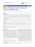
Figure 3
Asymmetry in kinematics of fast wrist flexion
movements in cerebellar patients exhibiting
hypermetria. V alues correspond to ratios of Accelerati on
Peaks d ivided by Deceleration Peaks. Mean +/- SD and
individual ratios are shown. Da ta from n = 7 ataxic patients;
mean age: 53.2 +/- 5.7 years. Control group: n = 7 s ubjects;
mean age: 54.5 +/- 6.1 years. Aimed target: 15 degrees;
n = 10 movements per subject.
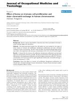
Figure 4
Wiring diagram of the cerebellar circuitry. Purkinje
neurons are the sole output of the cerebellar cortex. Basket
cells supply the inhibitory synapses via a synapse called
"pinceau", stellate cells supply the inhibition to Purkinje cell
dendrites. Lugaro cells are activated by serotoninergic fibers
and inhibit Golgi cells. In addition to the illustrated
serotoninergic afferences, cerebellar cortex receives other
aminergic inputs (acetylcholine, dopamine, norepinephrine,
histamine) or peptidergic projections (peptides such as
neurotensin). These fibers project sparsely t hroughout the
granular and molecular layers to contact directly the Pu rkinje
neurons and other cerebellar neurons. Abbreviations: ST:
serotoninergic fiber, pf: parallel fiber, Gran. c: granule cell,
MF: mossy fiber, br. c: unipolar brush cell, CF: climbing fiber,
IO: i nferior olive, Gc: Golgi cell, Lc: Lugaro cell, Bc: basket
cell, S c: stellate cell, PN: Purkinje neuron; CN: cerebellar
nucleus, mf: recurrent mossy fiber from nuclear cell.
Journal of NeuroEngineering and Rehabilitation 2009, 6:10 />Page 3 of 18
(page number not for citation p urposes)
than 400 Purkinje cells, making contacts with the dendritic
spines of at least 300 Purkinje neurons [24]. Dendrites of
Purkinje neurons are disposed within planes perpendicular
to the long axis of the folia. Each dendritic arborization of
Purkinje neuron enters in contact with more than 100.000
parallel fibers. Parallel fiber beams can bridge and make
functional links between cerebellar nuclei (Figure 5) [25],
with a beam exciting the dendrites of Purkinje, basket,
stellate and Golgi cells. Basket and stellate axons run
tangentially to either side of the transverse parallel fiber
beam, inhibiting Purkinje cells in the 'flanks' of the beam
[26]. Links across the interpositus and dentate nuclei would
effectively connect reach, grasp and reflex sensitivity. This is
based on the fact that each nucleus has a separate
somatotopical representation of the body. Head is caudal,
tail rostral, trunk lateral and extremities medial [27-29]. In
each nucleus, distal and proximal muscles are represented
and these regions can be coordinated by beams of parallel
fibers linking Purkinje cells belonging to distinct functional
units oriented along planes perpendicular to the long-
itudinal axis of the folia. This organization is the anatomical
substratum allowing the coordination of wrist, elbow and
shoulder joint during motion. Indeed, the length of parallel
fibers is sufficient to ensure the connection of Purkinje cells
projecting to different nuclei, permitting the coordination of
the corresponding functions such as control of locomotion,
modulation of reflex activity and reaching-grasping.
The inferior olive transmits signals to a well-defined cluster
of sagittally organized Purkinje cells, which project to given
areas in nuclei. These latter send a feedback projection to the
inferior olive (nucleo-olivary projections). Seven parallel
longitudinal zones are organized on each side of the
cerebellum (A, B, C1, C2, C3, D1, D2). The parasagittally
striped organization of the cerebellum is also found for the
expression of acetylcholinesterase and other molecules such
as zebrin II [see [30]]. The C3 zone receives inputs from the
receptive fields in forelimb skin and contains 30–40
longitudinal microzones,each50to150μm wide [16].
These microzones are the functional units of the cerebellar
cortex. Microcomplexes refertothecombinationofa
microzone and the related structures: small groups of
neurons in a cerebellar or vestibular nucleus, the inferior
olive and neurons in red nucleus [16]. The human
cerebellum might contain about 5000 microcomplexes.
Climbing fibers in nearby microzones are activated from
neighbouring skin areas, making a somatotopic map of the
ipsilateral forelimb skin [16]. The loop is closed in a way,
since microzones project to adjacent cell groups in the
anterior interpositus nucleus which controls movements
having a close relationship with the climbing fibers'
receptive fields.
Cerebellar nuclei
They represent the sole output from cerebellar circuits,
bringing si gnals in par ticular to bra instem nuclei,
thalamic nuclei, motor cortex, premotor cortex and
prefrontal association cortex via the cerebellothalamo-
cortical tracts (Figure 6, Figure 7). Cerebellar nuclei
project back to the overlying cerebellar cortex, with a
mediolateral and rostrocaudal pattern of nucleocortical
projections reflecting the corticonuclear projections [31].
Figure 5
Multiple body maps in the cerebellum. Each cerebellar
nucleus has a compl ete map of the body, with head located
posteriorly, limbs medially and trunk laterally. Thanks to the
parallel fibers (pf, issued from granule cells) linking together
Purkinje neurons (PN) projecting to distinct body areas,
myotomes can be interconnected during motor tasks.
Parallel fibers are long enough to link together Purkinj e
neurons projecting to different portions within one nuclear
body map, and multiple maps. The contacts between parallel
fibers and the dendrites of c ortical inhibitory interneurons
are not illustrated. Adapted from Thach, 2007.
Figure 6
Comparison of anatomical connections of the vermal
zone (A), the intermediate zone (B) and t he lateral
zone of the cerebe llum (C). The midline zone and the
intermediate zone receive direct informations from the
spinal cord, unlike the lateral cerebellum. Abbreviations:
IOC: inferior olivary complex, LVN: lateral vestibular
nucleus, FN: fastigial nu cleus, NI: nucleus interpositus, DN:
dentate nucleus.
Journal of NeuroEngineering and Rehabilitation 2009, 6:10 />Page 4 of 18
(page number not for citation p urposes)
In primates, fastigial nuclei project -although not exclu-
sively-onbothsidestothehindlimbareaofthemotor
cortex and th e pari etal cortex [32]. Interpositus nuclei are
connected with the trunk areas of the motor cortex/
premotor cortex [32]. Dentate nuclei have contralateral
projections to the forelimb zones of the motor cortex/
premotor cortex/prefrontal association cortex [32]. Ven-
tral areas of the dentate nuclei tend to project upon the
prefrontal cortex, in particular zone 9 and 46 which are
involved in working memory and guidance of behaviour
based on transiently stored information, while dorsal
areas send projections primarily to M1 area (Figure 7)
[33]. Functionall y, fastig ial nuclei are especially con -
cerned with eye movements, as well as upright stance
and gait; the interpositus nuclei play key-roles in the
modulation of reflexes, such as stretch, contact and
placing r eflexes; dentate nuclei are mainly involved in
voluntary movements of the extremities such as single-
joint and multi-joint goal-directed movements towards a
fixed or moving target [25].
Patterns of neuronal discharges in cerebellar circuits
Olivary cells fire between 1 and 10 H z, with a mean
frequency close to 1 Hz in most species [34]. The upper
frequency is limited by the long after-hyperpolarization
which lasts about 100 msec. Simple spikes of Purkinje
cells could determine the activity of the cerebellar nuclei,
and therefore govern cerebellar outflow. Simple spike
activity is mainly driven by the mossy fiber inputs to
granule cells. Its modulation is low during passive
movements and high during active movements [35,36].
The complex spikes would serve as error signals to adjust
the simple spike discharges if an error occurs [37].
Simul taneous electrical stimulation of mossy and climb-
ing fibers depresses the parallel fiber-Purkinje cell
synap ses whi ch are concur rently active (the so-called
long-term depression LTD, a form of synaptic plasticity
[37]. LTD is associated with a decrease of the post-
synaptic sensitivity to glutamate caused by removal of
AMPA receptors by endocytosis [38]. LTD plays an
essential role in the cerebellum's error-driven learning
mechanism [16]. In order to have a stable memory
process, an opposing process must balance LTD: long-
term potentiation (LTP). Post-synaptic LTP is able to
reset post-synaptic LTD [39]. Predominance of silent
granule synapses is in agreement with a key-role of LTP
for new learning [1]. For numerous tasks, learning must
initially proceed via LTP in either the direct or indirec t
pathway from granule cells to Purkinje neurons. The first
pathway would increase the excitability of the Purkinje
cell, by contrast with the second pathway.
Despite the inhibitory role exerted by Purkinje neurons
upon cerebellar nuclei, the neurons in these latter fire
spontaneously between 1 0 and 50 Hz. In absence of
motion, high rates of discharges of about 40–50 Hz are
common [25]. During mo tion, firing rates increase and
decrease above and below the baseline. This contributes
to the modulation of the sensitivity of given targets
according to a specific sensorimotor context.
Recordings in the fastigial nuclei indicate that they can
be divided into a rostral and a caudal zone [40]. The
rostral zone is in charge of the descending control of
somatic musculature, controls head orientation and
combined eye-head gaze shifts. The caudal zone controls
oculomotor functions (saccades, smooth pursuit) [41].
There are direct and indirect evidence that discharges in
the interpositus nucleus are related to the antagonist
muscle being used [25,4 2-44]. Interpositus neurons
modulate their activities in relation to sensory feedback
including that from oscillations in movements [45-47].
Figure 7
A: According to the model of Allen and Tsukahara
(1974), the intermediate zone of the cerebellar
hemisphere contributes to movement execution by
monitoring actual sensory feedback and processing
error signals that c ompensate for prediction errors
in movement planning. The lateral zone of the cerebellar
hemisphere part icipates in the pl anning and programming of
movements by integrating sensory information. B: Output
channels in the dentate nucleus. Distinct areas of the dentate
nucleus project predominantly upon different regions of the
contralateral cerebral cortex, via thalamic nuclei (MD/VLc:
medial dorsal/ventralis lateral pars caudalis nuclei, 'area X',
VPLo: nucleus ventralis posterio r lateral is pars oralis). Dorsal
portions of the dentate nucleus project mainly upon area 4.
Journal of NeuroEngineering and Rehabilitation 2009, 6:10 />Page 5 of 18
(page number not for citation p urposes)
Interpositus nucleus might select the degree of reciprocal
versus co-contraction pattern in a given task [43].
Moreover, the interpositus nuclei regulate the discharge
of gamma motor neurons [48] and the excitability of the
anterior horn in the spinal cord [49]. The temporary
inactivation of interpositus nucleus using a cooling
procedure induces tremor which is sensitive to proprio-
ceptive feedback but insensitive to vision [45]. The
cooling induces a 3–5 Hz action tremor as the animals
attempt to reach and grasp food, supporting the idea that
the interpositus nucleus uses abundant afferent inputs to
generate predictive signals. Monzée and colleagues have
shown in monkey that injections of muscimol in the
region corresponding to the anterior interpositus nucleus
induce a pronounced tremor and dysmetria of the
ipsilateral arm when the animal performs unrestrained
reaching and grasping movements [50]. Cells with
anticipatory and reflex-like responses in a lift and hold
task are located in the dorsal anterior interpositus and
not in the dentate nucleus [51]. Hore and Flament
(1986) have observed a te rminal tremor during targeted
limb movements after cooling of cerebellar nuclei [52 ].
They have hypothesized that cerebellum stabilizes limbs
during a maintained posture or after a brisk movement.
To counteract oscillati ons that would otherwise contam-
inate the termination o f movement, the CNS generates
bursts of muscle activity which anticipate the oscilla-
tions. Cooling of cerebellar nuclei interferes with the
normal predictive nature of these suppressive bursts [53].
In absence of adequately timed suppressive bursts, the
position of the limb is driven by non-anticipatory and
transcortical stretch response s [54]. Transcortical reflex
activities may even rein force oscillations, inste ad of
damping them. Repetitive TMS of the primary motor
cortex induces a cerebellar-like tremor which is attrib-
uted to the deficiency in the generation of predictive
responses [55].
Single-unit studies have demonstrated that the neuronal
activity in the dentate nucleus precedes t he onset o f
movement and may also start before the discharges in
the contralateral motor cortex [56]. In part icular, dentate
neurons are active preferentially when motion is
triggered by a mental association with visual or auditory
stimuli [25]. A key-experiment was performed by Thach
in 1978. The author recorded the activities in the motor
cortex, the dentate nucleus, the interpositus nucleus and
limbmusclesinmonkeys[56].Whenanexternalforce
disturbed wrist position, the order of firing was: muscles,
interpositus, motor cortex, dentate. When motion was
triggered by light, the order of activity was: dentate,
motor cortex, interpositus, muscles. These data strongly
suggest that the interpositus is involved in corrective
movements initiated by the feedback of the movement
itself, whereas the dentate nucleus contributes to the
initiation of a movement which is triggered by stimuli
mentally associated with the task. Anterior lesions might
impair more specif ically grasping, and posterior lesions
could generate especially reaching deficits [57]. Inactiva-
tion of the dentate nuclei result in delayed reaction times
in movements triggered by light or sound [58], similarly
to what is observed in cerebellar patients.
Cerebellar input exerts a facilitatory drive upon the
contralateral cerebral cortex. Experimentally, cerebellar
lesions depress the excitability of the contralateral motor
cortex, both in human and in rodents (Figure 8) [59,60].
Non-invasive transcranial activation of neural structures
using electrical and magnetic stimulation (TMS: tran-
scranial magnetic stimulation) has allowed the investi-
gation of the cerebello-thalamo-cortical pathway in
humans. Ugawa et al. have demonstrated significant
gain of EMG responses at an inter-stimulus interval (ISI)
of 3 ms (facilitatory effect) [61]. Conditioning magnetic
stimulus of the cerebellum suppresses motor cortex
excitability 5–8 msec later. This method activates the
unilateral cerebellar structures under the coil. Impaired
Figure 8
Decreased excitability of t he motor cortex
contralaterally to the ablation of the left
hemicerebellum in a r at, as revealed by the study of
recruitment curves of corticomotor responses in the
gastrocnemius muscle. Reco rdings in the gastrocnemius
muscle following incremental electrical stimulation of the
motor cortex. Plots correspond to the amplitude of motor
evoked potentials as a function of stimulus intensity. Filled
triangles: sti mulation of left motor cortex, open triangles:
stimulation of right motor cortex. Fitting with a sigmoidal
curve (3 para meters). 95% prediction band and 95%
confidence band are illustrated. Amplitudes of recorded
motor evoked potentials (MEPs) are expressed in mV.
Journal of NeuroEngineering and Rehabilitation 2009, 6:10 />Page 6 of 18
(page number not for citation p urposes)
facilitation and en hanced inhibition within mot or cortex
have been observed repeatedly in patients presenting
cerebellar lesions [62-66]. Hemicerebellectomy is asso-
ciated with higher motor thresholds contralateral to the
cerebellar lesion. The cerebellum influences also the
excitability of sensitive areas in the brain. Indeed, it has
been demonstrated that the N24 and later components
in somatosensory evoked potentials are markedly
reduced in case of absence of cerebellar input, suggesting
that the cer ebellar circuits influence directly the ex cit-
ability of the parietal cortex [67].
We recently found that trains of transcranial direct
current stimulation (tDCS) applied over the motor
cortex, a technique which is known to facilitate the
overall neural activity of the stimulated area [68,69], can
revert the decrease of excitability induced by an extensive
and acute unilateral cerebellar lesion [70]. tDCS prob-
ably restores the balance between excitatory and inhibi-
tory circ uits in case of hemi cerebell ar ablation. This
opens the possibility of treating human cerebellar
dysmetria with tDCS.
Computational models
The main theories of cerebellar function and their
respective assumptions are summarized in table 1
[25,71-77]. The works of Marr and Albus have exerted
a strong influence on computational models of cerebel-
lar functio n these last decades [16]. Another attractive
model is based upon the adaptative filter hypothesis.
The adaptative filter, developed by Fujita [71] following
the Marr-Albus framework, is a signal-processing device
transforming a set of temporally varying signals into
another [1]. Inputs to the filter are split into components
weighted individually and then recombined to generate
the filter's output. These weights determine the output.
This is a central task for the adaptative filt er [1]. This is
done by a teaching signal and a learning rule for
changing weight values. In case of the cerebellar circuitry,
if the firing of parallel fibers is positively correlated with
the firing of climbing fibers, the weight is reduced (LTD).
The reverse leads t o an increase in the weight (LTP). No
change occurs if the firings are uncorrelated. This
corresponds to the covariance learning rule [78]. This
rule does not distinguish LTP from LTD, considering that
both are part of the same computational process.
The adaptative-filter model has 2 main differences with
the Marr-Albus theory, making this a suitable candidate
for modelling cerebellar microcircuits.First,thesignal-
processing algo rithm is used in many practical applica-
tions. In this sense, it is considered as a model whose
functionality is demonstrated. It depends on the
connectivity with other structures, which is very con-
sistent with the anatomical organization of cerebellar
circuits. Second, it involves time-varying signals and
therefore addresses the key-issue of timing [1].
Internal models
It is widely accepted that expectations and estimates of
future motor states are critical for performing fast
coordinated movements . One of the main theories
addresses a central issue in motor control, namely the
intrinsic time delay of sensory feedback associated with
motor commands and motion. Sensory-motor delays
varyaccordingtothemodalityandcontext,andmaybe
Table 1: Theories of cerebellar func tion s
Theory Assumptions Selected referenceq
Adaptative filter hypothesis Based upon Marr-Albus theory.
Transformation of sets of signals into others. Components are weighted
individually and then recombined to minimise the errors in performance
caused by unavoidable noise.
Fujita, 1982 [ 71]
Internal models The cerebellum contains neural representations to emulate movement.
Internal models reproduce the dynamic properties of body parts.
Wolpert et al., 1998 [72]
Forward model The model predicts the next state given the current state and the motor
command.
Inverse model The model inverts t he system by providing the motor command that will cause
the desired change in state.
Tonic reinforcer The cerebellum tunes the intensities of agonist/antagonist/synergist muscles.
Cerebellum exerts an excitatory influence upon extra-cerebellar targets.
Eccles et al., 1967 [73]
Bastian and Thach, 2002 [25]
Cerebellar timer Cerebellum is the main site of temporal representation of action. Braitenberg, 1967 [74]
Ivry and Spencer, 2004 [75]
Wave-variable processor The cerebellum contributes to a servo-motor mechanism. Massaquoi and Slotine, 1996 [76]
Sensory processor The cerebellum monitors and adjusts the acquisition of sensory information. Bower, 1997 [77]
Journal of NeuroEngineering and Rehabilitation 2009, 6:10 />Page 7 of 18
(page number not for citation p urposes)
in the order of 50– 400 msec. Such delays imply that in-
flight updating of motor commands using sensory
feedback can never be ideal [4]. The cerebellum has
therefore been proposed to contain neural representa-
tions or 'internal models' to emulate fundamental
natural processes such as body movement [Figure 9]
[3]. According t o internal models, the motor cortex is
able to perform an accurate movement using an internal
feedback instead of the external feedback from the real
control object [16]. The internal feedback is closely
linked to the internal model of the object, built in the
cerebellum in close cooperation with the cerebral cortex.
This theory is supported b y fMRI studies, TMS experi-
ments and psychophysical studies. Indeed, the study of
Kawato et al [79] using fMRI strengthens the hypothesis
that the cerebellum implements a forward model for
coordination and accuracy in motor tasks, employing a
predictive information from one effector to e nsure
motor control of another one. Miall et al [80] have
studied the effects of disrupting the cerebellum during a
reach-to-target task using TMS. Stimuli were applied over
the ipsilateral cerebellum during the reaction time of the
subject who had to point to a previously observed target
location following an auditory cue. Errors in the initial
direction and the final position were consistent with the
pointing movements being planned from an estimated
hand position which was about 140 msec out of date.
These data suggest that the cerebellum predictively
updates a central sta te estimate . Accordin g to this
hypothesis, clumsiness in cerebellar patients and dysme-
tria are due to a malfunction in the predictive f eedforward
control and/or to a disorder in the accurate appraisal of
the consequences of motor commands. Internal models
have the advantage to allow the brain to precisely control
the movement without the need for sensory feedback
[16].
Forward models
The cerebellum may function similarly to a 'forward
model' by using efference copies of motor orders to predict
sensory effects of movements. Accurate predictions
would decrease the dependence on time-delayed sensory
signals. Cerebellar circuitry would be necessary to learn
to make appropriate predictions using error information
about the discrepancies between the actual and predicted
sensory consequences, not only for limb movements but
also for postural adjustments [81,82]. Figure 10 shows a
schematic view of the connections that could represent
important elements of the model. The cortico-ponto-
cerebellar tracts bring an efference copy of a motor
command to the cerebellar cortex. The cerebellum would
compute an expected sensory outcome, which would be
sent to cerebral cortical areas via excitatory connections
Figure 9
Forward model-based control scheme (top panel)
and inverse model-based control scheme (middle
panel). F orward model: the message dedicated to the
peripheral motor apparatus A is sent with an efference copy
transmitted to the cerebellum A'. Instructions originating
from higher motor centers (such as the premotor cortex)
reach a comparator (grey circle). The comparator drives the
motor cortex (a), which in turnsdriveslowermotorcenters
in the brainstem and spinal cord. Efference copies are used
to perform future p redictio ns. Cerebellar microcir cuits are
necessary to learn how to make appropriately these
predictive codes. Inverse mod el: A corresponds to the
motor appara tus/control object. Cerebellar cortex working
in parallel with the motor cortex and forming an internal
model with a transfer function a' reciprocally equal to the
dynamics of the con trol object (a' = 1/A). The input to the
cerebellum is the desired trajectory, t he output is the motor
command. T he bottom panel illustrates the model of the
wave-variable processor for the intermediate cerebellum and
the spinal cord gray matter. These structures contribute t o
motion control by processing control signals as wave
variables. These wave variables are combinations of forward
and return signals ensuring stable exchanges despite
destabilizing signal transmission delays (adapted from [76].
Journal of NeuroEngineering and Rehabilitation 2009, 6:10 />Page 8 of 18
(page number not for citation p urposes)
to the thalamus, and to the inferior olive via inhibitory
connections. The inferior olive, which may receive a
corollary discharge directly from the motor cortex, could
operate as a sort of comparator, signall ing errors to back
to cerebellar cortex and training it to make correct
predictions. Purkinje cell firings have several of the
characteristics of a forward internal model of the arm.
Indeed, Purkinje cell firing heralds the kinematics of
motion. Purkinje cell discharges anticipate the kine-
matics of motion, in agreement with a prediction activity
as demonstrated during circular manual t racking in
monkey [83]. Experimental data suggest that Purkinje
neurons from lobules IV-VI encode position, directional
parameters and velocities of arm movements [83,84].
Purkinje cells might provide a prediction signal of the
consequences of movement [85].
Some of the most convincing evidence that the central
nervous system (CNS) uses internal forward models in
human motor behavior comes from studies dedicated to
the control of grasping forces during manipulation of
objects [86]. The rate of grip force development and the
balance between the grip and load forces when grasping/
lifting an object is programmed i n order to meet the
requirements due to physical object properties, such as
weight, surface friction or shape. Cerebellar patients
generate excessive grip forces in relation to loads and
converging data suggest a distorted predictive force
control in cerebellar disorders [86].
Experimental evidence suggesting the use of internal
models for sensory signals has also been found in other
species. In sever al teleosts, cerebel lum-l ike structures
predict the sensory consequences of the behaviour of the
fish [87]. The suppression of self-generated electrosen-
sory noise (reafference) and other predictable signals is
performed partly by an adaptive filter mechanism,which
could represent a more ubiquitous form of the modifi-
able efference copy mechanism.
Inverse models
According to this theory, the cerebellum would lodge an
'inverse model'. Here the input to the cerebellum would
be the aimed trajectory, and the output would be a
motor command. In order to train this type of model,
error information would best be characterized in motor
coordinates in 3 directions. In the laboratory, cerebellar
patients exhibit difficulties in adapting to external force
field, in agreement with the inverse dynamics hypothesis
[88]. There are neurophysiological data supporting the
existence of inverse models: Shidara and colleagues have
shown that Purkinje cell activity during ocular move-
ments are consistent with signals of an inverse mo del
[89]. Although studies of the changes in Purkinje cell
firings occurring when an external f orce load is changed
from resistive to assistive during elbow movements are
suggestive of inverse dynamics model, it should be noted
that these experiments have not controlled limb kine-
matics or modified the magnitude of external loads [90].
To test the hypothesis that Purkinje cell firing is the
output of an inverse dynamics model, forces must be
changes while kinematics are kept constant. The study of
Pasalar and colleagues [91] is consistent with the idea
that Purkinje cells in cerebellar cortex code for kinematic
(i.e. sensory state) but not dynam ic information (i.e.
muscle commands). The majority of Purkinje cells do
not exhibit any modulation in the patterns of discharges
as a function of force type or load. In addition, the
directional tuning pattern seems unaffected, stre ngthen-
ing the idea of uncoupling between Purkinje cell firing
and electromyographic (EMG) activity in limbs. One of
Figure 10
Communication flows for information processing in
forward models of motor coding. Cerebellar modules
receive an efference copy of motor commands via the
corticopontocerebellar tract, in order to make predictions.
Reafference signals and corollary discharges reach the
comparator (inferior olive), which generates an error signal
updating the plastic cer ebellar microcircuits. Expected
sensory outcomes are conveyed to the primary motor
cortex via excitatory connections and to the inferior olive v ia
inhibitory pathways.
Journal of NeuroEngineering and Rehabilitation 2009, 6:10 />Page 9 of 18
(page number not for citation p urposes)
the differences between cerebellar simple spike responses
and those of motor cortical cells is the non uniform
distribution of preferred directions across the workspace
and the extensive overlap in the timing of the simple
spike correlations with movement direction, distance
and target position. These differences suggest that
Purkinje cells handle kinematic information in a
different way as compared to motor cortical neurons
[84].
The intermediate cerebellum might learn internal mod-
els of body mechanics, enabling the cerebellum to adapt
for the complex dynamics o f multi-joint movements
[92]. Cerebellar patients have difficulties in adjusting for
the interaction torques occurring during fast reaches
[12]. It has been repeatedly observed that during fast
goal-directed movements cerebellar patients are unable
to produce normal torque profiles. In particular, they
show abnormal profiles in shoulder muscle torques
varying inappropriately with the dynamic interaction
torques occurring at the elbow joint. Magnitudes of
dynamic interaction forces are scaled to the square of
movement speed, an observation which might explain
the worsening of dysmetria at higher velocities [53].
Inverse dynamic models allow for parsing t he net forces
acting at a joint into force components originating from
muscular activation (MUS), external forces (EXT) includ-
ing gravity, and dynamic inertial and interaction forces
(DYN) [53]. The net torque (NET) is the sum of all
positive and negative torque components:
NET MUS EXT DYN=++
In theory, dynamic interaction forces are the most critical
component amongst dynamic movement variables dur-
ing a coordination task or a multi-joint task. Dynamic
interaction forces have to be precisely computed b y the
CNS. Since muscles are the end effectors, the selection of
muscle activation patterns is a key step. Bernstein was the
first to suggest that muscle activation is selected to
compensate for physical consequences of motion [93].
Actually, the nervous system takes into account the fact
that external forces and interaction forces may support or
antagonize motion.
Given the numerous motor tasks and the huge number
of interactions with the environment, it is widely
accepted that the central ne rvous system must adapt
quickly to the context [86]. In o rder to process all the
contextual informations, it has been hypothesized that
multiple controllers are in charge of a context or a small
sets of contexts [72]. Indee d, a unique controller would
demand an enormous complexity and would need to
adapt each time to a new context, a potential source of
errors [86]. This hypothesis takes into account the need
to select the correct controller in a given circumstance
[86]. To m aster this task, multiple paired forward-inverse
models would be required.
Cerebellum and the adaptation of the magnitude of muscle
responses to inertia or damping
Cerebellum tunes the intensity o f the activities of
numerous antagonist and synergist muscles used auto-
matically in normal movements . It coordinates their
timing, duration and amplitudes of activity [25]. A
"tonic reinforcer" function seems suited for the interac-
tions between the cerebellum and vestibular nuclei,
reticular nuclei and motor cortex [25].
Fast single-joint monodirectional movements have been
studied to extract specific patterns of muscle discharges
in cerebellar p atients. These movements are normally
controlled by a triphasic pattern of EMG activity: a first
burst in the agonist muscle (providing the launching
torque) is followed by a second burst in the antagonist
muscle (providing the braking torque), followed by a
second burst i n the agonist muscle (to bring the limb
accurately to the target) [94,95]. Several deficits have
been discovered in cerebellar patients (Figure 11): (a) a
delayed onset latency of the antagonist EMG activity, (b)
a slower rate of rise in the agonist/antagonist EMG
activities, (c) an inability to tune the intensity of agonist/
antagonist EMG activities when the inertia of the limb is
increased [96,97].
Recently, deficits in reversal movements have been
found in ataxic p atients. Reversal movements refer to
Figure 11
Triphasic pattern of electromyographic (EMG)
activities in a control subject (left) and in a cerebellar
patient e xhibiting hypermetria (right). In the control
subject, the first agonist burst (AGO1) is followed by a burst
in the antagonist muscle (ANTA), followed by a seco nd burst
in the agonist muscle (AGO2). In the cerebellar patient,
threeEMGdeficitsareobserved:therateofriseofEMG
activities is depressed, the onset latency of the antagonist
EMG activity is delayed and the 2 agonist bursts are not
demarcated. FCR: flexor carpi ra dialis; ECR: extensor carpi
radialis. EMG traces are full-wave rectified and averaged
(n = 10 movements).
Journal of NeuroEngineering and Rehabilitation 2009, 6:10 />Page 10 of 18
(page number not for citation p urposes)
movement towards a fixed target immediately followed
by a return to the initial position. Reversal movements
are balanced in shape, and the agonist EMG a ctivity is
composed of 2 bursts which are clearly separated [98].
During a fast voluntary movement, muscle damping, a
non linear resistance to movement which depends on
velocity, is typically as ymmetrical, meaning that it
predominates in the direction of muscle shortening
[99]. For hand kinematics in the physiological range of
motion, the damping compensation signal (aiming at
compensating the asymmetry of the damping parameter)
is a crucial element for kinetic encoding by the motor
cortex [100]. The structures in the CNS regulating the
damping compensation signal have not been identified
so far, mainly due to technical constraints related to
methods of investigations. The elucidation of the
contribution of the cerebellar pathways in the damping
compensation signal has remained so far elusive. In
patients exhibiting a mild form of cerebellar ataxia, fast
single-joint movements in one direction may be accu-
rate. Thanks to the use of the haptic technology, it has
been observed that these patient s are unable to a dapt to
mechanical damping (addition of viscous forces) during
the ret urn to the in itial position (second phase of the
movement) [Figure 12]. The deficit is not dependent
upon the initial direction of movement. In complex
movements, the motor plan consists of asuperimposition
of elemental defined components [99,101]. In reversal
movements, these elemental components need (1) to be
selected and (2) to be superimposed sequentially. This
highlights the fact that a given muscle can exhibit a
normal behaviour facing mechanical damping during
the first part of a motor sequence, but is not able to
adapt appropriately for the next part. One implication is
that current rehabilitation strategies in patients with
cerebellar disorders should take into account the
differences in the motor strategies underlying pointing
movements and reversal movements in cerebe llar dis-
orders.
There are also experimental evidence that the cerebellum
modulates the gain of reflexes in human. One example is
that long-latency EMG responses (LLR) are abnormal in
cerebellar patients. Typically, the first component M1 (of
spinal origin) is spared in terms of latency/amplitude,
whereas the magnitude of the M3 respon se (long-latency
transcortical response) is increased [102,103]. This is
illustrated in Figure 13. The phenomenon is particularly
marked when lesions involve primarily the cerebellar
cortex, suggesting a loss of inhibition from Purkinje cells
Figure 12
Inability to adapt to damping in cerebellar
hypometria during fast reverse movements.
Movement (top panels) and the associated EMG bursts in a
control subject (left pan els) and in an ataxic patient (righ t
panels) for an aimed target of 0.3 rad are illustrated. Top
panels: superimposition of fast reversal movements
performed without damping (blue), with addition of 0.1
Nms/rad (black) or 0.2 Nms/rad (red). EMG bursts in the
flexor carpi radialis (FCR ) and the extensor carpi radialis
(ECR) are calibrated with a reference to a maximal isotonic
contraction (MIC) from 0 to 6 Nm (a.u.: arbitrary units). In
each position panel, grey areas correspond to the 99%
confidence interval of control values of movement
amplitudes in the basal mechanical state (no addition of
damping); dotted l ines in black and red delineate the 99%
confidence i nterval of control v alues during addition of 0.1
Nms/rad and 0.2 Nms/rad, respectively. In the patient, the
first phase of movement (from the starting position to the
target of 0.3 rad ) remains accurate but the second p hase
(from the target of 0.3 rad to the return to the initial
position) is hypometric. The hypometria is increased with
addition of damping. Arrowheads located near the EMG
traces indicate the o nset of EMG bursts (blue: no damping,
black: addition of 0.1 Nms/rad, red: addition of 0.2 Nms/rad).
AGO1, AGO2 and ANTA1 correspond to the first bur st in
the FCR, the second burst in the FCR and the antagonist
burst in the ECR, r espectivel y. Arrowheads near AGO1,
ANTA1, AGON2 and ANTA2 correspond to the onset of
the first burst in the FCR, the first phase of the burst in the
ECR, the second phase of the bur st in the ECR, and the
second burst in the FCR, respectively. AGO1 and ANTA2
are well demarcated in bottom left panel, unlike in the rig ht
bottom panel. Flex.: direction of flexion of the wrist; Ext.:
wrist extension.
Journal of NeuroEngineering and Rehabilitation 2009, 6:10 />Page 11 of 18
(page number not for citation p urposes)
leading to an overactivity of cerebellar nuclei. These data
confirm a contribution of the cerebellum in the tuning of
the magnitudes of muscle discharges.
Cerebellum as a movement timer
Another influential theory is that the ce rebellum acts as a
movement timer and is the main site of temporal
representation of action, thanks to numerous interactions
between the cerebellum and the inferior olive. Oscilla-
tions of inferior olive cells have been suggested to endow
the system with the capacity to create complex temporal
patterns, which might be applied for fine tuning of
motor output and motor adjustments. Experiments
showing that cerebellar lesions impair timing of motor
acts are convincing [75,104,105]. Patients with lateral
cerebellar lesions have difficulties in perceiving differ-
ences in intervals between tone pairs in the r ange of
0.5 sec, suggesting the presence of a general clock not only
for motion, but also for perception [106]. Although
apparently simple, the rhythmic synchronization
between a timed sensory stimulus and a motor response
step requires a highly complex signal processing proce-
dure for the brain [107]. The production of a motor
response time locked to a rhythmic stimulus implies an
extraction of the timing inform ation present in the
sensory stimulus. Subsequently, this information has to
be implemented to m ake predictions. Nevertheless, it is
nowclearthatthecerebellumisnotthesolesite
processing timing parameters in the brain [107]. The
cerebellum, basal ganglia and frontal cortex interact
strongly to pull out timing information and to funnel it
in 'operative' centres. Cerebellar circuitry might work as a
global supp ort system in sensory acquisition and
processing of timing procedures, facilitating the effi-
ciency of brain networks [107]. The cerebellum could be
seen as a sort of regulating clock.
Cerebellum and sensori-motor l earning
Damage to th e ce rebell um causes the inabil ity to learn
new complex movements [25]. Thanks to its high degree
of adaptation in its operational mechanisms, the
cerebellum contributes to various aspects of motor and
non-motor learning. Motor learning can be defined as
modifications in motor performance with practice, as an
increase in the repertoire of motor behaviour or as a new
behaviour maintained o ver a given period of time. In
agreement with the theory of error signals, an increase in
complex spikes f iring rates during the adaptation phase
to a novel load in a wrist-holding task has been
demonstrated [108]. Once the task was learned, complex
spike firing returned to baseline. According to Kitazawa
and colleagues, complex spikes occurring early during a
reaching task assist s in encoding the abso lute d irectio n
and destination of the arm, computing the relative
endpoint errors of the reach [109].
There is strong evidence that eyeblink conditioning is
dependent on the integrity of cerebellar networks.
Findings in human are in good agreement with findings
in animal studies [110]. Small lesions in the interpositus
nucleus induce a permanent loss of conditioned
responses. Several specie s have been used and several
models of the basic neural circuits required for the
acquisition and performance of classical eyeblink con-
ditioning h ave been discussed [111]. An intermediate
cerebellum-related network superimposed on the brain-
stem circuits regulating the inborn unconditioned eye-
blink response has been proposed. Neural plasticity
develops both in the cerebellar cortex and cerebellar
nuclei following training [112,113]. Recent experime ntal
observations are providing the first evidence that the
memory trace of motor learning may shift trans-
synaptically for consolidation to long-term memory
[114]. Neuroanatomical correlates of learning have
been studied in human. The majority of lesion studies
have investigated conditioned response acquisition. The
group of Timmann et al. has shown that the superior
cerebellar artery supplies critical zones for eyeblink
conditioning in human [110]. Cerebellar circuits are
also involved in the timing and extinction of condi-
tioned eyeblink responses. It should be mentioned here
that regarding the vestibulo-ocular reflex (VOR) learn-
ing, experiments suggest that short-term learning is
maintained by the cerebellum, while long-term learning
can continue also when the cerebell um is removed.
Figure 13
Long latency electromyographic (EMG) responses to
stretches of the first dorsal interosseous muscle in a
cerebellar patient (black line) and in a control subject
(grey line). L atencies of averaged rectified EMG responses
are normal, but the M3 response is increased in the
cerebellar patient. Surface EMG rectified and averaged 200
times. Responses are calibrated in arbitrary units (a.u.).
Journal of NeuroEngineering and Rehabilitation 2009, 6:10 />Page 12 of 18
(page number not for citation p urposes)
Other theories of cerebellar function
Coupling between the cerebellum and contralateral
thalamic nuclei/primary motor cortex is well established
[115]. Coherent oscillations betwee n the sensory cortex
and the cerebellar cortex have been reported. Activity in
cerebellofugal fibers is triggering oscillations in thalamic
nuclei and motor cortex [116]. The modulation of these
oscillations in terms of frequency and synchronicity
might be an important feature of the cerebello-cerebral
loops. Moreover, another and complementary role for
the cerebellum could be the tuning of the sensorimotor
coupling of n eural activities in a particular condition
combining reflex and voluntary movement [117]. This
theory is based on the fact that the relation between
sensory signals guiding motion and the movement itself
depends on the 'context', which takes into consideration
the re lative position of the limb segments, the position
of the body in the gravitational field and the external
forces interacting with the movement. This is also taken
into account in the hypothesis of the sensorimotor
coordinate transformer, according which the main function
of the cerebellum is to mathematically transform signals
from sensory to motor coordinates [118]. Cerebellar
operations would be represented by a matrix of gains,
leading to a prediction function.
The theory of the wave-variable processor attempts to
explain how the cerebellum deals with the issue of
feedback motor control in the presence of signal
transmission delays [76]. The central premise of the
wave-variable processor theory is that the interaction
between the intermediate cerebellum and t he spinal cord
represent a wave-variable-based communication. This is
based upon the teleoperation theory of Niemeyer and
Slotine (1991) [119]. According to this theory, the
cerebellum contributes to a servo-motor mechanism.
The "servo hypothesis" was originally proposed by
Merton [120]. Wave variables are linear combinations
of command/feedback signals that can exchanged
between a master unit and a slave unit to obtain a
stable control whatever the transmission delay. The
structure is consistent with the numerous combinations
of inputs, such as force feedback signals and corollary
discharges from internal pathways, ascending from the
spinal cord via the spinocerebellar tracts. The wave-
variable processing would allow the motor system to
work without complex internal models. Simulations of
rapid elbow movements hav e conf irmed that the model
mimics monkey's performance [76]. In addition, reduc-
tion of the cerebellar output induced a large oscillation
reminiscent of cerebellar tremor. Interestingly, simulated
signals of the interpositus nucleus matched the real
signals recorded in monkey. This model can take into
account the multiple reverberating loops existing
between the cerebellum and brainstem nuclei (such as
the cerebello-reticulo-cerebellar loops). However, due to
its linearity, this model cannot be applied to the non-
linear dynamics of a two-joint arm.
What did we learn from studies including patients with
cerebellar disorders?
Theories and models have been tested in cerebellar
disorders encountered in the clinic, such as cerebellar
stroke (acute focal lesion involving afferences and/or
efferences) or t he various forms of sporadic/i nher ited
cereb ell ar degenera tion (progressive loss of neurons in
the cerebellum, especially in the cerebellar cortex). As
mentioned in section III, the observation of the deficits
in patients with cerebellar disorders argues in favour of a
forward internal model [121]. Impaired adaptation in
anticipatory responses is observed during various experi-
mental paradigms [122,123]. For instance, it has been
shown that anticipatory pos tural adjustments are under
cerebellar supervision [82,122]. Horak and Diener have
assessed the adjustments in torque responses during
standing postural perturbations [122]. Healthy subjects
are able to scale the anticipatory postural responses
when perturbations are presented in a predictable order,
unlike cerebellar patient s. This suggests that the cerebel-
lum plays an integral role in using predictive feedfor-
ward control t o adapt postural responses [81]. Similar
deficits have been reported in locomotor-like adaptation
tasks [124]. Recent studies with a splitbelt treadmill (one
leg is forced to move faster than the other) have
demonstrated that the adaptation process includes
both a reactive and a predictive component [125].
Reactive adaptations arise and disappear quickly, respec-
tively after the perturbation and upon removal of the
splitbelt condition [81]. Regarding the predictive adap-
tations, they become apparent after several strides and
display after-effects suggesting t hat important informa-
tions related to the body-environment interactions have
been stored. Whereas reactive adaptations are spared in
case of cerebellar lesion, predictive adaptations are
impaired [121]. In human, lesions of the dentate nucleus
or lesions of the cerebellar cortex result in an uncoupling
of grip force-load force during a lifting and holding task
with objects of different weights [126]. By contrast,
lesions in the territory of the posterior inferior region of
the cerebellum do not cause any overshoot in grip force
nor a lack of coordination between grip and load force
profiles. The progressive and general loss of function
encountered in hereditary spinocerebellar ataxias is also
associated with impaired force adaptations during goal-
directed arm movements [88]. The failure to generalize
learning to untrained regions in the workspace suggests
that a chronic and progressive loss of cerebellar circuits
prevents the formation of the internal representation of
limb dynamics. These findings have direct implications
Journal of NeuroEngineering and Rehabilitation 2009, 6:10 />Page 13 of 18
(page number not for citation p urposes)
for daily rehabilitation of cerebellar dysmetria, but these
are currently underestimated.
Wearable devices and unobtrusive sensors
There are been limitations in the past to assess the
mechanisms of dysmetria due to technical constraints.
Wearable devices, unobtrusive sensors and body area
networks, as well as new techniques of assessments such
as haptic devices are emerging tools which will probably
modify our understanding and our methods of clinical/
laboratory evaluation of cerebellar dysmetria in the near
future. Exoskeletons are a typical example of wearable
devices with many potential applications in the field of
motion research and therapy. For instance, they can be
used to assess the effects of mechanical perturbations on
cerebellar deficits such as cerebellar tremor [127]. They
also can be used to evaluat e their potential usefulness in
restoring the metrics of motion in case of limb
dysmetria. Moreover, these devices open new perspec-
tives to assess the various theories of cerebellar functions
in a clinical environment, especially by modifying the
inertia or damping of individual s egments in a given
limb. Studies of eye-head-limbs coordination will
benefit from technological developments in the coming
years. Another example is the very recent application of
brain-computer interfaces (BCI) in this area of research.
Unobtrusive sensors are also becoming popular in
functional imaging studies, where they can bring critical
informations during data acquisition.
Conclusion
We have provided an overview of the current theories
underlying the roles of the cerebellum in motor control
and the mechanisms of cerebellar dysmetria. From the
anatomical point of view, the cerebellum is very well
positioned to process multi-modal sensory information.On
the basis of available experimental data, it can be
proposed that cerebellar dysmetria mainly results from
a deficit in the predictive feedforward control (Figure 14).
Predictions and subsequent updates based on sensory
events would be possi ble t hanks to the numerous
projections received by the cerebellum, the huge
computing capabilities of the cerebellar circuitry, and
the pertinent interaction of mossy and climbing fibres
[86]. Cerebellar cortex, especially Purkinje neurons,
plays a key-role in coding the kinematic features of
movement. Through the cerebello-thalamo-cortical
channel, inputs can modulate the efficacy of the
interconnections among cortical neurons, adjusting the
circuitry of the motor cortex in various contexts and
implementing predictions in the sensorimotor system.
As a result of a cerebellar lesion, patients have a
disorganized timing implementation in motor tasks
and exhibit difficulties in tuning the magnitudes of
motor responses. The generation o f inappr opriate
muscle torques may result from the errors in the
prediction of the mechanical consequences of move-
ments of one limb segment on adjacent joints [53].
Errors in predicting compensation torques may cause the
abnormal metrics of motion (dysmetria). Both the
defect in feedforward control and the abnormal ex cit-
ability of the motor cortex result in an inability for the
motor system to update motor programming based
upon sensory events. Cerebellar circuitry would coop-
erate with basal ganglia to generate smooth movements,
despite state changes and time delays (Figure 15). A
function of the cerebellum is system identification. The
cerebellum would construct internal models with the
aim of predicting sensory outcome of motor commands
and correct these commands via internal feedback [128].
Figure 14
Overview of the mechanisms of human cerebellar dysmetria.
Journal of NeuroEngineering and Rehabilitation 2009, 6:10 />Page 14 of 18
(page number not for citation p urposes)
Cerebellum acts both at the upper motor neuron and
lower motor neuron level to tune muscle discharges and
modulate the muscle reactivity to environmental
changes. Figure 16 summarizes the interactions between
the cerebellum and the motor system using the
component-based Hill muscle model, which is com-
monlyusedtopredictmuscleforcesandwhich
represents the active a nd passive properties of the
musculo-tendinous unit [129]. The model includes a
series elastic component and a neural input processor in
parallel with a viscous component . The figure also
shows an operational model for the cerebellar circuits.
The current theories of cerebellar functions can be under-
stood as complementary rather than mutually exclusive.
Some of them share commonalities. For goal-directed tasks,
predictive control is essential for fast executi on, bu t
predictions are also important for slow motion, due to the
increased reliance on time-delayed feedback signals [130].
The combination of both forward and inverse models
results in computational advantages for motor learning and
control. The context of the experiments, the biomechanical
features of the effectors being considered (eyes, limbs, ),
the motor task (reaching task, grasping, postural task,
gait, ), the way data have been collected, and the clinico-
radiological aspects (in case of studies with patients) should
all be taken into account and integrated when attempting to
extract the conceptual bases underlying cerebellar dysme-
tria. Quantitative lesion approach and theoretical motor
control provide complementary informations.
Figure 15
Overview of the motor control strategy for limb movements. Cerebellum builds internal models and corrects motor
commands, c omparabl e to a system identification function. Bas al ganglia ensures an optimal control of moti on, facilitating
motor commands. The parietal cortex integrates proprioceptive and visual outcomes, as well as sensory feedback, playing a
role of state estimator. Premotor cortex and motor cortex transforms predictions into sets of motoneur onal discharges,
encoding for force and direction of movement.
Journal of NeuroEngineering and Rehabilitation 2009, 6:10 />Page 15 of 18
(page number not for citation p urposes)
Competing interests
The author declares that they have no competing
interests.
Acknowledgements
Mario Manto is supported by the FNRS-Belgium.
References
1. Porrill J and Dean P: Silent synapses, LTP, and the indirect
parallel-fibre pathway: computational consequences of
optimal cerebellar noise-processing. PLoS Comput Biol 2008,
4(5):e1000085.
2. Stein RB, Gossen ER and Jones KE: Neu ronal variability: noise or
part of the signal?. Nature Re v Neurosci 2005 , 6:389–397.
3. Ramnani N: The primate cortico-cerebellar system: anato my
and function. Nat Rev Neurosci 2006, 7:511–522.
4. Manto M and Bastian AJ: Cerebellum and the deciphering of
motor coding. Cerebellum 2007, 6:3–6.
5. Allen GI and Tsukahara N: Cerebrocerebellar communication
systems. Physiol Rev 1974, 54:957–1006.
6. Kelly RM and Strick PL: C erebellar loops with motor cor tex
and prefrontal cortex of a nonhuman primate. JNeurosci2003,
23:8432–8444.
7. TimmannDandDaumI:Cerebellar contributions to cognitive
functions: A progress report after two decades of research.
Cerebellum 2007, 6:159–162.
8. Haines DE and Manto MU: Clinical symptoms of cer ebellar
disease and their interpretation. Cerebellum 2007, 6:360– 374.
9. Holmes G: The cerebellum of man. The Hughlings Jackson
memorial lecture. Brain 1939, 62:1–30.
10. Hallett M and Massaquoi S: Physiol ogic studies of dysmetria in
patients with cerebella r deficits. Can J Neurol Sci 1993, 20 S uppl
3:S83–S92.
11. Massaquoi SG and Hallett M: Kinematics of initiating a two-joint
arm movement in patients with cerebellar ataxia. Can J
Neurol Sci 1996, 23:3– 14.
12. Topka H, Konczak J, Schneider K, Boose A and Dichgans J:
Multijoint arm movement s in cere bellar ataxia : abnormal
control of movement dynamics. Exp Brain Res 1998,
119:493–503.
13. Bastian AJ, Martin TA and Keating JG: Cerebell ar ataxia:
abnormal control of interaction torques across multiple
joints. JNeurophysiol1996, 76:492–509.
14. Brown SH, Hefter H, Mertens M and Freund HJ: Disturbances in
human arm movement trajectory due to mi ld cerebellar
dysfunction. J Neurol Neurosurg Psychiatry 1990, 53:306–313.
15. Day BL, Thompson PD, Harding AE and Marsden CD: Influence of
vision on uppe r limb reaching movements in patients with
cerebellar ataxia. Brain 1998, 121:357 – 372.
16. Ito M: Cerebellar circuitry as a neuronal machine. Prog
Neurobiol 2006, 78:272–303.
17. Yarom Y and Cohen D: The olivocerebellar system as a
generator of temporal patterns. AnnNYAcadSci2002,
978:122–134.
18. Manto M, Haines D, Yoshida M, Obata K and Ito M: Cerebellar
classics I. Cerebellum 2007, 102–105.
19. Colin F, Ris L and Godaux E: Neuroanatomy of the cerebellum.
The cerebellum and its disorders Cambridge University Press, Cam-
bri dge, UK: Manto M, Pandolfo M 2002, 6–29.
20. Zagon IS, McLaughlin PJ and Smith S: Neural populations in the
human cerebellum: estimat ions from isolated cell nuclei.
Brain Res 1977, 127:279–82.
21. Brunel N, Hakim V, Isope P, Nodal JP and Barbour JP: Optimal
information storage and the distribution of synaptic weights:
perceptron versus Purkinje cells. Neuron 2004, 43:745–757.
22. Sch weighofer N, Doya K and Lay F: Unsupervised learning of
granule cell sparse codes enhances cerebellar adaptative
control. Neuroscience 2001, 103:35–50.
23. Mugnaini E: The leng th of cerebellar parallel fibers in chick en
and rhesus monkey. J Comp Neurol
1983, 220:7–15.
24. Eccles JC, Ito M and Szenthagothai J: The cerebellum as a
neuronal machine.Ne w York, Heidelberg, Springer-Verlag; 1967.
25. Bastian and Thach : Structure and function of the cerebellum.
The Cer ebellum and its disorders Cambridge University Press, Cam-
bri dge: Manto M, Pandolfo M 2002, 49–66.
26. Thach WT: On the mechanism of cereb ellar contributio ns to
cognition. Cerebellum 2007, 6:163–167.
27. Orioli PJ and Strick PL: Cerebellar connections with the motor
cor tex and the arcuate premotor area: an analysis employ-
ing retrograde transneuronal transport of WGA-HRP.
J Comp Neurol 1989, 288:612–626.
28. Asanuma C, Thach WT and Jones EG: Cytoarchitec tonic
delineation of the ventral lateral thalamic regi on in the
monkey. Brain Res 1983, 286(3):219–235.
29. Asanuma C, Thach WT and Jones EG: Distribut ion of cerebellar
terminations and their relation to other afferent termina-
tions in the ventral lateral thalami c region of the monkey.
Brain Res 1983, 286(3):237–265.
30. Sanchez M, Sillitoe RV, Attwell PJE, Ivarsson M, Rahman S, Yeo CH
and Hawkes R: Compart mentation of the rabbit cerebellar
cor tex. J Comp Neurol 2002, 444:159–173.
31. Gould BB and Graybiel AM: Afferents to the cerebellar cortex
in the cat: evidence for an intrinsic pathway leading from the
deep nuclei to the cortex. Brain Res 1976, 110:601– 611.
32. Sch mahmann JD: The cerebellum and cognition.Academic Press,
San Diego; 1997.
33. Middleton FA and Strick PL: Cerebellar output channels. The
cerebellum and cognition Academic Press, San Diego: Schmahmann JD
1997, 61–82.
34. Llinas R and Yarom Y: Electrophysiology of mammalian
inferior olivary neurons in vitro. Different types of voltage-
dependent ionic conductances. J Physiol (Lond) 1981,
315:549–567.
35. Bauswein E, Kolb FP, Leimbeck B and Rubia FJ: Simple and
complex spike activity of cerebellar Purkinje cells during
active and passive movements in the awake monkey. JPhysiol
1983,
339:379–94.
Figure 16
Representation of the sites of action of the
cerebellum. Hill's muscle model and an operational model
of the cerebellar circuitry are illustrated. Central and
peripheral loops in the central nervous system are shown,
with u pper motoneuron (UMN)/lower motoneuron (LMN).
IN indicates the pool of interneurons in the spinal cord.
Cerebellar influences on spinal motoneurons are mainly
indirect. DR corresponds to the dorsal root ganglia. The
rectangle in the bottom represents Hill's muscle model
(SE: series elastic component; NIP: neural input processor
in parallel with a viscous component PE). Operational model
of the cerebellar circuits: given inputs (IN) to a microzone
(Micro) elicit an output (OUT) which depends on the whole
complex of afferen t information impingin g upon the
microzone. Adapted from Manzoni, 2007.
Journal of NeuroEngineering and Rehabilitation 2009, 6:10 />Page 16 of 18
(page number not for citation p urposes)
36. Harvey RJ, Por ter R a nd Rawson JA: The natural discharges of
Purkinje cells in paravermal regions of lobules V and VI of
the monkey's cerebell um. J Physiol (Lond) 1977, 271:515–536.
37. Ito M: Long-term depression. Annu Rev Neurosci 1989, 12:85–102.
38. Montgomery JM, Selcher JC, Hanson JE and Madison DV: Dynamin-
dependent NMDAR endocytosis during LTD and its depen-
dence on synaptic state. BMC Neurosci 2005, 6:48.
39. Lev-Ram V, Mehta SB, Kleinfeld D and Tsien RY: Reversing
cerebellar long- term depression. Proc Natl Acad Sci USA 2003,
100:15989– 15993.
40. Buttner U, Fuchs A F, M arke rt -Sc hwa b G a nd B uck mast er P:
Fastigial nucleus activity in the alert monkey during slow
eye and head movements. J Neurophysiol 1991, 65:1360–1371.
41. Fuc hs AF, Robinson FR and Straube A: Participation of the caudal
fastigial nucleus in smooth-pursuit eye movements. I.
Neuronal activity. J Neur ophysiol 1994, 72:2714 – 2728.
42. Vilis T and Hore J: Effects of changes in mechanical state of
limb on cerebella r intention tremor. JNeurophysiol1977,
40:1214–1224.
43. Smith AM and Bourbonnais D: Ne uronal activity in cerebellar
cor tex related to control of prehensile force. JNeurophysiol
1981, 45:286–303.
44. Frysinger RC, Bourbonnais D, Kalaska JF and Smith AM: Cerebellar
cor tical activity during an tagonist cocontraction and reci-
procal inhibition of forearm muscles. J Neurophysiol 1984,
51:32–49.
45. Vilis T and Hore J: Central neuronal mechanisms contributing
to cerebellar tremor produced by limb perturbations.
JNeurophysiol1980, 43:279–291.
46. Soechting JF, Bur ton JE and Onoda N: Relationships between
sensory input, motor out put and unit activity in interpositus
and red nuclei during intentional movement. Brain Res 1978,
152:65–79.
47. Sch ieber MH and Th ach WT Jr:
Trained slow tracking II.
Bidirectional discharge patterns o f cer ebellar nuclear,
motor cortex, and spindle afferent neurons. JNeurophysiol
1985, 54(5):1228–1270.
48. Gilman S: The mechani sm of cerebellar hypotonia. Brain 1969,
92:621–638.
49. Oulad Ben Taib N, Laute MA, Pandolfo M and Manto MU:
Interactio n between repetitive stimulation of the sciatic
nerve and funct ional ablation of cerebel lar nucleus inter-
positus in the rat. Cerebellum 2004, 3:21–26.
50. Monzée J, Dre w T and Smith AM: Effects of muscimol
inactivation of the cerebellar nuclei on precision grip.
JNeurophysiol2004, 91:1240–1249.
51. Monzée J and Smith AM: Responses of cerebellar int erposit us
neurons to predictable perturbations applied to an object
held in a precis ion grip. J Neurophy siol 2004, 91:1230– 1239.
52. Hor e J and Flament D: Eviden ce that a disordered servo-like
mechanism contributes to tremor in movements during
cerebellar dysfunction. J Neurophysiol 1986, 56:123–136.
53. Topka H and Massaquoi SG: Pathophysiology of clinical
cerebellar signs. The Cerebellum and its disorders Cambridge
University Press, Cambridge: Manto M, Pandolfo M 2002, 121–135.
54. Flament D and Hore J: Movement and electromyographic
disorders associated with cerebellar dysmetria. JNeurophysiol
1986, 55:1221–1233.
55. Topka H, Mesc heriakov S an d Boose A, et al: A cerebellar-like
terminal and postural tremor induced in normal man by
tra nscranial magnetic stimulation. Brain 1999, 122:1551–1562.
56. Thach WT: Correlation of neura l discharge with pattern and
force of muscular activity, joint position, and direct ion of
intended next movement in motor cortex an d cerebellum.
JNeurophysiol1978, 41:654–678.
57. Mas on CR, Miller LE, Baker JF and Houk JC: Organiza tion of
reaching and grasping mo vements in the primate cerebel-
lar. JNeurophysiol1998,
79:537–554.
58. Trouche E and Beaubaton D: Initiation of a goal-directed
movement in the monkey. Exp Brain Res 1980, 40:311–321.
59. Di Lazza ro V, Restuccia D, Nardone R, Leggio MG, Olivi ero A,
Profice P, Tonali P and Molinari M: Motor cortex changes in a
patient with hemicerebellectomy. Electroencephalogr Clin Neu-
rop hysiol 1995, 97:259–263.
60. Oul ad Be n Taib N and Manto M: Effects of trains of high-
frequency stimulation of the premotor/suppleme ntary
motor area on conditioned corticomotor responses in
hemicerebellectomized rats. Exp Neurol 2008, 212:157–165.
61. Ugawa Y, Uesaka Y, Terao Y, Hanajima R and Kanazawa I: Magnetic
stimulation over the cerebellum in humans. Ann Neurol 1995,
37:703–713.
62. Wessel K, Tegenthoff M, Vorgerd M, Otto V, Nitsche MF and
Malin JP: Enhancement of inhibitory mechanisms in the
motor cortex of patients with c erebellar degeneration: a
study with transcranial magnetic brain stimulation. Electro-
encephalogr Clin Neurophysiol 1996, 101:273–280.
63. Liepert J, Kucinski T, Tuscher O, Pawlas F, Baumer T and Weiller C:
Motor cortex excitability after cerebell ar infar ction. Stroke
2004, 35:2484–2488.
64. Tamburin S, Fiaschi A, Marani S, Andreoli A, Manganotti P and
Zan ette G: Enhanced intracortical inhibition in cerebellar
patients. JNeurolSci2004, 217:205–210.
65. OliveriM,TorrieroS,KochG,SalernoS,PetrosiniLand
Caltagirone C: The role of transcra nial ma gnetic stimulation
in the study of cerebellar cognitive function. Cerebellum 2007,
6:95–101.
66. Di Lazzaro V, Restuccia D, Molinari M, Leggio MG, Nardone R and
Fogli D: Excitability of the motor cortex to magnetic
stimulation in patients with cerebellar lesions. JNeurol
Neurosurg Psychiatry 1994, 57:108–120.
67. Restuccia D, Valeriani M, Barba C, Le Pera D, Capecci M, Filippini V
and Molinari M: Functional changes of the primary somato-
sensory cortex in patients with unilater al cerebellar lesions.
Brain
2001, 124:757–768.
68. Bog gio PS, Castro LO, Savagim EA, Braite R, Cruz VC, Rocha RR,
Rig onatti SP, Silva MTA and Fregni F: Enhancement of non-
dominant hand motor function by anodal transcranial direct
cur rent stimulation. Neurosci Lett 2006, 404:232–236.
69. Nitsche MA, Seeber A, Frommann K, Klein CC, Rochford C,
Nitsche MS, Fricke K, Liebetanz D, Lang N, Antal A, Paulus W and
Tergau F: Modulating parameters of excitability during and
aft er transcranial direct current stimulation of the human
motor cortex. J Physiol (Lond) 2005, 568:291–303.
70. Oul ad Ben Taib N and Manto M: Trains of transcranial DC
stimulation antagonize motor cortex hypoexcitability
induced by acute hemicerebellectomy. J Neurosurg in press.
71. Fujita M: Adaptive filter model of the cerebellum. Biol Cybern
1982, 45:195–206.
72. Wolpert DM, Miall RC and Kawato M: Internal models in the
cerebellum. Trends Cogn Sci 1998, 2:338–347.
73. Eccles JC, Ito M and Szentagothai J: The cerebellum as a
neuronal machine.Springer-Verlag, Berlin; 1967.
74. Braintenbe rg V: Is the cerebellar cortex a biological clock in
the millisecond range?. Prog Brain Res 1967, 25:334–346.
75. Ivry RB and Spencer RM: The neural representation of time.
Curr Opin Neurobiol 2004, 14:225–232.
76. Massaquoi S and Slotine JE: The intermediate cerebellum may
function as a wave-variable processor. Neurosci Lett 1996,
215:60–64.
77. Bower JM: Control of sensory data acquisition. Int Rev Neurobiol
1997, 41:489–513.
78. Sejnowski TJ: Storing covariance with nonlinearly interacting
neurons. JMathBiol1977, 4:303–321.
79. Kawato M, Kuroda T, Imamizu H, Nakano E, Miyauchi S and
Yoshioka T: Internal forward models in the cerebellum: F MRI
study on grip force and load fo rce coupling.
Prog Brain Res
2003, 142:171–188.
80. Miall RC, Christensen LO, Cain O and Stanley J: Disruption of
state estimation in the human lateral cerebellum. Plos Biol
2007, 5(11):e316.
81. Morton SM and Bastian AJ: Mechanisms of cerebellar gait
ataxia. Cerebellum 2007, 6:79–86.
82. Ioffe ME, Chernikova LA and Ustinova KI: Role of cerebellum in
learning postural tasks. Cerebellum 2007, 6:87–94.
83. Roitman AV, Pasa lar S, Johnson MT and Ebner TJ: Position,
direction of movement, and speed tuning of cerebellar
Purkinje cells during circular manual tracking in monkey.
JNeurosci2005, 25:9244–9257.
84. Fu QG, Flament D, Coltz JD and Ebner TJ: Rela tionship of
cerebellar Purkinje cell simple spike discharge to movement
kinematics in the monkey. J Neurophysiol 1997, 78:478– 491.
85. Bell CC, Han V and Sawtell NB: Cerebellum-like structures and
their implications for cerebellar function. Annu Rev Neurosci
2008 in press.
86. Nowak DA, Topka H, Timmann D, Boecker H and Hermsdorfer J:
The role of the cerebellum for predictive control of
grasping. Cerebellum 2007, 6:7–17.
Journal of NeuroEngineering and Rehabilitation 2009, 6:10 />Page 17 of 18
(page number not for citation p urposes)
87. Bodznick D, Montgomery JC and Carey M: Adaptive mechanisms
in the elasmobranch hindbrain. JExpBiol1999, 202:1357–1364.
88. Mas chke M, Gomez CM, Ebn er TJ and Konczak J: Hereditary
cerebellar ataxia progressively impairs force adaptation
during goal-directed arm movements. JNeurophysiol2004,
91:230–238.
89. Shidara M, Kawano K, Gomi H and Kawato M: Inverse-dynamics
model eye movement control by Purkinje cells in the
cerebellum. Nature 1993, 365:50–52.
90. Yamamoto K, Kawato M, Kotosaka S and Kitazawa S: Encoding of
movement dynamics by Purkinje cell simple spike activity
during fast arm movements under resistive and assistive
force fields. JNeurophysiol2007, 97:1588–1599.
91. Pas alar S, Roitman AV, Durfee WK and Ebner TJ: Force field
effects on cerebel lar Purkinje cell discharge with implica-
tions for internal models. Nat Neurosci 2006, 9:1404–1411.
92. Sch weighofer N, Arbib MA and Kawato M: Role of the cerebellum
in reaching movements in human. I. Distribut ed inverse
dynamics control. Eur J Neurosci 1998, 10:86–94.
93. Bernstein N: The co-ordination and regulation of movement.
Pergamon Press, Oxford, United Kingdom; 1967.
94. Hannaford B a nd Stark L: Roles of the elements of the triphasic
control signal. Exp Neurol 1985, 90:619–634.
95. Hallett M, Shahani BT and Young RR: EMG analysis of stereo-
typed vo luntary movements in man. J Neurol Neurosurg
Psychiatry 1975, 38:1154–1162.
96. Manto M, Goda ux E and Jacquy J: Detection of silent cerebellar
les ions by increasing the inertial load of the moving hand.
Ann Neurol 1995, 37:344–350.
97. Manto M, Godaux E, Jacquy J and Hildebrand J: Cerebellar
hypermetria associated with a selective decreas e in the
rat e of rise of antagonist act ivity. Ann Neurol 1996, 39: 271– 274.
98. Gottlieb GL:
Muscle activation patterns during two types of
voluntary single-joint movement. JNeurophysiol1998,
80:1860–1867.
99. Feldman AG: Superposition of motor program. I. Rhythmic
forearm movements in man. Neuroscience 1980, 5:81–90.
100. Todorov E: Direct cort ical control of muscle activation in
voluntary arm movements: a model. Nature Neurosci 2000,
3:391–398.
101. Morasso P and Mussa Ivaldi FA: Tr ajectory formation and
handwriting: a computational model. Biol Cybern 1982,
45:131–142.
102. Mant o M: Cerebell ar ataxias. Movement Disorders. Handbook of
clinical neurophysiology Hallett M 2003, 1:491–520.
103. F riedmann HH, Noth J, Diener HC and Bacher M: Long latency
EMG responses in hand and leg muscles: cerebellar
disorders. J Neurol Neurosurg Psychiatry 1987, 50:71–77.
104. Timmann D, Watts S and Hore J: Failure of cerebellar patients
to time finger opening precisely causes ball high-low
ina ccuracy in overarm throws. J Neurophysiol 1999, 82:103–114.
105. Keating JG and Thach WT: Nonclock behaviour of inferior olive
neurons: interspike inter val of Purkinje cell compl ex spike
discharge in the awake behaving monkey is random.
JNeurophysiol1995, 73:1329–1340.
106. Ivry RB, Keele SW and Diener HC: Dissociation of the lateral
and medial cerebellum in movement timing and movement
execution. Exp Brain Res 1988 , 73:167–180.
107. Molinari M, Leggio MG and Thaut MH: The cerebellu m and
neural networks for rhythmic sensorimotor synchroniz ation
in the human brain. Cerebellum 2007, 6:18–23 .
108. Gilbert PFC a nd Thach WT: Purkinje cell activity during motor
learning. Brain Res 1977, 128:309
–328.
109. Kitazawa S, Kimura T and Yin PB: Cerebellar complex spikes
encode both destinations and errors in arm m ovements.
Nature 1998, 392:494–497.
110. Gerwig M, Kolb FP and Timmann D: The involvement of the
human cerebellum in eyeblink conditioning. Cerebellum 2007,
6:38–57.
111. Christian KM and Thompson RF: Neural substrates of eyeblink
conditioning : acquisition and retention. Learn Mem 2003, 10
(6):427–455.
112. De Zeeuw CI and Yeo CH: Time and tide in cerebellar
memory formation. Curr Opin Neurobiol 2005, 15:667–674.
113. McCormick DA, Clark GA, Lavond DG and T hompson RF: Initial
localization of the memory trace for a basic form of
learning. Proc Natl Acad Sci USA 1982, 79:2731–2735.
114. Hirano T: Motor control mechanism by the cerebellum.
Cerebellum 2006, 5:296–300.
115. Mant o M, Nowak D and Schutter DLJG: Coupling between
cerebellar hemispheres and sensory processing. Cerebellum
2006, 5:187–188.
116. Soteropoulos DS and Baker SN: Co rtico-cerebellar coh erence
during a precision grip task in the monkey. JNeurophysiol2006,
95:1194–1206.
117. Manzoni D: The cerebellum and sensorimotor coupling:
looking at the problem from the perspective of vestibular
reflexes. Cerebellum 2007, 6:24– 37.
118. Pellionisz AJ: Tensorial brain theory in cerebellar modelling.
Cerebellar functions New York, Springer-Verlag: Bloede l JR, Dichgans
J, Precht W 1985, 201–229, 100.
119. Niemeyer G and Slotine JE: Stable adaptative teleoperation.
IEEE Ocean Eng 1991, 16:152–162.
120. Merton PA: Speculations on the servo-control of movement.
The spinal cor d Little, Brown & Co, Boston: Malcom JL, Gray AB,
Wolstenholme GAW 1953.
121. Morton SM and Bastian AJ: Cerebellar contributions to
locomotor adaptations during splitbe lt treadmill walking.
JNeurosci2006, 26:9107–9116.
122. Horak FB and Diener HC: Cerebellar control of postural
scaling and central set. J Neurophysiol 1994, 72:479–493.
123. Lang CE and Ba stian AJ: Cerebellar subjects show impaired
adaptation of anticipatory EMG during catching.
JNeurophysiol1999, 82:2108–2119.
124. Eahart GM and Bastian AJ: Cerebellar gait ataxia: sele ction and
coordination of human locomotor forms. J Neurophysiol 2001,
85:759–769.
125. Reisman DS, Block HJ and Bastian AJ: Interlimb coordinatio n
during locomotion: what can be adapted and stored?.
JNeurophysiol2005, 94:2403–2415.
126. F ellows SJ, Ernst J, Schwarz M, Töpper R and Noth J: Precision grip
in ce rebellar disorders in man. Clin Neurophysiol 2001,
112:1793–1802.
127. Rocon E, Mant o M, Pons J, Camut S and Belda JM: Mechanical
suppression of essential tremor. Cerebellum 2007, 6:73–78.
128. Shadmehr R and Kraka uer JW: A comp utational neuroanatomy
for motor control. Exp Brain Res 2008, 185:359–381.
129. Scovil CY and Ronsky JL: Sensitivity of a Hill-based muscle
model to pertu rbations in model parameters. JBiomech2006,
39:2055–2063.
130. Bastian AJ: Learning to predict the future: the cerebellum
adapts feedforward movement control. Curr Opin Neurobiol
2006, 16:645–649.
Publish with BioMed Central and every
scientist can read your work free of charge
"BioMed Central will be the most significant development for
disseminating the results of biomedical research in our lifetime."
Sir Paul Nurse, Cancer Research UK
Your research papers will be:
available free of charge to the entire biomedical community
peer reviewed and published immediately upon acceptance
cited in PubMed and archived on PubMed Central
yours — you keep the copyright
Submit your manuscript here:
/>BioMedcentral
Journal of NeuroEngineering and Rehabilitation 2009, 6:10 />Page 18 of 18
(page number not for citation p urposes)