Human Musculoskeletal Biomechanics Part 6 doc
Bạn đang xem bản rút gọn của tài liệu. Xem và tải ngay bản đầy đủ của tài liệu tại đây (701.53 KB, 20 trang )
Biomechanical Studies on Hand Function in Rehabilitation
91
length, pennation angle and CSA in m.triceps brachii and m. vastus lateralis after 20 days of
bed rest. They found no significant changes in fascicle length and pennation angle even
though there was a significant reduction of the CSA (Kawakami, Muraoka et al.2000). Other
researchers have reported decreased muscle size, muscle strength and decreased pennation
angles after bed rest (Akima, Kuno et al.1997; Narici and Cerretelli 1998; Kawakami, Akima
et al. 2001). It has been claimed that one explanation for the different adaptations of muscle
architecture in different disused muscles (due to bed rest) is that the changes depends on the
individual muscle actions.
2.2.2 Micro-architecture
The skeletal muscles have a wide range of variations in size, shape, and arrangement of
fibres. Skeletal muscles are composed of muscle fibres that are bundled together in fascicles,
the fascicles are composed of about 200 muscle fibres. Each muscle fibre is surrounded by
the endomysium, which is connected to muscle fascia and tendons. The muscle fibres are
formed by myofilaments, comprised of myofibrils. A contractile myofibril is composed of
units, sarcomeres (Smith 1996; Marieb 1997). By using electron microscopy researchers have
observed the muscle structure (ultra-structure) and structures such as sarcomeres, actin and
myosin were analysed (Alberts 2002). These structures have become the basis of the theory
of sliding filaments during muscle contraction and later to the Cross-bridge theory, which
has become the accepted paradigm for muscle force production (Huxley 1954; Huxley 1957;
Huxley and Simmons 1971).
2.2.3 Muscle control
Muscles allow us to move our joints, to apply force and to interact with our world through
action. Muscles are important for us because they have the unique ability to shorten, and to
do that with enough force to perform movements. Muscle fibres are arranged into functional
groups; there, all fibres are innervated by one single motor neuron; these groups are called
motor units. Movements that are precisely controlled such as the finger movements are
produced by motor units with small numbers of fibres (Kandel, Schwartz et al. 1991). When
a muscle fibre is activated by a motor nerve impulse, the actin and myosin filaments in the
sarcomere connect strongly to each other, pulling the filaments together. Sarcomeres are
arranged in long chains that build up the muscle fibre, so when the sarcomeres contract,
become shorter, the whole fibre becomes shorter. To be able to produce force the muscle
must be innervated by a motor neuron, and the excitation-contraction coupling is along the
whole fibre length simultaneously through the T-tubule system. This leads to rapid release
of calcium ions from the sarcoplasmic reticulum. When the contraction signal ends, the
calcium is driven back to the sarcoplasmic reticulum through ATP-driven calcium pumps
(Kandel, Schwartz et al. 1991). Increase in neuromuscular function and muscle strength is
attained when the load intensity exceeds that of the normal daily activity of the individual
muscles (Hellebrandt and Houtz 1956; Karlsson, Komi et al. 1979). Increase in muscle
performance at the beginning of strength training can be explained by physiological and
neural adaptation, such as effective recruitment of motor units and reduction of inhibitory
inputs of the alpha motor neurons (Hakkinen, Malkia et al. 1997). Several researchers have
reported that muscle hypertrophy occurs after 6–8 weeks of strength training and that a
certain level of muscle strength is needed to prevent a decline in functional capacity
(Nygard, Luopajarvi et al. 1988; Sale 1988; Kannus, Jozsa et al. 1992). Inactivity or decrease
Human Musculoskeletal Biomechanics
92
in physical activity leads to loss of muscle strength and a decrease in neuromuscular
performance, this has been observed for patients with arthritis (Hakkinen, Hannonen et al.
1995). Some researcher claim that, during the early phase, muscle force production after
exercise is more related to improved innervations than increased CSA (Blazevich, Gill et al.
2007).
3. Non-invasive evaluation methods in rehabilitation
In this thesis, the effect of both the static and dynamic muscle architecture and the ability to
produce force is studied in the extensor muscle EDC in healthy subjects and RA patients;
either as physical performance or self-reported function. There are different evaluation
methods available to evaluate muscle architecture, force production and hand function in
rehabilitation.
3.1 Grip force measurements
Hand force is an important factor for determining the efficiency of interventions such as
physiotherapy and hand surgery. Hand force/grip strength is widely accepted as providing
an objective measure of the hand function (Balogun, Akomolafe et al. 1991; Incel, Ceceli et
al. 2002) and measurements of grip force have been used to evaluate patients with upper
extremity dysfunction. However, measurements have mainly been made of the flexion force
and pinch force. Even though flexion forces represent only 14 % and tripod pinch grip only
10 % of all daily hand grip activity (Adams, Burridge et al. 2004). Surprisingly little
measurements have been made of the finger extension force, despite the fact that extension
force is important in developing grip force. Furthermore, it has been difficult to evaluate
hand extension force impairment, since no commercially available measurement instrument
for finger extension force exists. Some research instruments have been designed. However
they are complicated, with little clinical potential and do not have the ability to measure
both whole hand extension force and single finger extension forces as the new force
measurement device, EX-it, has (Brorsson 2008 a, Kilgore, Lauer et al. 1998; da Silva 2002; Li,
Pfaeffle et al. 2003). Hand grip measurements have been seen to be a responsive measure in
relation to hand pain and correlate well with patients’ overall opinion of their hand ability;
these measurements provide a quick evaluation of patient’s progress throughout treatment
(Incel, Ceceli et al. 2002; Adams, Burridge et al. 2004). Grip force is influenced by many
factors including fatigue, time of day, hand dominance, pain, sex, age and restricted motion.
Interestingly, the synergistic action of flexor and extensor muscles is an important factor for
grip force production (Richards, Olson et al. 1996; Incel, Ceceli et al. 2002). It is widely
accepted that grip and pinch force measurements provide an objective index of the
functional integrity of the upper extremity. Today there are devices for measuring some
grips, such as Jamar™, Grippit™, MIE digital power and pinch grip analyser™ and
Pinchmeter ™ (Nordenskiold and Grimby 1993; Lagerstrom and Nordgren 1998; Mitsionis,
Pakos et al. 2008). Severe weaknesses in RA patients’ grip forces have been reported by
several authors. Nordenskiöld et al. (1993), reported reduced flexion force for RA women
compared to healthy controls using the Grippit device. Furthermore, Nordenskiöld (1997)
reported a relationship between significant grip force and daily activities (Nordenskiold and
Grimby 1993; Nordenskiold 1997). The activity limitations in relation to grip force and sex
after 3 years of RA has been claimed to be lower for women than for men. The authors
concluded that this result may be explained by reduced grip force rather than sex (Thyberg,
Biomechanical Studies on Hand Function in Rehabilitation
93
Hass et al. 2005). Fraser et al. (1999) reported weakness in three different grip types using an
MIE digital power and pinch grip analyser. They measured flexion force, pinch force and
tripod force. They also measured forearm parameters which they expected to be relevant for
producing forces, such as hand and forearm volume. They could however not find any
significant differences between healthy and RA parameters (Fraser, Vallow et al. 1999).
Buljina et al. (2001) reported the effectiveness of hand therapy for RA patients. They
evaluated grip strength with the measuring device called Jamar 1113 (Sammons-Preston,
Jackson, MI), then they analysed the tip-to-tip pinch, palmar pinch, key pinch, range of
motions in the MCP-joints while pain in the hands was measured by a visual analog scale
(VAS). They reported the effectiveness of therapy and that the RA patients significantly
increased their hand force (Buljina, Taljanovic et al. 2001). Jones et al. (1991) reported that
RA patients hand force was 75 % lower than healthy subjects (Jones, Hanly et al. 1991). Even
though hand exercises are used frequently for keeping and preventing loss of grip force for
RA patients, only few studies have evaluated the result of grip improvement (Hoenig, Groff
et al. 1993). Adams et al. (2004) reported flexion and tripod force recorded by an MIE digital
grip analyser, hand function was evaluated with the Grip ability test (GAT) and the
patient’s questionnaire Disability Arm Shoulder Hand (DASH). They concluded that grip
force was significantly correlated to self-reported assessment and hand function (Adams,
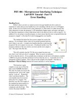
Burridge et al. 2004). Brorsson etal. (2008 a,b) showed that the extension force was
significantly reduced in the RA group (men, p < 0.05, and women p < 0.001) compared to
the control group. Furthermore, they showed that there was a significant difference between
the finger extension force for healthy men and women (p < 0.001), the finger extension force
and flexion force in the dominant hand for healthy subjects and RA patients are presented in
Figure 2.
Fig. 2. (A) Finger extension force in dominant-hand. (B) Flexion force in dominant-hand. The
box-plots represent healthy women (HW), healthy men (HM), women with RA (RAW) and
men with RA (RAM). The results are from participants in all papers (n=80 HW, n=47 HM,
n=65 RAW and n=12 RAM).
3.2 Ultrasound examination in skeletal muscle architechture
Ultrasound technology provides new and exciting possibilities to non-invasively access
physiological mechanisms inside the living body, both at rest and during muscle
contraction. Ultrasonic devices collect sound waves that are emitted by a probe after
Human Musculoskeletal Biomechanics
94
reflecting off the body’s internal tissues; this provides detailed images of the body
structures. The recent developments of the probes have enabled the use of US to examine
the joint and surrounding soft tissues such as the muscles. The increasing interest for US
among rheumatologists contributes to the understanding of the natural history of rheumatic
diseases, and US is today important in the early diagnosis of RA (Kane, Balint et al. 2004;
Grassi, Salaffi et al. 2005) . US has been used in several studies to provide in vivo information
about the muscle architecture of different muscles. Zheng et al. (2006) combined US with
surface electromyography for evaluating changes in muscle architecture after using
prosthetics (Zheng, Chan et al. 2006). US has also been used to study the differences
between men and women regarding muscle parameters such as muscle pennation angles
and muscle fascicle length (Kubo, Kanehisa et al. 2003). US allows for dynamic studies of
muscle architecture, Fukunaga et al. (1997) have developed a method to study the fascicle
length during contraction (Fukunaga, Ichinose et al. 1997). Furthermore, US has been used
to analyse the muscle architecture’s response to age, the authors concluded that some
muscles in the lower extremities decreased in thickness with aging but the fascicle length
did not decrees with aging (Kubo, Kanehisa et al. 2003). Loss of muscle mass with aging has
been reported to be greater in the lower extremities than in the upper extremities. Decreases
in CSA of the muscles have been reported to be 25-33 % lower in young compared to elderly
adults (Narici, Maganaris et al. 2003). However, several researchers have reported decreased
muscle strength but not decreased CSA, so the force, expressed per unit of muscle CSA, has
been reduced in older individuals (Young 1984; Macaluso, Nimmo et al. 2002; Narici,
Maganaris et al. 2003). US has been applied to the rotator cuff muscles to analyse the
dynamic contraction pattern of these muscles to confirm the neuromuscular intensity
(Boehm, Kirschner et al. 2005). Fukunaga et al. (1997) used US to measure muscle
architecture and function in human muscles. They pointed out that the use of cadavers for
studies of architecture and modelling of muscle functions would result in inaccurate and, in
some cases, misleading results (Fukunaga, Kawakami et al. 1997). Aagaard et al. (2001) used
US to measure the response to strength training and the changes in muscle architecture.
They concluded that the quadriceps muscle increased both its CSA and the pennation angle
after heavy resistance training (Aagaard, Andersen et al. 2001). Rutherford and Jones (1992)
did not find any increased pennation angles after resistance training, even though they
reported increased CSA and muscle force in the quadriceps muscle (Rutherford and Jones
1992). Brorsson et al. (2008) showed that there was a significant difference between the
muscle anatomy of healthy men and women. The results of the ultrasound measurements
and the differences in muscle architecture parameters between healthy men and women,
and healthy women and RA women are summarised in Table 1.
The overall shape changes in muscle CSA during contraction were more pronounced for
men than for women, (p < 0.01). US studies have also been performed on human skeletal
muscles to explore the changes in muscle architecture that occur during dynamic
contractions. The authors found that at a constant joint angle, the fascicle length and the
pennation angles changed significantly during muscle contraction (Reeves and Narici 2003).
3.3 Function test evaluation, patients’ questionnaires and visual analogue scale in
hand rehabilitation
The Grip Ability Test (GAT) is designed for individuals with RA; it measures ADL ability.
The test is based on three items chosen to represent different daily grip types. The test is
performed following a standardized protocol consisted of three items: to put a “sleeve”
Biomechanical Studies on Hand Function in Rehabilitation
95
(Flexigrip™ stocking) on their non-dominant hand, place a paper clip on an envelope and
pour 200 ml into a cup from a 1 litre water jug. GAT is a reliable, valid and sensitive ADL
test (Dellhag and Bjelle 1995). Hand function has been assessed by GAT for measuring grip
ability and activity limitations in several studies. Dellhag et al. (1992) reported that RA
patients have improved their hand function after just 4 weeks of hand exercise (Dellhag,
Wollersjo et al. 1992). Bjork et al. (2007) showed significant differences in activity limitations
between healthy controls and RA patients in there study using GAT (Bjork, Thyberg et al.
2007). The relationship between self-reported upper limb function and grip ability was
studied in an early rheumatoid population by Adams et al. (2004). They reported correlation
between GAT and the questioner DASH (Adams, Burridge et al. 2004). Dellhag et al. (2001)
reported in their study that patients with RA that have good hand function, low GAT score,
displayed normal or increased safety margin during precision grip-lift compared to healthy
controls (Dellhag, Hosseini et al. 2001).
Muscle parameters are presented as median (range)
*p < 0.05, ** p < 0.01 (significant differences between healthy men – healthy women and between
healthy women – RA women).
Table 1. Muscle architechture of EDC
Self-administered questionnaires are recommended for evaluating functional disability from
the patients’ perspective (Guillemin 2000; Liang 2000). The hand function is affected early on
in RA and can be evaluated with different methods. One widely used selfadministrated
extremity-specific questionnaire is the Disability of the Arm, Shoulder and Hand (DASH)
that is been reliable and validated for assessing upper limb functional ability in the RA
population (Atroshi, Gummesson et al. 2000). DASH has been used for evaluating the
effectiveness of patient-oriented hand rehabilitation programmes, and has shown significant
differences between two rehabilitation programmes and surgery (Gummesson, Atroshi et al.
2003; Harth, Germann et al. 2008). Furthermore, DASH has been used by Solem et al. (2006)
for evaluation of long-term results of arthrodesis (Solem, Berg et al. 2006). Adams et al.
(2004) showed in their study that DASH was useful to evaluate the relationship between
upper limb functional ability and structural hand impairment (Adams, Burridge et al. 2004).
Another commonly used generic questionnaire for evaluating functional disability in people
is the Short Form 36-item Health Survey (SF-36), there a validated Swedish version has been
developed (Sullivan, Karlsson et al. 1995). Generic healthy status measurements are
commonly used for evaluation of RA patients. SF-36 has been used to detect the treatment
effect in the study outcomes. Furthermore, use of SF-36 permits comparisons of physical and
mental aspects in the RA population, as well as comparison between patients with RA, other
patients groups and the general population (Tugwell, Idzerda et al. 2007). SF-36 has been
used in several studies to evaluate the clinical outcome and quality of life after arthroplasty,
Human Musculoskeletal Biomechanics
96
and concluded the health status and the overall physical functions with significant
improvements for RA patients (Angst, John et al. 2005; Ringen, Dagfinrud et al. 2008; Uhlig,
Heiberg et al. 2008).
Visual analog scale (VAS) pain is a method frequently used to measure perceived pain level
and the impact that high pain levels have on functional disability. Decreased functional
ability in patients with RA has been reported correlated with on disease activity, disease
duration, age, grip force and high pain level (Oken, Batur et al. 2008). Hand disabilities were
detected in 81 % of RA patients and strongly correlated to pain level, grip force and clinical
and laboratory activity. Female RA patients have reported more pain and worse disability
than men (Bodur, Yilmaz et al. 2006; Hakkinen, Kautiainen et al. 2006). Brorsson et al. (2008)
reported that neither the RA group nor the controls showed any significant improvement in
DASH score after 6 weeks of hand exercise therapy. However, after 12 weeks of hand
exercise the RA group showed a significant improvement in the DASH score, while there
was still no improvement in the control group. Neither group showed any significant
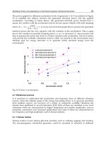
improvement in the SF-36 score after the hand exercises (Figure 3). However, some of the
RA patients reported “tiredness” in their hands after the exercise.
The exercises caused no significant change in the pain level (Table 2).
Fig. 3. SF-36 score pre- and post hand exercise therapy
Results of the SF-36 questionnaire, before (0) and after 12 weeks (12), of hand exercises. The
scale is 0–100, from worst to best. The questionnaire is designed for measuring the generic
health in the general population but is also useful for different patient groups. SF-36 is
divided into eight health profiles scales; physical function (PF), role physical (RP), bodily
pain (BP), general health (GH), vitality (VT), social functioning (SF), role emotional (RE) and
mental health (MH). All dimensions are independent of each other.
4. The hand in rheumatoid arthritis
RA is our most frequent autoimmune inflammatory disease, with prevalence of nearly 1 %. RA
is found throughout the world and affects all ethnic groups. It may strike at any age, but its
prevalence increases with age; the peak incidence being between the fourth and sixth decades.
The prevalence is about 2½ times higher in women than in men. The onset of symptoms
Biomechanical Studies on Hand Function in Rehabilitation
97
usually involves symmetrical joints in hand and feet, but RA is a systemic disease and might
affect any organ such as vessels, pleura or skin. There is often involvement of multiple joints
and surrounding tissues. It’s estimated that 80-90 % of the RA patients suffer from decreased
hand function (Maini 1998; O’Brien, Jones et al. 2006). The hand in most patients may develop
some typical pattern of deformity. These deformities are influenced by several factors, such as
inflammation in the joint with distension of the joint capsule and ligament attenuation.
Inflammation in and around tendons might distend tendon sheaths and cause tendon
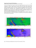
ruptures. The influence of disease by the characteristic MCP-joint deformity of ulnar drift
(Figure 4), results of local joint forces (Smith and Kaplan 1967; McMaster 1972; Tan, Tanner et
al. 2003; Bielefeld and Neumann 2005). Muscle involvement can lead to weakness and
contractures. RA patients are frequently affected by pain, weakness and restricted mobility:
the deformities of the hand, in various degrees, leads to limitation in activities of daily living
(ADL) (Chung, Kotsis et al. 2004; Mengshoel and Slungaard 2005; Masiero, Boniolo et al. 2007).
Median values of hand function tests before (week 0) and after 6 and 12 weeks of hand exercise. Median
and range are given for the grip ability test (GAT), disability, of arm shoulder and hand questionnaire
(DASH) and reported pain level (VAS). Number of participants (n=#)
*p < 0.05, **p < 0.01
Table 2. Hand function evaluations before and after hand exercise.
Fig. 4. The hand in most patients may develop some typical pattern of deformity; these
images show the characteristic MCP-joint deformity of ulnar drift. ©Sofia Brorsson
Human Musculoskeletal Biomechanics
98
The exact cause of RA is still unknown, however genetic, hormonal and environment factors
have been reported to be involved in autoimmune diseases such as RA (Ollier and
MacGregor 1995; Reckner Olsson, Skogh et al. 2001; Tengstrand, Ahlmen et al. 2004).
Diagnosis of RA are based on ACR criteria which include; pain and swelling in at least three
joint areas, symmetrical presentation, early morning joint stiffness for more than 1 hour,
involvement of MCP joint or PIP joint or wrists, subcutaneous nodules, positive rheumatoid
factor and radiological evidence of erosions. At least four of these signs or symptoms should
be present for six weeks (Arnett, Edworthy et al. 1988). Pain and tenderness of the joints are
well described and documented (Pearl and Hentz 1993), but there is less knowledge
concerning how the muscles are influenced by the disease. The most common histological
findings in RA are the pronounced muscle atrophy and nodular myositis. Magyar et al.
(1973) observed changes in the muscles consistent with denervation using electron
microscopy. These authors showed that the muscle changes might be due to a direct
involvement of the neuromuscular system and that the pathological changes affect the
contractile element in the muscles (Magyar, Talerman et al. 1973). An important part of hand
function is based on the function of the muscles which are involved in finger and wrist
motion and the ability to develop grip force. RA patients often report that they feel
weakness, particularly when performing flexion force. There are several possible reasons for
this weakness such as reduction in muscle fibre diameter, direct involvement of
inflammatory processes in the muscle, joint deformity influencing muscle function and pain
(Haslock, Wright et al. 1970; Leading 1984; Bruce, Newton et al. 1989). The muscle structure
(ultra-structure) and changes in rheumatoid arthritis have been recognised pathologically
and clinically. Although electron microscopy is valuable in investigating human skeletal
muscle both in normal and RA muscles, only a few data sources document muscle ultra-
structural alterations in RA patients (Haslock, Wright et al. 1970; Magyar, Talerman et al.
1973; Wollheim 2006). Furthermore, a non-invasive study on muscle architecture in RA
patients appears to be poorly investigated.
4.1 Rehabilitation and intervention of the Rheumatoid Arthritis hand
Treatment of RA is focused on reducing the inflammatory activity by medication,
rehabilitation and surgery (Stenstrom and Minor 2003). New disease modifying drugs for
RA patients administered early after onset have made it possible for people with this disease
to stay more active and more fit than 10-20 years ago (Pincus, Ferraccioli et al. 2002). Today’s
treatment options to increase hand function for RA patients include electrotherapy, injection
therapy, manual therapy and traditional exercise prescription, but the evidence base for
treatments remains weak, particularly when focusing on the hand (Weiss, Moore et al. 2004;
Plasqui 2008). In 1974, Lee et al. reported in their study that immobilization and/or physical
rest were beneficial in the treatment of RA, leading to a decrease in pain and joint swelling
(Lee, Kennedy et al. 1974). Other groups have reported that the forces involved in using the
hand lead to joint erosion and increased deformities (Ellison, Flatt et al. 1971; Kemble 1977).
Despite earlier fear of aggravating symptoms, there is now scientific evidence showing that
various forms of exercise are both safe and beneficial (Stenstrom and Minor 2003). However,
comparatively little research has evaluated the evidence for the benefits of hand exercise in
RA (O’Brien, Jones et al. 2006). Recently reviewed effectiveness on hand exercise therapy in
RA patients showed that only nine eligible studies have incorporated hand exercise therapy
as part of the intervention (Chadwick 2004; Wessel 2004). Hoening et al. (1993) showed in
Biomechanical Studies on Hand Function in Rehabilitation
99
their study that a home hand exercise program was effective for increasing the grip force in
the RA hand (Hoenig, Groff et al. 1993). Intensive hand exercise has previously been
reported to be effective for improving grip- and pinch force for RA patients (Ronningen and
Kjeken 2008). Brorsson et al. (2008) have showed that a regular home exercise programme
for the RA hand, evaluated with force measurements, ultrasound examination, function test
and patients questionnaires (Figure 5), is beneficial for grip (flexion and extension) force
production. Furthermore, they reported that hand exercise improves the relation between
flexion and extension forces as well as improved hand function. They also reported
improved flexion and extension force for the RA patients after 12-weeks of hand exercise
(Figure 6).
Fig. 5. The total study period was 18 weeks of home hand exercise, divided into 6-week
periods. Baseline values were determined at week 0 (Occasion I) and 6 (Occasion II).
Thereafter, the hand exercise programme was started, and the effects were measured after 6
weeks (Occasion III) and 12 weeks (Occasion IV). Evaluation methods used: (A) finger
extension force measurements (EX-it), (B) Flexion force measurements (Grippit™), (C) US
examination of the EDC muscle, (D) grip ability test, and (E) questionnaires.
Fig. 6. Illustrates the finger extension force (A) and flexion force (B) in the two groups of
participants in paper IV after 6 and 12 weeks of hand exercise. Both groups show significant
improvement after 6 and 12 weeks (* p < 0.05, **p < 0.01).
Hand surgery has been regarded as beneficial for some patients with RA. Arthroplastic
procedures of the wrist and fingers have been performed since 1960. An increasing number
of patients with RA receive joint replacements in the MCP joints of the hand. The purpose of
these operations is to improve the patients’ extension ability, extension force, and hand
Human Musculoskeletal Biomechanics
100
function as well as reduce pain (Weiss, Moore et al. 2004). At present, when the outcome of
surgery is evaluated, it is impossible to objectively test if the patients’ finger extension force
has been improved or not, since no force measurement device for finger extension force is
commercially available. It is necessary to find methods to objectively measure hand function
in order to be able to evaluate the functional impairment, as well as the results of
therapeutic interventions i.e. surgery or physical therapy.
5. Conclusion
To further our understanding of hand function, and specifically the extensor muscles’
function and ability to produce force in rehabilitation, this book chapter describes the
development and results of new non-invasive methods, a new finger extension force
measurement device, EX-it, and an ultrasound imaging method (Brorsson et al. 2008 a,b).
Furthermore, the results of this book chapter show that finger extension force measurements
and ultrasound are effective methods for evaluating improvement after the intervention
hand exercise. The effect of hand exercise on the extensor muscles could be objectively
evaluated with EX-it and ultrasonic imaging. This chapter also reported the usefulness of
short-term hand exercise for patients with RA and that a home exercise programme can
enhance hand function.
Various methods can be used to study muscle architecture, including ultrasound, magnetic
resonance imaging (Juul-Kristensen, Bojsen-Moller et al. 2000; Aagaard, Andersen et al.
2001) and laser diffraction. Laser diffraction is an invasive technique, while magnetic
resonance imaging is only suitable for static measurements. Ultrasound, on the other hand,
is non-invasive and clearly shows the movement of the muscle (Fukunaga, Ichinose et al.
1997). It is also harmless, can be repeated and offers the possibility of dynamic
examinations. The limitations with US are the quality of the examinations, which are
dependent on the investigator’s ability to reproduce the imaging conditions
(measurements), to find correct landmarks in both transverse and longitudinal direction and
standardise the procedures. Ultrasound has been shown to be a highly valuable tool to
assess in vivo muscle architecture for studying muscle function and relationships between
muscle force and muscle size (Maughan, Watson et al. 1984; Hakkinen and Keskinen 1989;
Kawakami, Abe et al. 1993; Fukunaga, Kawakami et al. 1997).
In rheumatoid arthritis, impaired finger extension is a common symptom; differences in
extension muscle force capacity as well as in muscle architectural parameters, between
normal and RA muscles are reported. Earlier studies have reported that RA patients also
have weaker grip, pinch and tripod force than healthy controls, and it has been suggested
that force assessment could be used as an accurate indicator of upper limb ability and that
grip force (i.e. flexion and pinch force) should be included in the evaluation and follow-up
of the patients with RA in hand rehabilitation units (Helliwell and Jackson 1994; Fraser,
Vallow et al. 1999; Adams, Burridge et al. 2004; Bodur, Yilmaz et al. 2006). The decrease in
force capacity could be explained by a direct effect of the disease on muscle function, disuse
or impaired neuromuscular transmission, or different medications, but the decrease could
also be due to the fact that the RA patients experienced more pain than the healthy subjects,
a situation which could influence their maximal muscle exertion. Loss of hand grip force has
been shown to result from pain, or fear of pain, or mechanical malfunction (Fraser, Vallow
et al. 1999).
Biomechanical Studies on Hand Function in Rehabilitation
101
Ultrasound is a non-invasive and harmless method that can be used to visualise functionally
important muscle parameters dynamically. Finger extension control is one of the most
difficult motions to regain after disease/injury and is also very important for prehensile
activities (Cauraugh, Light et al. 2000). Since both EX-it and ultrasound have been shown to
be sensitive in their evaluation of hand exercise, it can be expected that these methods can
be used to evaluate other interventions, such as surgical procedures, physiotherapy and/or
pharmacological treatment. With these new methods, arthroplastic interventions in the
MCP-joints of the fingers can objectively be evaluated. In a longer perspective it may be
possible to establish more efficient rehabilitation programmes for RA patients. Furthermore,
force measurements are a quick and easy measure of hand impairment and function, and
are useful when evaluating hand status. EX-it in combination with other non-invasive
evaluation methods (i.e.grip ability tests and health assessment questionnaires) will provide
more information on hand function. Patients with rheumatoid arthritis suffer from a variety
of functional deficiencies, of which impaired muscle function is a serious one. There is a
recent trend towards the use of non-invasive methods in studying disease-specific changes,
such as magnetic resonance imaging and ultrasound. Increased knowledge concerning
muscle morphology and function in RA will allow better diagnosis and evaluation of
interventions, such as surgical procedures, physiotherapy and/or pharmacological
treatment. In a longer perspective it may be possible to establish a more efficient
rehabilitation programme for RA patients. If combined with functional and clinical
measures of disability, information on muscle architecture could then be used as an
objective tool in the assessment of hand function after physical therapy and hand surgery.
In this thesis no negative effects of EX-it, ultrasound or the exercise programme on self
reported pain level were reported in the RA group. It is possible that RA patients need
continuous exercise to prevent loss of muscle strength and to improve the performance of
activities of daily living (Stenstrom 1994; Hakkinen, Malkia et al. 1997; O’Brien, Jones et al.
2006; Masiero, Boniolo et al. 2007). However, the response to exercise from RA patients must
be further evaluated to find out if longer exercise period can obliterate the differences
between healthy and rheumatoid arthritis muscle strength and function; or to find out if
these differences depend on a disease-specific effect on the rheumatoid arthritis muscles.
5.1 Future implications
Several questions have arisen during writing this book chapter and performed research in
this area and require further research. It would be of interest to analyse how EDC responds
during contraction at different locations of the muscle. Brorsson et al. (2008 a,b,
2009)reported that the inter muscle movement pattern in the muscle was observed, but were
unable to measure it with the methods used for this thesis. Further knowledge about in vivo
muscle pattern could provide information about the muscle as well as the elastic
characteristics of the aponeurosis and tendon.
Is it possible that the EDC, a muscle designed for precision tasks and grip control rather
than force exertion, is constructed differently from the large force-generating muscles?
Can US be used as a diagnostic tool for analysing muscle disease?
Are muscle movement patterns related to force production?
Does this muscle movement appear in other muscle groups?
RA patients significantly increased their hand force and hand function after exercise.
However, the response to exercise from RA patients must be further evaluated. It would be
Human Musculoskeletal Biomechanics
102
interesting to combine invasive and non-invasive methods to be able to answer the
following questions:
Would longer periods of hand exercise obliterate the differences between healthy and
rheumatoid arthritis muscle force and function?
Do the muscle’s architecture, force production and decreased function depend on
disease specific effects on the rheumatoid arthritis muscles?
It would be of great interest to investigate the possibility to objectively evaluate
interventions, such as surgical procedures, physiotherapy and/or pharmacological
treatment with the help of finger force measurements and ultrasound evaluations.
In a longer perspective, can it be possible to establish more efficient rehabilitation
programmes for RA patients through further knowledge about the muscle
biomechanics?
6. Acknowledgment
My research is a result of multidisciplinary collaboration between the School of Business
and Engineering, Halmstad University, Department of Hand Surgery, Sahlgrenska
University Hospital, Göteborg, Research and Development centre at Spenshult Hospital for
Rheumatic Diseases, Halmstad, Department of Diagnostic Radiology and Department of
Research and Education, Halmstad Central Hospital, Halmstad.
I would like to thank all the patients and healthy subjects, for your participation in the
studies, for performing the tests and answering the questioners. I extend my thanks to
Professor Marita Hilleges, for your inspiration, critical comments and clever suggestions.
7. References
1
Aagaard, P., J. L. Andersen, et al. (2001). “A mechanism for increased contractile strength of
human pennate muscle in response to strength training: changes in muscle
architecture.”J Physiol 534(Pt. 2): 613-23.
Adams, J., J. Burridge, et al. (2004). “Correlation between upper limb functional ability and
structural hand impairment in an early rheumatoid population.” Clin Rehabil
18(4): 405-13.
Akima, H., S. Kuno, et al. (1997). “Effects of 20 days of bed rest on physiological cross-
sectional area of human thigh and leg muscles evaluated by magnetic resonance
imaging.” J Gravit Physiol 4(1): S15-21.
Ashford, R. F., S. Nagelburg, et al. (1996). “Sensitivity of the Jamar Dynamometer in
detecting submaximal grip effort.” J Hand Surg [Am] 21(3): 402-5.
Atroshi, I., C. Gummesson, et al. (2000). “The disabilities of the arm, shoulder and hand
(DASH) outcome questionnaire: reliability and validity of the Swedish version
evaluated in 176 patients.” Acta Orthop Scand 71(6): 613-8.
Balogun, J. A., C. T. Akomolafe, et al. (1991). “Grip strength: effects of testing posture and
elbow position.” Arch Phys Med Rehabil 72(5): 280-3.
Berntson, L. and E. Svensson (2001). “Pain assessment in children with juvenile chronic
arthritis: a matter of scaling and rater.” Acta Paediatr 90(10):1131-6.
1
Since many references have several authors, only the two first authors are mentioned (in alphabetical order) in
this book chapter.
Biomechanical Studies on Hand Function in Rehabilitation
103
Bielefeld, T. and D. A. Neumann (2005). “The unstable metacarpophalangeal joint in
rheumatoid arthritis: anatomy, pathomechanics, and physical rehabilitation
considerations.” J Orthop Sports Phys Ther 35(8): 502-20.
Bjork, M. A., I. S. Thyberg, et al. (2007). “Hand function and activity limitation according to
health assessment questionnaire in patients with rheumatoid arthritis and healthy
referents: 5-year followup of predictors of activity limitation (The Swedish TIRA
Project).” J Rheumatol 34(2): 296-302.
Blazevich, A. J., N. D. Gill, et al. (2007). “Lack of human muscle architectural adaptation
after short-term strength training.” Muscle Nerve 35(1): 78-86.
Blazevich, A. J. and N. C. Sharp (2005). “Understanding muscle architectural adaptation:
macro- and micro-level research.” Cells Tissues Organs 181(1): 1-10.
Bodur, H., O. Yilmaz, et al. (2006). “Hand disability and related variables in patients with
rheumatoid arthritis.” Rheumatol Int 26(6): 541-4.
Boehm, T. D., S. Kirschner, et al. (2005). “Dynamic ultrasonography of rotator cuff muscles.”
J Clin Ultrasound 33(5): 207-13.
Brand, W. (1993). Clinical Mechanics of the Hand. St. Louis, Missouri.
Brorsson, S., A. Nilsdotter, et al. (2008a). “A new force measurement device for evaluating
finger extension function in the healthy and rheumatoid arthritic hand.” Technol
Health Care 16(4): 283-92.
Brorsson, S., A. Nilsdotter, et al. (2008b). “Ultrasound evaluation in combination with finger
extension force measurements of the forearm musculus extensor digitorum
communis in healthy subjects.” BMC Med Imaging 3:8:6.
Bruce, S. A., D. Newton, et al. (1989). “Effect of subnutrition on normalized muscle force and
relaxation rate in human subjects using voluntary contractions.” Clin Sci (Lond)
76(6): 637-41.
Buljina, A. I., M. S. Taljanovic, et al. (2001). “Physical and exercise therapy for treatment of
the rheumatoid hand.” Arthritis Rheum 45(4): 392-7.
Chadwick, A. (2004). “A review of the history of hand exercises in rheumatoid arthritis.”
Musculoskeletal Care 2(1): 29-39.
Chadwick, E. K. and A. C. Nicol (2001). “A novel force transducer for the measurement of
grip force.” J Biomech 34(1): 125-8.
Chung, K. C., S. V. Kotsis, et al. (2004). “A prospective outcomes study of Swanson
metacarpophalangeal joint arthroplasty for the rheumatoid hand.” J Hand Surg
[Am] 29(4): 646-53.
Debicki, D. B., P. L. Gribble, et al. (2004). “Kinematics of wrist joint flexion in overarm
throws made by skilled subjects.” Exp Brain Res 154(3): 382-94.
Dellhag, B. and A. Bjelle (1995). “A Grip Ability Test for use in rheumatology practice.” J
Rheumatol 22(8): 1559-65.
Dellhag, B., N. Hosseini, et al. (2001). “Disturbed grip function in women with rheumatoid
arthritis.” J Rheumatol 28(12): 2624-33.
Dellhag, B., I. Wollersjo, et al. (1992). “Effect of active hand exercise and wax bath treatment
in rheumatoid arthritis patients.” Arthritis Care Res 5(2): 87-92.
Doebelin, E. (1990). Measurements Systems, Application and Design. New York, Mc Graw-
Hill.
Ekdahl, C. and S. I. Andersson (1989). “Standing balance in rheumatoid arthritis. A
comparative study with healthy subjects.” Scand J Rheumatol 18(1): 33-42.
Human Musculoskeletal Biomechanics
104
Ellison, M. R., A. E. Flatt, et al. (1971). “Ulnar drift of the fingers in rheumatoid disease.
Treatment by crossed intrinsic tendon transfer.” J Bone Joint Surg Am 53(6): 1061-
82.
Fess, E. (1992). “Grip strength.” American Society of Hand Therapists. Clinical Assment
Recommendations Vol. 2nd: 41-45.
Fischer, H. C., K. Stubblefield, et al. (2007). “Hand rehabilitation following stroke: a pilot
study of assisted finger extension training in a virtual environment.”Top Stroke
Rehabil 14(1): 1-12.
Fitts, R. H. and J. J. Widrick (1996). “Muscle mechanics: adaptations with exercisetraining.”
Exerc Sport Sci Rev 24: 427-73.
Flatt, A. E. (1974). ”Letter: Shoulder-hand syndrome.” Lancet 1(7866): 1107-8.
Fransson, C. and J. Winkel (1991). “Hand strength: the influence of grip span and grip type.”
Ergonomics 34(7): 881-92.
Fraser, A., J. Vallow, et al. (1999). “Predicting ‘normal’ grip strength for rheumatoid arthritis
patients.” Rheumatology (Oxford) 38(6): 521-8.
Freilich, R. J., R. L. Kirsner, et al. (1995). “Isometric strength and thickness relationships in
human quadriceps muscle.” Neuromuscul Disord 5(5): 415-22.
Fukunaga, T., Y. Ichinose, et al. (1997). “Determination of fascicle length and pennation in a
contracting human muscle in vivo.” J Appl Physiol 82(1): 354-8.
Fukunaga, T., Y. Kawakami, et al. (1997). “Muscle architecture and function in humans.” J
Biomech 30(5): 457-63.
Fukunaga, T., M. Miyatani, et al. (2001). “Muscle volume is a major determinant of joint
torque in humans.” Acta Physiol Scand 172(4): 249-55.
Fung, Y. (1993). Biomechanics Mechanical properties of living tissues. New York, Springer-
Verlag.
Grassi, W., F. Salaffi, et al. (2005). “Ultrasound in rheumatology.” Best Pract Res Clin
Rheumatol 19(3): 467-85.
Guillemin, F. (2000). “Functional disability and quality-of-life assessment in clinical
practice.” Rheumatology (Oxford) 39 Suppl 1: 17-23.
Gummesson, C., I. Atroshi, et al. (2003). “The disabilities of the arm, shoulder and hand
(DASH) outcome questionnaire: longitudinal construct validity and measuring
self-rated health change after surgery.” BMC Musculoskelet Disord 4: 11.
Hakkinen, A., P. Hannonen, et al. (1995). “Muscle strength in healthy people and in patients
suffering from recent-onset inflammatory arthritis.” Br J Rheumatol 34(4): 355-60.
Hakkinen, A., H. Kautiainen, et al. (2006). “Muscle strength, pain, and disease activity
explain individual subdimensions of the Health Assessment Questionnaire
disability index, especially in women with rheumatoid arthritis.” Ann Rheum Dis
65(1): 30-4.
Hammer, A. and B. Lindmark (2003). “Test-retest intra-rater reliability of grip force in
patients with stroke.” J Rehabil Med 35(4): 189-94.
Harth, A., G. Germann, et al. (2008). “Evaluating the effectiveness of a patient oriented hand
rehabilitation programme.” J Hand Surg Eur Vol.
Haslock, D. I., V. Wright, et al. (1970). “Neuromuscular disorders in rheumatoid arthritis. A
motor-point muscle biopsy study.” Q J Med 39(155): 335-58.
Helliwell, P. S. and S. Jackson (1994). “Relationship between weakness and muscle wasting
in rheumatoid arthritis.” Ann Rheum Dis 53(11): 726-8.
Hoenig, H., G. Groff, et al. (1993). “A randomized controlled trial of home exercise on the
rheumatoid hand.” J Rheumatol 20(5): 785-9.
Biomechanical Studies on Hand Function in Rehabilitation
105
Hopkins, J. T., J. B. Feland, et al. (2007). “A comparison of voluntary and involuntary
measures of electromechanical delay.” Int J Neurosci 117(5): 597-604.
Huxley, A.F., R. Niedergerke (1954). “Structural Changes in Muscle During Contraction.
Interference Microscopy of Living Muscle Fibrers “ Nature 173: 971.
Huxley, A. F. and R. M. Simmons (1971). “Proposed mechanism of force generation in
striated muscle.” Nature 233(5321): 533-8.
Ichinose, Y., H. Kanehisa, et al. (1998). “Morphological and functional differences in the
elbow extensor muscle between highly trained male and female athletes.” Eur J
Appl Physiol Occup Physiol 78(2): 109-14.
Incel, N. A., E. Ceceli, et al. (2002). “Grip strength: effect of hand dominance.”Singapore
Med J 43(5): 234-7.
Innes, E. (1999). “Handgrip strength testing: A review of the literature.” Australian
Occupational Therapy Journal 46: 120-140.
Jones, E., J. G. Hanly, et al. (1991). “Strength and function in the normal and rheumatoid
hand.” J Rheumatol 18(9): 1313-8.
Juul-Kristensen, B., F. Bojsen-Moller, et al. (2000). “Muscle sizes and moment arms of rotator
cuff muscles determined by magnetic resonance imaging.”Cells Tissues Organs
167(2-3): 214-22.
Kandel, E.R., J.H. Schwartz, et al. (1991). Principles of neural science, Appleton & Lange.
Kane, D., P. V. Balint, et al. (2004). “Musculoskeletal ultrasound – a state of the art review in
musculoskeletal ultrasound in rheumatology.”Rheumatology (Oxford) 43(7): 823-8.
Kawakami, Y., T. Abe, et al. (1993). “Muscle-fiber pennation angles are greater in
hypertrophied than in normal muscles.” J Appl Physiol 74(6): 2740-4.
Kawakami, Y., H. Akima, et al. (2001). “Changes in muscle size, architecture, and neural
activation after 20 days of bed rest with and without resistance exercise.” Eur J
Appl Physiol 84(1-2): 7-12.
Kawakami, Y., Y. Muraoka, et al. (2000). “Changes in muscle size and architecture following
20 days of bed rest.” J Gravit Physiol 7(3): 53-9.
Kemble, J. V. (1977). “Functional disability in the rheumatoid hand.” Hand 9(3): 234-41.
Kubo, K., H. Kanehisa, et al. (2003). ”Muscle architectural characteristics in women aged 20-
79 years.” Med Sci Sports Exerc 35(1): 39-44.
Lieber, R. L. and J. Friden (2000). “Functional and clinical significance of skeletal muscle
architecture.” Muscle Nerve 23(11): 1647-66.
Maini, R. N. (1998). “Rheumatoid arthritis. A paradigm of inflammatory disease of the
musculoskeletal system.” Acta Orthop Scand Suppl 281: 6-13.
Marieb, E. (1997). Human anatomy and physiology. California, Benjamin/Cummings
Science.
Narici, M. and P. Cerretelli (1998). “Changes in human muscle architecture in disuseatrophy
evaluated by ultrasound imaging.” J Gravit Physiol 5(1): P73-4.
Nordenskiold, U. (1997). “Daily activities in women with rheumatoid arthritis. Aspects of
patient education, assistive devices and methods for disability and impairment
assessment.” Scand J Rehabil Med Suppl 37: 1-72.
Nordenskiold, U. M. and G. Grimby (1993). “Grip force in patients with rheumatoid arthritis
and fibromyalgia and in healthy subjects. A study with the Grippit instrument.”
Scand J Rheumatol 22(1): 14-9.
Otten, E. (1988). “Concepts and models of functional architecture in skeletal muscle.” Exerc
Sport Sci Rev 16: 89-137.
Human Musculoskeletal Biomechanics
106
Pearl, R. M. and V. R. Hentz (1993). ”Extensor digiti minimi tendon transfer to prevent
recurrent ulnar drift.” Plast Reconstr Surg 92(3): 507-10.
Qvistgaard, E., S. Torp-Pedersen, et al. (2006). “Reproducibility and inter-reader agreement
of a scoring system for ultrasound evaluation of hip osteoarthritis.” Ann Rheum
Dis 65(12): 1613-9.
Richards, L. G., B. Olson, et al. (1996). “How forearm position affects grip strength.” Am J
Occup Ther 50(2): 133-8.
Ringen, H. O., H. Dagfinrud, et al. (2008). “Patients with rheumatoid arthritis report greater
physical functional deterioration in lower limbs compared to upper limbs over 10
years.” Scand J Rheumatol 37(4): 255-9.
Ronningen, A. and I. Kjeken (2008). “Effect of an intensive hand exercise programme in
patients with rheumatoid arthritis.” Scand J Occup Ther: 1-11.
Schieber, M. H. and M. Santello (2004). “Hand function: peripheral and central constraints
on performance.” J Appl Physiol 96(6): 2293-300.
Solem, H., N. J. Berg, et al. (2006). “Long term results of arthrodesis of the wrist: a 6-15 year
follow up of 35 patients.” Scand J Plast Reconstr Surg Hand Surg 40(3): 175-8.
Sollerman, C. and A. Ejeskar (1995). “Sollerman hand function test. A standardised method
and its use in tetraplegic patients.” Scand J Plast Reconstr Surg Hand Surg 29(2):
167-76.
Stenstrom, C. H. (1994). “Home exercise in rheumatoid arthritis functional class II: goal
setting versus pain attention.” J Rheumatol 21(4): 627-34.
Stenstrom, C. H. and M. A. Minor (2003). “Evidence for the benefit of aerobic and
strengthening exercise in rheumatoid arthritis.” Arthritis Rheum 49(3): 428-34.
Sullivan, M., J. Karlsson, et al. (1995). “The Swedish SF-36 Health Survey.Evaluation of data
quality, scaling assumptions, reliability and construct validity across general
populations in Sweden.” Soc Sci Med 41(10): 1349-58.
Tan, A. L., S. F. Tanner, et al. (2003). ”Role of metacarpophalangeal joint anatomic factors in
the distribution of synovitis and bone erosion in early rheumatoid arthritis.”
Arthritis Rheum 48(5): 1214-22.
Tengstrand, B., M. Ahlmen, et al. (2004). “The influence of sex on rheumatoid arthritis: a
prospective study of onset and outcome after 2 years.” J Rheumatol 31(2): 214-22.
Thyberg, I., U. A. Hass, et al. (2005). “Activity limitation in rheumatoid arthritis correlates
with reduced grip force regardless of sex: the Swedish TIRA project.” Arthritis
Rheum 53(6): 886-96.
Trappe, S. W., T. A. Trappe, et al. (2001). ”Calf muscle strength in humans.” Int J Sports Med
22(3): 186-91.
Tugwell, P., L. Idzerda, et al. (2007). ”Generic quality-of-life assessment in rheumatoid
arthritis.” Am J Manag Care 13 Suppl 9: S224-36.
Weiss, A. P., D. C. Moore, et al. (2004). ”Metacarpophalangeal joint mechanics after 3
different silicone arthroplasties.” J Hand Surg [Am] 29(5): 796-803.
Wessel, J. (2004). “The effectiveness of hand exercises for persons with rheumatoid arthritis:
a systematic review.” J Hand Ther 17(2): 174-80.
Vliet Vlieland, T. P., T. P. van der Wijk, et al. (1996). “Determinants of hand function in
patients with rheumatoid arthritis.” J Rheumatol 23(5): 835-40.
Wollheim, F. A. (2006). “Aging, muscles, and rheumatoid arthritis.” Curr Rheumatol Rep
8(5): 323-4.
6
Cervical Spine Anthropometric and
Finite Element Biomechanical Analysis
Susan Hueston
1
, Mbulelo Makola
1
, Isaac Mabe
1
and Tarun Goswami
1,2
1
Biomedical and Industrial Human Factors Engineering
2
Department of Orthopedic Surgery, Sports Medicine and Rehabilitation
United States of America
1. Introduction
A multidisciplinary approach to the study of the cervical spine is presented. The cervical
spine provides higher levels of flexibility and motion as compared to the lumbar and
thoracic spine regions. These characteristics can be attributed to the anatomy of the specific
cervical vertebra. A statistical analysis of cervical vertebra anthropometry was performed in
order to determine if significant relationships exist between vertebral features. The analysis
was performed on a cohort of Chinese Singaporean cervical spines.
Mathematical analysis methods provide an extremely useful tool in the study of the cervical
spine. Analyses can provide force displacement response characteristics of the cervical
spine. Additionally, mathematical analysis methods can provide internal stress, and strain
response characteristics for cervical vertebra and intervertebral discs. Mathematical analyses
of the cervical spine require robust and accurate constitutive and geometric models. A
review of cervical spine finite element modeling techniques and approaches is presented in
order to help frame analysis and modeling best practices.
A finite element analysis study was performed focusing on vertebral endplate subsidence.
Subsidence is a failure mechanism in which a vertebral endplate fails after implantation of
an intra vertebral implant device. The effects of vertebral endplate morphology on stress
response were analyzed in order to better understand indicators for subsidence.
1.1 Analysis of Chinese Singaporean cervical spine anthropometry
With respect to biomechanics it is important to understand the anatomy of the body. In this
particular section the anatomy of the cervical spine will be presented, with investigation into
the morphometry of the vertebra themselves. To accomplish this, an investigation on how
the different dimensional anatomy of the cervical spine changes relates to each other will be
presented.
To begin, a brief explanation of the anatomy of spine will be presented in order to aid in
understanding of the anthropometry of the cervical spine. The spine consists of 5 sections:
cervical, thoracic, lumbar, sacrum, and coccyx (from top to bottom) (Saladin and Miller,
2004). There are 33 vertebrae in the whole spine: 7 in the cervical spine which is located in
the neck, 12 in the thoracic spine which is located in the chest, 5 in the lumbar spine which is
located in the lower back, 5 in the sacrum that is located at the base of the spine, followed by
the 4 small vertebrae in the coccyx (Saladin and Miller, 2004).
Human Musculoskeletal Biomechanics
108
As stated previously there are 7 vertebrae in the cervical spine. The first two vertebrae are
particularly unique and allow for movement of the head, the first is known as the Atlas (C1)
and the second the Axis (C2) (Saladin and Miller, 2004). Because of their unique features
analysis on correlations present in the dimensional anatomy was not completed. For the
remaining 5 vertebrae from C3-C7 an investigation in the correlation in the dimensional
anatomy was completed. The results of this investigation will allow for more accurate
modeling of this region, in order to assist in the development of improved spinal implants
as well as more efficient surgical device placement techniques. Additionally, these statistics
will lead to a better understanding of cervical spine functionality and its susceptibility to
failure. The different dimensional aspects that were analyzed were based on the
anthropometric measurements completed from a published study by Tan on Chinese
Singaporeans (Tan, Teo and Chua, 2004).
The present study involved the anthropometric measurements of linear, and angular
aspects, as well as area. The linear measurements included: upper and lower end plate
width (EPWu, and EPWl), upper and lower end plate depth (EPDu and EPDl), anterior and
posterior vertebral body height (VBHa and VBHp), spinal canal width (SCW), spinal canal
depth (SCD), left and right pedicle height (PDHl and PDHr), left and right pedicle width
(PDWl and PDWr), spinous process length (SPL), and the transverse process width (TPW).
The area measurements included: the upper and lower end plate area (EPAu and EPAl),
spinal canal area (SCA), and the left and right pedicle area (PDAl and PDAr). Finally, the
angular measurements included: upper and lower end plate transverse inclination (EPItu
and EPItl), left and right pedicle sagittal inclination (PDIsl and PDIsr), and the left and right
pedicle transverse inclination (PDItl and PDItr). Analysis was completed using the concepts
of linear regression, ANOVA, and parameter estimation. Utilizing these results an
investigation into any relationship that might be present between the previous
anthropometric measurements was completed for each segment. As an example, a
comparison between the EPWu of the C3 vertebra and the PDIsr of the C3 vertebra was
analyzed to determine if there was any statistically significant relationship present (Tan, Teo
and Chua, 2004).
Previous research, as discussed in this section, has been to provide quantitative
measurements for the cervical spine. The purpose of the analysis completed was to develop
any significant relationships present between the different anthropometrics of each vertebra.
Of these significant relationships it was important to see why they were significant, which
were significant in the opposite comparison (for example between EPWu vs. EPWl and
EPWl vs. EPWu), and which were found in more than just one vertebral segment.
1.2 Materials & methods
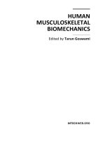
To begin the analysis of the correlations present in the cervical spine anthropometrics,
measurements were collected from Tan’s study on Chinese Singaporeans. The linear,
angular, and area measurements are depicted in Figure 1. In this analysis, a comparison of
just one vertebral body’s measurements was compared. A good example is comparing data
from the C3 vertebra to other C3 vertebral data. These comparisons totaled approximately
600 for each vertebral body segment. The statistical analysis was completed using linear
regression (including parameter estimation), and ANOVA with the use of SAS
®
9.2 TS Level
2M0. A regression analysis is a statistical technique used to explore relationships that are
present between two or more variables. In particular a linear regression analysis relates
these various variables into a straight-line relationship where the slope and the y-intercept
Cervical Spine Anthropometric and Finite Element Biomechanical Analysis
109
of the line are the regression coefficients. Not all points will lie on this line, but a majority of
the points will be within certain deviation of this line resulting in a model. For this
particular study, a simple linear regression was used. It involves just one independent
variable (x), also known as a regressor or predictor. With this linear regression analysis,
parameter estimation was used. Parameter estimation is a technique of statistical inference,
which is a way to make conclusions from random variation data. In this particular case,
parameter estimation was used to find the y-intercept and the slope of the linear
relationship between two anthropometric variables. ANOVA stands for Analysis of
Variance, and can be used in order to test the significance of regression analysis. For the
ANOVA, 95% confidence interval was used to test the significance between variables, while
a 97.5% interval was used for the parameter estimation. Another test of significance was
based off the R² value, which is also known as the correlation ratio. This correlation
coefficient is the proportion of total variance of the dependent variable that is explained by
the independent variable. Thus a higher value showing that the model is more accurate. In
the case of the analysis described in this paper if the R² value was >0.6, the model was
assumed to be a good fit (Montgomery and Peck, 1982; Gamst, Meyers and Guarino, 2008).
In the study completed by Tan on the Chinese Singaporeans, measurements of 10 cadaveric
males were completed based on the measurements defined in Figure 1. The measurements
mean and standard deviation found by Tan are displayed in Table’s 1-3 where Table 1
displays the linear measurements, Table 2 lists the area measurements, and Table 3 illustrates
the angular measurements that were taken in this study (Tan, Teo and Chua, 2004).
Fig. 1. Depiction of anthropometric measurements (Tan, Teo and Chua, 2004)
Human Musculoskeletal Biomechanics
110
C3 C4 C5 C6 C7
Mean Std dev Mean Std dev Mean Std dev Mean Std dev Mean Std dev
EPWu 13.8 0.1 14.7 0.1 14.9 0.1 15.8 0.0 19.0 0.1
EPWl 14.3 0.1 15.0 0.1 15.9 0.1 19.5 0.2 20.3 0.2
EPDu 13.6 0.1 14.0 0.1 14.3 0.1 14.6 0.2 15.1 0.2
EPDl 15.1 0.2 15.2 0.4 15.1 0.3 15.7 0.3 15.6 0.3
VBHa 10.0 0.2 9.9 0.3 9.6 0.2 10.4 0.3 11.2 0.2
VBHp 11.2 0.1 11.3 0.2 11.3 0.1 11.3 0.2 11.8 0.3
SCW 19.2 0.4 19.3 0.5 20.3 0.4 20.6 0.4 19.7 0.4
SCD 10.3 0.3 10.3 0.3 10.3 0.3 10.3 0.3 11.0 0.2
PDHl 6.7 0.2 6.6 0.2 6.3 0.3 6.0 0.3 6.5 0.2
PDHr 6.8 0.2 6.7 0.2 5.9 0.2 6.0 0.1 6.1 0.1
PDWl 4.5 0.2 4.6 0.2 4.7 0.1 5.1 0.2 5.6 0.2
PDWr 4.4 0.2 4.5 0.2 4.9 0.2 5.4 0.2 5.7 0.2
SPL 25.6 0.5 30.3 0.4 33.6 1.0 40.5 1.5 46.9 1.1
TPW 41.4 0.8 44.9 0.8 47.6 1.0 48.4 0.9 53.8 1.0
Table 1. Linear Measurements from Tan study (mm) (Tan, Teo and Chua, 2004)
C3 C4 C5 C6 C7
Mean Std dev Mean Std dev Mean Std dev Mean Std dev Mean Std dev
EPAu 154.7 3.8 169.2 4.9 187.4 6.6 210.5 10.0 220.8 9.0
EPAl 216.8 10.1 241.5 10.6 286.4 10.3 316.3 7.4 340.0 10.3
SCA 149.7 9.0 159.9 8.4 166.8 8.0 163.7 10.2 167.5 6.7
PDAl 27.6 1.0 27.7 0.8 27.4 1.1 29.4 1.5 33.7 2.6
PDAr 28.5 1.0 28.8 1.0 28.5 1.1 33.0 1.3 32.1 1.6
Table 2. Surface Area measurements from Tan study (mm²) (Tan, Teo and Chua, 2004)
Utilizing the mean and standard deviations from Tan’s study, SAS
®
random number
generation was used to create a normally distributed data set. From this random number
generation, 100 observations were simulated in order to make the comparisons more robust.
From this increase in sample size, linear regression analysis was completed simultaneously
with the ANOVA. The results of this analysis are shown and discussed in succeeding
paragraphs.