Hydrodynamics Advanced Topics Part 5 ppt
Bạn đang xem bản rút gọn của tài liệu. Xem và tải ngay bản đầy đủ của tài liệu tại đây (6.88 MB, 30 trang )
Laser-Induced Hydrodynamics in Water and Biotissues Nearby Optical Fiber Tip
105
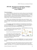
Knowing the level of the meniscus in a capillary it is possible to determine easily the total
volume of vapor-gas bubbles. Fig.10 shows change in the volume of generated bubbles at
different laser powers and different laser wavelengths. Our experiments show that the total
volume of bubbles rises gradually with time by a logarithmic low after the laser radiation
switching on. The total volume at 1 W of laser power rises with time monotonically for both
wavelengths, while at higher laser power quite strong fluctuations take place, with the
growing in time amplitude. As this takes place, at laser power of 3 W the strong eruption of
liquid from the capillary was observed after 4.7 s of laser irradiation (curve 3 at Fig. 10a). At
that moment the curve 3 interrupts, since the meniscus went out of visualization zone
because of the abrupt decrease of meniscus level.
The total volume of generated bubbles increases with laser power. Comparison of curves 1
and 2 at Fig.10b shows that twofold increase of laser power (from 1 to 2 W) causes about the
fourfold rise of the generated bubbles volume. After the laser radiation switching off, the
total volume of bubbles first rapidly decreases (vapor condensation inside bubbles), ant next
decreases more slowly. It should be noted that quite a strong low-frequency oscillations are
observed, caused by variation of total bubbles volume in a capillary.
In the case of 0.97m wavelength the fiber tip surface was covered by a thin carbon layer.
Arrows show the moments of laser on and laser off.
Digits at curves shows laser power in Watts.
Fig. 10. Change of the total bubbles volume at different powers of lasers with 0.97 m (a)
and 1.56 m (b) wavelengths of radiation.
Thus, the hydrodynamic processes related to the explosive boiling in the vicinity of the hot
tip surface are observed in the liquid even at medium laser powers. Note that the
intracapillary liquid exhibits effective mechanical oscillations with a frequency of 1– 5 Hz
and appears saturated with microbubbles. We expect the development of such laser-induced
hydrodynamic processes in water-saturated biotissues at medium laser powers.
On the one hand, such processes provide the saturation of cavities and fractures in a spinal
disc or bone with the water solution containing vapor-gas bubbles. On the other hand, they
give rise to high-power acoustic oscillations and vibrations in the organ containing the
connective tissue. Apparently, the filling of hernia with vapor-gas bubbles provides the
reproducible decrease in the density of herniation immediately after the laser treatment
(Sandler et al., 2004; Chudnovskii & Yusupov, 2008).
Hydrodynamics – Advanced Topics
106
It is known from (Bagratashvili et al., 2006) that the mechanical action on cartilages in the hertz
frequency range actively stimulates the synthesis of collagen and proteoglycans even at
relatively small amplitudes. The above estimations show that the pressure on biotissue
provided by the vapor-gas bubbles can reach tens of kilopascals. In accordance with
(Buschmann et al., 1995; Millward-Sadler & Salter, 2004), such pressures in the hertz frequency
range can lead to regenerative processes in cartilage owing to the activation of the interaction
of the extracellular matrix with the mechanoreceptors of chondrocytes (integrins).
3.3 Laser-induced generation of bubbles microjets
Note an interesting phenomenon in the experiments on the generation of bubbles in the
vicinity of the blackened tip surface of the fiber in the water cell: bubble microjets can be
generated at a laser power of less than 3 W (Fig. 11) (Yusupov et al., 2010). The lengths of the
microjets (Fig. 11a), which always start in the immediate vicinity of the fiber tip, reach
several millimeters, the transverse sizes normally range from 10 to 50 μm, and the sizes of
the bubbles that form the jets range from several to ten microns. The lifetime of the microjets
ranges from a few fractions of a second to tens of seconds. A microjet that emerges at a
certain spot on the tip surface remains attached to this spot and exhibits bending relative to
the mean position. Bubble microjets didn’t use to be continuous from start to end, the
discontinuities used to appear on them, which used to restore quite often. The observations
show (Yusupov et al., 2010) that the discontinuities are always related to the hydrodynamic
perturbations and are caused by relatively large bubbles that move in the vicinity of the
microjet. The appearance of quite a large bubble attached to the fiber tip caused the bubble
microjet bending around large bubble (Fig. 11b). Thus, we conclude that two conditions
must be satisfied for the generation of the bubble microjets. First, a hot spot must be formed
on the tip surface. Second, the neighborhood of such a spot must be free of the centers that
provide the generation and detachment of large bubbles. Note that the possibility of bubble
microjets in the vicinity of a point heat source is demonstrated in (Taylor & Hnatovsky,
2004).
Fig. 11. Bubble microjets in the vicinity of the tip surface of optical fiber.
A part of the blackened fiber tip is sown at the right upper corner.
4. Degradation of optical fiber tip
Laser-induced hydrodynamic effects in water and bio-tissues can lead to the significant
degradation of the fiber tip (Yusupov et al., 2011a). The most significant degradation of the
Laser-Induced Hydrodynamics in Water and Biotissues Nearby Optical Fiber Tip
107
fiber tip surface occurs in the regime of channel formation when the fiber is shifted inside the
wooden bar that mimics the biotissue. In this case, we observe substantial modifications and
distortion of tip surface. The comparison of the sequential photographs (Fig. 12) shows a
significant increase in the volume of the fiber fragment (swelling) in the vicinity of fiber tip.
Fig. 12. Modifications of the profile of the blackened fiber tip surface (side view) for regime
of channel formation (the channel is formed by the fiber that moves inside the wooden bar
with water and the radiation power is 5 W). The left-hand panel shows the original fiber just
after its blackening (Yusupov et al., 2011a).
SEM images (Fig. 13) show that the laser action in the regime of the channel formation in the
presence of water causes substantial modifications of the working surface: the sharp edge is
rounded and surface irregularities (craters) emerge on the tip surface. The image shows that
a thin shell (film) with circular holes is formed at the tip surface of the optical fiber. Multiple
cracks pass through some of the holes. In addition, we observe elongated crystal-like
structures on the surface (Fig. 13b). Looking through the largest hole in the film on the tip
surface (at the center of the lower part of the fragment at Fig. 13a), whose dimension in any
direction is greater than 10 µm, we observe the inner micron-scale porous structure.
Fig. 13. The microstructure of the fiber tip surface after laser action. a - SEM image of a
fragment of the fiber end surface; b - magnified SEM image of a fragment of the end surface
with the crystal-like structures on the surface (Yusupov et al., 2011a).
Hydrodynamics – Advanced Topics
108
Typical micron-scale circular holes on the film surface (Fig. 13a) can be caused by cavitation
collapse of single bubbles. It is well known that cavitation collapse of bubbles in liquid in
the vicinity of the solid surface gives rise to the high-speed cumulative microjets which can
destroy the solid surface (Suslick, 1994). Apparently, this effect leads to multiple cracks on
the film and the formation of the porous structure (Fig. 13a), since the cumulative microjets
can punch holes, cause cracks in the film, and destroy the structure of silica fiber tip.
Collapse of cavitation bubble apart from high pressure generation (up to10
6
MPa) can cause
overheating of gas up to temperatures as high as 10
4
К. Such high values of water pressure
and temperature can result in formation of supercritical water (critical pressure of water is
Р
c
=218 atm, critical temperature - T
c
=374ºС), which can dissolve silica fiber (Bagratashvili et
al., 2009).
Fig. 14 shows Raman spectra of some areas of laser irradiated fiber tip surface (curves 3-5)
compared with that of graphite (1) and diamond (2). Raman bands at 1590 cm
-1
and 1590 cm
-
1
to diamond and graphite nano-phases correspondingly (Yusupov et al., 2011a).
Fig. 14. Raman spectra from different areas of laser fiber tip surface (curves 3, 4 and 5)
compared with that of graphite (1) and diamond (2) (Yusupov et al., 2011a).
Formation of diamond nanophase at a fiber tip surface in this case is rationalized by
extremely high pressures and temperatures caused by cavitation processes stimulated by
laser irradiation (Yusupov et al., 2011a).
5. Laser-induced acoustic effects
Laser-induced hydrodynamics processes in water-saturated bio-tissues causes generation of
intense acoustic waves. We have studied the peculiarities of generation of such acoustic
waves in water and water-saturated biotissue (intervertebral disc, bone, et al.) in the vicinity
of blackened optical fiber tip using acoustic hydrophone (Brul and Kier 8100, Denmark). The
hydrophone with 0 – 200 KHz band was placed in water or biotissue at 1cm distance from
optical fiber tip. Fig. 15 demonstrates typical example of acoustic response to laser
irradiation for two different cases: in the bath of free water (Fig. 15a) and in the case of water
Laser-Induced Hydrodynamics in Water and Biotissues Nearby Optical Fiber Tip
109
The fiber tip surface is blackened before laser irradiation with 0.97 µm wavelength.
Fig. 15. Fragments of acoustic response to 3 W laser irradiation of water for two different
cases: in a bath of free water (a) and in a water-filled capillary (b).
filled capillary (Fig. 15b). In the case of the bath with free water, the short random laser-
induced acoustic spikes take place. At the same time, the acoustic response to laser
irradiation in the case of water-filled capillary (which imitates situation in real water-filled
biotissue channel) is different (Fig. 15b). Acoustic signal is amplitude-modulated by its
feature, and low-frequency modulation period is about 2 s.
Fig. 16 demonstrates acoustic response to laser irradiation of nucleus pulposus in vivo when
optical fiber was moved forward (regime of channels formation in the course of laser
healing of degenerated disc). The acoustic signal is non-stationary by its nature. The short-
pulse intense acoustic spikes take place and the signal itself is amplitude modulated
(similarly to that in water-filled capillary) with a modulation period of about 3 s.
Arrows show the moments of laser on and laser off.
Fig. 16. Acoustic response to 3 W laser irradiation with 0.97 µm wavelength of nucleus
pulposus in vivo, when optical fiber was moved forward in the intervertebral disc.
Hydrodynamics – Advanced Topics
110
The more detailed studies show that for both in vivo and in vitro cases laser-induced generation
of short-pulse intense quasi-periodic acoustic signals. The fragment of spectrogram of acoustic
response given at Fig. 17 clearly demonstrates temporal change of spectral components for
acoustic signal generated from laser irradiated nucleus pulposus in vitro when optical fiber
was moved forward in the intervertebral disc (similar to shown at Fig. 1).
Fig. 17. The fragment of spectrogram (a) ant temporal structure of single pulse (b) of
acoustic response generated from laser irradiated nucleus pulposus in vitro.
As one can see, the acoustic response in this case has the form of short, intense and broadband
(from 0 to 10 kHz) pulses of about 10 ms in duration combined into the series of pulses
generated with frequency of 40 Hz. Fig. 17b shows that the amplitude of single pulse is an
order of amplitude higher than the background acoustic noise. The most of acoustic power is
concentrated in such pulses. The broad spectrum of acoustic pulses and their low duration
indicate to shock-type of generated acoustic waves. The acoustic noise has broad spectral
maxima in the following spectral intervals: 600 – 700 Hz, 1 - 2 kHz and nearby 10 kHz.
Appearance of these bands are caused by the dynamics of vapor-gas mixture and are
associated with acoustic resonances of the system. Notice that laser-induced formation of
channels in degenerated spinal discs in vitro has been accompanied by 4 Hz in frequency
strong visual vibrations of needle with laser fiber.
Generation of such a strong acoustic vibrations is caused in our opinion by contact of
overheated (up to >1000 ºС (Yusupov et al., 2011a)) fiber tip with water and water-saturated
tissue of spinal disc. Such contact can result in explosive boiling of water solution nearby the
fiber tip and, also, in burning of collagen in cartilage tissues. Intense hydrodynamic
processes can take place nearby optical fiber tip, which are caused by fast heating of water
and tissue, by generation and collapse of vapor-gas bubbles (Chudnovskii et al., 2010a,
2010b; Leighton, 1994). As a result, the free space of disc or bone is filled by liquid saturated
by vapor-gas bubbles. Resonance vibrations are excited, since both disc and bone are quite
good acoustic resonators. These vibrations give rise to low-frequency modulation of acoustic
noise (Fig. 16) and to quasi-periodic generation of short intense pulses (Fig. 17)
(Chudnovskii et al., 2010a). The acousto- mechanic shock-type processes in resonance
conditions results in mixing and transport of gas-saturated degenerated tissue in the space
of defect (Chudnovskii et al., 2010b). These processes destroy hernia and decrease its density
(Fig. 2b), thus lowering the pressure to nervous roots. Another important impact of such
processes is the regeneration of disc tissues through the effects of mechanobiology
(Buschmann et al., 1995; Bagratashvili et al., 2006).
Laser-Induced Hydrodynamics in Water and Biotissues Nearby Optical Fiber Tip
111
6. Formation of filaments
In this division we will show that existence of strongly absorbed agents (in a form of Ag
nanoparticles, in particular) in laser irradiated water nearby optical fiber tip can result in
appearance of filamentary structures of these agents (Yusupov et al., 2011b). Medium power
(0.3 – 8.0 W) 0.97 µm in wavelength laser irradiation of water with added Ag nanoparticles
(in the form of Ag-albumin complexes) through 400 µm optical fiber stimulates self-
organization of filaments of Ag nanoparticles for a few minutes. These filaments represent
themselves long (up to 14 cm) liquid gradient fibers with unexpectedly thin (10 – 80 μm)
core diameter. They are stable in the course of laser irradiation, being destroyed after laser
radiation off. Such effect of filaments of Ag nanoparticles self-organization is rationalized by
the peculiarities of laser-induced hydrodynamic processes developed in water in presence of
laser light and by formation of liquid fibers.
Fiber laser radiation (LS-0,97 IRE-Polus, Russia) 0-10 W in output and 0.97 µm in wavelength
was delivered into water-filled plastic cell through 400 µm transport silica optical fiber, which
was placed horizontally in the cell. Low intensity (up to 1 mW) green pilot beam from the built
in diode laser was used to highlight the 0.97 µm laser irradiated zone in the cell. The cell was
placed at the sample compartment of optical microscope (MC300, MICROS, Austria) equipped
with color digital video-camera (Vision). Spectroscopic studies were performed with fiber-
optic spectrum analyzer (USB4000, Ocean Optics) and UV/vis absorption spectrometer (Cary
50, Varian). To measure the refraction index of collargol we have applied the fiber-optic
reflectometer FOR-11 (LaserChem, Russia), which provides 10
-4
precision of refraction index
measurements at 1256 nm wavelength. Cleavage of transport optical fiber has been always
produced just before each experiment. Ten minutes later (to provide reasonable attenuation of
hydrodynamic motions in the cell) the drop (0.01–1 ml in volume) of brown colored collargol
(complex of 25 nm in size Ag nanoparticles with albumin) has been smoothly introduced into
the water cell 0.5-10 mm aside from the optical fiber tip.
Our in situ optical microscopic studies of laser-induced filament formation were
accomplished in two different modes: 1) in transmission mode, using illumination with
white light from microscope lamp; 2) in scattering mode, using illumination with green light
of pilot laser beam through the same transport fiber.
Experiments show that 0.97 µm fiber laser irradiation of water in the cell with introduced
collargol drop causes (in some period of time from seconds to minutes) formation of thin
and long quite homogenous filaments, growing along the axis of 0.97 µm laser beam in
water. These filaments are brown colored (that gives the evidence of enhanced Ag
nanoparticles concentration in filament) and can be seen even with unaided eye.
Fig. 18 demonstrates the microscope image (in transmission mode) of one of such filaments.
This filament is located along the axis of output laser beam and is about 17 mm in length.
The measured profile of optical density of this filament is triangular in its shape with about
the same widths along filament (determined at half-maximum) of ~200 μm.
Fig. 18. Micro-image (in transmission mode) of filament of Ag nanoparticles fabricated in
water nearby optical fiber tip at 2.5 W of laser power (Yusupov et al, 2011b).
Hydrodynamics – Advanced Topics
112
Fig. 19a demonstrates the micro-image of another laser fabricated filament in scattering
mode. Intensity of light scattered from this filament decreases gradually with the distance
from fiber tip. Attenuation of green light in this case is caused by absorption and scattering
of green light in the course of its propagation through the filament. To reveal the
peculiarities of filament (given at Fig. 19a) we have performed the following processing of
its microscope image: all vertical profiles of image were normalized to local maximum (Fig.
19c); the microscope image was represented in shades of gray (Fig. 19b). As it follows from
figures 19b and 19c the length of given filament is about 6 mm, its average width is about 40
μm, and scattering intensity decreases rapidly with the distance from filament axis. Notice
that vertical profiles of all fabricated filaments (in both transmission and scattering modes)
are almost triangular with a sharp top. It was also established that the end of filament has
always a needle-like shape and, also, the width of filament obtained in transmission mode
measurements exceeds 3-5 times that obtained in scattering mode.
Fig. 19. a - Microscopic picture of filament (in scattering mode) of Ag nanoparticles
fabricated in water nearby optical fiber tip at 0.4 W of laser power. b - Image of this filament
represented in shades of gray after processing of (see text) of Fig. 19a. c - Normalized
vertical profiles of image given at Fig. 19b. (Yusupov et al, 2011b).
Laser-Induced Hydrodynamics in Water and Biotissues Nearby Optical Fiber Tip
113
It is of importance that filaments of Ag nanoparticles have been formed in our experiments
only in the case of existence of initial collargol concentration gradient in laser irradiated
water (when collargol drop was introduced initially into water aside from fiber tip). When
collargol drop was premixed in water cell before laser irradiation, formation of filaments has
never been observed (at any collargol concentrations in the cell and at any laser powers and
dozes).
The initial stage of filament self-organization process can be clearly seen in scattering mode
(Fig. 4). Some visible hydrodynamic flows take place nearby the fiber tip when laser power
is on. Such flows result in intrusion of collargol from neighboring area into the area in front
of the fiber tip. The slanting filament structure is clearly seen at Fig. 4. One can also see here
the initial process of new intrusion formation (outlined with dashed line). The rate of rise-up
front of a given intrusion (which is about 150 μm in average thickness) is found to be
described be exponential low (1): at 1 mm from laser fiber tip V= 1.5· 10
-2
cm/s, while at 2
mm from laser fiber tip V falls down to 3· 10
-3
cm/s.
We revealed that filaments of Ag nanoparticles self-organized in the course of 0.97 µm laser
irradiation can exist in the cell (in the presence of laser beam and with no external
mechanical distortions of liquid in the cell) for quite a long period of time. We have
supported such filaments for tens of minutes. Notice that both rectilinear and curved
filaments were self-organized in our experiments.
After 0.97 µm laser radiation being off, the filaments of Ag nanoparticles have been
completely destroyed for 10 – 30 s period of time. Notice that time Δt of diffusion blooming
of filament by value, estimated by formula
2
3
kT
xDt t
d
, (6)
where D – is diffusion coefficient of nanoparticle; k= 1.38· 10-23 J/K – Boltzmann constant;
T(K) – absolute temperature; μ = 1,002· 10-3 (N· s/m
2
) – dynamic viscosity of water; d=25
nm Ag nanoparticle diameter) gives Δt =25 s for =100 μm.
External mechanical distortions of filament of Ag nanoparticles results in its destruction.
However after mechanical distortion being off, the filament can be renewed completely in
presence of 0.97 µm laser radiation. Fig. 20 shows the dynamic of such filament renovation
after the distortion of self-organized filament (produced by its rapid crossing withthin a
metal needle). As one can see from Fig. 20, complete renewal took place for quite a short
period of time (~ 20 s).
Our experiments have shown that there is some range of 0.97 µm laser powers for which the
effect of laser-induced filament self-organization takes place and is, also, stable and
reproducible. At laser powers higher than 8 W we have newer observed filament formation.
At 0.2-0.5 W laser power filaments have been formed but have been unstable. The most
stable and long-living filaments were observed in 0.5-3 W laser power range. At laser power
less than 0.2 W we have never observed such filament formation. The instability of filaments
and even their absence at high laser powers is caused by intense laser-induced
hydrodynamic processes nearby the fiber tip. Our experiments show that the fiber tip
surface is gradually covered by a deposit, which absorbs laser radiation quite well. The wide
absorption band of deposit observed at fiber tip can be caused by island film of Ag
nanoparticles, and, possibly, by elementary carbon absorption (deposited at fiber tip due to
albumin thermo-decomposition). As a result of such deposits, the fiber tip becomes an
Hydrodynamics – Advanced Topics
114
Digits show the period of time from the beginning of filament destruction (Yusupov et al., 2011b).
Fig. 20. Renewal of destroyed filament of Ag nanoparticles in water nearby the tip of optical fiber.
intense heat source. That causes explosive water boiling, intense formation of micro-
bubbles, moving rapidly away from fiber tip to liquid (see for example Fig. 1,b) and
destroying filament.
We rationalize the observed effect of laser-induced self-organization of filaments from Ag
nanoparticles by following mechanisms. Initially (Fig. 21a), laser light absorption by water
(the absorption coefficient in water at 0.97 µm is about 0.5 cm-1) causes its heating with the
2-10ºС/s rate. Besides, the intense transfer of impulse to water takes place in this case. As a
result, the closed axis-symmetric liquid flows are developed being directed from fiber tip.
These flows promote Ag nanoparticles intrusion into the laser beam nearby the fiber tip
(Fig. 21b). Such intrusions are clearly seen in scattered green laser light (Fig. 4).
Another factor dominates at the second stage of filament self-organization. The refractive
index for collargol n
c
is higher than that for clean water n
w
. The value of n
c
-n
w
= 0.0044 at
wavelength λ=1256 nm was directly measured in our experiments using fiber-optic
densitometer. Due to the effect of total internal reflection laser light is concentrated inside
intrusion which work in fact as a liquid optical fiber. Channeling of laser light inside
intrusion with Ag nanoparticles results in deeper propagation of laser light into water. Light
pressure promotes faster movement of intrusion front giving rise to filament (Fig. 21c). As it
was shown in (Brasselet et al., 2008), for example, laser light pressure is also able to force
through the boundary between two unmixed liquids and to form thin channel of one liquid
inside another one, thus forming liquid optical fiber with gradient core. Thus, the image of
filament in transmission mode shows optical density of Ag nanoparticles. At the same time
the image of filament in scattering mode clearly demonstrate channeling effect in fabricated
filament which in fact is a liquid gradient fiber. Such liquid gradient fiber provides also
effective channeling of 970nm laser beam, thus promoting filament elongation and spatial
stability.
Laser-Induced Hydrodynamics in Water and Biotissues Nearby Optical Fiber Tip
115
a. Formation of water flow nearby the fiber tip.
b. Formation of Ag nanoparticles intrusions.
c. Fabrication of filaments from Ag nanoparticles.
d. Intense formation of micro-bubbles, hampering filament formation at high laser power.
Fig. 21. To the explanation of the effect of laser-induced formation of filaments of Ag
nanoparticles (Yusupov et al., 2011b).
Laser induced formation of 10-50 μm in thickness and up to few millimeters micro-bubble
streams (Fig. 11) can also promote the filaments fabrication observed in our experiments. It
is clear, however, that too intense chaotic formation of micro-bubble streams observed at
high laser power can hamper filament fabrication (Fig. 21d).
We believe that such filaments of nanoparticles can be developed not only in water media
but, also, in other fluids, with other laser wavelength and particles types. The indispensable
conditions in this case are the availability of sufficient level of laser light absorption in
irradiated medium nearby fiber tip and possibility of liquid fiber formation.
7. Conclusion
Hydrodynamic effects induced by a medium power (1–5 W) laser radiation in the vicinity of
the heated fiber tip surface in water and in water-saturated tissues are considered. A
threshold character of the dynamics of liquid is demonstrated. At a relatively low laser
power (about 1 W), the slow formation of vapor-gas bubbles with sizes of hundreds of
microns are observed at the optical fiber tip surface. The bubbles can be attached to the tip
surface in the course of laser radiation. At higher laser power increases, effective
hydrodynamic processes related to the explosive boiling in the vicinity of the overheated
fiber tip surface take place. The resulting bubbles with sizes ranging from a few microns to
several tens of microns provide the motion of liquid. The estimated velocities of bubbles in
Hydrodynamics – Advanced Topics
116
the vicinity of the fiber tip surface can be as high as 100 mm/s. Generation of bubbles in the
capillary leads to the circulating water flows with periods ranging from 0.2 to 1 s. Such
circulation intensity increases with the laser power. For the laser radiation with a
wavelength of 0.97 μm, we observe such effects only for the blackened fiber tip surface,
which serves as a local heat source. At a laser power of less than 3 W, stable bubble
microjets, which consist of the bubbles (ranging from several to ten microns) can be
generated in the vicinity of the blackened tip surface.
Laser-induced hydrodynamic effects in water and bio-tissues can cause the significant
degradation of the fiber tip. Cavitation collapse of bubbles in liquid in the vicinity of fiber
tip surface gives rise to the high-speed cumulative microjets which can destroy the solid
surface. This effect leads to multiple cracks on the film and the formation of the porous
structure, formation of supercritical water and even generation of diamonds nano-crystal.
Laser-induced hydrodynamics processes in water and water-saturated bio-tissues are
accompanied by generation of intense acoustic waves in resonance conditions, even of
shock-type waves. The acousto-mechanic processes results in mixing and transport of gas-
saturated degenerated tissue in the space of defect.
We found that medium power (0.3- 8 W) 0.97 µm in wavelength laser irradiation of water
with added Ag nanoparticles (in the form of Ag-albumin complexes) through 400μm
optical fiber stimulates self-organization of unexpectedly thin (10-80 µm) and lengthy (up
to 14 cm) filaments of Ag nanoparticles in the form of liquid gradient fibers. These
filaments in water are stable in the course of laser irradiation being destroyed after laser
radiation off. Such effect of filaments of Ag nanoparticles self-organization is rationalized
by the peculiarities of laser-induced hydrodynamic processes developed in water in
presence of laser light.
8. Acknowledgment
This work is supported by Russian Foundation for Basic Research (grant № 09-02-00714).
9. References
Bagratashvili V.N., Sobol E.N., Shekhter A.B. (Eds). (2006). Laser Engineering of Cartilage.
Fizmatlit, ISBN 5-9221-0729-1, Moscow
Bagratashvili V.N., Konovalov A.N., Novitskiy A.A., Poliakoff M., and Tsypina S.I. (2009).
Reflectometric studies of the etching of a silica fiber with a germanium silicate core
in sub- and supercritical water. Russian Journal of Physical Chemistry B, Focus on
Physics, Vol. 3, No. 8, pp. 1154-1164, ISSN 1990-7931
Berry D.W., Heckenberg N.R., and Rubinszteindunlop H. (2000). Effects associated with
bubble formation in optical trapping. Journal of Modern Optics, Vol. 47, No. 9, pp.
1575 — 1585, ISSN 0950-0340
Brasselet E., Wunenburger R., and Delville J P. (2008). Liquid optical fibers with multistable
core actuated by light radiation pressure. Physical Review Letters, Vol. 101, pp. 1-5,
ISSN 1079-7114
Buschmann M.D., Gluzband Y.A., Grodzinsky A.J., and Hunziker E.B. (1995). Mechanical
Compression Modulates Matrix Biosynthesis in Chondrocyte Agarose Culture.
Journal of Cell Science, Vol. 108, pp. 1497-1508, ISSN 0021-9533
Laser-Induced Hydrodynamics in Water and Biotissues Nearby Optical Fiber Tip
117
Chudnovskii V.M. and Yusupov V.I. (2008). Method of Laser Intervention Effects in
Osteochondrosis, Patent RF No. 2321373
Chudnovskii V., Bulanov V., and Yusupov V. (2010a). Laser Induction of Acoustic
Hydrodynamical Effects in Medicine. Photonics, Vol. 1, pp. 30-36, ISSN 1993-
7296
Chudnovskii V.M., Bulanov V.A., Yusupov V.I., Korskov V.I., and Timoshenko V.S. (2010b).
Experimental justification of laser puncture treatment of spine osteochondrosis.
Laser Medicine, Vol. 14, No. 1, pp. 30-35, ISSN 2071-8004
Hale G.M. and Querry M.R. (1973). Optical constants of water in the 200-nm to 200-μm
wavelength region. Applied Optics, Vol. 12, pp. 555–563, ISSN 0003-6935
Leighton T. G. (Ed.). (1994). The Acoustic Bubble, Academic Press Limited, ISBN 0124419208
9780124419209,London
Millward-Sadler S.J. and Salter D.M. (2004). Integrin-dependent signal cascades in
chondrocyte mechanotransduction. Annals of Biomedical Engineering, Vol. 32, No. 3,
pp. 435-446, ISSN 0090-6964
Privalov V.A., Krochek I.V., and Lappa A.V. (2001). Diode laser osteoperforation and its
application to osteomyelitis treatment. Proceedings of the SPIE, Vol. 4433, pp. 180-
185, ISSN 0277-786X
Rokhsar C.K. and Ciocon D.H. (2009). Fractional Photothermolysis for the Treatment of
Postinflammatory Hyperpigmentation after Carbon Dioxide Laser Resurfacing.
Dermatologic Surgery, Vol. 35, No. 3, (March 2009), pp. 535-537, ISSN 1524-4725
Sandler B.I., Sulyandziga L.N., Chudnovskii V.M., Yusupov V.I., and Galin Y.M. (2002).
Bulletin physiology and pathology of respiration, Vol. 11, pp. 46-49, ISSN 1998-5029
Sandler B.I., Sulyandziga L.N., Chudnovskii V.M., Yusupov V.I., Kosareva O.V., and.
Timoshenko V.C. (2004). Prospects for Treatment of Compression Forms of Discogenic
Lumbosacral Radiculitis by Means of Puncture Nonendoscopic Laser Operations
(Skoromec A.A.), Dalnauka, ISBN 5-8044-0443-1, Vladivostok
Suslick K.S. (1994). The chemistry of ultrasound. The Yearbook of Science & the Future, pp 138-
155, Encyclopaedia Britannica, ISBN 0852294026, Chicago
Taylor R.S. and Hnatovsky C. (2004). Growth and decay dynamics of a stable microbubble
produced at the end of a near-field scanning optical microscopy fiber probe.
Journal of Applied Physics, Vol. 95, No. 12, (June 2004), pp. 8444-8449, ISSN 0021-
8979
Van den Bos R., Arends L., Kockaert M., Neumann M., Nijsten T. (2009). Endovenous
therapies of lower extremity varicosities: a meta-analysis. Journal of Vascular
Surgery, Vol. 49, No. 1, pp. 230-239, ISSN 0741-5214
Yusupov V.I., Chudnovskii V.M., and Bagratashvili V.N. (2010). Laser-induced
hydrodynamics in water-saturated biotissues. 1. Generation of bubbles in liquid.
Laser Physics, Vol. 20, No. 7, pp.1641-1646, ISSN 1054 660X
Yusupov V.I., Chudnovskii V.M., and Bagratashvili V.N. (2011a). Laser-induced
hydrodynamics in water-saturated biotissues. 2. Effect on Delivery Fiber. Laser
Physics, Vol. 21, No. 7, pp. 1230-1234, ISSN 1054 660X
Hydrodynamics – Advanced Topics
118
Yusupov V.I., Chudnovskii V.M., Kortunov I.V., Bagratashvili V.N. (2011b). Laser-induced
self-organization of filaments from Ag nanoparticles. Laser Physics Letters, Vol. 8,
No. 3, (March 2011), pp. 214–218, ISSN 1612-2011
6
Endocrine Delivery System
of NK4, an HGF-Antagonist and
Anti-Angiogenic Regulator, for Inhibitions
of Tumor Growth, Invasion and Metastasis
Shinya Mizuno
1
and Toshikazu Nakamura
2
1
Division of Virology, Department of Microbiology and Immunology,
Osaka University Graduate School of Medicine, Osaka
2
Kringle Pharma Joint Research Division for Regenerative Drug Discovery,
Center for Advanced Science and Innovation, Osaka University, Osaka
Japan
1. Introduction
Estimates of the worldwide incidence and mortality from 27 cancers in 2008 have been
prepared for 182 countries by the International Agency for Research on Cancer (Ferlay et al.,
2010). Overall, an estimated 12.7 million new cancer cases and 7.6 million cancer deaths
occur in 2008, with 56% of new cancer cases and 63% of the cancer deaths occurring in the
less developed regions of the world. The most commonly diagnosed cancers worldwide are
lung (1.61 million, 12.7% of the total), breast (1.38 million, 10.9%) and colorectal cancers (1.23
million, 9.7%). Cancer is neither rare anywhere in the world, nor mainly confined to high-
resource countries. Many cancer subjects die from cancer as a result of organ failure due to
“metastasis” (Geiger & Peeper, 2009), thus indicating that medical control of tumor
metastasis leads to a marked improvement in cancer prognosis.
The acquisition of the metastatic phenotype is not simply the result of oncogene mutations,
but instead is achieved through an interstitial stepwise selection process (Mueller & Fusenig,
2004). The dissociation and migration of cancer cells, together with a breakdown of
basement membranes between the parenchyme and stroma, are a prerequisite for tumor
invasion. The next sequential events involved in cancer metastasis include the following: (i)
penetration of cancer cells to adjacent vessels (i.e., intravasation); (ii) suppressed anoikis (i.e.,
suspension-induced apoptosis) of cancer cells in blood flow; and (iii) an extravascular
migration and re-growth of metastatic cells in the secondary organ. For an establishment of
anti-metastasis therapy, it is important to elucidate the basic mechanism(s) whereby tumor
metastasis is achieved through a molecular event(s).
Hepatocyte growth factor (HGF) was discovered and cloned as a potent mitogen of rat
hepatocytes in a primary culture system (Nakamura et al., 1984, 1989; Nakamura, 1991).
Beyond its name, HGF is now recognized as an essential organotrophic regulator in almost
all tissues (Nakamura, 1991; Rubin et al., 1993; Zarnegar & Michalopoulos, 1995; Birchmeier
& Gherardi, 1998; Nakamura & Mizuno, 2010). Actually, HGF induces mitogenic, motogenic
Hydrodynamics – Advanced Topics
120
and morphogenic activities in various types of cells via its receptor, MET (Bottaro et al., 1991;
Higuchi et al., 1992). HGF is required for organogenesis in an embryonic stage and for tissue
repair in adulthood during various diseases (Nakamura, 1991; Birchmeier & Gherardi, 1998;
Nakamura & Mizuno, 2010). Several lines of in vitro studies indicate that HGF stimulates
scattering and migration of cancer cells (Matsumoto et al., 1994, 1996a; Nakamura et al.,
1997). In malignant tumors, HGF is expressed by stromal cells, such as fibroblasts, while
MET is over-expressed by cancer cells, thus suggesting in the mid-1990s that a paracrine
signal from HGF-producing stroma cells to carcinomas may cause malignant behaviors,
such as invasion and metastasis (Matsumoto et al., 1996b).
NK4 is an intra-molecular fragment of HGF, which is generated by a chemical cleavage of
mature form HGF (Date et al., 1997; Nakamura et al., 2010). NK4 includes an N-terminal
hairpin domain and 4-kringle domains (K1-K4) of HGF α-chain, which binds to MET. Thus,
NK4 antagonizes HGF activities as a competitive inhibitor. Using NK4 as an HGF-
antagonist in rodents with malignant tumors, we have accumulated evidence showing that
endogenous HGF-MET cascade is a key conductor for tumor metastasis, while inhibition of
MET signals leads to the arrests of tumor growth. Unexpectedly, NK4 prohibits tumor
angiogenesis through a MET-independent mechanism. This review focuses on the roles of
HGF in cancer biology and pathology. We also emphasize the effectiveness of NK4 in
experimental cancer models where NK4 is supplemented via a “hydrodynamics-based”
gene therapy.
2. Effects of HGF on intra-tumor cells during cancer progression
In the mid-1980s, MET was identified as a mutated oncogene from carcinogen-induced
osteosarcoma cells (MNNG-HOS) that transform NIH3T3 fibroblasts (Cooper et al., 1984).
MET-encoding protein has a tyrosine kinase activity (Dean et al., 1985), suggesting that MET
may be an orphan receptor of growth factors. In the early 1990s, MET-coding product was
demonstrated to be a high-affinity receptor for HGF (Bottaro et al., 1991; Higuchi et al., 1992).
Scatter factor (SF) stimulates tumor cell movement, as its name indicates, and is shown
molecularly identical to HGF (Konishi et al., 1991; Weidner et al., 1991). HGF has several
activities required for tumor cell invasion and metastasis, as described below. In this section,
we summarize the direct effects of HGF on intra-tumor cells, including carcinoma, and on
vascular and lymphatic cells prior to discussion of the contribution of HGF-MET cascades
during tumor malignancy.
2.1 Scattering and migration of tumor cells
Initial events for the metastatic spread of tumors involve loss of cell-cell contact within the
primary tumor mass. The integrity and morphology of epithelial tumor cell colonies are
maintained by cell-cell contact mediated by cadherins and its associated intracellular catenin
molecules. Cancer cells must lose their tight cell-to-cell contact by down-regulation of
cadherin-cadherin complex during invasion into adjacent tissues. HGF induces scattering (i.e.,
dispersion of cluster cells into single cells) via an endocytosis of E-cadherin from cell surface to
cytoplasma (Watabe et al., 1993; Miura et al., 2001). During cell migration, HGF activates the
Ras-Rab5 pathway for endocytosis of cadherins (Kimura et al., 2006), which triggers nuclear
localization of β-catenin, a transcription factor of genes responsible for cell motility (Hiscox &
Jiang, 1999). Stimulation of an Rho small G protein cascade and activation of cdc42, rac and
PAK by HGF leads to the disassembly of stress fiber or focal adhesions, while lamellipodia
Endocrine Delivery System of NK4, an HGF-Antagonist and
Anti-Angiogenic Regulator, for Inhibitions of Tumor Growth, Invasion and Metastasis
121
formation and cell spreading are enhanced by HGF (Royal et al., 2000). These changes confer a
down-stream mechanism of MET-mediated cancer invasion.
2.2 Breakdown of basement membranes
During cancer invasion, tumor cells must move across a basement membrane between
epithelium and lamina propria (i.e., sub-epithelium). HGF stimulates motility in a biphasic
process: cells spread rapidly and form focal adhesions, and then they disassemble these
condensations, followed by increased cell locomotion. In the early phase (i.e., within a few
minutes post-stimulation), HGF induces phosphorylation of focal adhesion kinase (FAK)
together with a tight bridge between the extra-cellular matrix (ECM) and integrins of cancer
cells (Matsumoto et al., 1994; Parr et al., 2001). In the later phase, HGF-stimulated cancer cells
invade into matrix-based gels in vitro, or across basement membrane ECM in vivo
(Nakamura et al., 1997). In this process, HGF up-regulates several types of matrix
metalloproteinase (MMP), such as MMP-1, -2, and -9, through activation of Ets, a
transcriptional factor of MMPs (Li et al., 1998; Nagakawa et al., 2000; Jiang et al., 2001).
Considering that MMP-inhibitors diminish HGF-mediated migration, the induction of MMP
through HGF-Ets cascade is essential for tumor invasion into adjacent normal tissues.
2.3 Endothelial attachment and extravasation of cancer cells
Needless to say, tumor angiogenesis as well as lymphatic vessel formation are important for
delivery of cancer cells from the primary tumor to secondary organs. HGF enhances
angiogenesis via induction of the proliferation and morphogenesis of endothelial cells (EC)
(Bussolino et al., 1992; Nakamura et al., 1996). Actually, HGF supplementation leads to the
enhancement of tumor angiogenesis in vivo (Laterra et al., 1997). Recent studies delineated
the capacity of HGF to induce lymphatic morphogenesis (Kajiya et al., 2005; Saito et al.,
2006). Thus, HGF is considered to facilitate cancer metastasis via neo-induction of vascular
or lymphatic vessel beds. HGF has a direct effect on EC for enhancing tight adhesion of
tumor cells on endothelium via FAK phosphorylation (Kubota et al., 2009a). Furthermore,
HGF decreases endothelial occludin, a cell-cell adhesion molecule (Jiang et al., 1999a). Under
such a loss of EC-EC integrity, HGF decreases the trans-endothelial resistance of tumor
vessels and enhances cancer invasion across an EC barrier (i.e., intravasation in primary
tumors and extravasation in metastatic organs) (Fig. 1).
2.4 Prevention of cancer cell anoikis
Anoikis, also known as suspension-induced apoptosis, is a term used to describe
programmed cell death (apoptosis) of epithelial cells induced by loss of matrix attachment.
In addition to gaining functions of invasion and angiogenesis, cell resistance to anoikis also
appears to play an important role in tumor progression and metastasis as tumor cells lose
matrix attachment during metastasis. However, it is unknown how cancer cells escape from
anoikis-like death during metastasis. It was demonstrated, in a non-adherent culture
models, that HGF is a key molecule inhibiting suspension-induced anoikis, and this effect is
mediated via a crosstalk that is, in turn, mediated by phosphatidyl-inositol 3-kinase (PI-3K)
and extracellular signal-regulated kinase (ERK)-1/2 (Zeng
et al., 2002; Kanayama et al., 2008).
A recent report described that tetraspanin CD151-knockdown abolishes preventive effect of
HGF on tumor anoikis (Franco et al., 2010). Thus, it is likely that cell surface tetraspanins are
important for signaling complexes between MET and integrin-β4, a known amplifier of
HGF-mediated cell survival.
Hydrodynamics – Advanced Topics
122
Fig. 1. Various effect of HGF on cancer cells and endothelial cells (EC) during tumor
progression. For example, sequential events during the lung metastasis of hepatic carcinoma
are summarized as follows: (A) dissociation and scattering of hepatocellular cancer cells
through an HGF-induced endocytosis of cadherins; (B) tumor migration into stromal areas
across the basement membrane (BM) is mediated via MMP-dependent matrix degradation
and Rho-dependent cell movement; (C) invasion of tumor cells into neighboring vessels (i.e.,
intravasation) where the tight junction between ECs is lost by HGF-MET signaling; (D)
inhibition of tumor cell anoikisis by MET-AKT cascades during blood flow, and out-flux of
tumor cells across vessel walls (i.e., extravasation); and (E) in the lung, HGF supports
growth of metastatic nodules via providing vascular beds as an angiogenic factor.
Overall, HGF is shown to take direct action on carcinoma cells: (i) cell spreading via an
endocytosis of cadherins; (ii) enhancement of invasion across basement membranes via Rho-
dependent and MMP-dependent pathways; and (iii) anti-anoikis activity during blood
circulation. Toward tumor vessels, HGF elicits vascular and lymphatic EC proliferation and
branching angiogenesis, while intravasation and extravasation are achieved through HGF-
induced reduction of EC-EC integrity. These HGF-MET-mediated biological functions seem
advantageous for invasion and metastasis of malignant tumors, including carcinoma and
sarcoma (Fig. 1).
[Note] Long-term administration of recombinant HGF does not elicit tumor formation in
healthy animals, and this result supports a rationale of HGF supplement therapy for treating
chronic organ diseases, such as liver cirrhosis, at least in cancer-free patients.
3. Regulation of HGF production by cancer cells
Several lines of histological evidence indicate that HGF is produced in stroma cells, such as
fibroblasts, vascular EC and smooth muscle cells in tumor tissues. In contrast, MET is over-
expressed mainly by tumor cells, particular near invasive areas, implying a possible
paracrine signal from HGF-producing stroma cells to MET-expressing carcinoma cells
Endocrine Delivery System of NK4, an HGF-Antagonist and
Anti-Angiogenic Regulator, for Inhibitions of Tumor Growth, Invasion and Metastasis
123
(Matsumoto et al., 1996b). Herein, we will discuss the molecular basis whereby stromal HGF
production is up-regulated by tumor cells during cancer invasion and metastasis.
3.1 Stroma as a microenvironment to determine behaviors of tumors
The important roles of stroma during tumor progression are demonstrated through several
independent studies. Carcinoma-associated fibroblasts, but not normal fibroblasts, stimulate
tumor progression of initiated non-tumorigenic epithelial cells both in an in vivo tissue
recombination and in an in vitro co-culture system (Olumi et al., 1999). Transforming growth
factor (TGF)-β signaling is critical for down-regulating HGF production (Matsumoto et al.,
1992). Of note, an inactivation of TGF-β type II receptor gene in stromal fibroblasts leads to
the onset of epithelial growth and invasion (Bhowmick et al., 2004). In this process,
activation of paracrine HGF is a key mechanism for stimulation of epithelial proliferation
(Bhowmick et al., 2004). Thus, the suppression of HGF production by TGF-β seems to be
important for an escape from cancer metastasis (Matsumoto & Nakamura, 2006).
3.2 Regulation of HGF production in stroma by tumor cells
As repeated, a major source of HGF in tumors is stromal cells (including fibroblasts,
endothelium, macrophages and neutrophils) (Wislez et al., 2003; Matsumoto & Nakamura,
2006; Grugan et al., 2010). Thus, how stromal HGF is up-regulated during tumor progression
should be discussed. There is now ample evidence that numerous types of carcinoma cells
secrete soluble factors that induce HGF production in stromal cells (i.e., HGF-inducers). For
example, conditioned medium obtained from breast cancer cells enhances HGF production
in fibroblasts, along with a raise in prostaglandin-E2 (Matsumoto-Taniura et al., 1999). Of
note, suppression of prostaglandin-E2 production by indomethacin leads to down-
regulation of stromal HGF production and suppression of tumor migration in vitro
(Matsumoto-Taniura et al., 1999), indicating that cancer-derived prostaglandins are
important for up-regulating HGF in stromal cells (Matsumoto-Taniura et al., 1999; Pai et al.,
2003). Other carcinoma-derived HGF-inducers are interleukin-1β (IL-1β), basic fibroblast
growth factor (b-FGF), platelet-derived growth factor (PDGF), and TGF-α (Hasina et al.,
1999; Matsumoto & Nakamura, 2003). These results indicate a crosstalk between carcinoma
and stroma, mediated via a paracrine loop of HGF-inducers produced by carcinoma and
HGF secreted from stroma cells, such as fibroblasts (Matsumoto et al., 1996a).
3.3 Inflammation-mediated HGF up-regulation mechanism
In addition to stromal fibroblasts, tumor-associated macrophages (TAM) are known to
highly produce HGF during non-small lung cancer invasion (Wang et al
., 2011). It is
reported that TAM isolated from 98 primary lung cancer tissues show the higher production
of HGF, along with the concomitant increases in urokinase-type plasmin activator (uPA),
cyclooxygenase-2 (Cox2) and MMP-9 (Wang et al., 2011). Anti-MMP-9 antibody largely
diminishes TAM-induced invasion, while Cox2 and uPA are critical for HGF production
and activation, respectively, suggesting that Cox2-uPA-HGF-MMP cascades in TAM
participate in non-small lung cancer invasion. Likewise, HGF production is enhanced by
neutrophils infiltrating bronchiolo-alveolar subtype pulmonary adenocarcinoma (Wislez et
al., 2003).
Clinical studies demonstrate that serum levels of HGF are elevated in patients with
recurrent malignant tumors (Wu et al., 1998; Osada et al., 2008), thus suggesting an
Hydrodynamics – Advanced Topics
124
endocrine mechanism of the HGF delivery system. In this regard, it is known that peripheral
blood monocytes produce HGF, contributing to the increase in blood HGF levels via an
endocrine mechanism (Beppu et al., 2001). Overall, production of HGF by inflammatory cells
is involved in carcinoma invasion and metastasis (i.e., local system), while peripheral blood
monocytes seem to prevent tumor cell anoikis during metastasis, possibly by a release of
HGF into blood (i.e., systemic system).
4. Structure and activity of NK4 as HGF antagonist
HGF is a stromal-derived paracrine factor that has stimulated cancer invasion at least in vitro
(Matsumoto et al., 1994; Matsumoto et al., 1996a; Nakamura et al., 1997). Clinical studies
suggest that the degree of serum HGF and Met expressions in cancer tissues appears to
correlate with a given prognosis (Yoshinaga et al., 1993; Osada et al., 2008). Thus, it is
hypothesized that in vivo inhibition of HGF-MET signaling may be a reasonable strategy to
prohibit cancer metastasis. To test this hypothesis, we prepared NK4 as an intra-molecular
fragment of HGF via a chemical digestive process (Date et al., 1997; Matsumoto et al., 1998).
As expected, NK4 bounded to MET and inhibited HGF-MET coupling as a competitive
inhibitor. An additional “unexpected” value was that NK4 inhibited tumor angiogenesis via
a MET-independent pathway. This section focuses on the biological value of NK4 as an
HGF-antagonist and as an angiogenesis inhibitor.
4.1 Structure and anti-invasive function of NK4
NK4 was initially purified as a fragment from elastase-digested samples of recombinant
human HGF (Date et al., 1997). The N-terminal amino acid sequence of NK4 and of the
remnant fragment, assumed to be composed of an HGF β-chain, revealed that NK4 is
cleaved between the 478
th
valine and the 479
th
asparagine. The N-terminal amino acid
sequence of NK4 revealed that the N-terminal structure of NK4 is the same as undigested
HGF (i.e., 32
nd
pyroglutamate), indicating that NK4 is composed of the N-terminal 447
amino acids of the α-chain of HGF and contains the N-terminal hairpin domain and four
kringle domains (thus designated NK4) (Fig. 2A). The binding domains that are
responsible for high-affinity binding to MET are the N-terminal hairpin and the first
kringle domains in NK4 (and HGF). MET tyrosine phosphorylation occurs in A549 lung
carcinoma within 10 minutes after HGF addition, while NK4 inhibits the HGF-mediated
MET activation (Fig. 2B). Actually, NK4 functions as an HGF-antagonist: HGF induces
invasion and migration of the gallbladder and bile duct carcinoma cells in ECM-based
gels, while NK4 inhibits HGF-induced invasion in a dose-dependent manner (Fig. 2C)
(Date et al., 1998). These anti-invasive effects of NK4 are seen in distinct types of cancer
cells (Hiscox et al., 2000; Maehara et al., 2001; Parr et al., 2001), strengthening the common
role of NK4 during cancer migration.
4.2 Perlecan-dependent anti-angiogenic mechanism by NK4
Vascular EC highly express MET, while HGF stimulates mitogenic and morphogenic
activities in EC (Nakamura et al., 1996), thus suggesting that NK4 could inhibit HGF-
induced angiogenesis. Actually, NK4 potently inhibited the HGF-mediated proliferation of
EC in vitro (Jiang et al., 1999b). Strikingly, NK4 also inhibited microvascular EC proliferation
and migration, induced by other angiogenic factors, such as b-FGF and vascular endothelial
Endocrine Delivery System of NK4, an HGF-Antagonist and
Anti-Angiogenic Regulator, for Inhibitions of Tumor Growth, Invasion and Metastasis
125
Fig. 2. Preparation of NK4 as an HGF-antagonist and its inhibitory effects on tumor invasion
in vitro. (A) Preparation and structure of NK4. NK4 is generated via a cleavage of HGF
between 478
th
Val and 479
th
Asn. (B) Inhibition of HGF-mediated MET tyrosine
phosphorylation by NK4 in lung carcinoma cells. (C) Biological activity of NK4. Cancer cell
invasion (upper chamber) is induced across a Matrigel layer when fibroblasts (FB) are
placed on a lower chamber. In this co-culture system, NK4 inhibits FB-induced tumor cell
invasion in a dose-dependent manner.
growth factor (VEGF) (Fig. 3A) (Kuba et al., 2000). When a pellet containing b-FGF was
implanted under the rabbit cornea, angiogenesis was rapidly induced. In this model, NK4
inhibited b-FGF-induced angiogenesis (Fig. 3B). In vitro models of EC proliferation, HGF
and VEGF phosphorylate MET and KDR/VEGF receptor, respectively, whereas NK4
inhibits HGF-induced MET tyrosine phosphorylation, but not VEGF-induced KDR
phosphorylation (Kuba et al., 2000). Nevertheless, NK4 inhibited the VEGF-mediated EC
proliferation without modification of VEGF-mediated ERK1/2 (p44/42 mitogen-activated
protein kinase) activation. These results suggest the presence of another mechanism
whereby NK4 inhibits VEGF- and b-FGF-mediated angiogenesis.
The fibronectin-integrin signal is essential for the spreading and proliferation of EC. Based
on this background, we demonstrated that NK4-mediated growth arrest of EC is due to a
loss of the fibronectin-integrin signal. Affinity purification with NK4-immobilized beads
revealed that NK4 binds to perlecan (Sakai et al., 2009). Consistent with this result, NK4 was
co-localized with perlecan in EC. Perlecan is a multi-domain heparan sulfate proteoglycan
that interacts with basement membrane components such as fibronectin. Of interest,
knockdown of perlecan expression by siRNA diminished the fibronectin assembly and EC
spreading, indicating an essential role of fibronectin-perlecan interaction during EC
movement. A recent report described that NK4-perlecan interaction suppressed the normal
assembly of fibronectin by perlecan (Sakai et al., 2009). As a result, FAK activation became
faint in EC after NK4 treatment. Under such a loss of fibronectin-integrin signaling by NK4,
EC growth and motility were suppressed, even in the presence of b-FGF or VEGF. This is
the reason why NK4 arrests b-FGF- or VEGF-mediated angiogenesis (Fig. 3C).
Hydrodynamics – Advanced Topics
126
Fig. 3. Anti-angiogenic effects of NK4 via a perlecan-dependent mechanism. (A) NK4
suppresses HGF-, b-FGF-, and VEGF-induced proliferation of EC in vitro (Kuba et al., 2000).
(B) Inhibition of b-FGF-induced corneal neovascularization by NK4 treatment in rabbits. (C)
Involvement of perlecan (PC) in NK4-mediated growth arrest of EC. Left: Cell surface PC is
required for the binding of fibronectin and α5β1-integrin, leading to FAK phosphorylation
and crosstalk of VEGF-VEGF receptor (KDR) signaling. Right: NK4 binds to PC, and then
the binding of integrin to fibronectin is impaired. As a result, VEGF fails to elicit G1/S
progression of EC in the presence of NK4 (Sakai et al., 2009).
We have accumulated in vitro evidence showing that HGF-MET system may elicit cancer
invasion via a paracrine loop of stroma-carcinoma interaction. This phenomenon is also
demonstrated in vivo: anti-HGF antibody potently suppressed the tumor invasion in a
mouse model of pancreas cancer (Tomiola et al., 2001). On the other hand, several
investigators proposed, in the late-1990’s, a new concept that tumor angiogenesis inhibition
leads to the arrest of cancer growth and metastasis (Yancopoulos et al., 1998). Inhibition of
tumor angiogenesis leads to local hypoxia, and then apoptotic death of cancer cells is
associated with the arrests of tumor growth and metastasis (i.e., cytostatic therapy). In this
regard, NK4 also elicits an anti-angiogenic effect via perlecan-dependent mechanism. Thus,
bi-functional properties of NK4 as an HGF antagonist and angiogenesis inhibitor raise a
possibility that NK4 may prove therapeutic for cancer patients, as follows.
5. Anti-cancer therapy using NK4 in animal models
Carcinoma and sarcoma show malignant phenotypes prompted by a stroma-derived HGF-
MET signal at least in vitro. If NK4 could block MET signaling as an HGF-antagonist in vivo,
supplemental therapy with NK4 would be a pathogenesis-based strategy to counteract
Endocrine Delivery System of NK4, an HGF-Antagonist and
Anti-Angiogenic Regulator, for Inhibitions of Tumor Growth, Invasion and Metastasis
127
tumor invasion and metastasis. This hypothesis is widely demonstrated through extensive
studies using tumor-bearing animals, as described below.
5.1 First evidence of NK4 for inhibition of carcinoma progression in vivo
HGF, or co-cultured fibroblasts, are known to induce invasion of gallbladder carcinoma cells
(GB-b1) across Matri-gel basement membrane components (Li et al., 1998). NK4
competitively inhibits the binding of HGF to MET on GB-d1 cells. As a result, NK4
diminishes HGF-induced, or fibroblast-induced, motogenic activities (Date et al., 1998), thus
suggesting that stroma-derived HGF is a key conductor for provoking tumor invasion. Such
an important role of HGF was also demonstrated in vivo. Subcutaneous inoculations of
human gallbladder carcinoma GB-d1 cells in nude mice allow for primary tumor growth
and invasion to adjacent muscular tissues. Using this conceptual model, we provided the
first evidence of NK4 as an anti-tumor drug (Date et al., 1998). Recombinant NK4 has
inhibited the growth and muscular invasion in a mouse model of gallbladder carcinoma.
Consistent with tumor growth arrest, apoptotic change becomes evident during NK4
injections. Since HGF has an anti-apoptotic effect on cancer cells (Zeng et al., 2002), reverse
of HGF-induced protection by NK4 may be one of the mechanisms whereby carcinoma
growth can be suppressed during NK4 supplemental therapy.
5.2 Inhibition of tumor angiogenesis by NK4 treatment
In a culture of EC, NK4 produces anti-angiogenetic effects via a MET-independent pathway
(Kuba et al., 2000; Nakabayashi et al., 2003). These effects are also observed in animal models
of malignant tumors: administration of recombinant NK4 suppressed primary tumor
growth, metastasis of Lewis lung carcinoma, and Jyg-MC(A) mammary carcinoma
implanted into mice (Kuba et al., 2000), although neither HGF nor NK4 affected proliferation
and survival of these tumor cells in vitro. NK4 treatment resulted in a remarkable decrease
in microvessel density and an increase in apoptotic tumor cells in primary tumors,
suggesting that the inhibition of tumor growth by NK4 may be achieved by the suppression
of tumor angiogenesis (Kuba et al., 2000). The anti-angiogenic effects of NK4 are widely
demonstrated in various types of cancers [see our review articles (Matsumoto & Nakamura,
2005; Matsumoto et al., 2008a,b)]. Because the inhibition of angiogenesis by NK4 leads to
tumor hypoxia, hypoxia-primed apoptosis may contribute to a reduction in tumor size
during NK4 supplemental therapy.
5.3 Delayed NK4 therapy for attenuation of end-stage pancreas carcinoma
Anti-tumor effect of NK4 is also observed in a mouse model of advanced pancreas
carcinoma (Tomioka et al., 2001). When NK4 treatment was initiated on day 10, a time when
cancer cells were already invading surrounding tissues, NK4 potently inhibited the tumor
growth, peritoneal dissemination, and ascites accumulation at 4 weeks after the inoculation.
Such an anti-tumor effects of NK4 correlated with decreased vessel density in pancreatic
tumors. In an end-stage of pancreas cancer, NK4 inhibited the malignant phenotypes, such
as peritoneal dissemination, invasion of cancer cells into the peritoneal walls and ascites
accumulation (Tomioka et al., 2001). As a result, NK4 prolonged the survival time of mice at
an end-stage of cancer (Fig. 4). Because effective systemic therapy for pancreatic cancer is
currently not available, and diagnosing pancreatic cancer in its early stages is difficult, the
highly invasive and metastatic behaviors of pancreatic cancer lead to difficulty in attaining a
Hydrodynamics – Advanced Topics
128
Fig. 4. Anti-tumor effects of NK4 on advanced pancreas cancer in mice. (A) Schedules for
NK4 treatment of mice with pancreatic cancer. NK4 was injected into mice between 3 and 28
days after the inoculation of human pancreatic cancer cells (SUIT-2). (B) Inhibition of
primary tumor growth by NK4. Photographs show appearance of the primary pancreatic
cancer. (C) Histological analysis of the effect of NK4-treatment on tumor angiogenesis (left)
and apoptosis (right). NK4-treatment reduced the number of vessel numbers, while
apoptotic death of cancers was enhanced by NK4. (D) Inhibitory effects of NK4 on
peritoneal metastasis. Left: Typical macroscopic findings. Middle: Changes in the number of
metstatic nodules. Right: Changes in the ascite volumes. (E) Prolonged survival of tumor-
bearing mice treated with NK4.
long-term survival and a recurrence-free status. Targeting tumor angiogenesis and blockade
of HGF-mediated invasion of cancer cells may prove to be potential therapy for patients
with pancreatic cancer.
5.4 Therapy combining NK4 with other treatments
Anti-cancer chemotherapy is widely used for the suppression of malignant tumors with or
without surgical treatment. Therapy regimens that combine anti-cancer chemo drugs and
NK4 enhance their anti-tumor effect (Matsumoto et al., 2011). Irradiation therapy often
enhances cancer metastasis, especially in cases of pancreatic carcinoma, and this is
associated with the irradiation-induced up-regulation of HGF in fibroblasts (Qian et al.,
2003; Ohuchida et al., 2004). Thus, NK4 may overcome these irradiation-associated side
effects.
Epidermal growth factor receptor (EGFR) kinase inhibitors, such as Gefitinib, are used to
treat non-small cell lung cancers that have activating mutations in the EGFR gene, but most
of these tumors become resistant to EGFR-kinase inhibitors due to enhancement of HGF-
MET signals (Engelman et al., 2007; Yano et al., 2008; Okamoto et al., 2010). Thus, NK4
treatment may reverse HGF-induced resistance to Gefitinib.
Endocrine Delivery System of NK4, an HGF-Antagonist and
Anti-Angiogenic Regulator, for Inhibitions of Tumor Growth, Invasion and Metastasis
129
Recently, it was demonstrated that NK4-mediated tumor regression depends on the
infiltration of cytotoxic T lymphocytes (Kubota et al., 2009b). Importantly, depletion of CD8+
cells markedly abrogated the anti-tumor activity of NK4 in a mouse model of colon cancer.
NK4 enhances immune responses in dendritic cells in vitro. Thus, NK4 may also have utility
for anti-tumor immunotherapy.
There is now ample evidence that NK4 is useful for the inhibition of growth, invasion and
metastasis in various types of tumors, such as gastric carcinoma (Hirao et al., 2002), pancreas
cancer (Tomioka et al., 2001), prostate cancer (Davies et al., 2003), multiple myeloma (Du et
al., 2007) and melanoma (Kishi et al., 2009) (Table-1). These results support our hypothesis
that HGF is a key determinant of tumor malignancy (Matsumoto et al., 1996b).
Tumor diseases NK4 therapy Outcome Literature
(Cell lines and treatment)
A. Digestive system:
Gastric carcinoma Adeno-NK4, ip Inhibitions of growth Ueda K et al.,
(TMK1 cells, and metastasis, Eur J Cancer
ip, Mouse) Anti-angiogenesis, 40: 2135-2142
Reduced ascites (2004)
Hepatic carcinoma Adeno-NK4, iv Inhibitions of growth, Son G et al.,
(HUH7 cells, Anti-angiogenesis, J Hepatol 45:
portal vein, Mouse) Prolonged survival 688-695 (2006)
Gallbladder cancer NK4, sc Inhibitions of growth Date K et al.,
(GB-d1 cells, and invasion Oncogene 17:
sc, Mouse) 3045-354 (1998)
Pancreatic carcinoma r-NK4, ip Inhibitions of growth, Tomioka Det al.,
(SUIT-2 cells, invasion and metastasis, Cancer Res 61:
intra-pancreas, Anti-angiogenesis, 7518-7524
Mouse) Reduced ascites, (2001)
Prolonged survival
Colon carcinoma NK4 cDNA, Inhibitions of growth, Wen J et al.,
(MC-38 cells, bolus iv invasion and metastasis, Cancer Gen Ther
intra-spleen, (hydrodynamics) Anti-angiogenesis, 11: 419-430
Mouse) Prolonged survival (2004)
B. Respiratory system:
Lung carcinoma r-NK4, sc Inhibitions of growth Kuba K et al.,
(Lewis carcinoma, and metastasis, Cancer Res 60:
sc, Mouse) Anti-angiogenesis, 6737-6743
Enhanced apoptosis (2000)
Lung carcinoma Adeno-NK4, Inhibition of growth, Maemondo M
(A549 cells, intra-tumor Anti-angiogenesis et al., Mol Ther 5:
sc, Mouse) or ip 177-185 (2002)
Mesothelioma Adeno-NK4, Inhibition of growth, Suzuki Y et al.,
(EHMES-10 cells, intra-tumor Enhanced apoptosis, Int J Cancer 127:
sc, Mouse) Anti-angiogenesis 1948-1957
(2010)