báo cáo hóa học: "Reduction of motion artifact in pulse oximetry by smoothed pseudo Wigner-Ville distribution" doc
Bạn đang xem bản rút gọn của tài liệu. Xem và tải ngay bản đầy đủ của tài liệu tại đây (885.39 KB, 9 trang )
BioMed Central
Page 1 of 9
(page number not for citation purposes)
Journal of NeuroEngineering and
Rehabilitation
Open Access
Research
Reduction of motion artifact in pulse oximetry by smoothed pseudo
Wigner-Ville distribution
Yong-sheng Yan
†
, Carmen CY Poon and Yuan-ting Zhang*
Address: Joint Research Center for Biomedical Engineering, The Chinese University of Hong Kong, Shatin, Hong Kong
Email: Yong-sheng Yan - ; Carmen CY Poon - ; Yuan-ting Zhang* -
* Corresponding author †Equal contributors
Abstract
Background: The pulse oximeter, a medical device capable of measuring blood oxygen saturation
(SpO2), has been shown to be a valuable device for monitoring patients in critical conditions. In
order to incorporate the technique into a wearable device which can be used in ambulatory
settings, the influence of motion artifacts on the estimated SpO2 must be reduced. This study
investigates the use of the smoothed psuedo Wigner-Ville distribution (SPWVD) for the reduction
of motion artifacts affecting pulse oximetry.
Methods: The SPWVD approach is compared with two techniques currently used in this field, i.e.
the weighted moving average (WMA) and the fast Fourier transform (FFT) approaches. SpO2 and
pulse rate were estimated from a photoplethysmographic (PPG) signal recorded when subject is in
a resting position as well as in the act of performing four types of motions: horizontal and vertical
movements of the hand, and bending and pressing motions of the finger. For each condition, 24 sets
of PPG signals collected from 6 subjects, each of 30 seconds, were studied with reference to the
PPG signal recorded simultaneously from the subject's other hand, which was stationary at all
times.
Results and Discussion: The SPWVD approach shows significant improvement (p < 0.05), as
compared to traditional approaches, when subjects bend their finger or press their finger against
the sensor. In addition, the SPWVD approach also reduces the mean absolute pulse rate error
significantly (p < 0.05) from 16.4 bpm and 11.2 bpm for the WMA and FFT approaches, respectively,
to 5.62 bpm.
Conclusion: The results suggested that the SPWVD approach could potentially be used to reduce
motion artifact on wearable pulse oximeters.
Introduction
Wearable medical devices are capable of continuously
monitoring an individual's vital signs in real time. These
devices are particularly important to the world's increas-
ingly aging population, whose health conditions have to
be assessed regularly or monitored continuously. The
devices can warn individuals of symptoms of deteriora-
tion, e.g. alerting them when their blood pressure is
increasing to a level above a predetermined threshold. The
devices can also automatically notify emergency services
in critical situations. In order to make wearable devices
practical, a series of technical problems have to be solved.
Published: 01 March 2005
Journal of NeuroEngineering and Rehabilitation 2005, 2:3 doi:10.1186/1743-0003-2-3
Received: 20 January 2005
Accepted: 01 March 2005
This article is available from: />© 2005 Yan et al; licensee BioMed Central Ltd.
This is an Open Access article distributed under the terms of the Creative Commons Attribution License ( />),
which permits unrestricted use, distribution, and reproduction in any medium, provided the original work is properly cited.
Journal of NeuroEngineering and Rehabilitation 2005, 2:3 />Page 2 of 9
(page number not for citation purposes)
For example, these devices need to be miniature in size,
must possess a user-friendly interface and be efficient in
power consumption. Most importantly, these devices
need to have a low failure rate and must report minimal
false alarms. In other words, these devices are required to
provide an accurate estimate of the monitored vital sign
under normal daily life situations. This leads to the impor-
tant topic on the reduction of motion artifacts [1-4]. In
this paper, the smoothed pseudo Wigner-Ville distribu-
tion (SPWVD) is investigated as a novel motion artifacts
resistant approach for estimating one of the most impor-
tant vital signs – the blood oxygen saturation level
(SpO2).
The paper is organized as follows. Section 2 reviews the
techniques commonly used for attenuating motion arti-
facts in pulse oximetry. Section 3 discusses the basic the-
ory for SpO2 computation and the techniques used in this
study for reducing motion artifacts. Section 4 compares
the performance of two time-frequency techniques, i.e.
the short-time Fourier transform (STFT) and the SPWVD.
Section 5 presents the protocol and the results of an exper-
iment to assess motion artifact reduction in real data. Sec-
tion 6 discusses the performance of the SPWVD approach
as compared to the traditional time domain and spectral
methods. Lastly, the major findings of this paper are sum-
marized in section 7.
Background
SpO2 is commonly monitored by a pulse oximeter, which
has been widely adopted as a standard measure during
anesthesia, neonatal care and post-operative recovery
[5,6]. Pulse oximeters currently available on the market
normally perform remarkably well when the monitored
subject is in the resting position. However, their reliability
is significantly reduced when the subject moves, even
when movements are only involuntary, such as shivering
[1-4,7]. Therefore, the reduction of motion artifacts is of
particular concern in the development of pulse oximeters
to be applied in ambulatory, pediatric and trauma set-
tings, as well as for implementing them into wearable
devices for personal home healthcare [8].
A number of attempts have been made in the past decade
to improve the accuracy of pulse oximeters when subjects
move. Typical methods can be generally classified into
three categories: (1) based on an independent measure of
motion; (2) based on a model of the ideal signal or the
noise; and (3) based on features recognized from the cor-
rupted signal. For techniques based on an independent
measure of motion, one or more transducers (e.g. piezo or
optical sensors) are employed to record the user's motion.
By assuming that the artifact is a linear addition to the pul-
satile photoplethysmographic (PPG) signal, the original
signal can be reconstructed from the corrupted signal [9-
11]. This hypothesis is however often doubted when
inspecting PPG signals under typical artifact-producing
forces [12]. This observation drives researchers to develop
more realistic models for the PPG signal or the artifact.
A recently proposed PPG artifact reduction methodology
was based on the inversion of a nonlinear physical artifact
model and could significantly reduce the effect of changes
of probe coupling [8,12]. However, model-based tech-
niques suffer inherently from the specificity of the model
design and are unable to cope with all aspects of real-life
scenarios.
On the other hand, techniques based on feature recogni-
tion are free of the generic problem of model designs.
Instead, these techniques often utilize some predeter-
mined criteria to separate regions of corrupted and uncor-
rupted PPG signal and estimate the desired parameters
from the uncorrupted portion of it. For example, Swedlow
et al. calculated the derivative of a signal and identified a
portion of it as a motion artifact whenever the ratio of
adjacent positive and negative peaks of the derivative is
below a threshold [13]. J.E. Scharf et al. evaluated the use
of spectral analysis to separate the cardiac physiologic
components from the recorded PPG signal that is contam-
inated by motion artifact for SpO2 estimation [14-16].
The above methodologies employ techniques in the time
domain or frequency domain. However, due to the non-
stationary nature of PPG signals, the use of time-fre-
quency analysis appears to be extremely attractive. Dowla
et al. proposed using a neural network together with a
wavelet transform (WT) to estimate SpO2 in the presence
of a motion artifact, and found out that this technique
performs better than conventional algorithm that detects
peaks and troughs of the PPG signal for estimating SpO2
levels [17]. In their method, a neural network was trained
to identify the motion level, which was then fed into a sec-
ond neural network together with the amplitude ratios at
different scales of WT of the PPG signal to estimate SpO2
levels. It has been pointed out by another researcher [16]
that using WT for SpO2 computation requires careful
analysis and additional testing. WT does not result in a
spectrum where the amplitude of a unique cardiac fre-
quency can be directly obtained for SpO2 estimation. On
the other hand, although such a unique component is
available on the spectrum obtained from fast Fourier
transform (FFT), the time-frequency resolution of FFT or
STFT is relatively low when compared to other time-fre-
quency techniques such as the Wigner-Ville distribution.
The goal of this study is to investigate the use of SPWVD,
a high resolution time-frequency transformation where
the amplitude of a unique cardiac frequency is apparent,
for the estimation of SpO2 levels.
Journal of NeuroEngineering and Rehabilitation 2005, 2:3 />Page 3 of 9
(page number not for citation purposes)
Methods
Basic theory
The traditional algorithms for estimating SpO2 detect
peaks and troughs of the PPG signal in the time domain.
Based on the Beer-Lambert law, which relates the optical
path length and effective absorbance to the intensity of
transmitted light, the relationship between intensity of
transmitted light and SpO2 is commonly described as:
I (
λ
, t) = I
0
(
λ
) exp[(-s
ε
HbO2
(
λ
) + (1 - s)
ε
Hb
(
λ
))·c·d (t)],
(1)
where,
ε
HbO2
and
ε
Hb
are the extinction coefficients of oxy-
genated and de-oxygenated hemoglobin, and s, c, and d
represent SpO2, total concentration of hemoglobin and
the optical path length respectively.
By using two light sources – red and infrared lights – and
calculating a normalized ratio of the AC component to the
DC component for each light source, SpO2 can be com-
puted from the ratio of ratios R, i.e. the normalized ratio
of the red to the infrared transmitted light intensity. That
is,
In practice, SpO2 can be obtained from equation (3)
directly or by an empirical equation that relates SpO2 and
R. In this study, SpO2 is estimated directly from equation
(3).
SpO2 computation by weighted moving average (WMA)
By calculating the ratio of the AC components and the
ratio of the DC components of the two light sources,
SpO2 can be obtained from every single pulse of a PPG
signal. To stabilize the reading, the weighted moving aver-
age (WMA) is often used [18]. Typical averaging methods,
e.g. the median averaging and standard arithmetic averag-
ing, are applied to every several samples or samples in
every several-second intervals. In this study, the SpO2
obtained by the WMA approach was the average of SpO2
samples in an 8-second period. Overlap processing was
performed at 1-second interval. The 8-second period is
selected in order to satisfy clinical requirements [15,16].
SpO2 computation by fast Fourier transform (FFT)
Based on the hypothesis that cardiac rate can be estimated
more easily by spectral analysis than time domain analy-
sis, techniques in the frequency domain have been widely
investigated as alternatives in pulse oximetry. For exam-
ple, the FFT and discrete cosine transform (DCT) were
proposed for estimating SpO2 [14-16]. These techniques
calculate the spectrogram of the PPG signal in a fixed time
period and select the strongest spectral line in the cardiac
frequency band as the AC component. The cardiac fre-
quency band is usually predetermined by certain thresh-
olds or obtained from an independent pulse rate
estimator, e.g. by applying electrocardiography in parallel.
In this study, FFT is applied to every 8-second PPG signal
at 1-second interval. The cardiac frequency band is prede-
termined as 0.8–2 Hz, i.e. corresponding to 48–120 bpm.
SpO2 computation by SPWVD
The Wigner-Ville distribution (WVD) of a signal x(t) is
given as:
where x(t) and x*(t) are the time series of the signal and
its complex conjugate respectively.
The problem of the WVD is the so-called cross-term inter-
ference, which appears as frequencies that lie between the
frequencies of any two strong components. In order to
suppress cross-term interference, the smoothed pseudo
WVD is often used:
The two windowing operations h and g are equivalent to
smoothing the WVD in the frequency and time domain
respectively. Selection of the window is a compromise
between the joint time-frequency resolution and the level
of cross-term interference. Common choices of window
include the rectangular and Kaiser windows [19-21]. In
our experiment, we chose the Hamming window as both
the time and frequency smoothing windows, g(t) and
h(
τ
).
The maximum magnitude within the cardiac frequency
band of the SPWVD in each second was used for SpO2
computation. Since SPWVD represents energy distribu-
tion, the square root of the magnitude was used for calcu-
lating the ratio of ratios R and SpO2. Moreover, as the rate
of change of SpO2 is relatively slow, SpO2 that changed
by more than 2% per second was considered to be physi-
ologically impossible, and was rejected from the calcula-
tion [22].
Simulation: STFT spectrogram versus SPWVD
The performance of SpO2 computation by the SPWVD
approach was evaluated in a simulation using PPG signals
collected from a volunteer under modest random
R
II
II
II
II
AC DC RED
AC DC IR
RED IR AC
RED IR DC
==
(/)
(/)
(/)
(/)
, and, 2
(()
s
R
Hb R Hb IR
Hb R HbO R HbO IR Hb IR
=
−⋅
−+ −
ελ ελ
ελελελελ
() ( )
() ()[ ( ) ( )
22
]]⋅
()
R
3
WVD t x t x t e d
x
j
,()()
*
ω
ττ
τ
ωτ
()
=+ −
()
−∞
+∞
−
∫
22
4
SPW t h g s t x t x t e d
x
j
(, ) () ( )( ) ( )
*
ωτ
ττ
τ
ωτ
=−+−
()
−
−∞
+∞
−∞
+∞
∫∫
22
5
Journal of NeuroEngineering and Rehabilitation 2005, 2:3 />Page 4 of 9
(page number not for citation purposes)
motions. For comparison, SpO2 was also estimated from
the spectrogram obtained by STFT.
The noise-mixing-composition (NMC) method was
applied to mimic the clinical situation and to induce a
range of signal-to-noise ratios (SNR) [23]. To synthesize a
noise-contaminated signal, an artifact noise that has been
verified to be similar to the real noise was added to an
undisturbed basis signal. The synthesized signal and SNR
are formulated as:
with the basis episode S and artifact episode N and a mix
parameter
ε
, which was adjusted to achieve the desired
SNR.
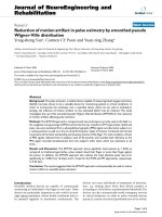
512 samples of the PPG signal were recorded at 20 Hz
from a volunteer. The signal was contaminated with in-
band noise but with the cardiac frequency recognizable.
The artifact noise was extracted by a filtering technique in
the frequency domain. The spectrogram of the PPG signal
was calculated using an FFT-based algorithm. Coefficients
of the spectrum within 0.5 Hz of the primary cardiac fre-
quency or its harmonics were set to zeros. The inverse FFT
of the modified spectrum allowed us to obtain a pure arti-
fact noise. Typical spectra of the contaminated signal
(solid) and the resulting pure artifact noise (starred) are
shown in Figure 1.
A set of synthesized signals with different SNR values was
obtained by changing the value of the mix parameter
ε
in
equation (6). The undisturbed signal was also estimated
from equation (6) by setting
ε
= 0. SpO2 were estimated
from the set of synthesized signals in an 8-second period
at 1-second interval by using the STFT and SPWVD
approaches. The mean SpO2 error during the complete
25.6 seconds is shown in Figure 2 as a function of SNR.
Figure 2 suggests that the two approaches lead to similar
results for high SNR values (e.g. SNR>-5 dB). However,
the SPWVD method outperforms the STFT-based tech-
nique for low SNR (e.g. SNR<-5 dB). Also, it is observed in
this simulation that the errors are randomly positive or
negative for high SNR values, but is mostly positive for
low SNR values, i.e. the approaches consistently overesti-
mate the SpO2 level. When the SNR value decreases,
energy in the side-bands of the noise artifact that over-
lapped with the cardiac frequency components increases.
And therefore, the resultant SpO2 level approaches a
value that would have been estimated from the pure noise
artifact, which differed by 7.2% from the actual SpO2
level for this specific trial.
Typical spectra of contaminated signal and extracted artifactFigure 1
Typical spectra of contaminated signal and extracted artifact.
Spectrum of the contaminated signal (solid) is obtained by
FFT. Coefficients of the spectrum within 0.5 Hz of the pri-
mary cardiac frequency or its harmonics were set to zeros to
result in the spectrum of pure artifact noise (starred).
SSNS S N
SNR
SS
N
RR RIRIR IR
RIR
R
**
,
var( ) var( )
var( ) var(
=+ = +
=
+
+
εε
ε
1
NN
IR
)
,6
()
The mean SpO2 error obtained by the STFT and SPWVD approaches at different levels of SNRFigure 2
The mean SpO2 error obtained by the STFT and SPWVD
approaches at different levels of SNR. The SPWVD approach
outperforms the STFT-based technique for low SNR.
Journal of NeuroEngineering and Rehabilitation 2005, 2:3 />Page 5 of 9
(page number not for citation purposes)
It should be noted that the pulse rate was predetermined
in the simulation, which helped both approaches to deter-
mine the cardiac frequency band more accurately. In prac-
tical situations, the electrocardiogram can be recorded
simultaneously and used as a reliable pulse rate estimator.
As for the computational cost, the SPWVD approach can
be implemented efficiently by making use of its symmetry
properties, and thus, it can reduce the computational cost
to a quarter of that of the STFT technique [20].
Experiment and Results
Experimental protocol
The purpose of this experiment is to compare the perform-
ance of three different methods (WMA, FFT, and SPWVD)
in estimating SpO2 on subjects when they are (a) in a rest-
ing position and (b) in motion.
Six healthy subjects participated in the study. Four kinds
of motions have been investigated: horizontal movement
(M1) and vertical movement (M2) of the hand, as well as
the bending motion (M3) and pressing motion (M4) of
the finger. These motions were selected because they are
some of the common movements attributable to the
motion artifact in pulse oximetry [7,24]. Subjects were
asked to perform all four movements, 4 times each, and
each time for a duration of 30 seconds. When performing
each movement, subjects were asked to move their right
hand, or the index finger of their right hand, for a magni-
tude of 2–5 cm at a frequency of 0.5–4 Hz, while keeping
their left hand stationary. Signals were recorded simulta-
neously from the index fingers of both hands. Throughout
the analysis, SpO2 or pulse rate estimated from the left
hand, which was stationary at all times, was used as the
reference. The reference estimates were obtained by the
WMA method.
The collected signals were separated into an AC and a DC
component. The AC component was filtered out by a 4th
order Butterworth band-pass filter with cut-off frequen-
cies at 0.5 Hz and 20 Hz. The ratio of the DC components
was computed directly in the time domain and the same
value was used for the three different approaches, i.e. the
WMA, FFT and SPWVD approach. On the other hand, a
different ratio of the AC components was computed using
each of the three approaches.
To evaluate the performance of the different approaches,
the SpO2 bias and precision, the pulse rate error, the
dropout rate and the SpO2 performance index (PI) were
calculated. The bias and precision are defined as the mean
and standard deviation of the difference between refer-
ence and estimated SpO2 respectively. The pulse rate error
is the difference between reference and estimated pulse
rate. The dropout rate and SpO2 PI are evaluation param-
eters adopted from previous work by S.J. Barker [24]. The
dropout rate is the percentage of time during which the
technique fails to give a SpO2 reading, and SpO2 PI is the
percentage of time during which the SpO2 level was
within 7% of the reference reading.
Results
Table 1 shows the composite values from all the experi-
ments when subjects were in a resting position and in
motion. As indicated in Table 1, all three approaches can
achieve 100% SpO2 PI, 0.0% dropout rate and less than 3
bpm mean absolute pulse rate error in this experiment
with a limited dataset.
However, the SPWVD approach shows significant
improvement in both SpO2 and pulse rate estimation as
compared to the WMA and FFT approaches when subjects
were in motion. SpO2 estimated from the SPWVD, WMA
and FFT approaches differed from the reference by -1.07 ±
2.42%, -1.31 ± 3.58% and -1.42 ± 3.18%, respectively.
The mean absolute pulse rate error is reduced significantly
(p < 0.05) from 16.4 bpm and 11.2 bpm for the WMA and
FFT approaches, respectively, to 5.62 bpm for the SPWVD
approach. The SpO2 PI also has the highest SpO2 PI
Table 1: Performance statistics of the different approaches. The bias, precision and performance index (PI) of SpO2, as well as the
mean absolute pulse rate error and dropout rate, are used to evaluate the performance of the WMA, FFT and SPWVD approaches
when subjects are in a resting position and in motion.
State Approach SpO2 bias (%) SpO2 precision (%) SpO2 PI (%) Mean absolute pulse
rate error (bpm)
Dropout rate (%)
Resting WMA 0.19 0.34 100 1.25 0.0
FFT 0.24 0.53 100 2.51 0.0
SPWVD 0.21 0.41 100 1.35 0.0
Motion WMA -1.31 3.58 81 16.4 4.6
FFT -1.42 3.18 83 11.2 0.0
SPWVD -1.07 2.42 91 5.62 0.0
Journal of NeuroEngineering and Rehabilitation 2005, 2:3 />Page 6 of 9
(page number not for citation purposes)
(91%). Both the SPWVD and FFT approaches achieve
0.0% dropout rate. The WMA approach sometimes failed
to give a reading during bending or pressing motions
(dropout rate = 4.6%), which would lead to instrument
"dropout" or "freeze" in clinical situations.
Figure 3 shows the distribution of SpO2 bias and pulse
rate error of the three approaches. As shown in Figure
3(a), the SpO2 errors obtained by the SPWVD approach
have a higher incidence (72%) in the main error band (-
3%, 3%), which is the range of bias commonly accepted
by most pulse oximeter manufacturers, as compared to
that obtained by the WMA (55%) and FFT (56%)
approaches.
For the estimation of pulse rate, 90% of the pulse rate
error falls in the error band (-10 bpm, 10 bpm) when the
SPWVD approach is used (see Figure 3(b)). When com-
pared to the WMA and FFT approaches, where only 36%
and 40% of the error fall in this error band respectively,
the SPWVD significantly outperforms the other two
approaches.
Figure 4 shows the SpO2 output bias and precision under
conditions with different kinds of motions: horizontal
and vertical movements of the hand, as well as bending
and pressing motions of the finger. It can be seen that the
estimation of SpO2 by the SPWVD approach improved
significantly (p < 0.05) as compared to the WMA and FFT
approaches when subjects bend their finger or press their
finger against the sensor. The three approaches show no
significant differences (p > 0.05) when subjects move
their hand horizontally or vertically.
Figure 5 gives the error distribution of SpO2, obtained by
the SPWVD approach, when subjects were in different
types of motions. It is found that the bending (M3) and
pressing motions (M4) of the finger have a relatively
broader error distribution than the horizontal and vertical
movements of the hand (M1 and M2). It can also be seen
that the error distribution of M2 is slightly more concen-
trated than that of the M1.
Discussion
Spectral analysis is useful for separating motion artifact
and cardiac physiologic spectra [14-16]. However, these
techniques will not be applicable to spectra that contain
frequency bands close to each other. Moreover, since both
the motion and cardiac frequency are nonstationary in
nature, simply using techniques in the frequency domain
would not be able to separate them when one of the spec-
tra varies within the fixed time window (i.e. an 8-second
period in this study). Therefore, a time-frequency
representation of the corrupted signal would be useful.
The SPWVD approach is proposed for the reduction of
motion artifacts because it can suppress cross-term inter-
ference while maintaining a good time-frequency concen-
tration [19]. In addition, the approach utilizes the fact
The distributions of (a) SpO2 bias and (b) pulse rate error obtained by the WMA, FFT and SPWVD approachesFigure 3
The distributions of (a) SpO2 bias and (b) pulse rate error obtained by the WMA, FFT and SPWVD approaches
Journal of NeuroEngineering and Rehabilitation 2005, 2:3 />Page 7 of 9
(page number not for citation purposes)
that SPWVD is an energy distribution and directly calcu-
lates the magnitude of the AC component from the spec-
trum. The approach solves the problem of WT, where a
unique value for the cardiac frequency may not always be
available [14-16]. Moreover, the approach does not
require a large amount of samples for training, as the
back-propagation neural network approach proposed in
[17].
Standard parameters used to evaluate the performance of
the techniques in pulse oximetry have been adopted in
this study. S.J. Barker [24] evaluated 20 commercial pulse
oximeters on 70 subjects, where data were recorded on
each subject for 6 minutes during normal situation and 3
minutes during a hypoxemic episode. A motorized
motion table was used to induce rubbing or tapping
motions of the finger, with amplitude of ± 2 cm and fre-
quency either fixed at 3 Hz or randomly varied between
1–4 Hz. As compared to the performance of some of the
commercial products evaluated in [24], which have SpO2
bias in the range of 0.4–12%, SpO2 precision in the range
of 2–6%, and SpO2 PI in the range of 27–94%, the pro-
posed SPWVD approach reports comparable
performance.
The four motions investigated in this study are some of
the common movements associated with motion artifacts
affecting pulse oximetry [24]. By studying the effect of
each component on the estimated SpO2, one would have
a clearer picture of what kind of motion induces the larg-
est error on SpO2 estimation. In future studies, it would
be interesting to develop a model that specifically deals
with one type of motion. As suggested by Figure 4 and Fig-
ure 5, bending the finger (M3) or pressing the finger
against the sensor (M4) induces a larger error on SpO2
estimation than horizontal or vertical movements of the
hand (M1 or M2). In fact, this is consistent with the clini-
cal findings discussed in [7], which suggested that bend-
ing and/or pressing the finger may cause the irregular
compression of the vascular bed between the emitter and
detector of pulse oximeter sensor, and thus inducing
higher errors in the estimated SpO2. A potential solution
would be to place multiple sensors around or along the
finger so that the ratio of the light intensity received or a
pressure reading could be an indication of the degree of
bending, pressure exerted or even the level of distortion
made on the peripheral blood vascular bed.
Compared with the WMA and FFT approaches, the
SPWVD approach showed a significant improvement (p <
0.05) in pulse rate estimation when subjects were in
motion. Although such a significant improvement is not
found in the estimation of SpO2, this is attributed to the
fact that erroneous SpO2 estimates above the 100% upper
bound were always rejected. It is hypothesized that when
patients with SpO2 much lower than 100% are recruited
as subjects for evaluating the different approaches, the
performance of each approach will be more notably dif-
SpO2 (a) bias and (b) precision when subjects performed different types of motions: horizontal movement and vertical move-ment of the hand, as well as the bending motion and pressing motion of the fingerFigure 4
SpO2 (a) bias and (b) precision when subjects performed different types of motions: horizontal movement and vertical move-
ment of the hand, as well as the bending motion and pressing motion of the finger.
Journal of NeuroEngineering and Rehabilitation 2005, 2:3 />Page 8 of 9
(page number not for citation purposes)
ferent from each other. However, this hypothesis remains
to be proven in a clinical study involving a significantly
large patient population.
Conclusion
Estimation of SpO2 by a time-frequency representation,
the SPWVD, has been investigated in this study. The
approach has been tested on four kinds of motions that
are found in common movements associated with motion
artifacts in pulse oximetry [7,24], i.e. the horizontal move-
ment and vertical movement of the hand, as well as the
bending motion and pressing motion of the finger. When
compared with the WMA and FFT techniques, the SPWVD
approach shows significant improvement (p < 0.05) when
subjects bend their finger or press their finger against the
sensor. When subjects were in motion, SpO2 levels esti-
mated from the SPWVD, WMA and FFT approaches dif-
fered from the reference by -1.07 ± 2.42%, -1.31 ± 3.58%
and -1.42 ± 3.18% respectively. The SPWVD approach
achieves 0.0% dropout rate and 91% SpO2 PI when sub-
jects were in motion. For the estimation of pulse rate, the
SPWVD approach results in a mean absolute pulse rate
error of 5.62 bpm, as compared to 16.4 bpm and 11.2
bpm by the WMA and FFT approaches respectively. The
results of the study suggested that the SPWVD approach
could potentially be used to improve the performance of
wearable pulse oximeters by reducing the influence of
The error distributions of SpO2, obtained by the SPWVD approach, when subjects performed different types of motions: hor-izontal movement and vertical movement of the hand, as well as the bending motion and pressing motion of the fingerFigure 5
The error distributions of SpO2, obtained by the SPWVD approach, when subjects performed different types of motions: hor-
izontal movement and vertical movement of the hand, as well as the bending motion and pressing motion of the finger.
Publish with BioMed Central and every
scientist can read your work free of charge
"BioMed Central will be the most significant development for
disseminating the results of biomedical research in our lifetime."
Sir Paul Nurse, Cancer Research UK
Your research papers will be:
available free of charge to the entire biomedical community
peer reviewed and published immediately upon acceptance
cited in PubMed and archived on PubMed Central
yours — you keep the copyright
Submit your manuscript here:
/>BioMedcentral
Journal of NeuroEngineering and Rehabilitation 2005, 2:3 />Page 9 of 9
(page number not for citation purposes)
motion artifacts, in particular when subjects bend their
finger or press it against the sensor.
Competing interests
The author(s) declare that they have no competing
interests.
Authors' contributions
YSY designed and carried out the experiment, analyzed
and interpreted the data, and drafted the manuscript.
CCYP helped to analyze and interpret the data, and
assisted in drafting the manuscript. YTZ conceived of the
study, and participated in its design and coordination and
helped to finalize the manuscript. All authors read and
approved the final manuscript.
Acknowledgements
We would like to acknowledge the support of Hong Kong Innovation and
Technology Fund. We are also grateful to Standard Telecommunications
Ltd., IDT Technology Ltd. and Jetfly Technology Ltd. for their support of
the ITF project.
References
1. Visram AR, Jones RDM, Irwin MG, Bacon-Shone J: Use of two pulse
oximeters to investigate a method of movement artifact
rejection using photo-plethysmographic signals. Brit J Anaesth
1994, 72:288-392.
2. Runciman WB, Webb RK, Barker L, Curriie M: The pulse oxime-
ter: application and limitations: an analysis of 2000 incident
reports. Anaesth Intens Care 1993, 21:543-550.
3. Lawless ST: Crying wolf: false alarms in a pediatric intensive
care unit. Crit Care Med 1994, 22:981-985.
4. Sokwoo Rhee, BH Yang, HH Asada: Artifact-resistant power-effi-
cient design of finger-ring plethysmographic sensors. IEEE
Trans Biomed Eng 2001, 48(7):795-805.
5. Severinghaus JW, Honda Y: History of blood gas analysis. VII.
Pulse oximetry. J Clin Monit 1987, 3(2):135-138.
6. Severinghaus JW, Kelleher JF: Recent developments in pulse
oximetry. Anesthesiology 1992, 76(6):1018-1038.
7. Tobin RM, Pologe JA, Batchelder PRB: A characterization of
motion affecting pulse oximetry in 350 patients. Anesthesia and
Analgesia 2002:S54-61.
8. Hayes MJ, Smith PR: A new method for pulse oximetry process-
ing inherent insensitivity to artifact. IEEE Trans Biomed Eng 2001,
48(4):452-461.
9. Diab MK: Signal processing apparatus. International Patent Applica-
tion WO 96/12435 1996.
10. Matthews GR: Pulse responsive device. International Patent Applica-
tion WO 91/18550 1991.
11. Parker D: Optical monitor (oximeter, etc) with motion arti-
fact suppression. International Patent Application WO 94/03102
1994.
12. Hayes MJ, Smith PR: Artifact reduction in
photoplethysmography. Appl Opt 1998, 37(31):7427-7446.
13. Swedlow DB: Oximeter with motion detection for alarm
modification. International Patent Application WO 94/22360 1994.
14. Scharf JE, Athan S, Cain D: Pulse oximetry through spectral
analysis. Proceedings of the Twelfth Southern Biomedical Engineering
Conference 1993.
15. Rusch TL, Scharf JE, Sankar R: Alternate pulse oximetry algo-
rithms for SpO2 computation. Proceedings of the Sixteenth Annual
International Conference of the IEEE Engineering in Medicin and Biology
Society 1994.
16. Rusch TL, Sankar R, Scarf JE: Signal processing methods for pulse
oximetry. Comput Biol Med 1996, 26(2):143-159.
17. Dowla FU, Skokowski PG, Leach RR: Neural Networks and
Wavelet Analysis in the Computer Interpretation of Pulse
Oximetry Data. In Proceedings of IEEE Workshop on Neural Networks
and Signal Processing Kyoto, Japan; 1996.
18. Webster JG: Design of pulse oximeters Philadelphia: Institute of Physics
Pub; 1997.
19. Ricanmato AL, Absher RG, Moffroid MT, Tranowski JP: A time fre-
quency approach to evaluate electromyographic recordings.
proceedings of the fifth Annul IEEE symposium on computer based medical
systems 1992:520-527.
20. Velez EF, Absher RG: Smoothed Wigner-Ville parametric
modeling for the analysis of nonstationary signals. Proceedings
of 1989 International Symposium on Circuits and Systems 1989.
21. Hlawatsch F, Boudreaux-Bartels GF: Linear and quadratic time-
frequency signal representations. IEEE Signal Processing Magzine
1992, 4:21-67.
22. Coetzee FM, Elghazzawi Z: Noise-resistant pulse oximetry using
a synthetic reference signal. IEEE Trans Biomed Eng 2000,
47(8):1018-1026.
23. Kastle SW, Konecny E: Determine the artifact sensitivity of
recent pulse oximeters during laboratory benchmarking.
Journal of Clinical monitoring and computing 2000, 16:509-522.
24. Barker SJ: Motion resistant pulse oximetry: a comparison of
new and old models. Anesthesia and Analgesia 2002, 95:967-72.