báo cáo hóa học: " Efficacy of motor imagery in post-stroke rehabilitation: a systematic review" ppt
Bạn đang xem bản rút gọn của tài liệu. Xem và tải ngay bản đầy đủ của tài liệu tại đây (290.27 KB, 10 trang )
BioMed Central
Page 1 of 10
(page number not for citation purposes)
Journal of NeuroEngineering and
Rehabilitation
Open Access
Review
Efficacy of motor imagery in post-stroke rehabilitation: a systematic
review
Andrea Zimmermann-Schlatter*
1,2
, Corina Schuster
2,3
, MiloAPuhan
4
,
Ewa Siekierka
5
and Johann Steurer
4
Address:
1
Zurich University of Applied Sciences, Winterthur, Switzerland,
2
Reha Rheinfelden, Salinenstrasse 98, 4310 Rheinfelden, Switzerland,
3
Oxford Brookes University, Oxford, United Kingdom ,
4
Horten Centre for patient-oriented research and knowledge transfer, University of Zurich,
Switzerland and
5
Department of Neurology, University Hospital Zurich, Switzerland
Email: Andrea Zimmermann-Schlatter* - ; Corina Schuster - ;
Milo A Puhan - ; Ewa Siekierka - ; Johann Steurer -
* Corresponding author
Abstract
Background: Evaluation of how Motor Imagery and conventional therapy (physiotherapy or
occupational therapy) compare to conventional therapy only in their effects on clinically relevant
outcomes during rehabilitation of persons with stroke.
Design: Systematic review of the literature
Methods: We conducted an electronic database search in seven databases in August 2005 and also
hand-searched the bibliographies of studies that we selected for the review.
Two reviewers independently screened and selected all randomized controlled trials that compare
the effects of conventional therapy plus Motor Imagery to those of only conventional therapy on
stroke patients.
The outcome measurements were: Fugl-Meyer Stroke Assessment upper extremity score (66
points) and Action Research Arm Test upper extremity score (57 points).
Due to the high variability in the outcomes, we could not pool the data statistically.
Results: We identified four randomized controlled trials from Asia and North America. The
quality of the included studies was poor to moderate. Two different Motor imagery techniques
were used (three studies used audiotapes and one study had occupational therapists apply the
intervention). Two studies found significant effects of Motor Imagery in the Fugl-Meyer Stroke
Assessment: Differences between groups amounted to 11.0 (1.0 to 21.0) and 3.2 (-4 to 10.3)
respectively and in the Action Research Arm Test 6.1 (-6.2 to 18.4) and 15.8 (0.5 to 31.0)
respectively. One study did not find a significant effect in the Fugl-Meyer Stroke Assessment and
Color trail Test (p = 0.28) but in the task-related outcomes (p > 0.001).
Conclusion: Current evidence suggests that Motor imagery provides additional benefits to
conventional physiotherapy or occupational therapy. However, larger and methodologically
sounder studies should be conducted to assess the benefits of Motor imagery.
Published: 14 March 2008
Journal of NeuroEngineering and Rehabilitation 2008, 5:8 doi:10.1186/1743-0003-5-8
Received: 6 August 2007
Accepted: 14 March 2008
This article is available from: />© 2008 Zimmermann-Schlatter et al; licensee BioMed Central Ltd.
This is an Open Access article distributed under the terms of the Creative Commons Attribution License ( />),
which permits unrestricted use, distribution, and reproduction in any medium, provided the original work is properly cited.
Journal of NeuroEngineering and Rehabilitation 2008, 5:8 />Page 2 of 10
(page number not for citation purposes)
Background
Annually 15 million people worldwide suffer from a
stroke. Of these, five million remain permanently disa-
bled, despite intensive rehabilitation programs, and are
no longer capable to care for themselves [1].
During the first few days following the incident, lifesaving
and thrombolytic therapies have priority. However, as
soon as possible [2,3], patients should exercise to activate
the process of recovery and neural re-organization [4-6].
Different rehabilitative approaches are used for post-
stroke treatment. One of them is Motor Imagery (MI). MI
was initially developed to improve the performance of
athletes [7-9] and has been adopted in rehabilitation pro-
grams for persons with stroke [10] to support motor
recovery [11,12].
Mental imagery refers to the active process by which
humans experience sensations with or without external
stimuli [13]. It is an active process during which a specific
action is reproduced within working memory without any
real movements [13,14]. Studies [15,16] demonstrate that
during MI sessions partially the same brain areas are as
activated as during functional tasks.
Function, behavior, and performance are rehearsed men-
tally as if the person is actually performing them [17].
From sports literature it is well known that MI, when
applied in addition to functional training, is more effec-
tive than MI or functional training alone [18]. However,
Sharma [12] has pointed out that MI training alone pro-
duces less improvement than functional training.
An advantage of MI is that patients can practice it inde-
pendently during the regeneration phase between two
physical therapy sessions. MI can also be practiced in all
stages of stroke recovery [13]. In an early stage of recovery,
MI allows patients to mentally practice a task which they
cannot yet carry out physically due to motor impairment.
However, it has not been determined yet, when it is best
to start with MI.
Although there is sufficient evidence that MI can improve
function in healthy subjects [13], only a few, small rand-
omized controlled trials have evaluated the effect of MI in
stroke patients. To explore the potential role of MI in post-
stroke rehabilitation and to outline a potential research
agenda, we conducted a systematic review of all rand-
omized controlled trials that analyze the effect of MI on
patients after a cortical stroke.
Methods
Identifications of studies
We searched the following databases for relevant studies:
Ovid MEDLINE (Ovid version, from inception to August
2005), PEDRO (online version, University of Sydney,
Australia, August 2005), PsycINFO (from 1967 to July
2005), Psyndexplus (from 1977 to June 2005), CINAHL
(Cumulative Index to Nursing & Allied Health Literature,
from 1982 to July 2005), Cochrane Central Register of
Controlled Trials (Oxford, UK, 2004, issue 1), and Scopus
(from inception to August 2005).
The detailed search strategy for the MEDLINE search is
described in the appendix.
The search was conducted without restrictions to language
or year of publication.
We also hand-searched the bibliography of all studies
ordered in full text.
Selection criteria
We included all randomized controlled trials that com-
pare conventional physiotherapy or occupational therapy
to MI combined with conventional physiotherapy or
occupational therapy in post-stroke rehabilitation. We
excluded mental practice based on computer-animated
techniques, because these techniques are not available in
most rehabilitation settings. Only studies about patients
with a first episode of stroke were considered with no
restrictions concerning age or time since onset of stroke.
The outcome assessment had to be clinically and func-
tionally relevant, for example performance of specific
tasks and activities or health-related quality of life.
Study selection
After the electronic database search, the two reviewers (AZ
and CS) screened the titles and the abstracts of all result-
ing references (N = 2116) independently. They recorded
their decision about in- or exclusion in an EndNote
(Thomson Wintertree Software Inc) file. In cases where
reading the abstracts was not enough to determine
whether or not to include a study, the entire study was
ordered. The reviewers then evaluated the retrieved full-
text studies and made a decision on inclusion or exclusion
according to the criteria specified above.
The reviewers also hand-searched the bibliographies of
the full-text studies and reviews to identify further relevant
studies. Each reviewer's decisions as well as the final deci-
sions on journal articles were recorded in the EndNote
file. Studies that did not fulfill all of the predefined criteria
were excluded and their bibliographic details were listed
with the specific reason for their exclusion.
Journal of NeuroEngineering and Rehabilitation 2008, 5:8 />Page 3 of 10
(page number not for citation purposes)
Data extraction
The reviewers independently recorded details about study
design, interventions, outcome measurement methods,
and results in a predefined form. Both also separately eval-
uated the quality of the included trials based on a detailed
list of quality items (see table 1). A third reviewer (JS)
resolved any discrepancies when the two reviewers disa-
greed. We tried to contact the authors of the selected stud-
ies for further information about missing data but did not
get any response.
Quality assessment
The two reviewers appraised all included trials based on a
pre-defined list of selected quality items assessing compo-
nents of internal validity [19] (Table 1). In case of any dis-
crepancy, we obtained the opinion of a third reviewer. We
divided all quality items into the following four catego-
ries: 1 = item is properly addressed; 2 = item is partially
addressed (authors mentioned that this quality item was
fulfilled but did not describe the procedure); 3 = item is
not properly addressed or not stated (the item was not ful-
filled or the authors did not mention it); 4 = item is not
applicable.
Analysis
We summarized the results of the data extraction and the
quality assessments in structured tables. This compilation
allowed us to examine the variation in patient characteris-
tics, study quality and results.
Because of the heterogeneity in the studies we could not
perform a data pooling for a meta-analysis. Wherever pos-
sible, we presented point estimates and 95% confidence
intervals of single study results. Since not all results were
presented with a confidence interval of 95%, we used the
standard deviation from one study [20] to estimate the
confidence interval of the other studies [21].
Results
Study selection
Figure 1 shows the study selection process and the review-
ers' agreement on study inclusion. Our search yielded
2116 potentially relevant citations after removing dupli-
cates. 113 articles were selected for closer evaluation. Of
these, we included four RCTs [22-25]. Reasons for the
exclusion of the other 109 studies were: no RCTs (n = 58),
study population differed from the pre-defined study pop-
ulation (n = 26), MI was not used as an intervention (n =
25). The two reviewers agreed in 96% of the cases on
inclusion or exclusion of the studies.
Characteristics of the included studies
(Table 2 provides descriptive data for the included stud-
ies)
Time elapsed since stroke ranged from a few days (mean:
12.3 days) to several years (mean: 23.8 months). Three
studies [23-25] were carried out in North-America and
one in Asia [22]. The study populations were quite homo-
geneous in terms of age but heterogeneous in aspects such
as gender, dominant limb, affected side, and time elapsed
since the incident (table 2).
Only one study [24] assessed the individual's ability to
imagine using the Movement Imagery Questionnaire
(MIQ) [26].
Duration and frequency of MI interventions varied
between ten minutes [24] and one hour a day [22] with
three to five sessions per week. The shortest intervention
period lasted three weeks [22], the longest six weeks
[23,24].
All studies compared MI plus conventional physiotherapy
or occupational therapy to only conventional physiother-
apy or occupational therapy. None of the included studies
analyzed the effect of MI alone.
Table 1: Quality assessment of the included studies
Liu [22] Page [24] Page [25] Page [23]
Selection of prognostic homogenous study population (disease progression) 1 3 3 3
Concealment of random allocation 1 3 3 3
Prestratification of prognostically relevant variables 3 3 3 3
Random allocation (description of procedure) 2 1 1 3
Registration of loss to follow-up 1 3 4 4
Blinding of patients 4444
Blinding of persons who implement interventions 4 4 4 4
Registration of co-interventions that bear on outcome for each group 3 3 1 3
Blinding of persons who assess treatment effects 3 1 1 3
Check to what extent blinding was successful 3 3 3 3
1 = Item is properly addressed, 2 = Item is partially addressed, 3 = Item is not properly addressed or not stated, 4 = Item is not applicable
Journal of NeuroEngineering and Rehabilitation 2008, 5:8 />Page 4 of 10
(page number not for citation purposes)
Flow diagram of study selection processFigure 1
Flow diagram of study selection process.
Potentially relevant studies identified from electronic databases
(Medline, PsycINFO, Psyndex, CINAHL, Cochrane, Scopus, PEDRO)
N=2116 references
Studies excluded after
title and abstract
screening: n=2042
Studies retrieved for detailed evaluation:
x From electronic databases: n=74
x From hand searching (conference proceedings, reference lists of
reviews and full text studies): n=39
Total: n=113
Studies excluded after full text assessment
Reasons for exclusion:
x No RCTs n=58
x The study population differs from the defined
study population n=26
x MI is not used as an intervention n=25
Inclusion n=4
Total exclusion: n=113
Initial agreement on inclusion and exclusion: 96%
All studies included in the review: n=4
From electronic databases: n=3
From hand searching: n=1
Journal of NeuroEngineering and Rehabilitation 2008, 5:8 />Page 5 of 10
(page number not for citation purposes)
Table 2: Characteristics of included randomized controlled trials
Study Number of patients Gender (% male) Mean age in years (±
SD if available) range
(if available)
Time since stroke
(months)
Intervention Outcomes
Liu [22] 46 with a first unilateral
cerebral infarction
48 MI group: 71.0 (± 6.0)
Controls: 72.7 (± 9.4)
0.5 Intervention group:
60 minutes PT sessions five days a week for 3 weeks. Plus: motor
imagery five 60 minutes sessions per week for 3 weeks. OT's
provided the motor imagery training.
FMSA
CTT2
Task performance test
Controls:
60 minutes PT sessions five days a week for 3 weeks. Plus instead
of imagery: a demonstration-then-practice method for the same
tasks as in the MI group for five 60 minutes sessions per week for
3 weeks. OT's provided the demonstration than practice training.
Page [24] 13 with a unilateral
cerebral infarction
77 64.6 range:54–79 6.5 Intervention group:conventional therapy (OT and PT) 3 times/
week, in 60 minutes segments for 6 weeks. Plus: 10 minutes
audiotape with cognitive visual images + using such a tape at
home twice a week.
FMSA
ARAT
Controls:
Conventional therapy (OT and PT) 3 times/week, in 60 minutes
segments for 6 weeks.
Plus instead of imagery: 10-minutes tape containing stroke
information + using such a tape at home twice a week.
Study Number of patients Gender (% male) Mean age in years (±
SD if available)
Time since stroke
(months)
Intervention Outcomes
Page [23] 11 with a stroke 82 62.3 (± 5.1) range: 53–71 24 Intervention group:
A set of ADL's is practiced through PT 2 times/week for 30
minutes for 6 weeks. Plus after PT participants received 30
minutes MP intervention. And they also practice it mentally at
home.
ARAT
MAL
Controls:
A set of ADL's is practiced through PT 2 times/week for 30
minutes for 6 weeks. Plus instead of MP: after the PT session they
received a session of relaxation techniques for 30 minutes.
Page [25] 16 with a unilateral
cerebral infarction
100 63.2 (± 4) 22 Intervention group: OT: 3 times/week in 30 minutes sessions
for 4 weeks.
Plus: an imagery intervention lasting 20 minutes after the OT
session.
FMSA
Controls: OT: 3 times/week in 30 minutes sessions for 4 weeks.
Plus instead of MP: after OT session a 20 minutes tape with
instructions and information requiring the patients' attention and
participation and on the causes and the pathology of strokes.
Journal of NeuroEngineering and Rehabilitation 2008, 5:8 />Page 6 of 10
(page number not for citation purposes)
One study [22] trained the patients to carry out specific
tasks using the technique of MI. In the first week, the pri-
mary objective was task analysis enhancement: Patients
had to identify each step of the task with the help of MI
and picture cards showing the task. In the second week,
the primary objective was problem identification: patients
had to visualize their own performance and identify the
problems encountered and the solutions in each task step
by means of MI. The third week focused on task perform-
ance: Patients had to imagine performing the task and
then carry it out. In this study occupational therapists
applied MI. As control intervention Liu et al. used a so-
called "demonstration than practice method" (an occupa-
tional therapist demonstrated the same tasks as used in
the MI group, afterwards patients had to practice this dem-
onstrated tasks.
In the studies published by Page [23-25], patients had to
listen to an audiotape with an introduction on relaxation,
some suggestions for external, cognitive visual images,
and instructions to refocus into the room. Duration of the
tapes varied from ten [24] to 30 minutes [23].
All studies published by Page used conventional therapy
plus information about strokes [24,25] or relaxation tech-
niques [23] whereas Liu et al. [22] used a session with
demonstration than practice method of the trained task as
a control intervention.
None of the included studies evaluated the patients' satis-
faction with the intervention.
Quality assessment
The quality of the studies was moderate to poor. Table 1
summarizes the methodological quality of all the
included studies.
None of the included studies used pre-stratification.
Two items (blinding of persons who implemented the
interventions and blinding of patients) were not applica-
ble in any studies. We applied the quality assessment in a
restrictive manner and considered audiotapes with infor-
mation about strokes and relaxation techniques as not
being a "blinding procedure".
The registration of any co-intervention(s) was properly
addressed in one study [23], yet three studies did not
address this issue [22-25].
Blinding of the assessors was properly addressed in two
studies [23,24] but not at all addressed in the others
[22,25].
Effects of motor imagery
In three studies the outcomes were measured with Fugl-
Meyer Stroke Assessment (FMSA) and in two studies with
the Action Research Arm Test (ARAT). The ARAT is an
assessment for measuring specific changes in function of
the upper extremity (grasp, grip, pinch and gross move-
ments) for persons with hemiplegia. The test has a total
score of 57 points [27]. In addition the FMSA upper
extremity subscale is an assessment for movement,
reflexes, coordination and speed with a total score of 66
points. For the ARAT the minimal clinically important dif-
ference is estimated to be 5.7 [28] whereas for the FMA the
minimal clinically important difference is not estimated
yet, but Gladstone proposes a 10% change of total score
to be relevant.
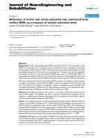
Table 3 summarizes the results of the included studies and
figure 2 shows the forest plots of the ARAT and the FMSA.
We draw a dotted line to point out the minimal clinically
important difference of ARAT and FMSA upper extremity
score.
Liu [22] found no significant difference between the
FMSA upper extremity subscale and the Color Trail Test
(CTT), but did find a significantly higher level of perform-
ance in the trained as well as untrained tasks for the
imagery group. The trained tasks in week three were also
evaluated in a one-month follow-up and the difference
between the two groups was considered significant for the
intervention group. Since Liu et al. [22] only reported that
the FMSA was not significant. We were not able to display
their results in figure 2. Page [24] reported substantial
increases in the FMSA upper extremity subscales and the
ARAT scores for the intervention group. The difference
between the two groups exceeds the clinically important
difference [28-30].
Page [23] detected a significant change in the ARAT score
for the intervention group and remarkable changes con-
cerning the Amount of Use (AOU) and the Quality of
Movement (QOM) of the Motor Activity Log (MAL) [31].
Page [25] found a 35.98% (± 10.17%) improvement in
the FMSA upper extremity subscale for the intervention
group compared to 21.15% (± 4.87%) for the control
group, but no significance levels were reported in this
study.
Discussion
Our systematic review indicates that there is modest evi-
dence supporting the additional benefit of MI compared
to only conventional physiotherapy in patients with
stroke. Three studies [23-25] proved the positive effects of
MI interventions on the ARAT and the FMSA and one
study [22] stated significant effects on task-related out-
Journal of NeuroEngineering and Rehabilitation 2008, 5:8 />Page 7 of 10
(page number not for citation purposes)
comes, but not on the ARAT and the FMSA. Two studies
[23,24] found higher mean change scores than the mini-
mal clinically relevant difference in the ARAT and in the
FMSA respectively.
The methodological quality of included randomized con-
trolled trials with small sample size (n = 11 – 46) limits
the findings of this review. The results of this review are
only valuable for short-term effects of MI on functional
outcomes. The presentation of data in the analyzed stud-
ies (for example: p-values of differences between the
groups) complicated the data extraction and further anal-
ysis. This review cannot answer questions concerning the
best time for an MI intervention because of the variability
of time elapsed since the stroke event in the different
patient samples. For the same reason, this review can also
not respond to questions concerning the optimal dura-
tion or frequency of the intervention or the fatigue appear-
ance in stroke patients. Since none of the included studies
assessed how patients coped with the treatment, this
review cannot draw any conclusions about the effect of
the patients' motivation on the efficacy of MI. The authors
of this review are not aware of any study which assesses
whether a patient's ability to take part in the decision-
making process influences the effectiveness of MI.
Although evidence exists that patients should start exercis-
ing as soon as possible [2,3], Byl et al. [32] found evidence
that individuals > 6 months after stroke can achieve high
levels of function following directed practice based on the
principles of neuroplasticity. Since these results are based
on functional exercises, it is unclear if they can be adapted
to MI. From studies with athletes it is well known that it is
an advantage for motor learning if the athlete is familiar
with MI techniques. Isaac et al. [33] noted that subjects
with a specialization such as elite athletes, air traffic con-
trollers or pilots achieve significantly better results in vivid
imagery than matched controls.
In June 2006, a systematic review [34] on the same topic
also included one Controlled Clinical Trial (CCT) [35],
two patient series [36,37], and three single case reports
[38-40]. The results of theses studies support the results
found in the four RCTs. Braun et al. [34] applied different
quality assessment criteria and judged the quality of the
included studies moderate to sufficient. They found
"some evidence that mental practice as an additional ther-
apy has effects on recovery after a stroke" but also stated
that "mental practice and the outcome measurement are
not standardized and thus difficult to compare." They
advise further research based on a clear definition of the
content of mental practice using standardized measure-
ment methods. In contrast to Braun et al. [34] we pre-
sented data of three studies in a quantitative manner with
forest plots of the ARAT and the FMSA to facilitate the
interpretation of the effects of MI.
For further research, the authors recommend studies of
better methodological quality, bigger sample size, and
longer follow-up. Further research is also necessary to
determine the optimum time for the intervention and the
duration of the intervention, and to analyze the influence
of motivation on the efficacy of MI.
MI appears to be an attractive treatment opinion, easy to
learn and to apply and the intervention is neither physi-
Table 3: Effects of MI
Study Assessment Time of measurement Results
Liu [22] FMSA upper extremity subscales,
CTT
Pretest, Posttest after the inter-vention,
follow-up after one month
Not significant
Trained Tasks, set 1 Not significant
Trained tasks, set 2 significant
Trained tasks, set 3 significant
Untrained tasks significant
Trained tasks, set 3, follow up significant
Page [25] FMSA, upper extremity subscales Two pretests within one week, one
posttest after the intervention
% Improvement MI group: 35.98 (10.17)
Controls: 21.15 (4.87)
No significance level is reported in this study.
Page [24] FMSA, upper extremity subscales Two pretest within one week, one
posttest after the intervention
Improvement: MI group: 13.8 Controls: 2.9
No significance level is reported in this study.
ARAT Improvement: MI group: 16.4 Controls: 0.7 No
significance level is reported in this level.
Page [23] ARAT Two pretests within one week, one
posttest after the intervention
significant
Motor Activity Log Amount of
Use (AOU)
Improvement: MI group: 1.6 Controls: 0.4
No significance level is reported in this study.
Motor Activity Log Quality of
Movement (QOM)
MI group: 2.2 Controls: 0.2
No significance level is reported in this study.
Journal of NeuroEngineering and Rehabilitation 2008, 5:8 />Page 8 of 10
(page number not for citation purposes)
cally exhausting nor harmful. Therefore, the authors
believe that MI may generate additional benefit for
patients.
Competing interests
The author(s) declare that they have no competing inter-
ests.
Authors' contributions
AZ participated in the study design, the study selection
process the data extraction, performed the data analysis,
and drafted the manuscript. CS participated in the study
selection process, the data extraction and revised the man-
uscript. MP participated in the study design, the data anal-
ysis and revised the manuscript. ES revised the
manuscript. JS participated in the study design and revised
the manuscript. All authors read and approved the final
manuscript.
Appendix
We used the following search terms for MEDLINE, Psy-
cINFO, Psyndex, Cochrane, CINAHL, Scopus, PEDRO
1 imagery.mp,hw. (953)
2 (imaginat$ or imagine$).mp,hw. (784)
3 (mental adj (practice or preparation or rehearsal or ther-
apy)). mp,hw. (2006)
4 biofeedback$.mp,hw. (1023)
5 motor learning.mp,hw. (294)
6 neuronal plasticity.mp,hw. (254)
7 nerve cell plasticity.mp,hw. (2)
8 stroke?.mp,hw. (9395)
9 hemipare$.mp,hw. (493)
10 hemiple$.mp,hw. (1295)
11 apople$.mp,hw. (20)
12 cerebrovascular disorder$.mp,hw. (597)
13 exp "intracranial embolism and thrombosis"/(95)
14 exp intracranial hemorrhages/(1212)
15 exp carotid artery disease/(427)
Differences between ARAT and FMSA upper extremity change scoresFigure 2
Differences between ARAT and FMSA upper extremity change scores.
Differences (95% CI)
Assessment Study
Difference
between FMA
and ARA change
scores
Favors usual
therapy
Favors Mental
imagery
FMSA
Page [24]
Page [25]
ARAT
Page [24]
Page [23]
-
20
0
-10-15
-5 5 10 15 20
6.1(-6.2 to 18.4)
15.8(0.5 to
31.0)
25
30
11(1 to 21)
3.2(-4 to
10.3)
-25-30
The difference between pre and posttest for FMSA and ARAT between controls and motor imagery group is bigger than the
minimal clinically important difference (dotted line) which is for both assessments about 10% of the total score (ARAT: 5.7 and
FMSA: 6.6).
Change score
Journal of NeuroEngineering and Rehabilitation 2008, 5:8 />Page 9 of 10
(page number not for citation purposes)
16 exp cerebral ischemia/(791)
17 exp cerebral vascular accident/(9144)
18 exp brain ischemia
19 exp basal ganglia cerebrovascular disease
20 exp cerebral hemorrhage
21 exp cerebral ischemia
22 exp cerebrovascular accidents
23 exp paralysis/(3528)
24 exp paresis
25 or/1–7 (5113)
26 or/8–18 (16704)
27 19 and 20 (265)
Acknowledgements
The authors thank Dr Pius Estermann, Information Officer, University Hos-
pital of Zurich, who designed and conducted the electronic database
search, Jan Kool, Martina Spiess and Cornelia Barth for critical remarks and
Katharina Schlatter and Arianne Knüsel for English corrections.
References
1. World OH: The Atlas of Heart Disease and Stroke. .
2. Biernaskie J, Chernenko G, Corbett D: Efficacy of rehabilitative
experience declines with time after focal ischemic brain
injury. J Neurosci 2004, 24:1245-1254.
3. Kwakkel G, Kollen B, Twisk J: Impact of time on improvement
of outcome after stroke. Stroke 2006, 37:2348-2353.
4. Cauraugh JH, Summers JJ: Neural plasticity and bilateral move-
ments: A rehabilitation approach for chronic stroke. Prog
Neurobiol 2005, 75:309-320.
5. Schaechter JD: Motor rehabilitation and brain plasticity after
hemiparetic stroke. Prog Neurobiol 2004, 73:61-72.
6. Komitova M, Johansson BB, Eriksson PS: On neural plasticity, new
neurons and the postischemic milieu: an integrated view on
experimental rehabilitation. Exp Neurol 2006, 199:42-55.
7. Callow N: Types of Imagery Associated with Sport Confi-
dence in Netball Players of Varying Skill Levels. Taylor & Fran-
cis 2001, 13:1-17.
8. Ryan ED, Simons J: Cognitive demand, imagery, and frequency
of mental rehearsal as factors influencing acquisition of
motor skills. 1985, 3:35-45.
9. Williams JM: . In Applied sport psychology: personal growth to peak per-
formance Mayfield publishing company; 1998.
10. Van Leeuwen RIJ: Mental practice and imagery: a potential role
in stroke rehabilitation. Phys Ther Rev 1998, 3:47-52.
11. Jeannerod M, Frak V: Mental imaging of motor activity in
humans. Curr Opin Neurobiol 1999, 9:735-739.
12. Sharma N, Pomeroy VM, Baron JC: Motor imagery: a backdoor
to the motor system after stroke? Stroke 2006, 37:1941-1952.
13. Jackson P: Potential Role of Mental Practice Using Motor
Imagery in Neurologic Rehabilitation. Arch Phys Med Rehabil
2001, 83:1133-1141.
14. Decety J, Grezes J: Neural mechanisms subserving the percep-
tion of human actions. Trends Cogn Sci 1999, 3:172-178.
15. Hanakawa T, Immisch I, Toma K, Dimyan MA, Van Gelderen P, Hallett
M: Functional properties of brain areas associated with
motor execution and imagery. J Neurophysiol 2003, 89:989-1002.
16. Gerardin E, Sirigu A, Lehericy S, Poline JB, Gaymard B, Marsault C,
Agid Y, Le Bihan D: Partially overlapping neural networks for
real and imagined hand movements. Cereb Cortex 2000,
10:1093-1104.
17. Driskell J: Does mental practice enhance Performance? Amer-
ican Journal of Psychological Association 1994, 79:481-492.
18. Feltz D: The Effects of Mental Practice on Motor Skill Learn-
ing and Performance. A Meta-analysis. J Sport Psychol 1983,
5:25.
19. ter-Riet G, Kessels AGH: Commentary on Rampes et all 'Does
electroacupunture reduce craving for alcohol? A rand-
omized controlled study'. Complement Therap Med 1997:116-118.
20. Page S: Mental practice combined with physical practice for
upper-limb motor deficit in subacute stroke. Phys Ther 2001,
81:1455-1462.
21. Higgins JPTGS, editors: Cochrane Handbook for Systematic
Reviews of Interventions 4.2.6 (updated September 2006). In
The Cochrane Library Chichester, UK: John Wiley & Sons, Ltd; 2006.
22. Liu KP, Chan CC, Lee TM, Hui-Chan CW: Mental imagery for
promoting relearning for people after stroke: a randomized
controlled trial. Archives of Physical Medicine and Rehabilitation 2004,
85:1403-1408.
23. Page J: Effects of Mental Practice on Affected Limb Use and
Function in Chronic Stroke. Arch Phys Med Rehabil 2005,
86:399-402.
24. Page J: A randomized efficacy and feasibility study of imagery
in acute stroke. Clinical Rehabilitation 2001, 15:
233-240.
25. Page S: Imagery improves upper extremity motor function in
chronic stroke: a pilot study. Occup Ther J Res 2000, 20:200-215.
26. Hall EGPJ: Movement Imagery Questionnaire. London, ON:
University of Western Ontario; 1983.
27. Lyle R: A performance test for assessment of upper limb func-
tion in physical rehabilitation treatment and research. Int J
Rehabil Res 1981, 4:483-492.
28. Van der Lee JH, de Groot V, Beckermann H, Wagenaar RC,
Lankhorst GJ, Bouter LM: The intra- and interrater reliability of
the action research arm test: a practical test of upper
extremity function in patients with stroke. Arch Phys Med Reha-
bil 2001, 81:14-19.
29. Gladstone DJ, Danells CJ, Black SE: The fugl-meyer assessment of
motor recovery after stroke: a critical review of its measure-
ment properties. Neurorehabil Neural Repair 2002, 16:232-240.
30. Krebs HI, Buerger SP, Newbery MJ, Hogan N, Volpe BT, Ferraro M,
Lynch D, Makiyama A, Sandmann M: Rehabilitation robotics: Pilot
trial of a spatial extension for MIT-Manus. Journal of NeuroEngi-
neering and Rehabilitation 2004, 1:.
31. Van der Lee JH, Beckerman H, Knol DL, de Vet HC, Bouter LM: Clin-
imetrics Properties of the Motor Activity Log for the Assess-
ment of Arm Use in Hemiparetic Patients. Stroke 2004,
35:1410-1414.
32. Byl N, Roderick J, Mohamed O, Hanny M, Kotler J, Smith A, Tang M,
Abrams G: Effectiveness of sensory and motor rehabilitation
of the upper limb following the principles of neuroplasticity:
patients stable poststroke. Neurorehabilitation and Neural Repair
2003, 17:176-191.
33. Isaac AR, Marks DF: Individual differences in mental imagery
experience: developmental changes and specialization. Br J
Psychol 1994, 85(Pt 4):479-500.
34. Braun Susy MBAJ, Borm Paul J, Schack Thomas, Wade Derick T: The
Effects of Mental Practice in Stroke Rehabilitation: A Sys-
tematic Review. Arch Phys Med Rehabil 2006,
87:842-852.
35. Dijkerman HC, Ietswaart M, Johnston M, MacWalter RS: Does
motor imagery training improve hand function in chronic
stroke patients? A pilot study. Clinical Rehabilitation 2004,
18:538-549.
36. Crosbie JH, McDonough SM, Gilmore DH, Wiggam MI: The adjunc-
tive role of mental practice in the rehabilitation of the upper
limb after hemiplegic stroke: a pilot study. Clinical Rehabilitation
2004, 18:60-68.
37. Liu KPY: Mental imagery for relearning of people after brain
injury. Brain Injury 2004, 18:1163-1172.
Publish with BioMed Central and every
scientist can read your work free of charge
"BioMed Central will be the most significant development for
disseminating the results of biomedical research in our lifetime."
Sir Paul Nurse, Cancer Research UK
Your research papers will be:
available free of charge to the entire biomedical community
peer reviewed and published immediately upon acceptance
cited in PubMed and archived on PubMed Central
yours — you keep the copyright
Submit your manuscript here:
/>BioMedcentral
Journal of NeuroEngineering and Rehabilitation 2008, 5:8 />Page 10 of 10
(page number not for citation purposes)
38. Page SJ, Levine P, Sisto SA, Johnston MV: Mental practice com-
bined physical practicefor upper-limb motor deficit in suba-
cute stroke. Phys Ther 2001, 81:1455-1462.
39. Dickstein R, Dunsky A, Marcovitz E: Motor imagery for gait reha-
bilitation in post-stroke hemiparesis. Phys Ther 2004,
84:1167-1177.
40. Jackson PL, Doyon J, Richards CL, Malouin F: The Efficacy of Com-
bined Physical and Mental Practice in the Learning of a Foot-
Sequence Task after Stroke: A Case Report. Neurorehabilitation
& Neural Repair 2004, 18:106-111.