Theo dõi tưới máu não trong và sau hồi sức ngưng tim
Bạn đang xem bản rút gọn của tài liệu. Xem và tải ngay bản đầy đủ của tài liệu tại đây (2.07 MB, 25 trang )
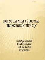
THEO DÕI TƯỚI MÁU NÃO
TRONG VÀ SAU HỒI SỨC NGƯNG TIM
Nguyễn Vinh Anh
TpHCM, tháng 09/2023
Dàn trình bày
• Rối loạn tưới máu não trong – sau ngưng tim
• Các phương pháp khơng xâm lấn
• Các phương pháp xâm lấn
• Kết luận
Cơ chế điều hòa tưới máu não Cerebral autoregulation
Zeiler et al. 2019 British Journal of Anaesthesia
Đơn vị Thần kinh – Mạch máu
Neurovascular unit
Iadecola 2017
Neurovascular
dysfunction
Idacola 2017
Khả năng dung nạp nội sọ Intracranial compliance
Volume-pressure interaction
Transcranial Doppler (TCD)
Lưu lượng máu não (CBF) trong ngưng tim
Lewis, (1994) Journal of Critical Care
Transcranial Doppler Pulsatility Index
Thiết bị quang phổ cận hồng ngoại
(Near Infra Red Spectroscopy: NIRS)
• INVOS 3000 COx probe (Somanetics) to the frontal skull.
Duration of cerebral oximetry recordings.
rSO2, regional cerebral oxygen saturation.
Scatter plots of serum neuron-specific
enolase (NSE) concentration at 48 h
after cardiac arrest vs. median regional
cerebral oxygen saturation (rSO2)
during the first 36 h in intensive care
unit in patients with good (Cerebral
Performance Category [CPC] 1–2) and
poor (CPC 3–5) neurological outcome
The probability for a good outcome (CPC 1–2) and the area under the
receiver operating characteristic curve for the lowest 60-min median rSO2
to predict good outcome overall and in tertiles based on the lowest 60-min
median rSO2 during the first 36 h in ICU
Ryan L. Hoiland 2023
MAP was increased by an
average of 32 - 16 mm Hg
with the infusion of
phenylephrine.
H LATKY ET AL.
N EUROSURGERY
2005
TRÂN TRỌNG CẢM ƠN