Nuclear Power Operation Safety and Environment Part 12 ppt
Bạn đang xem bản rút gọn của tài liệu. Xem và tải ngay bản đầy đủ của tài liệu tại đây (651.56 KB, 30 trang )
Long-Term Effects of Exposure to Low-Levels of Radioactivity:
a Retrospective Study of
239
Pu and
90
Sr from Nuclear Bomb Tests on the Swiss Population
319
because these databases contain autopsy tissues from both the general public and workers
of the nuclear industry. Similar values to Switzerland were determined in Germany (Bunzl
and Kracke, 1983) and in the UK (Popplewell et al., 1985) for the years around 1980. Higher
values were obtained at the Semipalatinsk test site (STS) during the 2000’s, indicating an
effect of the test site fallout in the plutonium body burden of the population (Yamamoto et
al., 2006). Using ICP-MS, (Yamamoto et al., 2008) found a significantly lower
240
Pu/
239
Pu
isotopic ratio of 0.125 in autopsy tissues (bone) of individuals from the STS, confirming the
influence of the STS fallout on plutonium incorporation.
There were too few bone ash samples in our study to separate individuals from different
regions, especially the ones potentially affected by the Swiss NPPs. Accordingly, our data
represent a pool of bone samples from all over Switzerland. Nevertheless, the
240
Pu/
239
Pu
isotopic ratio of 0.18 indicates, beyond any reasonable doubts, that the plutonium inhaled by
the Swiss population comes from the fallout of the NBTs of the sixties.
7. Retention half-times in the skeleton of
90
Sr and plutonium
The retention half-time in the skeleton of bone-seeking radionuclides such as
90
Sr and
plutonium is a key parameter used for their dosimetry in humans. Currently, only a partial
answer is given to the question of how long plutonium will stay in the body. Values found
in the literature are situated between 15 to 100 years, with a proposed value by ICRP 56 or
Kathren (1995) of 50 y. Our long-term study of
90
Sr and plutonium in the vertebrae allowed
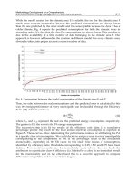
us to determine, with a high statistical significance, the retention half-time of both
radionuclides in cancellous bones. It is of 40±15 y (95% confidence) for plutonium and
13.5±1.5 for
90
Sr (Figure 9). Meanwhile, the retention time of
90
Sr is very close to the retention
time found in milk teeth, milk, grass and soil (0-5 cm, Table 1).
Fig. 9. The use of the data from our long-term study for the determination of the retention
time of
90
Sr and plutonium in cancellous bones.
Nuclear Power – Operation, Safety and Environment
320
Site Soil (0-5 cm) Grass Milk milk teeth Vertebrae
Grangeneuve
12.3±3.6 11.6 ±3.9 14.8 ±2.3
Mühleberg
9.0 ±1.3 7.6 ±1.3 14.5 ±2.6
Gösgen
7.8 ±0.9 6.7 ±1.1 10.1 ±2.7
Leibstadt
8.9 ±1.5 12.3 ±3.9 12.5 ±2.5
Switzerland
9.5±2 9.5±3 13±2
10.0
±
3 13.0 ±1
Table 1. Retention half-time of
90
Sr in different compartments of the environment, food and
human for different locations in Switzerland.
These results demonstrate that the calculated retention half-time for
90
Sr is in fact an
apparent retention half-time because
90
Sr is still incorporated in bones after the Nuclear Test
Ban Treaty, due to ingestion of contaminated food, especially milk. In this respect, the
90
Sr
activity in vertebrae is a better reflection of the contamination of the food chain and the
environment rather than any mechanism of
90
Sr excretion. Consequently, bones remain
contaminated by
90
Sr as long as environmental contamination lasts (Froidevaux et al., 2010).
8. Conclusion
In this work we show that plutonium and
90
Sr from NBTs fallout have contaminated the
Swiss population. The level of the contamination is very low and the potential effect of this
contamination can be classified within the very low dose effects. In this respect, the NBTs
contamination can be viewed as a surrogate for the potential effect that a NPP could have on
a nearby population in case of accidental release of low intensity. Compared to other studies
conducted worldwide on the same problem, we see that the Swiss population received
NBTs fallout similar to other Northern Hemisphere regions but that the incorporation of
90
Sr
might have been slightly higher because the diet of the Swiss population includes a
significant portion of dairy products. The determination of plutonium in milk teeth at a very
low-level using sensitive sf-ICP-MS technique allowed us to demonstrate that plutonium
does not cross the placental barrier and that the babies were probably born free of
plutonium. Nevertheless, the determination of significant amounts of plutonium in bones of
adults shows that the incorporation of NBT plutonium in the skeleton of the babies starts as
soon as they begin to breathe and continues as long as the plutonium is present in air.
90
Sr
has been incorporated as a consequence of food contamination, as demonstrated by the
strong correlation between the milk activity and the milk teeth activity, and
90
Sr in the body
will stay in equilibrium with the
90
Sr present in the environment. We also show that the
analytical part of such a study has to be handled with great care because the levels
measured are so low that contamination of the samples by other radionuclides easily
happens. In this respect, careful radiochemical work must be carried out on the samples,
either for
90
Sr or plutonium analyses, otherwise results are submitted to significant bias. In
addition, our long-time study allowed us to determine the retention half-time of plutonium
and
90
Sr in the skeleton. We think that this kind of study forms a very good basis for
epidemiological studies involving the effects of a low dose of radiation (Wakeford et al.,
2010). We thus conclude that a survey of the population by yearly sampling of milk teeth
and vertebrae is very useful to demonstrate an increase in the population body burden that
may be attributed to air and/or environmental contamination. In view of the presence of 5
NPPs in Switzerland, this program helps to determine any potential negative effect of the
NPPs on the population in case of accidental release. This survey program is well accepted
Long-Term Effects of Exposure to Low-Levels of Radioactivity:
a Retrospective Study of
239
Pu and
90
Sr from Nuclear Bomb Tests on the Swiss Population
321
by the population and offers reassurance that people are not submitted to unacceptable
doses of radiation.
9. Acknowledgment
Research funding was provided by the Swiss Federal Office of Public Health for PF and MH
and by the University of Lausanne (PF and FBo). We thank J J. Geering for his long-term
collection of vertebrae and milk teeth, in collaboration with pathologists and dentists from
different regions of Switzerland, and for
90
Sr analyses from 1960 to 2001. F. Barraud is
acknowledged for her careful work in the
90
Sr analyses of teeth and bones samples. We
thank A. Alt for instrumental assistance with the sf ICP-MS.
10. References
Aarkrog, A. (1971) Prediction Models for Sr-90 in Shed Deciduous Teeth and Infant Bone.
Health Physics, Vol.21, No.6, pp. 803-&, ISSN 0017-9078.
Agarande, M., Benzoubir, S., Neiva-Marques, A. M., & Bouisset, P. (2004) Sector field
inductively coupled plasma mass spectrometry, another tool for plutonium
isotopes and plutonium isotope ratios determination in environmental matrices.
Journal of Environmental Radioactivity, Vol.72, No.1-2, pp. 169-176, ISSN 0265-931X.
Atkinson, W. D., Law, D. V., Bromley, K. J., & Inskip, H. M. (2004) Mortality of employees of
the United Kingdom atomic energy authority, 1946-97. Occupational and
Environmental Medicine, Vol.61, No.7, pp. 577-585, ISSN 1351-0711.
Badie, C., Arnould, C., Sarbach, J., Arnaud, M., Lemercier, R., Bernard, C., Howell, P., &
Texier, T. (1987) Sr-90 Levels in Human-Teeth Collected in French-Polynesia.
Radioprotection, Vol.22, No.4, pp. 325-332, ISSN 0033-8451.
Baglan, N., Hemet, P., Pointurier, F., & Chiappini, R. (2004) Evaluation of a single collector,
double focusing sector field inductively coupled plasma mass spectrometer for the
determination of U and Pu concentrations and isotopic compositions at trace level.
Journal of Radioanalytical and Nuclear Chemistry, Vol.261, No.3, pp. 609-617, ISSN
0236-5731.
Bauman, A., Franic, N., Baumstark, M., & Popovic, V. (1977) Sr-90 in Human Bone. Health
Physics, Vol.32, No.4, pp. 318-321, ISSN 0017-9078.
Becker, J. S. (2003) Mass spectrometry of long-lived radionuclides. Spectrochimica Acta Part B-
Atomic Spectroscopy, Vol.58, No.10, pp. 1757-1784, ISSN 0584-8547.
Becker, J. S., Soman, R. S., Sutton, K. L., Caruso, J. A., & Dietze, H. J. (1999) Determination of
long-lived radionuclides by inductively coupled plasma quadrupole mass
spectrometry using different nebulizers. Journal of Analytical Atomic Spectrometry,
Vol.14, No.6, pp. 933-937, ISSN 0267-9477.
BEIR7 (2006) Health Risks from Exposure to Low Levels of Ionizing Radiation: BEIR VII -
Phase 2. National research council of the national academies, the national
academies press, Washington DC, ISBN 10-309-53040-7 (2006).
Black,R.J., Sharp,L., Harkness, E.F., & McKinney, P.A. (1994) Leukaemia and non-Hodgkin’s
lymphoma: incidence in children and young adults resident in the Dounreay area
of Caithness, Scotland in 1968–91. J Epidemiology and Community Health, Vol.48, pp.
232–236, ISSN 0143-005X.
Nuclear Power – Operation, Safety and Environment
322
Bithell, J. F., Dutton, S. J., Draper, G. J., & Neary, N. M. (1994) Distribution of Childhood
Leukemias and Non-Hodgkins-Lymphomas Near Nuclear Installations in England
and Wales. British Medical Journal, Vol.309, No.6953, pp. 501-505, ISSN 0959-8138.
Boulyga, S. F. & Becker, J. S. (2001) Determination of uranium isotopic composition and U-
236 content of soil samples and hot particles using inductively coupled plasma
mass spectrometry. Fresenius Journal of Analytical Chemistry, Vol.370, No.5, pp. 612-
617, ISSN 0937-0633.
Boulyga, S. F., Desideri, D., Meli, M. A., Testa, C., & Becker, J. S. (2003) Plutonium and
americium determination in mosses by laser ablation ICP-MS combined with
isotope dilution technique. International Journal of Mass Spectrometry, Vol.226, No.3,
pp. 329-339, ISSN 1387-3806.
Bunzl, K. & Kracke, W. (1983) Fallout Pu-239-240 and Pu-238 in Human-Tissues from the
Federal-Republic of Germany. Health Physics, Vol.44, No.pp. 441-449, ISSN 0017-
9078.
Cardis, E., Kesminiene, A., Ivanov, V., Malakhova, I., Shibata, Y., Khrouch, V., Drozdovitch,
V., Maceika, E., Zvonova, I., Vlassov, O., Bouville, A., Goulko, G., Hoshi, M.,
Abrosimov, A., Anoshko, J., Astakhova, L., Chekin, S., Demidchik, E., Galanti, R.,
Ito, M., Korobova, E., Lushnikov, E., Maksioutov, M. A., Masyakin, V., Nerovnia,
A., Parshin, V., Parshkov, E., Piliptsevich, N., Pinchera, A., Polyakov, S., Shabeka,
N., Suonio, E., Tenet, V., Tsyb, A., Yamashita, S., & Williams, D. (2005a) Risk of
thyroid cancer after exposure to I-131 in childhood. Journal of the National Cancer
Institute, Vol.97, No.10, pp. 724-732, ISSN 0027-8874.
Cardis, E., Vrijheid, M., Blettner, M., Gilbert, E., Hakama, M., Hill, C., Howe, G., Kaldor, J.,
Muirhead, C. R., Schubauer-Berigan, M., & Yoshimura, T. (2005b) Risk of cancer
after low doses of ionising radiation - retrospective cohort study in 15 countries.
British Medical Journal, Vol.331, No.7508, pp. 77-80B, ISSN 0959-8146.
Christensen, G. C., Alstad, J., Kvale, E., & Pappas, A. C. (1975) Strontium-90 in Human Bone
in Norway 1956-1972. Health Physics, Vol.28, No.6, pp. 677-684, ISSN 0017-9078.
Culot, J. P., Blommaert, W., Hurtgen, C., & Minon, J. P. (1997) Radioactivity of teeth in the
follow-up of old contamination cases. Journal of Radioanalytical and Nuclear
Chemistry, Vol.226, No.1-2, pp. 139-143, ISSN 0236-5731.
Dahl, S. G., Allain, P., Marie, P. J., Mauras, Y., Boivin, G., Ammann, P., Tsouderos, Y.,
Delmas, P. D., & Christiansen, C. (2001) Incorporation and distribution of strontium
in bone. Bone, Vol.28, No.4, pp. 446-453, ISSN 8756-3282.
Degteva, M. O., Kozheurov, V. P., Tolstykh, E. I., Vorobiova, M. I., Anspaugh, L. R., Napier,
B. A., & Kovtun, A. N. (2000) The Techa River Dosimetry System: Methods for the
reconstruction of internal dose. Health Physics, Vol.79, No.1, pp. 24-35, ISSN 0017-
9078.
Dehos, R. & Kistner, G. (1980) Sr-90 Content in Human-Bone of West-German Residents.
Health Physics, Vol.39, No.4, pp. 682-683, ISSN 0017-9078.
Dolphin, A. E., Goodman, A. H., & Amarasiriwardena, D. D. (2005) Variation in elemental
intensities among teeth and between pre- and postnatal regions of enamel.
American Journal of Physical Anthropology, Vol.128, No.4, pp. 878-888, ISSN 0002-
9483.
Doyle, P., Maconochie, N., Roman, E., Davies, G., Smith, P. G., & Beral, V. (2000) Fetal death
and congenital malformation in babies born to nuclear industry employees: report
from the nuclear industry family study. Lancet, Vol.356, No.9238, pp. 1293-1299,
ISSN 0140-6736.
Long-Term Effects of Exposure to Low-Levels of Radioactivity:
a Retrospective Study of
239
Pu and
90
Sr from Nuclear Bomb Tests on the Swiss Population
323
Fisenne, I. M., Cohen, N., Neton, J. W., & Perry, P. (1980) Fallout Plutonium in Human-
Tissues from New-York-City. Radiation Research, Vol.83, No.1, pp. 162-168, ISSN
0033-7587.
Froidevaux, P., Bochud, F., & Haldimann, M. (2010) Retention half times in the skeleton of
plutonium and Sr-90 from above-ground nuclear tests: A retrospective study of the
Swiss population. Chemosphere, Vol.80, No.5, pp. 519-524, ISSN 0045-6535.
Froidevaux, P., Dell, T., & Tossel, P. (2006a) Radionuclides in Food and Foodstuff, In
Radionuclide concentrations in food and the Environment, Pöschl and Nollet ed, pp 225-
268, Taylor and Francis, ISBN 0-8493-3594-9, Boca Raton.
Froidevaux, P., Geering, J. J., & Valley, J. F. (2006b) Sr-90 in deciduous teeth from 1950 to
2002: The Swiss experience. Science of the Total Environment, Vol.367, No.2-3, pp.
596-605, ISSN 0048-9697.
Froidevaux, P. & Haldimann, M. (2008) Plutonium from Above-Ground Nuclear Tests in
Milk Teeth: Investigation of Placental Transfer in Children Born between 1951 and
1995 in Switzerland. Environmental Health Perspectives, Vol.116, No.12, pp. 1731-
1734, ISSN 0091-6765.
Gulson, B. L. & Gillings, B. R. (1997) Lead exchange in teeth and bone - A pilot study using
stable lead isotopes. Environmental Health Perspectives, Vol.105, No.8, pp. 820-824,
ISSN 0091-6765.
Heasman, M. A., Kemp, I. W., Urquhart, J. D., & Black, R. (1986) Childhood Leukemia in
Northern Scotland. Lancet, Vol.1, No.8475, pp. 266-266, ISSN 0140-6736.
Hodgson, S. A., Ham, G. J., Youngman, M. J., Etherington, G., & Stradling, G. N. (2004) A
review of measurements of radionuclides in members of the public in the UK.
Journal of Radiological Protection, Vol.24, No.4, pp. 369-389, ISSN 0952-4746.
Hoffmann, W., Dieckmann, H., Dieckmann, H., & SchmitzFeuerhake, I. (1997) A cluster of
childhood leukemia near a nuclear reactor in northern Germany. Archives of
Environmental Health, Vol.52, No.4, pp. 275-280, ISSN 0003-9896.
Hunter, N. & Muirhead, C. R. (2009) Review of relative biological effectiveness dependence
on linear energy transfer for low-LET radiations. Journal of Radiological Protection,
Vol.29, No.1, pp. 5-21, ISSN 0952-4746.
ICRP (International Commission on Radiological Protection) (1986) The Metabolism of
Plutonium and Related Elements.
Kaatsch, P., Spix, C., Schulze-Rath, R., Schmiedel, S., & Blettner, M. (2008) Leukaemia in
young children living in the vicinity of German nuclear power plants. International
Journal of Cancer, Vol.122, No.4, pp. 721-726, ISSN 0020-7136.
Kalmykov, L., Paschenko, G., & Gur, E. (1997) Sr-90 internal irradiation of population
residing in the north-east region of Ukraine. Radiation Protection Dosimetry, Vol.71,
No.1, pp. 57-60, ISSN 0144-8420.
Kathren, R. L. (1995) The United-States Transuranium and Uranium Registries - 1968-1993.
Radiation Protection Dosimetry, Vol.60, No.4, pp. 349-354, ISSN 0144-8420.
Kathren, R. L. (2004) A review of contributions of human tissue studies to biokinetics,
bioeffects and dosimetry of plutonium in man. Radiation Protection Dosimetry,
Vol.109, No.4, pp. 399-407, ISSN 0144-8420.
Kelley, J. M., Bond, L. A., & Beasley, T. M. (1999) Global distribution of Pu isotopes and Np-
237. Science of the Total Environment, Vol.238, No.pp. 483-500, ISSN 0048-9697.
Ketterer, M. E., Hafer, K. M., Link, C. L., Kolwaite, D., Wilson, J., & Mietelski, J. W. (2004a)
Resolving global versus local/regional Pu sources in the environment using sector
Nuclear Power – Operation, Safety and Environment
324
ICP-MS. Journal of Analytical Atomic Spectrometry, Vol.19, No.2, pp. 241-245, ISSN
0267-9477.
Ketterer, M. E., Hafer, K. M., & Mietelski, J. W. (2004b) Resolving Chernobyl vs. global
fallout contributions in soils from Poland using Plutonium atom ratios measured
by inductively coupled plasma mass spectrometry. Journal of Environmental
Radioactivity, Vol.73, No.2, pp. 183-201, ISSN 0265-931X.
Kim, C. S., Kim, C. K., Lee, J. I., & Lee, K. J. (2000) Rapid determination of Pu isotopes and
atom ratios in small amounts of environmental samples by an on-line sample pre-
treatment system and isotope dilution high resolution inductively coupled plasma
mass spectrometry. Journal of Analytical Atomic Spectrometry, Vol.15, No.3, pp. 247-
255, ISSN 0267-9477.
Krestinina, L., Preston, D. L., Davis, F. G., Epifanova, S., Ostroumova, E., Ron, E., & Akleyev,
A. (2010) Leukemia incidence among people exposed to chronic radiation from the
contaminated Techa River, 1953-2005. Radiation and Environmental Biophysics,
Vol.49, No.2, pp. 195-201, ISSN 0301-634X.
Kulev, Y. D., Polikarpov, G. G., Prigodey, E. V., & Assimakopoulos, P. A. (1994) Sr-90
Concentrations in Human Teeth in South Ukraine, 5 Years After the Chernobyl
Accident. Science of the Total Environment, Vol.155, No.3, pp. 215-219, ISSN 0048-
9697.
Laurier, D., Jacob, S., Bernier, M. O., Leuraud, K., Metz, C., Samson, E., & Laloi, P. (2008)
Epidemiological studies of leukaemia in children and young adults around nuclear
facilities: a critical review. Radiation Protection Dosimetry, Vol.132, No.2, pp. 182-190,
ISSN 0144-8420.
Lindahl, P., Keith-Roach, M., Worsfold, P., Choi, M. S., Shin, H. S., & Lee, S. H. (2010) Ultra-
trace determination of plutonium in marine samples using multi-collector
inductively coupled plasma mass spectrometry. Analytica Chimica Acta, Vol.671,
No.1-2, pp. 61-69, ISSN 0003-2670.
Mangano, J.J & Sherman, J. D. (2011) Elevated In Vivo Strontium-90 From Nuclear Weapons
Test Fallout Among Cancer Decedents: A Case-control Study Of Deciduous Teeth.
International Journal of Health Sciences, Vol. 41, No. 1, pp 137-158.
Martin, T. J. & Seeman, E. (2008) Bone remodelling: its local regulation and the emergence of
bone fragility. Best Practice & Research Clinical Endocrinology & Metabolism, Vol.22,
No.5, pp. 701-722, ISSN 1521-690X.
McInroy, J. F., Campbell, E. E., Moss, W. D., Tietjen, G. L., Eutsler, B. C., & Boyd, H. A.
(1979) Plutonium in Autopsy Tissue - Revision and Updating of Data Reported in
La-4875. Health Physics, Vol.37, No.1, pp. 1-136, ISSN 0017-9078.
Nussbaum, R. H. (2009) Childhood Leukemia and Cancers Near German Nuclear Reactors:
Significance, Context, and Ramifications of Recent Studies. International Journal of
Occupational and Environmental Health, Vol.15, No.3, pp. 318-323, ISSN 1077-3525.
ODonnell, R. G., Mitchell, P. I., Priest, N. D., Strange, L., Fox, A., Henshaw, D. L., & Long, S.
C. (1997) Variations in the concentration of plutonium, strontium-90 and total
alpha-emitters in human teeth collected within the British Isles. Science of the Total
Environment, Vol.201, No.3, pp. 235-243, ISSN 0048-9697.
Papworth, D. G. & Vennart, J. (1984) The Uptake and Turnover of Sr-90 in the Human
Skeleton. Physics in Medicine and Biology, Vol.29, No.9, pp. 1045-1061, ISSN 0031-
9155.
Popplewell, D. S., Ham, G. J., Johnson, T. E., & Barry, S. F. (1985) Plutonium in Autopsy
Tissues in Great-Britain. Health Physics, Vol.49, No.2, pp. 304-309, ISSN 0017-9078.
Long-Term Effects of Exposure to Low-Levels of Radioactivity:
a Retrospective Study of
239
Pu and
90
Sr from Nuclear Bomb Tests on the Swiss Population
325
Pourcelot, L., Steinmann, P., & Froidevaux, P. (2007) Lower variability of radionuclide
activities in upland dairy products compared to soils and vegetation: Implication for
environmental survey. Chemosphere, Vol.66, No.8, pp. 1571-1579, ISSN 0045-6535.
Qiao, J. X., Hou, X. L., Roos, P., & Miro, M. (2010) Rapid and simultaneous determination of
neptunium and plutonium isotopes in environmental samples by extraction
chromatography using sequential injection analysis and ICP-MS. Journal of
Analytical Atomic Spectrometry, Vol.25, No.11, pp. 1769-1779, ISSN 0267-9477.
Renaud, P., Pourcelot, L., Metivier, J. M., & Morello, M. (2003) Mapping of Cs-137 deposition
over eastern France 16 years after the Chernobyl accident. Science of the Total
Environment, Vol.309, No.1-3, pp. 257-264, ISSN 0048-9697.
Rogel, A., Carre, N., Amoros, E., Bonnet-Belfais, M., Goldberg, M., Imbernon, E., Calvez, T.,
& Hill, C. (2005) Mortality of workers exposed to ionizing radiation at the French
National Electricity company. American Journal of Industrial Medicine, Vol.47, No.1,
pp. 72-82, ISSN 0271-3586.
Roman, E., Doyle, P., Maconochie, N., Davies, G., Smith, P. G., & Beral, V. (1999) Cancer in
children of nuclear industry employees: report on children aged under 25 years
from nuclear industry family study. British Medical Journal, Vol.318, No.7196, pp.
1443-1450, ISSN 0959-8138.
Rosenthal, H. L., Austin, S., Oneill, S., Takeuchi, K., Bird, J. T., & Gilster, J. E. (1964)
Incorporation of Fall-Out Strontium-90 in Deciduous Incisors + Foetal Bone. Nature,
Vol.203, No.494, pp. 615-&, ISSN 0028-0836.
Rytomaa, I. (1972) Sr-90 in Deciduous Teeth Collected in Northern Finland from Children Born
in 1952-1964. Acta Odontologica Scandinavica, Vol.30, No.2, pp. 219-&, ISSN 0001-6357.
Schmitz, M., Schmitz, K., & Aumann, D. C. (2004) A simple radiochemical method for
determining bone-seeking radionuclides in bone and teeth: Sr-90 in teeth of
children from Ukraine and Germany. Journal of Radioanalytical and Nuclear
Chemistry, Vol.260, No.3, pp. 451-457, ISSN 0236-5731.
Schmitz-Feuerhake, I., Dieckmann, H., Hoffmann, W., Lengfelder, E., Pflugbeil, S., &
Stevenson, A. (2005) The Elbmarsch leukemia cluster: Are there conceptual
limitations in controlling immission from nuclear establishments in Germany?
Archives of Environmental Contamination and Toxicology, Vol.49, No.4, pp. 589-600,
ISSN 0090-4341.
Shore, R. E. (2009) Low-Dose Radiation Epidemiology Studies: Status and Issues. Health
Physics, Vol.97, No.5, pp. 481-486, ISSN 0017-9078.
Sokolnikov, M. E., Gilbert, E. S., Preston, D. L., Ron, E., Shilnikova, N. S., Khokhryakov, V.
V., Vasilenko, E. K., & Koshurnikova, N. A. (2008) Lung, liver and bone cancer
mortality in Mayak workers. International Journal of Cancer, Vol.123, No.4, pp. 905-
911, ISSN 0020-7136.
Stamoulis, K. C., Assimakopoulos, P. A., Ioannides, K. G., Johnson, E., & Soucacos, P. N.
(1999) Strontium-90 concentration measurements in human bones and teeth in
Greece. Science of the Total Environment, Vol.229, No.3, pp. 165-182, ISSN 0048-9697.
Svendsen, E. R., Kolpakov, I. E., Stepanova, Y. I., Vdovenko, V. Y., Naboka, M. V.,
Mousseau, T. A., Mohr, L. C., Hoel, D. G., & Karmaus, W. J. J. (2010) (137)Cesium
Exposure and Spirometry Measures in Ukrainian Children Affected by the
Chernobyl Nuclear Incident. Environmental Health Perspectives, Vol.118, No.5, pp.
720-725, ISSN 0091-6765.
Takizawa, Y., Hisamatsu, S., Abe, T., & Yamashita, J. (2000) Actinides and long-lived
radionuclides in tissues of the Japanese population: Summary of the past 20-year
Nuclear Power – Operation, Safety and Environment
326
studies. Journal of Radioanalytical and Nuclear Chemistry, Vol.243, No.2, pp. 305-312,
ISSN 0236-5731.
Taylor, D. M. (1995) Environmental Plutonium in Humans. Applied Radiation and Isotopes,
Vol.46, No.11, pp. 1245-1252, ISSN 0969-8043.
Taylor, R. N., Warneke, T., Milton, J. A., Croudace, I. W., Warwick, P. E., & Nesbitt, R. W.
(2001) Plutonium isotope ratio analysis at femtogram to nanogram levels by
multicollector ICP-MS. Journal of Analytical Atomic Spectrometry, Vol.16, No.3, pp.
279-284, ISSN 0267-9477.
Tolstykh, E. I., Degteva, M. O., Vorobiova, M. I., & Kozheurov, V. P. (2001) Fetal dose
assessment for the offspring of the Techa Riverside residents. Radiation and
Environmental Biophysics, Vol.40, No.4, pp. 279-286, ISSN 0301-634X.
Tolstykh, E. I., Shagina, N. B., Peremyslova, L. M., Degteva, M. O., Phipps, A. W., Harrison,
J. D., & Fell, T. P. (2008) Reconstruction of Sr-90 intake for breast-fed infants in the
Techa riverside settlements. Radiation and Environmental Biophysics, Vol.47, No.3,
pp. 349-357, ISSN 0301-634X.
Tolstykh, E. I., Shishkina, E. A., Degteva, M. O., Ivanov, D. V., Shved, V. A., Bayankin, S. N.,
Anspaugh, L. R., Napier, B. A., Wieser, A., & Jacob, P. (2003) Age dependencies of
Sr-90 incorporation in dental tissues: Comparative analysis and interpretation of
different kinds of measurements obtained for residents on the Techa River. Health
Physics, Vol.85, No.4, pp. 409-419, ISSN 0017-9078.
Wakeford, R., Darby, S. C., & Murphy, M. F. G. (2010) Temporal trends in childhood
leukaemia incidence following exposure to radioactive fallout from atmospheric
nuclear weapons testing. Radiation and Environmental Biophysics, Vol.49, No.2, pp.
213-227, ISSN 0301-634X.
Warneke, T., Croudace, I. W., Warwick, P. E., & Taylor, R. N. (2002) A new ground-level
fallout record of uranium and plutonium isotopes for northern temperate latitudes.
Earth and Planetary Science Letters, Vol.203, No.3-4, pp. 1047-1057, ISSN 0012-821X.
Wyse, E. J., Lee, S. H., La Rosa, J., Povinec, P., & de Mora, S. J. (2001) ICP-sector field mass
spectrometry analysis of plutonium isotopes: recognizing and resolving potential
interferences. Journal of Analytical Atomic Spectrometry, Vol.16, No.9, pp. 1107-1111,
ISSN 0267-9477.
Yamamoto, M., Hoshi, M., Sakaguchi, A., Shinohara, K., Kurihara, O., Apsalikov, K. N., &
Gusev, B. I. (2006) Plutonium and uranium in human bones from areas
surrounding the Semipalatinsk nuclear test site. Journal of Radiation Research, Vol.47,
No.pp. A85-A94, ISSN 0449-3060.
Yamamoto, M., Oikawa, S., Sakaguchi, A., Tomita, J., Hoshi, M., & Apsalikov, K. N. (2008)
Determination of Pu-240/Pu-239 isotopic ratios in human tissues collected from
areas around the Semipalatinsk nuclear test site by sector-field high resolution ICP-
MS. Health Physics, Vol.95, No.3, pp. 291-299, ISSN 0017-9078.
Zebaze, R. M. D., Ghasem-Zadeh, A., Bohte, A., Iuliano-Burns, S., Mirams, M., Price, R. I.,
Mackie, E. J., & Seeman, E. (2010) Intracortical remodelling and porosity in the
distal radius and post-mortem femurs of women: a cross-sectional study. Lancet,
Vol.375, No.9727, pp. 1729-1736, ISSN 0140-6736.
Zoriy, M. V., Halicz, L., Ketterer, M. E., Pickhardt, C., Ostapczuk, P., & Becker, J. S. (2004)
Reduction of UH+ formation for U-236/U-238 isotope ratio measurements at
ultratrace level in double focusing sector field ICP-MS using D2O as solvent.
Journal of Analytical Atomic Spectrometry, Vol.19, No.3, pp. 362-367, ISSN 0267-9477.
15
The Biliprotein C-Phycocyanin Modulates the
DNA Damage Response in Lymphocytes from
Nuclear Power Plant Workers
K. Stankova
1
, K. Ivanova
1
, V. Nikolov
1
, K. Minkova
2
,
L. Gigova
2
, R. Georgieva
1
and R. Boteva
1
1
National Center of Radiobiology and Radiation Protection
2
Institute of Plant Physiology and Genetics
Bulgaria
1. Introduction
The biliprotein C-phycocyanin (C-PC) is a light-harvesting photoreceptor in cyanobacteria
and in red algae (Rhodophyta and Cryptophyta) with applications as a natural colorant in
nutritional industry and cosmetics (Prasanna et al., 2007) and as a fluorescent marker in
medical and biological studies (Glazer, 1994; Sun et al., 2003). The protein is composed of
two homologous subunits - α and β (Stec et al., 1999; Contreras-Martel et al., 2007),
respectively with one and two phycocyanobilin chromophores, covalently attached to
cysteine residues. The subunits form αβ complexes which aggregate into α3β3 trimers and
α6β6 hexamers, the latter being the functional unit of the protein. C-PC has been shown to
display a variety of pharmacological activities, related to the antioxidant, anti-inflammatory,
neuro- and hepato-protective, anti-tumour and wound-healing mechanisms (Romay et al.,
2003; Ge et al., 2006; Li et al., 2005; Madhyastha et al., 2008). These properties have attracted
attention to the compound as a possible radio-protective agent. It has been demonstrated
that rats exposed to 5 Gy of X-rays and fed phycocyanin normalized their antioxidant
system within 4 weeks after exposure (Karpov et al., 2000).
Recently, we studied the effects of C-PC in combination with ionizing radiation on
lymphocytes, isolated from nuclear power plant workers, exposed to low doses of ionizing
radiation (IR), and compared them with the effects on lymphocytes from nonexposed controls
(Ivanova et al., 2010). We found that the biliprotein stimulated the expression of the
antioxidant enzymes manganese superoxide dismutase (MnSOD), catalase and glutathione-S-
transferase (GST) during the early radiation response of lymphocytes from workers, but not
from controls. Since the biliprotein positively affects the antioxidant defense pathways, it
might be of interest for the radioprotection of occupationally exposed people.
In this study we have further characterized the effects of C-PC on the early radiation response
of lymphocytes from unexposed controls and from workers, exposed to low doses of radiation.
We quantified the level of persisting radiation-induced DNA double-strand breaks (DSBs) in
the presence and absence of C-PC. DSBs are the most dangerous type of DNA lesions, induced
by several genotoxic agents, including gamma IR (γ-IR). The ability of cells to readily process
DSBs is of vital importance for genomic integrity, as failure to repair these lesions results in
Nuclear Power – Operation, Safety and Environment
328
chromosomal breakage, fragmentation and translocation. Moreover, impaired or defective
rejoining of radiation-induced DNA strand breaks usually correlates strongly with the
individual susceptibility to cancer (Alapetite et al., 1999; Berwick & Vineis, 2000).
The amount of persisting DSBs in cells was determined by the comet assay (CA), a quick,
simple and reliable method for analyzing DNA damage and repair that requires a small
number of cells and can be performed on both freshly isolated and cryopreserved cells
(Decordier et al., 2010). Due to its sensitivity, the method is preferred in human
epidemiological studies related to biomonitoring (Möller et al., 2000; Touil et al., 2002).
Additionally, the CA is able to provide information on different types of DNA damage/repair
and detect cellular damage in a wide dose range of exposures from 0.05 to 10 Gy (Kalthur et
al., 2008; Mohseni-Meybodi et al., 2009; Palyvoda et al., 2003). The experiments were
performed on human lymphocytes, which, due to their radiosensitivity and circulation
throughout the body, reflect the overall state of the organism and are the cellular type most
frequently used for assessment of the systematic radiation response (Collins et al., 2008;
Decordier et. al., 2010). A major problem with CA is that its sensitivity often leads to detection
of a high variation within a single individual. A reliable methodology should be able to detect
differences between individuals, but should show a minimal intra-individual variation.
Therefore, prior to the epidemiological experiment, in an attempt to achieve minimal intra-
individual variation and a linear dose-response curve, we carefully tested a number of
conditions. We attained a stable linear dose-response dependence of DNA lesions, persisting
2h after exposure in the dose range from 0.5 to 8 Gy gamma rays.
Our data indicated that C-PC might stimulate the repair of radiation-induced DNA lesions
in lymphocytes from both occupationally exposed subjects and non-exposed controls.
Moreover, the biliprotein seems to limit the manifestations of high radiosensitivity.
Interestingly, we registered a pronounced lower genotoxicity of C-PC in lymphocytes from
workers with cumulative doses higher than 20 mSv. Additionally, the effects of C-PC were
age-dependent.
2. Experimental procedures
2.1 Subjects and sampling
The exposed group consisted of 44 workers aged between 26 and 62 years, employed at the
“Kozloduy” Nuclear Power Plant (NPP), Bulgaria. Cumulative exposure to ionizing
radiation (IR), estimated from personal dosimeter records, ranged from 0.32 to 330.77 mSv
and represented the sum of the doses collected for the whole period of occupation in the
“strictly controlled area”. The control group included 12 non-exposed subjects from the NPP
administrative staff, aged between 42 and 58 years. In order to exclude external effect on the
results of this study, we recorded information on the smoking habits, alcohol consumption,
use of medications and previous diagnostic exposure to X-rays. The studied groups were
homogenous on the aforementioned criteria and the statistical analysis found no significant
effects due to any factor. The study was performed under the National Program
“Genomics” of the Ministry of Health and Ministry of Education, Youth and Science of
Bulgaria. Informed consent was obtained from all participants.
Blood (2 ml) drawn by venipuncture and collected in EDTA-coated tubes (Vakutainer,
Benton Dickinson, Oxford, UK) was delivered to the laboratory and stored at 4
0
C for up to
24h before processing. The samples from the control and exposed subjects were handled
concurrently and the assays were run on coded samples.
The Biliprotein C-Phycocyanin Modulates the
DNA Damage Response in Lymphocytes from Nuclear Power Plant Workers
329
2.2 Isolation, treatment with C-PC and irradiation of lymphocytes from human
peripheral blood
C-PC was extracted, purified and concentration-adjusted as previously described (Ivanova
et al., 2010).
Peripheral blood mononuclear cells were isolated by density-gradient centrifugation
(Lymphoflot, Biotest, Dreieich, Germany), suspended in 1 ml RPMI 1640 supplemented with
10% fetal calf serum (RPMI, Sigma, St Louis, MO, USA) and counted on haemocytometer.
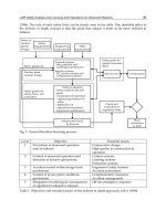
The lymphocytes from each subject were then split and subjected to four different
treatments, using the conditions, described by Ivanova (Ivanova et al., 2010) as shown in
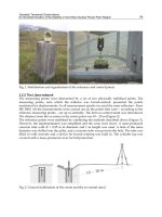
Fig. 1a: (A) 4 hours of incubation with RPMI before lysis; (B) 2 hours of incubation in RPMI,
followed by irradiation with 2 Gy (
137
Cs gamma source, dose rate 2.07 Gy/min), incubation
for another 2 hours and lysis; (C) 4 hours of incubation in RPMI, supplemented with 5μM C-
PC (RPMI-C-PC) before lysis; (D) 2 hours of incubation in RPMI supplemented with 5μM C-
PC, followed by irradiation (as described), incubation for another 2 hours and lysis. All
above procedures were carried out at room temperature.
2.3 Single Cell Gel Electrophoresis (Comet assay)
The neutral comet assay was applied for analysis of radiation- and/or C-PC-induced strand
breaks in DNA. Three comet test slides were prepared from each treatment, described in
Section 2.2 and Fig. 1a. Lymphocytes (5 x 10
5
cells/ml) were suspended in low melting
point agarose (final concentration 0.7% in phosphate buffered saline), dropped onto frosted
glass slides which had been precoated with 0.5% normal melting point agarose, then
refrigerated (4ºC) for 15 min. To dissolve cellular proteins and lipids, the slides were
immersed in lysis buffer (10 mM Tris, 100 mM EDTA, 2.5 M NaCl, 1% Triton X-100, pH 8.0)
for 40 min at 4ºC, and washed 3 times for 5 min in pre-cooled TBE buffer, pH 8.0.
Electrophoresis was performed in TBE for 20 min at 0.5 V/cm
2
.
Finally, the slides were
washed in ethanol and air-dried, stained with ethidium bromide (5 µg/ml) and analyzed
under a fluorescence microscope (Olympus BX41). Double-strand breaks were analyzed by
the parameter “tail moment” (TM), determined by the Comet Score 1.5 Software for fifty
cells per slide. This parameter is the product of tail length and % DNA in the tail and is
considered most informative when low levels of damage are present (Collins et al., 2008).
2.4 Statistical analysis
Distributions of variables were determined using Kolmogorov-Smirnoff test (Marques de Sá
& Frias, 2007). Lilefors and Levene tests were used to determine the homogeneity of
variance. The effects of different treatments (such as exposure to IR, C-PC treatment or the
combination of C-PC treatment plus irradiation) were analyzed using one way ANOVA.
Student t-test for dependent variables was carried out in order to compare every factor pair
in each group. Results showing p<0.05 were considered significant. As a null hypothesis it
was presumed that there is no difference between groups.
3. Results
3.1 C-PC induces changes in DNA response to irradiation in non-irradiated subjects
included in the control group
First we wanted to analyze whether the cells of each individual responded with an increase
in DNA lesions to the different treatments. For this we determined the standard deviation
Nuclear Power – Operation, Safety and Environment
330
for the TM values which we had calculated from each triplet of comet test slides. Average
TM values which increased for different treatments with more than two standard
deviations, were considered elevated. Thus, as evident from Table 1, in vitro irradiation
alone generates elevated levels of persisting DNA lesions in the lymphocytes from all
(100%) of the non-exposed subjects included in the control group. 5 μM C-PC by itself also
causes an increase in the lesions in more than half (67%) of the cases suggesting that
treatment with C-PC is toxic for more than half of the subjects, included in the control
group. Notably, when incubated with C-PC prior to irradiation, the samples from only half
of the subjects show levels of DNA lesions, higher than those of the non-treated samples.
This means that C-PC treated cells do not accumulate additional lesions upon radiation
exposure. Thus, despite the fact that C-PC shows some toxicity, it also seems to protect cells
from additional radiation damage.
There was a significant increase in the median value of the parameter TM, upon irradiation of
cells which were grown in the absence of C-PC (Fig. 1b, B vs. A, t
11
=6.4). In contrast, for cells
grown in C-PC supplemented medium, the median value slightly decreased upon irradiation
(Fig. 1b, D vs. C, t
11
=2.36). The lower median TM value, calculated for the combined treatment,
C-PC plus irradiation (D), in comparison to the separate treatments with C-PC (C) or
irradiation (B), suggested that the biliprotein exerted radio-protection. Residual damage
calculations (B minus A vs. D minus C) confirmed these findings (data not shown).
The relatively high levels of data dispersion (wide confidence intervals), observed in all
conditions (Fig. 1b) are consistent with high inter-individual variations in the cellular
response of the control subjects. Notably, the cells irradiated after treatment with C-PC (D)
showed a lower level of data scattering than that of treatments (B) or (C), which was
comparable to the dispersion range of values found for the non-treated samples (A). This is
evidently due to a reduction of the maximal TM values in (D). This observation suggests
that the biliprotein limits the manifestations of high radiosensitivity.
3.2 C-PC induces changes in the DNA response of lymphocytes from workers with
very low cumulative doses of radiation
The average annual exposure of 17 subjects with very low cumulative doses, ranging from
0.32 to 12.12 mSv, did not exceed 1 mSv/year - the public dose limit, mandated by ICRP
(ICRP 60, 1990), and these workers were unified in a group with very low dose occupational
exposure. Cumulative doses and data on the levels of DNA damage in the workers are
summarized in Table 2. Similar to the non-exposed control group, the additional, in vitro
irradiation of the cells generated а significant increase in the levels of persisting lesions (Fig.
1c, B vs. A, t
16
=3.63) in the majority of cases (76%). Treatment of the cells with C-PC also
generated elevated levels of unrepaired DNA strand breaks in the majority (82%) of the
subjects (Fig. 1c, C vs. A, t
16
=3.11). Notably, after irradiation, samples, which had been pre-
incubated with C-PC, showed lower median levels of DNA breaks as well as a reduction in
the number of the subjects with higher levels of persisting DNA lesions (59% of the subjects)
when compared with the samples which were irradiated only (Fig. 1c, D vs. B, t
16
=2.68) or
incubated with C-PC without in vitro irradiation (Fig. 1c, D vs. C, t
16
=2.77). This result was
similar to the effect of the protein on the non-exposed control group and demonstrated its
radio-protective effect on the subjects with a very low dose occupational exposure.
As seen in Fig. 1c, the exposure of the cells only to C-PC or to IR (B and C) elevated the
median values of TM and extended the range of the data dispersion. This is consistent with
the cellular toxicity of the two agents. The data dispersion towards the higher break
The Biliprotein C-Phycocyanin Modulates the
DNA Damage Response in Lymphocytes from Nuclear Power Plant Workers
331
extremes was more drastic with C-PC (C) than with irradiation alone (B), although the
median values of damage levels in C-PC treated cells (C) was lower than that of irradiated
cells (B). Notably, in combination, C-PC and radiation (D) induced a well pronounced
decrease in the median values of TM, which were brought down almost to the levels of the
controls (A). Additionally, the combination of the two agents (D) narrowed the range of
data dispersion, again bringing it close to that of controls (A). In conclusion, for this group
we observed a beneficial effect of C-PC on lymphocytes treated prior to radiation exposure,
despite the toxicity of the protein. This conclusion was further confirmed by residual
damage calculations (B minus A vs. D minus C).
(a) Lymphocytes from each subject were treated as follows: A - 4 hours of incubation with
RPMI before lysis (controls); B - 2 hours of incubation in RPMI, followed by irradiation,
incubation for another 2 hours and lysis; C - 4 hours of incubation in RPMI, supplemented
with 5μM C-PC; D - 2 hours of incubation in RPMI supplemented with 5μM C-PC, followed
by irradiation, incubation for another 2 hours and lysis.
(b) TM for the different treatments of lymphocytes from non-exposed subjects
(c) TM for the different treatments of lymphocytes from workers with cumulative doses,
ranging from 0.32 to 12.12 mSv
(d) TM for the different treatments of lymphocytes from workers with cumulative doses,
ranging from 26.77 to 330.77 mSv
Whiskers represent non-outlier range, boxes: 25-75% confidence intervals (CI), (■) median
value and (●) outlier values.
Fig. 1. Treatment patterns and their effects on subjects from different exposure groups
Nuclear Power – Operation, Safety and Environment
332
3.3 C-PC induces changes in the DNA response of lymphocytes from workers with
higher cumulative doses of radiation
This group included 27 professionals with cumulative doses, ranging from 26.77 to 330.77
mSv. Data, summarized in Table 3, showed, that in this group, in comparison with the two
previous groups (non-exposed controls and exposed to very low doses of radiation), which
were characterized by high levels of radiation-induced DNA lesions in the majority of
samples (100 and 76%, respectively), the number of workers with persisting DNA lesions,
induced after the in vitro exposure of the cells to 2 Gy gamma rays or treatment with C-PC
was reduced by half to 48% and 44%, respectively. This is consistent with improved repair
capacity of the subjects included in this group, which is probably relevant to their chronic
low dose radiation exposure, which may have acted as in vivo adaptive dose. C-PC showed
the lowest cytotoxicity in this group of workers since the median TM values and the range
of data scattering were similar to those in untreated samples (Fig. 1d, C vs. A). This is also
consistent with a general robustness of the cellular DNA repair capacity of this group of
subjects, which is evident from the similar TM median of treatments A and B (Fig. 1d) - a
sign of possible protective adaptation to toxic exposures, developed in subjects with higher
cumulative doses of radiation. Significant differences in the levels of persisting lesions were
detected only between the cells, irradiated in vitro and those treated with C-PC (Fig. 1d, C
vs. B, t
24
=2.44). It is important to note, however, that regardless of the similarity of TM
median values of B and A (Fig. 1d), irradiation of cells caused a definite increase in the data
scattering towards the higher TM values, as compared to non-irradiated cells. Such an
increase was not evident in the cells treated with C-PC only (Fig. 1d, C vs. A), rendering the
C-PC treatment in this group less toxic to DNA than in the previous two groups (Fig. 1b and
1c). However, in contrast to the other two groups of subjects, C-PC treatment in this case did
not cause a decrease in the amount of radiation-induced DSBs (Fig. 1d, D) – a finding that
was confirmed by residual damage calculations (B minus A vs. D minus C).
3.4 The magnitude of the C-PC effect depends on the cumulative doses of exposure
We compared the ТМ values for each treatment among the three subject groups. As seen in
Fig. 2, the only significant differences found were for treatment of cells with C-PC only (C),
which showed that the protein was less toxic for workers with cumulative doses higher than
20 mSv (Fig. 2, group 3) and this effect contrasted with the toxicity registered for the
controls and the group of professionals with very low dose radiation exposure (Fig. 2,
groups 1 and 2). This indicates that chronic occupational exposure might stimulate the
cellular defense mechanisms and induce resistance to DNA damage, caused by agents, such
as C-PC. The workers with higher cumulative doses might also be more resistant to
radiation-induced toxicity since in the same group (Fig. 1d, B) we registered lower median
values of TM in the lymphocytes irradiated with 2 Gy gamma rays as compared to the TM
values in the other two groups (Fig. 1b, B and 1c, B).
It is worth noting the differences between the control (Fig. 1b) and the two groups of
workers (Fig. 1c and 1d) regarding the median values of the parameter TM. For both groups
of professionals, we found lower median values of TM upon each of the exposures (C-PC, 2
Gy or the combination of the two agents) in comparison with the median TM values of the
non-exposed controls (Group 1). This result suggests that workers possess lower levels of
persisting DNA lesions than the controls, which is probably due to improved DNA repair
capacity induced by the low dose professional exposure. This may also be relevant to radio-
adaptive phenomena, mobilizing and activating repair of DNA damage in the groups of the
professionals.
The Biliprotein C-Phycocyanin Modulates the
DNA Damage Response in Lymphocytes from Nuclear Power Plant Workers
333
Fig. 2. TM in non-exposed controls (Group 1) and in subjects with cumulative doses, ranging
from 0.32 to 12.12 mSv (Group 2) or from 26.77 to 330.77 mSv (Group 3) treated with 5 μM
C-PC. Vertical bars represent 95% CI.
3.5 Age dependence of the DNA response of lymphocytes treated with C-PC and/or
irradiated with 2 Gy gamma rays
All individuals, non-exposed controls and occupationally irradiated workers, were divided
into two groups. The first one included 24 subjects (3 controls and 21 occupationally
exposed) of the age from 26 to 46 years. The second group consisted of 32 subjects, all of
them older than 46 years (9 controls and 23 occupationally exposed). Comparison of all
mean TM values in the first group showed significant differences between the levels of
DNA damage in the non-treated samples and the in vitro irradiated lymphocytes in the
presence and absence of C-PC (Fig. 3a, A vs. B and D, t
23
=2.35 and t
23
=2.03, respectively).
We also observed a significant narrowing of the dispersion of the TM values for the cells,
irradiated after pre-treatment with C-PC (Fig. 3a, D), indicating reduction of the inter-
individual variability and unification of the radiation responses by C-PC. Notably, the
dispersion was narrowed predominantly by reducing the non-outlier range from the top –
indicating that C-PC, combined with radiation, selectively improves the repair capacity of
cells which, in all other conditions (A, B and C) demonstrate impaired DNA repair
mechanisms. The last observation suggested that the protein stimulated better the repair of
the radiation-induced DNA lesions in lymphocytes of the susceptible individuals. This
observation may be important for the maintenance of genomic integrity in this high-risk
subgroup of the population.
Comparison of the TM values obtained for the older group (age 46-62 years, Fig. 3b) showed
increased levels of persisting DNA lesions (p=0.05) in the cells irradiated in vitro (B, t
31
=
3.54), incubated with C-PC (C, t
31
=2.26), or incubated with C-PC prior to radiation exposure
(D, t
31
=2.93), when compared to the control setting in this group (A). As with the younger
group, the median values of the TM for treatment B, C and D were similar. However, the
significant top-down reduction in the TM value scattering, described for the younger group
after irradiation of the cells, pre-treated with C-PC (Fig.3a, D) , was not evident in this group
of subjects.
Nuclear Power – Operation, Safety and Environment
334
Fig. 3. Effect of different treatment patterns on subjects, grouped according to age: (a) from
26 to 46 years and (b) from 47 to 62 years. Note that both groups included exposed subjects
and non-exposed controls. Whiskers represent non-outlier range, boxes: 25-75% confidence
intervals (CI), (■) median value and (●) outlier values.
No. Age TM (Ctrl.) TM (2Gy) TM (C-PC) TM (C-PC + 2Gy)
1 50 1.85 ± 0.21 3.41 ± 0.32 6.16 ± 0.11 3.88 ± 0.19
2 48 3.10 ± 0.22 6.80 ± 0.10 6.86 ± 0.07 7.15 ± 0.16
3 50 3.86 ± 0.13 5.38 ± 0.18 2.38 ± 0.51 4.91 ± 0.29
4 45 7.71 ± 0.15 9.82 ± 0.13 6.65 ± 0.24 6.24 ± 0.10
5 58 7.23 ± 0.17 8.24 ± 0.13 8.36 ± 0.08 6.80 ± 0.16
6 50 3.23 ± 0.04 3.78 ± 0.09 3.93 ± 0.04 4.13 ± 0.04
7 47 5.38 ± 0.01 7.45 ± 0.04 4.52 ± 0.14 4.12 ± 0.06
8 53 4.44 ± 0.09 7.28 ± 0.05 5.84 ± 0.01 3.91 ± 0.05
9 49 1.61 ± 0.09 1.79 ± 0.02 2.07 ± 0.03 2.02 ± 0.09
10 42 5.21 ± 0.04 7.10 ± 0.12 5.32 ± 0.07 5.13 ± 0.07
11 54 4.62 ± 0.02 6.68 ± 0.04 6.39 ± 0.05 7.70 ± 0.05
12 42 5.46 ± 0.02 7.13 ± 0.41 5.28 ± 0.03 5.00 ± 0.26
Table 1. Levels of persisting DNA strand breaks determined by TM in lymphocytes of non-
exposed controls treated like in Fig. 1a.
The Biliprotein C-Phycocyanin Modulates the
DNA Damage Response in Lymphocytes from Nuclear Power Plant Workers
335
No. Age
Cumulative
dose (mSv)
TM
(Ctrl.)
TM
(2Gy)
TM
(C-PC)
TM
(C-PC + 2Gy)
1 33 0.32 3.32 ± 0.28 8.44 ± 0.17 11.29 ± 0.48 4.77 ± 0.51
2 32 1.15 2.82 ± 0.02 3.75 ± 0.08 3.08 ± 0.04 2.08 ± 0.10
3 37 1.33 4.36 ± 0.03 11.19 ± 0.13 8.78 ± 0.16 8.34 ± 0.05
4 35 1.67 4.15 ± 0.03 5.29 ± 0.08 4.45 ± 0.08 4.11 ± 0.17
5 32 1.79 3.46 ± 0.02 3.98 ± 0.07 3.69 ± 0.02 3.78 ± 0.05
6 38 1.80 2.35 ± 0.09 2.30 ± 0.04 2.97 ± 0.04 3.08 ± 0.08
7 55 2.29 4.22 ± 0.14 5.05 ± 0.44 7.77 ± 0.39 4.64 ± 0.18
8 55 2.29 3.62 ± 0.16 7.35 ± 0.12 7.96 ± 0.13 6.62 ± 0.08
9 55 2.59 1.24 ± 0.45 1.01 ± 0.18 1.16 ± 0.42 1.69 ± 0.73
10 55 3.50 5.47 ± 0.02 6.18 ± 0.04 6.58 ± 0.10 4.84 ± 0.03
11 26 4.71 3.62 ± 0.53 3.90 ± 0.31 5.67 ± 0.09 4.55 ± 0.15
12 46 6.55 4.50 ± 0.07 6.21 ± 0.09 6.15 ± 0.04 6.38 ± 0.04
13 52 8.37 4.14 ± 0.03 5.82 ± 0.06 4.39 ± 0.05 3.80 ± 0.10
14 26 9.32 5.57 ± 0.02 7.07 ± 0.03 7.11 ± 0.04 6.36 ± 0.06
15 44 9.71 2.14 ± 0.10 4.16 ± 0.09 4.21 ± 0.04 3.56 ± 0.08
16 30 10.06 1.86 ± 0.27 3.72 ± 0.26 0.86 ± 0.16 1.77 ± 0.09
17 49 12.12 1.08 ± 0.12 0.91 ± 0.20 0.82 ± 0.02 1.33 ± 0.27
Table 2. Levels of persisting DNA strand breaks (TM) in lymphocytes of workers with very
low cumulative doses (0.32-12.12 mSv) treated like in Fig. 1a.
4. Discussion
Our study has addressed three main questions: (i) are there significant differences in the
DNA strand break repair in lymphocytes of workers, chronically exposed to low doses of IR
and of non-exposed controls; (ii) can the biliprotein C-PC modify the DNA repair capacity of
lymphocytes and the early cellular radiation response; (iii) is the level of the chronic
exposure of significance for the impact of the biliprotein on the repair of DNA lesions. To
answer the questions we assessed the amount of unrepaired DNA lesions in lymphocytes by
the neutral comet assay 2h after irradiation of the cells in the presence or absence of C-PC.
The experiments were performed with freshly drawn human G
0
phase lymphocytes
(Kaczmarek et al., 1987), which are known to utilize the NHEJ pathway for the repair of
DSBs in DNA, supposedly the main lesions, detected by the neutral CA. NHEJ is a relatively
fast process, which is completed within 2h after the exposure (Lankoff et al., 2006; Palyvoda
et al., 2003). This was also confirmed by kinetics, utilizing the comet test in our laboratory
(data not shown). With our setup we have detected changes in the cellular capacity to mend
breaks in DNA generated after exposure to toxic agents, but not the differences in initial IR-
induced lesions.
Nuclear Power – Operation, Safety and Environment
336
No. Age Cumulative dose (mSv) TM (Ctrl.)
TM
(2Gy)
TM
(C-PC)
TM
(C-PC + 2Gy)
1 38 26.77 0.90 ± 0.05 0.78 ± 0.11 3.46 ± 0.08 3.82 ± 0.04
2 44 28.47 2.05 ± 0.06 4.10 ± 0.10 1.79 ± 0.06 2.80 ± 0.15
3 53 31.14 4.65 ± 0.18 2.92 ± 0.09 2.44 ± 0.07 4.14 ± 0.28
4 54 31.43 7.82 ± 0.09 8.54 ± 0.09 6.23 ± 0.05 7.73 ± 0.18
5 48 36.39 3.47 ± 0.17 2.63 ± 0.20 0.94 ± 0.11 2.27 ± 0.11
6 56 37.06 1.61 ± 0.15 0.58 ± 0.05 0.76 ± 0.13 1.06 ± 0.06
7 48 38.05 5.76 ± 0.08 6.84 ± 0.12 6.33 ± 0.03 5.25 ± 0.16
8 32 38.72 3.74 ± 0.18 5.64 ± 0.26 4.11 ± 0.19 3.90 ± 0.05
9 45 44.66 1.44 ± 0.20 1.09 ± 0.08 1.30 ± 0.23 1.33 ± 0.30
10 50 53.19 0.81 ± 0.20 0.58 ± 0.12 0.67 ± 0.10 1.07 ± 0.01
11 36 56.07 3.97 ± 0.02 3.47 ± 0.16 2.90 ± 0.06 2.39 ± 0.10
12 49 67.76 4.06 ± 0.02 5.36 ± 0.07 5.17 ± 0.07 5.36 ± 0.03
13 41 70.46 5.36 ± 0.11 4.05 ± 0.13 3.88 ± 0.15 3.56 ± 0.06
14 50 73.21 2.54 ± 0.03 3.40 ± 0.12 3.04 ± 0.08 3.24 ± 0.05
15 45 84.57 4.34 ± 0.45 10.16 ± 0.54 6.16 ± 0.33 4.10 ± 0.15
16 54 85.80 1.04 ± 0.22 3.21 ± 0.14 1.43 ± 0.08 1.76 ± 0.08
17 48 87.48 3.95 ± 0.10 6.05 ± 0.03 4.66 ± 0.04 5.88 ± 0.07
18 56 88.28 3.60 ± 0.05 2.89 ± 0.09 5.00 ± 0.14 7.41 ± 0.05
19 50 95.02 2.35 ± 0.10 2.76 ± 0.17 2.19 ± 0.18 2.38 ± 0.12
20 52 95.14 2.10 ± 0.35 10.85 ± 0.96 11.05 ± 1.11 9.01 ± 1.64
21 62 116.82 2.95 ± 0.12 3.03 ± 0.09 2.98 ± 0.05 2.88 ± 0.17
22 42 125.17 2.82 ± 0.03 1.65 ± 0.21 0.91 ± 0.07 2.48 ± 0.32
23 48 126.39 2.55 ± 0.28 11.12 ± 0.41 2.76 ± 0.22 5.06 ± 0.11
24 41 130.23 1.22 ± 0.80 1.61 ± 0.38 3.45 ± 0.24 4.26 ± 0.32
25 41 180.61 3.37 ± 0.39 7.97 ± 0.34 4.03 ± 0.43 9.29 ± 0.37
26 40 246.94 8.21 ± 0.13 4.82 ± 0.14 3.79 ± 0.08 5.02 ± 0.02
27 48 330.77 2.42 ± 0.16 2.68 ± 0.15 2.52 ± 0.02 2.22 ± 0.11
Table 3. Levels of persisting DNA strand breaks (TM) in lymphocytes of workers with
cumulative doses ranging from 26.77 to 330.77 mSv treated like in Fig. 1a.
The comet assay which we have used is an approach, applied in a number of studies to assess
the repair capacity of occupationally exposed populations and recently reviewed by Decordier
(Decordier et al. 2010). The CA method has also been applied in several small pilot studies,
comparing the DNA repair capacity between cancer patients and healthy subjects (Alapetite et
al., 1999; Leprat et al., 1998; Djuzenova, et al., 2006; Zhang et al., 2006). In these studies, the CA
has demonstrated an impaired DNA lesion repair capacity in the lymphocytes of cancer
The Biliprotein C-Phycocyanin Modulates the
DNA Damage Response in Lymphocytes from Nuclear Power Plant Workers
337
patients, when compared to that of the healthy controls. It has also been shown that CA could
be a useful approach to study the radiosensitivity and individual risk of radiation therapy
induced toxicity in cancer patients after in vitro challenging of the cells with ionizing radiation.
The results of our work have indicated large inter-individual variations in the baseline
endogenous levels of DNA lesions in lymphocytes from workers and non-exposed controls.
Notably, the data dispersion covered a relatively large range of values and was registered in
all three experimental groups, including the non-exposed control group. In the control group
we observed the highest variation in persisting baseline levels of DNA lesions (Fig. 1b, A). In
vitro irradiation with 2 Gy γ-rays or treatment with C-PC of the cells additionally enlarged the
range of data dispersion, indicating significant differences in the individual susceptibility of
the subjects to toxic exposures. Exposure to 2 Gy gamma rays exerted the strongest effects on
data scattering in the group of professionals with cumulative doses higher than 20 mSv (Fig.
1d, B) whereas C-PC generated highest level of data dispersion in samples from the group of
workers, exposed to very low doses of occupational IR (Fig. 1c, C).
The biliprotein elevated the number of persisting, unrepaired DNA lesions in the group of
subjects with lower professional irradiation and in the control group. Such toxicity was not
observed in the group of professionals with higher doses of occupational radiation exposure
(Fig. 2). This effect might be attributed to induction of adaptive processes, due to the chronic
exposure to low doses of radiation of the subjects in the last group. Studies of other
laboratories on the repair capacity of nuclear power plant workers (Toili et al., 2002) or
workers exposed to xenobiotics, lead or pesticides (Restreppo et al., 2000; Vodicka et al.,
2004; Piperakis et al., 2009) have shown, similar to our results, that workers repair DNA
damage more efficiently than the non-exposed controls. The authors attributed this
phenomenon to adaptive response by the sub-chronic genotoxic exposures. The adaptive
protection, shown in this study is also consistent with the conclusion of our previous study
(Ivanova et al., 2010).
Notably, in combination with radiation exposure, C-PC exerted protective effects on
lymphocytes of controls and of workers exposed to very low doses of radiation. For these
groups, the lymphocytes, treated with C-PC and exposed to gamma rays, showed reduced
levels of DNA lesions (in comparison to C-PC treatment alone), which suggested that the
protein neutralized the genotoxic effects of IR and exerted radio-protection. It seems that the
biliprotein selectively improved the DNA repair capacity of the individuals with higher
radiosensitivity, which could be of particular interest for the radio-protection of high-risk
subgroups of the population and workers.
The toxic effect of C-PC, registered in this study, is probably linked to the photosensitizing
properties of the protein (Padula & Boiteaux, 1999; Zhang et al., 1999; Paul et al., 2006). It has
been shown that visible light, absorbed by the tetrapyrrolic chromophores of
phycobiliproteins, can generate reactivate oxygen species (ROS), such as hydroxyl radical
and singlet oxygen, which induce oxidative damage to DNA. The photosensitizing
properties of C-PC may also contribute to the apoptotic effects of the protein in cancerogenic
cell lines (Roy et al., 2007; Subhashini et al., 2004) and macrophages (Reddi et al., 2003).
Apoptotic activity, however, is evident only for higher protein concentrations (>20 μM) and
longer incubation times of the cells with the protein (>24h). The trends in DNA damage,
induced by C-PC, were found quite similar to those, induced by ionizing radiation or by
hydrogen peroxide in the presence of transition metals (Epe, 1995; Padula & Boiteaux, 1999).
Notably, for subjects with low cumulative doses of IR and for those from the control group,
we registered higher levels of unrepaired DNA lesions both in cells, treated with C-PC and
in those, irradiated with 2 Gy.
Nuclear Power – Operation, Safety and Environment
338
5. Conclusion
The data from this and from a previous study, carried out in our laboratory, which was
focused on the effects of C-PC on the enzymatic anti-oxidant defense system of lymphocytes
(Ivanova et al., 2010), suggest that C-phycocyanin can affect the anti-oxidant and repair
mechanisms in lymphocytes, and modulate the early radiation response. They showed that
the protein impacts the repair of deleterious forms of DNA damage, generated upon
exposure of the cells to γ-rays, which is essential for the preservation of genomic integrity.
Notably, the biliprotein might selectively improve the DNA repair capacity of the
individuals with higher radiosensitivity, which could be of particular interest for the radio-
protection of the high-risk subgroups of population and workers. The protein induced
significantly less persisting DNA lesions in the group of workers with higher doses (>20
mSv, Fig. 2, group 3) in comparison with the other two groups, which is relevant to adaptive
phenomena induced by the chronic occupational exposure of the subjects. However, the
present study should be considered a pilot one and additional experiments are needed to
decide whether the protein could be applied for the protection of occupationally exposed
individuals (such as nuclear power plant workers, miners, some medical doctors and pilots),
and subgroups of the population with higher susceptibility to the toxic effect of ionizing
radiation.
6. Acknowledgement
This study was carried out as part of the National Program “Genomics” of the Republic of
Bulgaria and is partially financed by the Bulgarian Ministry for Education, Youth and
Science (Grant G-3-10/05). We would like to thank Savina Stoitsova for her critical reading
of the text.
7. References
Alapetite, C., Thirion, P., De La Rochefordiere, A., Cosset, J.M. & Moustacchi, E. (1999).
Analysis by Alkaline Comet Assay of Cancer Patients with Severe Reactions to
Radiotherapy: Defective Rejoining of Radioinduced DNA Strand Breaks in
Lymphocytes of Breast Cancer Patients. Int J Cancer, Vol. 83, No. 1, (Sep 24), pp. (83-
90)
Berwick, M. & Vineis, P. (2000). Markers of DNA Repair and Susceptibility to Cancer in
Humans: An Epidemiologic Review. J Natl Cancer Inst, Vol. 92, No. 11, (Jun 7), pp.
(874-897)
Collins, A.R., Oscoz, A.A., Brunborg, G., Gaivao, I., Giovannelli, L., Kruszewski, M., Smith,
C.C. & Stetina, R. (2008). The Comet Assay: Topical Issues. Mutagenesis, Vol. 23, No.
3, (May), pp. (143-151)
Contreras-Martel, C., Matamala, A., Bruna, C., Poo-Caamano, G., Almonacid, D., Figueroa,
M., Martinez-Oyanedel, J. & Bunster, M. (2007). The Structure at 2Ǻ Resolution of
Phycocyanin from Gracilaria Chilensis and the Energy Transfer Network in a Pc-Pc
Complex. Biophys Chem, Vol. 125, No. 2-3, (Feb), pp. (388-396)
Decordier, I., Loock, K.V. & Kirsch-Volders, M. (2010). Phenotyping for DNA Repair
Capacity. Mutat Res, Vol. 705, No. 2, (Oct), pp. (107-129)
Epe, B. (1995). DNA Damage Profile Induced by Oxidizing Agents Rev Phys Bioch Pharm,
Vol. 127, pp. (223-249)
The Biliprotein C-Phycocyanin Modulates the
DNA Damage Response in Lymphocytes from Nuclear Power Plant Workers
339
Ge, B., Qin, S., Han, L., Lin, F. & Ren, Y. (2006). Antioxidant Properties of Recombinant
Allophycocyanin Expressed in Escherichia Coli. J Photochem Photobiol B, Vol. 84, No.
3, (Sep 1), pp. (175-180)
Glazer, A.N. (1994). Phycobiliproteins - a Family of Valuable, Widely Used Fluorophores. J
of Appl Phycol, Vol. 6, pp. (105-112)
ICRP (1991). 1990 Recommendations of the International Commission on Radiological
Protection. ICRP Publication 60. Ann ICRP 21 (1-3)
Ivanova, K.G., Stankova, K.G., Nikolov, V.N., Georgieva, R.T., Minkova, K.M., Gigova, L.G.,
Rupova, I.T. & Boteva, R.N. (2010). The Biliprotein C-Phycocyanin Modulates the
Early Radiation Response: A Pilot Study. Mutat Res, Vol. 695, No. 1-2, (Jan), pp. (40-
45)
Kaczmarek, L., Calabretta, B., Elfenbein, I.B. & Mercer, W.E. (1987). Cell Cycle Analysis of
Human Peripheral Blood T Lymphocytes in Long-Term Culture. Exp Cell Res, Vol.
173, No. 1, (Nov), pp. (70-79)
Kalthur, G., Kumar, P., Devi, U., Ali, S., Upadhya, R., Pillai, S. & Rao, A. (2008).
Susceptibility of Peripheral Lymphocytes of Brain Tumour Patients to in Vitro
Radiation-Induced DNA Damage, a Preliminary Study. Clin Exp Med, Vol. 8, No. 3,
(Sep), pp. (147-150)
Karpov, L.M., Brown, Ii, Poltavtseva, N.V., Ershova, O.N., Karakis, S.G., Vasil'eva, T.V. &
Chaban Iu, L. (2000). The Postradiation Use of Vitamin-Containing Complexes and
a Phycocyanin Extract in a Radiation Lesion in Rats]. Radiats Biol Radioecol, Vol. 40,
No. 3, (May-Jun), pp. (310-314)
Lankoff, A., Bialczyk, J., Dziga, D., Carmichael, W.W., Gradzka, I., Lisowska, H., Kuszewski,
T., Gozdz, S., Piorun, I. & Wojcik, A. (2006). The Repair of Gamma-Radiation-
Induced DNA Damage Is Inhibited by Microcystin-Lr, the Pp1 and Pp2a
Phosphatase Inhibitor. Mutagenesis, Vol. 21, No. 1, (Jan), pp. (83-90)
Leprat, F., Alapetite, C., Rosselli, F., Ridet, A., Schlumberger, M., Sarasin, A., Suarez, H.G. &
Moustacchi, E. (1998). Impaired DNA Repair as Assessed by the "Comet" Assay in
Patients with Thyroid Tumors after a History of Radiation Therapy: A Preliminary
Study. Int J Radiat Oncol Biol Phys, Vol. 40, No. 5, (Mar 15), pp. (1019-1026)
Li, B., Zhang, X., Gao, M. & Chu, X. (2005). Effects of Cd59 on Antitumoral Activities of
Phycocyanin from Spirulina Platensis. Biomed Pharmacother, Vol. 59, No. 10, (Dec),
pp. (551-560)
Madhyastha, H.K., Radha, K.S., Nakajima, Y., Omura, S. & Maruyama, M. (2008). Upa
Dependent and Independent Mechanisms of Wound Healing by C-Phycocyanin. J
Cell Mol Med, Vol. 12, No. 6B, (Dec), pp. (2691-2703)
Marques De Sa, J.P. & Frias, R. (2007). Applied Statistics (2), Springer-Verlag, 978-3-540-71971-
7, Berlin-Heidelberg
Mohseni-Meybodi, A., Mozdarani, H. & Mozdarani, S. (2009). DNA Damage and Repair of
Leukocytes from Fanconi Anaemia Patients, Carriers and Healthy Individuals as
Measured by the Alkaline Comet Assay. Mutagenesis,
Vol. 24, No. 1, (Jan), pp. (67-73)
Moller, P., Knudsen, L.E., Loft, S. & Wallin, H. (2000). The Comet Assay as a Rapid Test in
Biomonitoring Occupational Exposure to DNA-Damaging Agents and Effect of
Confounding Factors. Cancer Epidemiol Biomarkers Prev, Vol. 9, No. 10, (Oct), pp.
(1005-1015)
Padula, M. & Boiteaux, S. (1999). Photodynamic DNA Damage Induced by Phycocyanin and
Its Repair in Saccharomyces Cerevisiae. Braz J Med Biol Res, Vol. 32, No. 9, (Sep). pp.
(1063-1071)
Nuclear Power – Operation, Safety and Environment
340
Palyvoda, O., Polanska, J., Wygoda, A. & Rzeszowska-Wolny, J. (2003). DNA Damage and
Repair in Lymphocytes of Normal Individuals and Cancer Patients: Studies by the
Comet Assay and Micronucleus Tests. Acta Biochim Pol, Vol. 50, No. 1, pp. (181-190)
Paul, B.T., Patel, A., Selvam, G.S., Mishra, S., Ghosh, P.K. & Murugesan, R. (2006).
Photodynamic Action of C-Pcs Obtained from Marine and Fresh Water
Cyanobacterial Cultures: A Comparative Study Using Epr Spin Trapping
Technique. Free Radical Res., Vol. 40, No.8, (Aug) pp. (821-825)
Piperakis, S.M., Kontogianni, K., Karanastasi, G., Iakovidou-Kritsi, Z., Cebulska-
Wasilewska, A. & Piperakis, M.M. (2009). Investigation of the Genotoxic Effect of
Pesticides on Greenhouse Workers' Lymphocytes. Environ Mol Mutagen, Vol. 50,
No. 2, (Mar), pp. (121-126)
Prasanna, R., Sood, A., Suresh, A. & Kaushik, B.D. (2007). Potentials and Applications of
Algal Pigments in Biology and Industry. Acta Bot Hung, Vol. 49, pp. (131-156)
Reddy, M.C., Subhashini, J., Mahipal, S.V., Bhat, V.B., Srinivas Reddy, P., Kiranmai, G.,
Madyastha, K.M. & Reddanna, P. (2003). C-Phycocyanin, a Selective Cyclooxygenase-
2 Inhibitor, Induces Apoptosis in Lipopolysaccharide-Stimulated Raw 264.7
Macrophages. Biochem Biophys Res Commun, Vol. 304, No. 2, (May 2), pp. (385-392)
Restrepo, H.G., Sicard, D. & Torres, M.M. (2000). DNA Damage and Repair in Cells of Lead
Exposed People. Am J Ind Med, Vol. 38, No. 3, (Sep), pp. (330-334)
Romay, C., Gonzalez, R., Ledon, N., Remirez, D. & Rimbau, V. (2003). C-Phycocyanin: A
Biliprotein with Antioxidant, Anti-Inflammatory and Neuroprotective Effects. Curr
Protein Pept Sci, Vol. 4, No. 3, (Jun), pp. (207-216)
Roy, K.R., Arunasree, K.M., Reddy, N.P., Dheeraj, B., Reddy, G.V. & Reddanna, P. (2007).
Alteration in Mitochondrial Membrane Potential by Spirulina Platensis C-
Phycocyanin Induces Apoptosis in the Doxorubicin-Resistant Human
Hepatocellular-Carcinoma Cell Line Hepg2. Biotechnol Appl Biochem, Vol. 47, No.3,
(Jul). pp. (159-167)
Stec, B., Troxler, R.F. & Teeter, M.M. (1999). Crystal Structure of C-Phycocyanin from
Cyanidium Caldarium Provides a New Perspective on Phycobilisome Assembly.
Biophys J, Vol. 76, No. 6, (Jun), pp. (2912-2921)
Subhashini, J., Mahipal, S.V., Reddy, M.C., Mallikarjuna Reddy, M., Rachamallu, A. &
Reddanna, P. (2004). Molecular Mechanisms in C-Phycocyanin Induced Apoptosis
in Human Chronic Myeloid Leukemia Cell Line-K562. Biochem Pharmacol, Vol. 68,
No. 3, (Aug 1), pp. (453-462)
Sun, L., Wang, S., Chen, L. & Gong, X. (2003). Promising Fluorescent Probes from
Phycobiliproteins. IEEE J. Cel Top Quantum Electron, Vol. 9, pp. (177-188)
Touil, N., Aka, P.V., Buchet, J.P., Thierens, H. & Kirsch-Volders, M. (2002). Assessment of
Genotoxic Effects Related to Chronic Low Level Exposure to Ionizing Radiation
Using Biomarkers for DNA Damage and Repair. Mutagenesis, Vol. 17, No. 3, (May),
pp. (223-232)
Vodicka, P., Kumar, R., Stetina, R., Musak, L., Soucek, P., Haufroid, V., Sasiadek, M.,
Vodickova, L., Naccarati, A., Sedikova, J., Sanyal, S., Kuricova, M., Brsiak, V.,
Norppa, H., Buchancova, J. & Hemminki, K. (2004). Markers of Individual
Susceptibility and DNA Repair Rate in Workers Exposed to Xenobiotics in a Tire
Plant. Environ Mol Mutagen, Vol. 44, No. 4, pp. (283-292)
Zhang, C., Naftalis, E. & Euhus, D. (2006). Carcinogen-Induced DNA Double Strand Break
Repair in Sporadic Breast Cancer. J Surg Res, Vol. 135, No. 1, (Sep), pp. (120-128)
16
Effects of Gamma-Ray Irradiation on Tracking
Failure of Polymer Insulating Materials
Boxue Du, Yu Gao and Yong Liu
Tianjin University
China
1. Introduction
Electrical insulation and dielectrics play a key role in the performance and reliability of most
electrical systems, where a single-point system failure may prove catastrophic or even fatal
to the electrical equipment. Polymers are the most commonly used dielectrics because of
their reliability, availability, ease of fabrication and low cost. The selection of the proper
polymer dielectric for a desired application depends on the requirements and operating
conditions of the system. Voltage surges are known to be one of the factors leading to
tracking failure, which often appear in lighting, switching and circuit breaker operation.
Tracking failure is a dielectric breakdown phenomenon occurring on polymer surfaces
comprising carbonized conductive paths. When a sufficiently intense discharge lasts for a
considerable time, the decomposed carbon products, with some parts of the channel
carbonized, are progressive and rapidly form on the sample surface. When the carbonized
deposits bridge the interval between electrodes, a sudden decrease in the insulating
resistance occurs. The importance of this information has been revealed for preventing fires,
short-circuit and insulation failure in electrical appliances and devices, and especially the
tracking and ignition of polymers have been examined.
Polymer materials are widely used in radiation environments, such as scientific research
fields and nuclear power stations, where the high safety and reliability are demanded.
The polymer insulating materials are inevitably exposed to various kinds of radiation. The
changes in their physical and chemical properties could prematurely terminate the useful
life of the dielectric. Outside and inside of the secondary shield in the containment vessel
of nuclear power plants, the maximum dose rates of irradiation are 0.01 Gy/h and 1
Gy/h. The dose rate in nuclear power plants varies widely from 10 µGy/h to 10 kGy/h
with a potential of total exposure 1000 kGy or greater. The high reliability is based on
construction safety and system safety during the use of electrical equipment. Accordingly,
it is important to investigate the influence of radiation on polymeric insulating materials
used in the radioactive environments. Presently, the researches on irradiation aging are
mostly concentrated on electrical and mechanical performance, but the effect of radiation,
especially the radiation aging theory is rarely studied. Kuriyama et al have measured the
physical and electrical properties of gamma-ray irradiated PVC jacketed cable and
concluded that the electrical receptivity of PVC is reduced obviously. We have
investigated the tracking resistance of gamma-ray irradiated polyethylene and modified
Nuclear Power – Operation, Safety and Environment
342
polycarbonate materials, and the results reveal that the tracking resistance is improved by
irradiation for cross-linking type materials, but the conclusions for degradation type
materials are opposite.
The polymers used in the field of electrical and electronic engineering are not only subject to
an electric field alone, quite often they operate under the influence of both electric and
magnetic fields. In addition to shielding effects against space plasma flux, magnetic field
changes the electron kinematics and the gas desorption rate, and hence the flashover
potential. The effect of a magnetic field parallel to an applied electric field has been
considered and reported that a magnetic field can shorten the formative time lag of
breakdown. Magnetic field might influence dielectric breakdown in different ways. The
basic mechanisms and physical phenomena providing magnetic insulation have been
investigated in the past mainly for vacuum-insulator interfaces in high-power transmission
lines and vacuum diodes. The influence of magnetic field on the sparking voltage has been
examined for a slightly non-uniform electric field applied perpendicularly to a magnetic
field, including its effects on the formative time lag and on the cycloidal movement of
electrons. Low energy non-disruptive discharge carbonized “track” marks have been
identified by a number of users on gas insulating switchgear (GIS) insulating surfaces and
the cause of this phenomenon has not yet been identified. However, there have been very
few investigations concerning the influence of magnetic field on the tracking failure of
polymer materials used in electrical equipments. Therefore, it is necessary to study how
their electrical characteristics change under magnetic field.
Polymers are used on space equipment with the examples of these devices working at high dc
voltage on satellites such as ionic propulsion systems, photomultiplier tubes, scientific
electronic instruments, communication systems and solar cell power supply systems.
Energetic electromagnetic radiation and low pressure are two main factors in the space
environment. We have reported that surface discharge occurs earlier at low pressures than
atmospheric pressure. In fact, surface discharge is one of the principal causes for premature
failure in electronic equipments used in low pressure regions. The dielectric properties of the
polymers under the environments of low pressure and radiation may change. It is necessary to
study the way how their electrical characteristics change under the combined environments.
As technology advances, increasing demands on the reliable operation under various
operating and environmental conditions are made on materials and components. Therefore,
it is necessary to study how their electrical characteristics change with consideration of
radiation, low pressure and magnetic field. However, knowledge of influence of irradiation
under combined conditions of radiation, low pressure and magnetic field is limited, and
systematic study is desirable.
Polymer materials are widely used in electrical insulation because of their high breakdown
strength, high resistivity and low dielectric loss. Polybutylene naphthalate is widely used in
aerospace electronics, electric engineering, chemical, metallurgy and medical equipment.
Polybutylene terephthalate is usually used for electric coupler, blind plug, switch and
insulating cover. Polyethylene terephthalate is one of the most widely used polymers for
electrical and electronic industries. Whether the dielectric property of the polymers is
changed by irradiation and magnetic field or not are worth investigating. In this chapter,
effects of total dose of gamma-ray irradiation on tracking failure of polymer materials are
described. The results reveal that the tracking failure properties are greatly changed by
gamma-ray irradiation.
Effects of Gamma-Ray Irradiation on Tracking Failure of Polymer Insulating Materials
343
2. Experiment setup and procedure
2.1 Test samples
Polybutylene naphthalate (PBN) and polybutylene terephthalate (PBT) are produced with
butanediol by reaction with 2, 6-naphthalic acid and dimethyl terephthalate, respectively.
Polyethylene terephthalate (PET) is produced with ethylene glycol by reaction with purified
terephthalic acid. For comparison, both polycarbonates mixed with 3% polyethylene (M-PC)
and polyethylene (PE) samples are also tested.
The samples are irradiated in air up to 100 kGy and then up to 1000 kGy with a dose rate of
10 kGy/h by using a
60
Co gamma-source. The thickness and dimensions are 3 mm and 20
mm×20 mm. The sample surface is cleaned with ethyl alcohol and dried in a desiccator in
air at room temperature for 24 h or longer before the testing.
2.2 Experimental apparatus and procedure for pulse voltage application
Schematic diagram of experimental set-up is shown in Fig. 1. Test sample is stressed
electrically under a HV pulse voltage at the level of 30 kV and the pulse width is 1 ms. The
pulse interval is adjusted from 5 ms to 10 ms. The electrode geometry is needle-plate
electrode pattern, where the needle electrode is connected to the HV voltage and the plate
electrode is grounded through the discharge detecting circuit. The length of each stainless
steel electrode is 45 mm, the radius of the semicircle plate electrode is 5 mm and the
thickness is 0.5 mm. The diameter of the needle electrode is 0.65 mm and the interval
between the electrodes is 3 mm. The magnetic environment is produced by the
electromagnet (PEM-50). The magnetic flux density (MFD) is 495 mT and the direction of
E×B is 0, 90 and 270 degrees with respect to the sample surface. The vacuum chamber is
made from a glass tube with two sheets of acrylic board for the cover and bottom.
(a) Relative angles
(b) Experimental apparatus (c) Experiment setup for reduced pressure
Fig. 1. Schematic diagram of test system for pulse voltage
Tracking failure is defined as the insulation failure under electric field with consequent lack
of dielectric properties, which is determined when the surface is burning or short-circuit of
the electrodes has persisted for 2s. The tracking failure is caused by decomposed carbon on
the sample surface due to heat generated by the discharge, which starts locally across the