INTERNAL MEDICINE BOARDS - PART 1 ppt
Bạn đang xem bản rút gọn của tài liệu. Xem và tải ngay bản đầy đủ của tài liệu tại đây (1.12 MB, 67 trang )
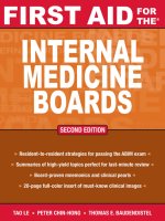
FIRSTAID
INTERNAL
MEDICINE
BOARDS
Second Edition
TAO LE, MD, MHS
Assistant Clinical Professor of Medicine and Pediatrics
Chief, Section of Allergy and Immunology
Department of Medicine
University of Louisville
Louisville, Kentucky
PETER CHIN-HONG, MD
Assistant Professor, Division of Infectious Diseases
Department of Medicine
University of California at San Francisco
San Francisco, California
THOMAS E. BAUDENDISTEL, MD, FACP
Associate Director, Internal Medicine Residency
Department of Medicine
California Pacific Medical Center
San Francisco, California
New York / Chicago / San Francisco / Lisbon / London / Madrid / Mexico City
Milan / New Delhi / San Juan / Seoul / Singapore / Sydney / Toronto
FOR
THE
®
Copyright © 2008 by Tao Le. All rights reserved. Manufactured in the United States of America. Except as permitted under the United States Copyright Act of 1976,
no part of this publication may be reproduced or distributed in any form or by any means, or stored in a database or retrieval system, without the prior written permis-
sion of the publisher.
0-07-164320-6
The material in this eBook also appears in the print version of this title: 0-07-149913-X.
All trademarks are trademarks of their respective owners. Rather than put a trademark symbol after every occurrence of a trademarked name, we use names in an
editorial fashion only, and to the benefit of the trademark owner, with no intention of infringement of the trademark. Where such designations appear in this book, they
have been printed with initial caps.
McGraw-Hill eBooks are available at special quantity discounts to use as premiums and sales promotions, or for use in corporate training programs. For more
information, please contact George Hoare, Special Sales, at or (212) 904-4069.
TERMS OF USE
This is a copyrighted work and The McGraw-Hill Companies, Inc. (“McGraw-Hill”) and its licensors reserve all rights in and to the work. Use of this work is subject
to these terms. Except as permitted under the Copyright Act of 1976 and the right to store and retrieve one copy of the work, you may not decompile, disassemble,
reverse engineer, reproduce, modify, create derivative works based upon, transmit, distribute, disseminate, sell, publish or sublicense the work or any part of it without
McGraw-Hill’s prior consent. You may use the work for your own noncommercial and personal use; any other use of the work is strictly prohibited. Your right to use
the work may be terminated if you fail to comply with these terms.
THE WORK IS PROVIDED “AS IS.” McGRAW-HILL AND ITS LICENSORS MAKE NO GUARANTEES OR WARRANTIES AS TO THE ACCURACY, ADE-
QUACY OR COMPLETENESS OF OR RESULTS TO BE OBTAINED FROM USING THE WORK, INCLUDING ANY INFORMATION THAT CAN BE
ACCESSED THROUGH THE WORK VIA HYPERLINK OR OTHERWISE, AND EXPRESSLY DISCLAIM ANY WARRANTY, EXPRESS OR IMPLIED,
INCLUDING BUT NOT LIMITED TO IMPLIED WARRANTIES OF MERCHANTABILITY OR FITNESS FOR A PARTICULAR PURPOSE. McGraw-Hill and
its licensors do not warrant or guarantee that the functions contained in the work will meet your requirements or that its operation will be uninterrupted or error free.
Neither McGraw-Hill nor its licensors shall be liable to you or anyone else for any inaccuracy, error or omission, regardless of cause, in the work or for any damages
resulting therefrom. McGraw-Hill has no responsibility for the content of any information accessed through the work. Under no circumstances shall McGraw-Hill
and/or its licensors be liable for any indirect, incidental, special, punitive, consequential or similar damages that result from the use of or inability to use the work, even
if any of them has been advised of the possibility of such damages. This limitation of liability shall apply to any claim or cause whatsoever whether such claim or cause
arises in contract, tort or otherwise.
DOI: 10.1036/007149913X
We hope you enjoy this
McGraw-Hill eBook! If
you’d like more information about this book,
its author, or related books and websites,
please click here.
Professional
Want to learn more?
DEDICATION
To the contributors to this and future editions, who took time to share their
knowledge, insight, and humor for the benefit of residents and clinicians.
and
To our families, friends, and loved ones, who endured and assisted in the
task of assembling this guide.
Copyright © 2008 by Tao Le. Click here for terms of use.
This page intentionally left blank
v
Authors vii
Preface ix
Acknowledgments xi
How to Contribute xiii
Introduction to the ABIM xv
Chapter 1 Allergy and Immunology 1
Raffi Tachdjian, MD, MPH
Marc Riedl, MD, MS
Chapter 2 Ambulatory Medicine 23
Deborah Lindes, MD
Cindy J. Lai, MD
Chapter 3 Cardiovascular Disease 83
Ankush Goel, MD
Sanjiv Shah, MD
Chapter 4 Critical Care 149
Elliott Dasenbrook, MD
Christian A. Merlo, MD, MPH
Chapter 5 Dermatology 161
Tara Miller, MD
Siegrid S. Yu, MD
Chapter 6 Endocrinology 203
Melissa Weinberg, MD
Diana M. Antoniucci, MD
Karen Earle, MD
Chapter 7 Gastroenterology and Hepatology 251
Ma Somsouk, MD
Scott W. Biggins, MD, MAS
Chapter 8 Geriatrics 313
Abigail Holley, MD
Param Dedhia, MD
Chapter 9 Hematology 341
Andrea Harzstark, MD
Thomas Chen, MD, PhD
Chapter 10 Hospital Medicine 387
Jesse Liu, MD
Robert L. Trowbridge, MD
CONTENTS
For more information about this title, click here
Chapter 11 Infectious Diseases 421
Brian S. Schwartz, MD
José M. Eguía, MD
Chapter 12 Nephrology 479
Carmen A. Peralta, MD, MAS
Alan C. Pao, MD
Chapter 13 Neurology 509
Joey English, MD, PhD
S. Andrew Josephson, MD
Chapter 14 Oncology 549
Andrea Harzstark, MD
Jonathan E. Rosenberg, MD
Chapter 15 Psychiatry 581
Demian Rose, MD, PhD
Amin N. Azzam, MD, MA
Chapter 16 Pulmonary Medicine 599
Elliott Dasenbrook, MD
Christian A. Merlo, MD, MPH
Chapter 17 Rheumatology 629
Mary E. Margaretten, MD
Jonathan Graf, MD
Chapter 18 Women’s Health 665
Deborah Lindes, MD
Linda Shiue, MD
Color insert
715-734
Appendix: Abbreviations and Symbols 689
Index 697
About the Authors 714
DIANA M. ANTONIUCCI, MD
Assistant Professor, Division of Endocrinology and Metabolism
Department of Medicine
University of California, San Francisco
AMIN N. AZZAM, MD, MA
Assistant Professor
Department of Psychiatry
University of California, San Francisco
SCOTT W. BIGGINS, MD, MAS
Assistant Professor, Division of Gastroenterology
Department of Medicine
University of California, San Francisco
THOMAS CHEN, MD, PhD
Staff Physician
Associated Medical Specialists
San Mateo Medical Center
PARAM DEDHIA, MD
Clinical Instructor
Associate Director of Geriatric Education Committee
Department of Medicine
Johns Hopkins Bayview Medical Center
KAREN EARLE, MD
Medical Director, Center for Diabetes Services
Department of Internal Medicine
California Pacific Medical Center
JOSÉ M. EGUÍA, MD, MPH
Associate Physician
Positive Health Program
Department of Medicine
University of California, San Francisco
JONATHAN GRAF, MD
Assistant Professor, Division of Rheumatology
Department of Medicine
University of California, San Francisco
CINDY J. LAI, MD
Assistant Professor
Department of Medicine
University of California, San Francisco
CHRISTIAN A. MERLO, MD, MPH
Assistant Professor, Division of Pulmonary and Critical Care
Medicine
Department of Medicine
Johns Hopkins University
ALAN C. PAO, MD
Assistant Professor, Division of Nephrology
Department of Medicine
University of California, San Francisco
MARC RIEDL, MD, MS
Assistant Professor, Division of Clinical Immunology and Allergy
Department of Medicine
University of California, Los Angeles
JONATHAN E. ROSENBERG, MD
Assistant Professor, Division of Hematology and Oncology
Department of Medicine
University of California, San Francisco
SANJIV SHAH, MD
Assistant Professor, Division of Cardiology
Department of Medicine
Northwestern University
LINDA SHIUE, MD
Physician, Palo Alto Medical Foundation
Assistant Professor
Department of Medicine
University of California, San Francisco
ROBERT L. TROWBRIDGE, MD
Assistant Professor
University of Vermont College of Medicine
Maine Hospitalist Service
Maine Medical Center
SIEGRID S. YU, MD
Assistant Professor
Department of Dermatology
University of California, San Francisco
SENIOR REVIEWERS
viii
Copyright © 2008 by Tao Le. Click here for terms of use.
This page intentionally left blank
This page intentionally left blank
This page intentionally left blank
INTRODUCTION
xvi
INTRODUCTION
For house officers, the ABIM is the culmination of three years of hard work. For
practicing physicians, it becomes part of their maintenance of certificate. How-
ever, the certification and recertification process does not simply represent yet
another set of exams in a series of expensive tests. To your patients, it means that
you have the level of clinical knowledge and competency required to provide
good clinical care. In fact, according to a poll conducted for the ABIM, about
72% of adult patients are aware of their physicians’ board-certification status.
In this chapter we talk more about the ABIM and provide you with proven ap-
proaches to conquering the exam. For a detailed description of the ABIM,
visit www.abim.org or refer to the Certification Examination in Internal Medi-
cine Information Booklet, which can also be found on the ABIM Web site.
ABIM—THE BASICS
How Do I Register to Take the Exam?
You can register for the exam online by going to “On-Line Services” at
www.abim.org. The registration fee is currently about $1135. If you miss the
application deadline, a $400 nonrefundable late fee is tacked on. Check the
ABIM Web site for the latest registration deadlines, fees, and policies.
What If I Need to Cancel the Exam or Change Test Centers?
The ABIM currently provides partial refunds if a written cancellation is re-
ceived before certain deadlines. You can also change your test center with a
written request for a specific deadline. Check the ABIM Web site for the latest
information on its refund and cancellation policy as well as procedures.
How Is the ABIM Test Structured?
As of August 2006, the ABIM exam is a one-day computer-based test (CBT).
The exam is divided into four two-hour sections with 60 questions in each
section, for a total of 240 questions. Images (blood smears, x-rays, ECGs, pa-
tient photos) are embedded in certain questions. During the time allotted for
each block, examinees can answer test questions in any order as well as review
responses and change answers. However, examinees cannot go back and
change answers from previous blocks. The CBT format (see Figure 1) allows
you to make your own notes on each question using a pop-up box, and it also
permits you to click a box to designate which questions you might wish to re-
view before the end of the session (time permitting).
Please check the ABIM Web site to check for Web demos, updates, and de-
tails about the new format, as well as to identify testing centers nearest to you.
What Types of Questions Are Asked?
All questions are single-best-answer types only. You will be presented with a
scenario and a question followed by four to six options. Virtually all questions
on the ABIM are vignette based. A substantial amount of extraneous informa-
Register early to avoid an
extra $400 late fee.
The majority of your patients
will be aware of your
certification status.
INTRODUCTION
xvii
tion may be given, or a clinical scenario may be followed by a question that
could be answered without actually requiring that you read the case. Some
questions require interpretation of photomicrographs, radiology studies, pho-
tographs of physical findings, and the like. It is your job to determine which
information is superfluous and which is pertinent to the case at hand.
Question content is based on a content “blueprint” developed by the ABIM
(see Table 1). This blueprint may change from year to year, so check the
ABIM Web site for the latest. About 75% of the primary content focuses on
traditional subspecialties such as cardiology and gastroenterology. The re-
maining 25% pertains to certain outpatient or related specialties and subspe-
cialties such as allergy/immunology, dermatology, and psychiatry. There are
also cross-content questions that may integrate information from multiple pri-
mary content areas.
How Are the Scores Reported?
The passing scores are set before the exam administration, so your passing is
not influenced by the relative performance of others taking the test with you.
Scoring and reporting of test results may take up to three months. In addi-
tion, your pass/fail status will be available to you on the ABIM Web site
through the “On-Line Services” within a day of the results being mailed to
you. Note that you need to register to access this feature.
FIGURE 1.
ABIM CBT format.
Virtually all questions are
case based.
INTRODUCTION
xviii
Your score report will give you a “pass/fail” decision, the overall number of ques-
tions you answered correctly with a corresponding percentile, and the number
of questions you answered correctly with a corresponding percentile for the pri-
mary and cross-content subject areas noted in the blueprint. Each year, be-
tween 20 and 40 questions on the exam do not count toward your final score.
Again, these may be “experimental” questions or questions that are later disqual-
ified. Historically, between 85% and 90% of first-time examinees pass on the
first attempt (see Table 2). About 90% of examinees who are recertifying pass on
the first attempt, and about 97% are ultimately successful with multiple at-
tempts. There is no limit on the number of retakes if an examinee fails.
TABLE 1.
ABIM Certification Blueprint
PRIMARY CONTENT AREAS RELATIVE PROPORTIONS
Cardiovascular Disease 14%
Gastroenterology 9%
Pulmonary Disease 10%
Infectious Disease 9%
Rheumatology/Orthopedics 8%
Endocrinology/Metabolism 8%
Oncology 7%
Hematology 6%
Nephrology/Urology 6%
Allergy/Immunology 3%
Psychiatry 4%
Neurology 4%
Dermatology 4%
Obstetrics/Gynecology 3%
Ophthalmology 2%
Miscellaneous 3%
Total 100%
CROSS-CONTENT AREAS RELATIVE PROPORTIONS
Critical Care Medicine 10%
Geriatric Medicine 10%
Prevention 6%
Women’s Health 6%
Clinical Epidemiology 3%
Ethics 3%
Nutrition 3%
Palliative/End-of-Life Care 3%
Adolescent Medicine 2%
Occupational Medicine 2%
Substance Abuse 2%
Source: www.abim.org, 2007.
INTRODUCTION
xix
THE RECERTIFCATION EXAM
The recertification exam is given twice per year, typically in the spring (April
or May) and in the fall (October). It is a one-day exam that consists of three
modules lasting two hours each. Each module has 60 questions, for a total of
180 questions. You get two minutes per question. The recertification exam is
currently administered as a CBT at a Pearson VUE testing site. Performance
on the recertification exam is similar to that of the certification exam; how-
ever, pass rates have been trending downward over the last few years (see
Table 3).
TEST PREPARATION ADVICE
The good news about the ABIM is that it tends to focus on the diagnosis and
management of diseases and conditions that you have likely seen as a resident
and that you should expect to see as an internal medicine specialist. Assuming
that you have performed well as a resident, First Aid and a good source of
practice questions may be all you need. However, consider using First Aid as a
guide and using multiple resources, including a standard textbook, journal re-
view articles, MKSAP, or a concise electronic text such as UpToDate, as part
TABLE 2.
First-Time Test Taker Performance
YEAR NUMBER TAKING PERCENTAGE PASSED
2006 7006 91%
2005 7051 91%
2004 7056 92%
2003 6751 92%
2002 7074 87%
Source: www.abim.org, 2007.
TABLE 3.
Recertification Performance
YEAR PERCENT PASSED
2002 91%
2003 85%
2004 86%
2005 84%
2006 79%
Check the ABIM Web site for
the latest passing
requirements.
INTRODUCTION
xx
of your studies. Original research articles are low yield, and very new research
(i.e., research done less than one to two years before the exam) will not be
tested. In addition, there are a number of high-quality board review courses
offered around the country. Board review courses are very expensive but can
help those who need some focus and discipline.
Ideally, you should start your preparation early in your last year of residency,
especially if you are starting a demanding job or fellowship right after resi-
dency. Cramming in the period between end of residency and the exam is
not advisable.
As you study, concentrate on the nuances of management, especially for diffi-
cult or complicated cases. For common diseases, learn both common and
uncommon presentations; for uncommon diseases, focus on the classic pre-
sentations and manifestations. Draw on the experiences of your residency
training to anchor some of your learning. When you take the exam, you will
realize that you’ve seen most of the clinical scenarios in your three years of
wards, clinics, morning reports, case conferences, or grand rounds.
Other High-Yield Areas
Focus on topic areas that are typically not emphasized during residency train-
ing but are board favorites. These include the following:
■
Topics in outpatient specialties (e.g., allergy, dermatology, ENT, ophthal-
mology)
■
Formulas that are needed for quick recall (e.g., alveolar gas, anion gap,
creatinine clearance)
■
Basic biostatistics (e.g., sensitivity, specificity, positive predictive value,
negative predictive value)
■
Adverse effects of drugs
TEST-TAKING ADVICE
By this point in your life, you have probably gained more test-taking expertise
than you care to admit. Nevertheless, here are a few tips to keep in mind
when taking the exam:
■
For long vignette questions, read the question stem and scan the options;
then go back and read the case. You may get your answer without having
to read through the whole case.
■
There’s no penalty for guessing, so you should never leave a question
blank.
■
Good pacing is key. You need to leave adequate time to get to all the ques-
tions. Even though you have two minutes per question on average, you
should aim for a pace of 90 to 100 seconds per question. If you don’t know
the answer within a short period, make an educated guess and move on.
■
It’s okay to second guess yourself. Research shows that our “second
hunches” tend to be better than our first guesses.
■
Don’t panic with “impossible” questions. They may be experimental
questions that won’t count. Again, take your best guess and move on.
■
Note the age and race of the patient in each clinical scenario. When eth-
nicity is given, it is often relevant. Know these well, especially for more
common diagnoses.
The ABIM tends to focus on
the horses, not the zebras.
Use a combination of First Aid,
textbooks, journal reviews,
and practice questions.
Never, ever leave a question
blank! There’s no penalty for
guessing.
INTRODUCTION
xxi
■
Questions often describe clinical findings instead of naming eponyms
(e.g., they cite “tender, erythematous bumps in the pads of the finger” in-
stead of “Osler’s nodes”).
TESTING AND LICENSING AGENCIES
American Board of Internal Medicine
510 Walnut Street, Suite 1700
Philadelphia, PA 19106-3699
215-446-3500 or 800-441-2246
Fax: 215-446-3633
www.abim.org
Educational Commission for Foreign Medical Graduates (ECFMG)
3624 Market Street
Philadelphia, PA 19104-2685
215-386-5900
Fax: 215-386-9196
www.ecfmg.org
Federation of State Medical Boards (FSMB)
P.O. Box 619850
Dallas, TX 75261-9850
817-868-4000
Fax: 817-868-4099
www.fsmb.org
This page intentionally left blank
ALLERGY & IMMUNOLOGY
2
DIAGNOSTIC TESTING IN ALLERGY
Allergy Skin Testing
■
A confirmatory test for the presence of allergen-specific IgE antibody.
■
Prick-puncture skin testing: Adequate for most purposes. A drop of al-
lergen extract is placed on the skin surface, and epidermal puncture is
performed with a specialized needle.
■
Intradermal skin testing: Used for venom and penicillin testing; 0.02 mL
of allergen is injected intracutaneously using a 26- to 27-gauge needle.
■
All skin testing should use ᮍ (histamine) and ᮎ (saline) controls.
■
Skin-testing wheal-and-flare reactions are measured 15–20 minutes after
placement.
Laboratory Allergy Testing
■
Radioallergosorbent (RAST) serologic testing is performed to confirm the
presence of allergen-specific IgE antibody.
■
Results are comparable to skin testing for pollen- and food-specific IgE.
■
Recommended when the subject has anaphylactic sensitivity to the anti-
gen; useful when skin testing is either not available or not possible owing
to skin conditions or interfering medications (e.g., antihistamine use).
■
RAST testing alone is generally not adequate for venom or drug allergy
testing.
Delayed-Type Hypersensitivity Skin Testing
■
An effective screening test for functional cell-mediated immunity.
■
Involves intradermal injection of 0.1 mL of purified antigen. The stan-
dard panel includes Candida, mumps, tetanus toxoid, and PPD.
■
The injection site is examined for induration 48 hours after injection.
■
Approximately 95% of normal subjects will respond to one of the above-
mentioned antigens.
■
The absence of a response suggests deficient cell-mediated immunity or
anergy.
Allergen Patch Testing
■
The appropriate diagnostic test for allergic contact dermatitis.
■
Suspected substances are applied to the skin with adhesive test strips for 48
hours.
■
The skin site is examined 48 and 72 hours after application for evidence of
erythema, edema, and vesiculation (reproduction of contact dermatitis).
DIAGNOSTIC TESTING IN IMMUNOLOGY
Complement Deficiency Testing
■
The complement pathway consists of the classic, alternative, and man-
nose-binding lectin pathways (see Figure 1.1).
■
CH50 is a screening test for the classic complement pathway.
■
All nine elements of C1–C9 are required to produce a normal CH50.
■
A normal CH50 does not exclude the possibility of low C3 or C4.
ALLERGY & IMMUNOLOGY
3
■
Deficiencies that lead to disease include the following:
■
CH50: Any C1–C9.
■
C1-INH: Hereditary angioedema.
■
C1, C2, C4: Recurrent sinopulmonary infections (encapsulated bacte-
ria).
■
C2: SLE.
■
C3: Pyogenic bacterial infections.
■
C5–C9: Neisserial infections.
Humoral (B-Cell) and Cellular (T-Cell) Deficiency Testing
Testing for B- and T-cell deficiency is as follows:
■
CD19: For B-cell immunity.
■
IgG, IgM, IgA, IgD, IgE: For antibody.
■
CD3, CD4, CD8: For T-cell immunity.
■
CD16, CD56: For natural killer cell immunity.
FIGURE 1.1.
The complement reaction sequence.
(Reproduced, with permission, from Brooks GF et al. Jawetz, Melnick, & Adelberg’s Medical
Microbiology, 24th ed. New York: McGraw-Hill, 2004, Fig. 8-9.)
Membrane attack complex
C5b–C9
Cell lysis
C3 convertases
C5b
C6,C7,C8,C9
Factor B
Factor D
Properdin
C4
C2
Activated
C1
Immune
complex
Microbial
surfaces
Classic
pathway
Alternative
pathway
C3
[C4b2b] [C3bBb]
C5 convertases
[C4b2b3b] [C3bBbC3b]
Anaphylatoxins
C3b
C3a
C3
C5a
C5
MB lectin
pathway
Microbial
surfaces
MGL
Think of terminal complement
deficiency (C5–C9) in an
otherwise healthy patient
presenting with recurrent
Neisseria meningitis.
ALLERGY & IMMUNOLOGY
4
GELL AND COOMBS CLASSIFICATION OF IMMUNOLOGIC REACTIONS
The traditional framework that is used to describe immune-mediated reac-
tions. It is not inclusive of all complex immune processes.
Type I: Immediate Reactions (IgE Mediated)
■
Specific antigen exposure causes cross-linking of IgE on mast cell/basophil
surfaces, leading to the release of histamine, leukotrienes, prostaglandins,
and tryptase.
■
Mediator release leads to symptoms of urticaria, rhinitis, wheezing, diar-
rhea, vomiting, hypotension, and anaphylaxis, usually within minutes of
antigen exposure.
■
Late-phase type I reactions may cause recurrent symptoms 4–8 hours af-
ter exposure.
Type II: Cytotoxic Reactions
■
Mediated by antibodies, primarily IgG and IgM, directed at cell surface
or tissue antigens. Antigens may be native, foreign, or haptens (small for-
eign particles attached to larger native molecules).
■
Antibodies destroy cells by opsonization (coating for phagocytosis), com-
plement-mediated lysis, or antibody-dependent cellular cytotoxicity.
■
Clinical examples include penicillin-induced autoimmune hemolytic
anemia and certain forms of autoimmune thyroiditis.
Type III: Immune Complex Reactions
■
Exposure to antigen in genetically predisposed individuals causes antigen-
antibody complex formation.
■
Antigen-antibody complexes activate complement and neutrophil infil-
tration, leading to tissue inflammation that most commonly affects the
skin, kidneys, joints, and lymphoreticular system.
■
Clinically presents with symptoms of “serum sickness” 10–14 days after
exposure; most frequently caused by β-lactam antibiotics or nonhuman
antiserum (antithymocyte globulin, antivenoms).
Type IV: Delayed Hypersensitivity Reactions (T-Cell Mediated)
■
Exposure to antigen causes direct activation of sensitized T cells, usually
CD4+ cells.
■
T-cell activation causes tissue inflammation 48–96 hours after exposure.
■
The most common clinical reaction is allergic contact dermatitis such as
that resulting from poison ivy.
ASTHMA
A chronic inflammatory disorder of the airway resulting in airway hyper-
responsiveness, airflow limitation, and respiratory symptoms. Often begins
in childhood, but may have adult onset. Atopy is a strong identifiable risk fac-
Gell-Coombs classi-
fication system—
ACID
Anaphylactic: type I
Cytotoxic: type II
Immune complex:
type III
Delayed
hypersensitivity:
type IV
ALLERGY & IMMUNOLOGY
5
tor for the development of asthma. Subtypes include exercise-induced, occu-
pational, aspirin-sensitive, and cough-variant asthma.
SYMPTOMS
Symptoms include dyspnea (at rest or with exertion), cough, wheezing, mu-
cus hypersecretion, chest tightness, and nocturnal awakenings with respiratory
symptoms. Symptoms may have identifiable triggers (e.g., exercise, exposure
to cat dander, NSAIDs, exposure to cold).
EXAM
■
Acute exacerbations: Expiratory wheezing; a prolonged expiratory phase;
↑ respiratory rate.
■
Severe exacerbations: Pulsus paradoxus, cyanosis, lethargy, use of acces-
sory muscles of respiration, silent chest (absence of wheezing due to lack
of air movement).
■
Chronic asthma without exacerbation: Presents with minimal to no
wheezing. Signs of allergic rhinosinusitis (boggy nasal mucosa, posterior
oropharynx cobblestoning, suborbital edema) are commonly found. Exam
may be normal between exacerbations.
DIFFERENTIAL
■
Upper airway obstruction: Foreign body, tracheal compression, tracheal
stenosis, vocal cord dysfunction.
■
Other lung disease: Emphysema, chronic bronchitis, CF, allergic bron-
chopulmonary aspergillosis, Churg-Strauss syndrome, chronic eosinophilic
pneumonia, obstructive sleep apnea, restrictive lung disease, pulmonary
embolism.
■
Cardiovascular disease: CHF, ischemic heart disease.
■
Respiratory infection: Bacterial or viral pneumonia, bronchiectasis, si-
nusitis.
DIAGNOSIS
Diagnosed by the history and objective evidence of obstructive lung disease.
■
PFTs: Show a ↓ FEV
1
/FVC ratio with reversible obstruction (> 12% ↑ in
FEV
1
after bronchodilator use) and normal diffusing capacity.
■
Methacholine challenge: Useful if baseline lung function is normal but
clinical symptoms are suggestive of asthma. A
ᮍ methacholine challenge
test is not diagnostic of asthma, but a
ᮎ test indicates that asthma is un-
likely (high sensitivity; lower specificity).
TREATMENT
Acute exacerbations are treated as follows:
■
Initial treatment: Inhaled rapid-acting β
2
-agonists (albuterol), one dose
q 20 min × 1 hour; O
2
to keep saturation > 90%.
■
Good response: With a peak expiratory flow (PEF) > 80% of predicted
or personal best after albuterol, continue albuterol q 3–4 h and insti-
tute appropriate chronic therapy (see below).
■
Incomplete response: With a PEF 60–80% of predicted or personal
best, consider systemic corticosteroids; continue inhaled albuterol q 60
min × 1–3 hours if continued improvement is seen.
The role of a methacholine
challenge is to exclude the
diagnosis of asthma. A
ᮍ challenge can be 2° to
numerous etiologies and thus
may not be definitive.
ALLERGY & IMMUNOLOGY
■
Poor response or severe episode: With a PEF < 60% of predicted or
personal best, give systemic corticosteroids and consider systemic epi-
nephrine (preferably IM), IV theophylline, and/or IV magnesium.
■
Patients with improved symptoms, a PEF > 70%, and O
2
saturation > 90%
for 60 minutes after the last treatment may be discharged home with ap-
propriate outpatient therapy and follow-up. Oral corticosteroids are appro-
priate in most cases.
■
Patients with incomplete responses after the initial two hours of treatment
(persistent moderate symptoms, PEF < 70%, O
2
saturation < 90%) should
be admitted for inpatient therapy and monitoring with inhaled albuterol,
O
2
, and systemic corticosteroids.
■
Patients with a poor response to initial therapy (severe symptoms, lethargy,
confusion, PEF < 30%, P
O
2
< 60, PCO
2
> 45) should be admitted to the
ICU for treatment with inhaled albuterol, O
2
, IV corticosteroids, and
possible intubation and mechanical ventilation.
Chronic asthma therapy is based on asthma severity as defined by the Na-
tional Asthma Education and Prevention Program (NAEPP) guidelines (see
Table 1.1). The treatment regimen should be reviewed every 1–6 months,
with changes made depending on symptom severity and clinical course.
TABLE 1.1. NAEPP Guidelines for the Treatment of Chronic Asthma
ASTHMA
CLASSIFICATION SYMPTOMS
a
PULMONARY FUNCTION RECOMMENDED TREATMENT
Mild intermittent ≤ 2 days per week, ≤ 2 nights PEF ≥ 80% Bronchodilator 2–4 puffs q 4 h
per month. as needed. No daily medications
necessary.
Mild persistent > 2 days per week but < 1 time PEF ≥ 80% Add low-dose inhaled
per day or > 2 nights per month. corticosteroids.
Leukotriene modifiers,
theophylline, and cromolyn
may also be added.
Moderate Daily symptoms or > 1 night per PEF 60–80% ↑ to medium-dose inhaled
persistent week. corticosteroids and add a
long-acting inhaled β
2
-agonist.
Leukotriene modifiers or
theophylline may also be
added.
Severe persistent Continuous symptoms. PEF < 60% ↑ to high-dose inhaled
corticosteroids.
Daily oral corticosteroids may
be added if necessary (60 mg
QD).
a
Dyspnea (at rest or with exertion), cough, wheezing, mucus hypersecretion, chest tightness, and nocturnal awakenings with respi-
ratory symptoms.
6
ALLERGY & IMMUNOLOGY
7
■
All asthma patients should use 2–4 puffs of a short-acting bronchodilator
as needed for symptoms. Use of a short-acting bronchodilator ≥ 2 times
per week may indicate the need for ↑ control therapy.
■
Mild intermittent asthma (symptoms ≤ 2 days per week and ≤ 2 nights
per month; PEF ≥ 80%): No daily medication is needed.
■
Mild persistent asthma (symptoms > 2 days per week but < 1 time per
day or > 2 nights per month; PEF ≥ 80%):
■
Low-dose inhaled corticosteroids are preferred.
■
Other medications include leukotriene modifiers, theophylline, and
cromolyn.
■
Moderate persistent asthma (daily symptoms or > 1 night per week and
PEF 60–80%):
■
Low- to medium-dose inhaled corticosteroids and long-acting in-
haled β
2
-agonists are preferred.
■
Alternative therapy involves inhaled corticosteroids and leukotriene
modifiers or theophylline.
■
Severe persistent asthma (continuous symptoms; PEF < 60%):
■
High-dose inhaled corticosteroids and long-acting inhaled β
2
-
agonists.
■
Oral corticosteroids at a dosage of up to 60 mg QD may be necessary.
Additional treatment considerations include the following:
■
Recognize the exacerbating effects of environmental factors such as aller-
gens, air pollution, smoking, and weather (cold and humidity).
■
Use potentially exacerbating medications with caution (aspirin, NSAIDs,
β-blockers).
■
Always consider medication compliance and technique as possible com-
plicating factors in poorly controlled asthma.
■
Treatment of coexisting conditions (e.g., rhinitis, sinusitis, GERD) may
improve asthma.
COMPLICATIONS
■
Hypoxemia, respiratory failure, pneumothorax or pneumomediastinum.
■
Frequent hospitalization and previous intubation are warning signs of po-
tentially fatal asthma.
■
A subset of patients with chronic asthma develop airway remodeling, lead-
ing to accelerated, irreversible loss of lung function.
ALLERGIC RHINITIS
The most common cause of chronic rhinitis. Allergic factors are present in
75% of rhinitis cases. May be seasonal or perennial; incidence is greatest in
adolescence and ↓ with advancing age. Usually persistent, with occasional
spontaneous remission.
SYMPTOMS
■
Sneezing, nasal itching, rhinorrhea, nasal congestion, sore throat, throat
clearing, itching of the throat and palate.
■
Sleep disturbance; association with obstructive sleep apnea.
■
Concomitant conjunctivitis with ocular itching, lacrimation, and puffi-
ness.
Asthma symptoms that occur
more than twice weekly
generally indicate the need for
inhaled corticosteroid therapy.
